rr947 - investigation of techniques to discriminate between tdi and tda exposures in biological
TRANSCRIPT

Health and Safety Executive
Investigation of techniques to discriminate between TDI and TDA exposures in biological samples
Prepared by the Health and Safety Laboratory for the Health and Safety Executive
RR947 Research Report

Health and Safety Executive
Investigation of techniques to discriminate between TDI and TDA exposures in biological samples
Kate Jones Harpur Hill Buxton Derbyshire SK17 9JN
Isocyanates continue to be one of the leading causes of occupational asthma in the UK and biological monitoring (BM) is a valuable tool in assessing exposure to them. BM for toluene diisocyanate (TDI) is based on the measurement of toluene diamine (TDA) in urine after acid hydrolysis of any conjugates (‘total TDA’). The work described here has shown some progress in being able to differentiate between TDA and TDI exposures through the analysis of urine samples. Although none of the outcomes are conclusive, there are some tests that can be applied to positive ‘total TDA’ samples to determine the most likely exposure source. Detection of free TDA is likely to indicate TDA exposure rather than TDI exposure. This technique is limited to samples where the ‘total TDA’ level exceeds about 30 nmol/l. Where a more definitive assessment of exposure to TDI is required, the measurement of TDI-specific lysine conjugates is possible. Currently this would require blood samples to achieve the necessary sensitivity although we have detected this metabolite in urine. This is an area for further work. These techniques also have application for other isocyanate exposures.
This report and the work it describes were funded by the Health and Safety Executive (HSE). Its contents, including any opinions and/or conclusions expressed, are those of the author alone and do not necessarily reflect HSE policy.
HSE Books

© Crown copyright 2012
First published 2012
You may reuse this information (not including logos) free of charge in any format or medium, under the terms of the Open Government Licence. To view the licence visit www.nationalarchives.gov.uk/doc/open-government-licence/, write to the Information Policy Team, The National Archives, Kew, London TW9 4DU, or email [email protected].
Some images and illustrations may not be owned by the Crown so cannot be reproduced without permission of the copyright owner. Enquiries should be sent to [email protected].
ii

KEY MESSAGES
Isocyanates continue to be one of the leading causes of occupational asthma in the UK and biological monitoring is a valuable tool in assessing exposure to isocyanates.
This project arose from an investigation into a company using a foam blowing technique. Cases of occupational asthma had been identified. The process involved the use of toluene diisocyanate (TDI) however the possibility was raised that toluene diamine (TDA) could be generated during the process and monitoring confirmed this.
Biological monitoring (BM) for TDI is based on the measurement of TDA in urine after acid hydrolysis of any conjugates (‘total TDA’). However exposure to TDA will also result in measurable TDA in urine after acid hydrolysis of any conjugates. Because of the detection of TDA in the work environment, there was some uncertainty as to whether measured total TDA in urine reflected exposure to TDI, TDA or a mixture of both. The control approaches to each chemical will differ so clarification of the actual chemical exposure was important.
This work has shown some progress in being able to differentiate between TDA and TDI exposures through the analysis of urine samples. Although none of the outcomes are conclusive (due to the limited nature of the samples available for testing), there are some tests can be applied to positive ‘total TDA’ samples to determine the most likely exposure source.
It is reasonable to assume that where urine samples from workers contain detectable levels of free TDA, this is likely to indicate that TDA exposure (or a mixed TDA/TDI exposure) has occurred. This technique is limited to samples where the ‘total TDA’ level exceeds about 30 nmol/l.
Where a more definitive assessment of exposure to TDI is required, the measurement of TDI-specific lysine conjugates is possible. Currently this would require blood samples to achieve the necessary sensitivity although we have detected this metabolite in urine. This is an area for further work.
Analysis of the samples taken as part of an incident investigation into polyurethane foam blowing, found no free TDA in any single sample but a did find a 2,6-TDI-lysine conjugate in a pooled urine sample. This suggests that the primary exposure was likely to be TDI.
These techniques also have application for other isocyanate exposures such as HDI (where there may be potential interference from an unknown source of HDA exposure), MDI and IPDI (where both MDA and IPDA are widely used industrial chemicals and co-exposures may well be possible or need to be clarified).
Further work on the determination of isocyanate-specific lysine conjugates in urine, is on-going through HSE’s strategic research programme looking at current and future exposure issues. This will enable conclusive demonstration of actual isocyanate exposure, resulting in absorption into the body and therefore the potential to cause health effects. Such a method would not be confounded by potential co-exposure to diamines.
iii

iv
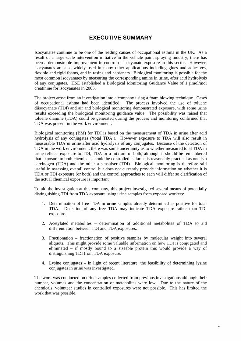
EXECUTIVE SUMMARY
Isocyanates continue to be one of the leading causes of occupational asthma in the UK. As a result of a large-scale intervention initiative in the vehicle paint spraying industry, there has been a demonstrable improvement in control of isocyanate exposure in this sector. However, isocyanates are also widely used in many other applications including glues and adhesives, flexible and rigid foams, and in resins and hardeners. Biological monitoring is possible for the most common isocyanates by measuring the corresponding amine in urine, after acid hydrolysis of any conjugates. HSE established a Biological Monitoring Guidance Value of 1 µmol/mol creatinine for isocyanates in 2005.
The project arose from an investigation into a company using a foam blowing technique. Cases of occupational asthma had been identified. The process involved the use of toluene diisocyanate (TDI) and air and biological monitoring demonstrated exposure, with some urine results exceeding the biological monitoring guidance value. The possibility was raised that toluene diamine (TDA) could be generated during the process and monitoring confirmed that TDA was present in the work environment.
Biological monitoring (BM) for TDI is based on the measurement of TDA in urine after acid hydrolysis of any conjugates (‘total TDA’). However exposure to TDA will also result in measurable TDA in urine after acid hydrolysis of any conjugates. Because of the detection of TDA in the work environment, there was some uncertainty as to whether measured total TDA in urine reflects exposure to TDI, TDA or a mixture of both; although it should be remembered that exposure to both chemicals should be controlled as far as is reasonably practical as one is a carcinogen (TDA) and the other a sensitiser (TDI). Biological monitoring is therefore still useful in assessing overall control but does not currently provide information on whether it is TDA or TDI exposure (or both) and the control approaches to each will differ so clarification of the actual chemical exposure is important
To aid the investigation at this company, this project investigated several means of potentially distinguishing TDI from TDA exposure using urine samples from exposed workers:
1. Determination of free TDA in urine samples already determined as positive for total TDA. Detection of any free TDA may indicate TDA exposure rather than TDI exposure.
2. Acetylated metabolites – determination of additional metabolites of TDA to aid differentiation between TDI and TDA exposures.
3. Fractionation – fractionation of positive samples by molecular weight into several aliquots. This might provide some valuable information on how TDI is conjugated and eliminated – if mostly bound to a sizeable protein this would provide a way of distinguishing TDI from TDA exposure.
4. Lysine conjugates – in light of recent literature, the feasibility of determining lysine conjugates in urine was investigated.
The work was conducted on urine samples collected from previous investigations although their number, volumes and the concentration of metabolites were low. Due to the nature of the chemicals, volunteer studies in controlled exposures were not possible. This has limited the work that was possible.
v
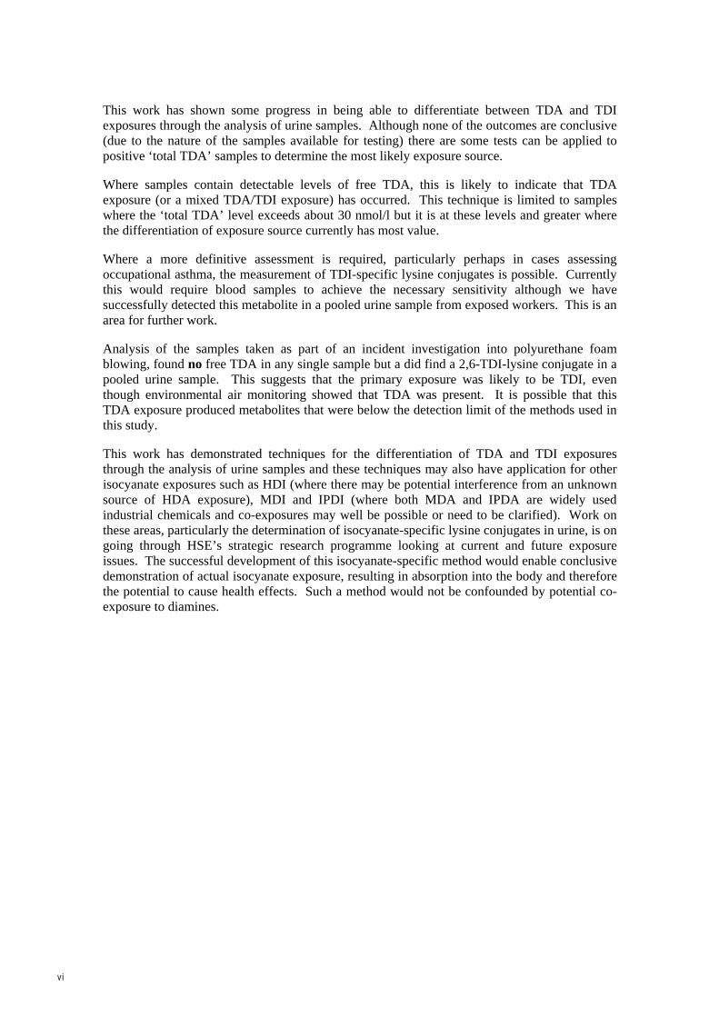
This work has shown some progress in being able to differentiate between TDA and TDI exposures through the analysis of urine samples. Although none of the outcomes are conclusive (due to the nature of the samples available for testing) there are some tests can be applied to positive ‘total TDA’ samples to determine the most likely exposure source.
Where samples contain detectable levels of free TDA, this is likely to indicate that TDA exposure (or a mixed TDA/TDI exposure) has occurred. This technique is limited to samples where the ‘total TDA’ level exceeds about 30 nmol/l but it is at these levels and greater where the differentiation of exposure source currently has most value.
Where a more definitive assessment is required, particularly perhaps in cases assessing occupational asthma, the measurement of TDI-specific lysine conjugates is possible. Currently this would require blood samples to achieve the necessary sensitivity although we have successfully detected this metabolite in a pooled urine sample from exposed workers. This is an area for further work.
Analysis of the samples taken as part of an incident investigation into polyurethane foam blowing, found no free TDA in any single sample but a did find a 2,6-TDI-lysine conjugate in a pooled urine sample. This suggests that the primary exposure was likely to be TDI, even though environmental air monitoring showed that TDA was present. It is possible that this TDA exposure produced metabolites that were below the detection limit of the methods used in this study.
This work has demonstrated techniques for the differentiation of TDA and TDI exposures through the analysis of urine samples and these techniques may also have application for other isocyanate exposures such as HDI (where there may be potential interference from an unknown source of HDA exposure), MDI and IPDI (where both MDA and IPDA are widely used industrial chemicals and co-exposures may well be possible or need to be clarified). Work on these areas, particularly the determination of isocyanate-specific lysine conjugates in urine, is on going through HSE’s strategic research programme looking at current and future exposure issues. The successful development of this isocyanate-specific method would enable conclusive demonstration of actual isocyanate exposure, resulting in absorption into the body and therefore the potential to cause health effects. Such a method would not be confounded by potential coexposure to diamines.
vi
CONTENTS PAGE
1. INTRODUCTION ........................................................................ 1
2. IMPLICATIONS.......................................................................... 3
3. METHODS AND RESULTS ....................................................... 4 3.1 Investigation of free Diamine Metabolites 4 3.2 Identification of Acetylated Metabolites 5 3.3 Molecular Weight Fractionation 11 3.4 Isocyanate-specific Lysine Conjugate 12
4. DISCUSSION ........................................................................... 16 4.1 Investigation of free Diamine Metabolites 16 4.2 Identification of Acetylated Metabolites 16 4.3 Molecular Weight Fractionation 16 4.4 Isocyanate-specific Lysine Conjugate 16
5. REFERENCES ......................................................................... 18
vii

viii

1. INTRODUCTION
Isocyanates continue to be one of the leading causes of occupational asthma in the UK. As a result of a large-scale intervention initiative in the vehicle paint spraying industry, there has been a demonstrable improvement in control of isocyanate exposure in this sector. However, isocyanates are also widely used in many other applications including glues and adhesives, flexible and rigid foams, and in resins and hardeners. Biological monitoring is possible for the most common isocyanates by measuring the corresponding amine in urine, after acid hydrolysis of any conjugates. HSE established a Biological Monitoring Guidance Value of 1 µmol/mol creatinine for isocyanates in 2005.
The project arose from an investigation into a company using a foam blowing technique. Cases of occupational asthma had been identified. The process involved the use of toluene diisocyanate (TDI) and air and biological monitoring demonstrated exposure, with some urine results exceeding the biological monitoring guidance value. The possibility was raised that toluene diamine (TDA) could be generated during the process and monitoring confirmed that TDA was present in the work environment.
Biological monitoring (BM) for TDI is based on the measurement of ‘total’ TDA in urine after acid hydrolysis of any conjugates. However exposure to TDA will also result in measurable TDA in urine after acid hydrolysis of any conjugates. Because of the detection of TDA in the work environment, there was some uncertainty as to whether measured TDA in urine reflects exposure to TDI, TDA or a mixture of both; although it should be remembered that exposure to both chemicals should be controlled as far as is reasonably practical as one is a carcinogen (TDA) and the other a sensitiser (TDI). Biological monitoring is therefore still useful in assessing overall control but does not currently provide information on whether it is TDA or TDI exposure (or both) and the control approaches to each will differ so clarification of the actual chemical exposure is important.
It has been reported (Timchalk, Smith et al. 1994) that in rats much less of a TDI dose (10% of an inhalation dose, 35% of an oral dose) is excreted as free and acetylated TDA than for a TDA dose (~85% of an oral dose). For free TDA, the results are even clearer, with no free TDA detected in urine as a result of TDI exposures. It is known from the literature (Brorson, Skarping et al. 1991) and our own work that free TDA is not detected in urine samples from workers exposed to TDI. This raises the possibility that being able to measure free and/or acetylated TDA could help distinguish TDI from TDA exposures. Whereas TDA is simply metabolised to acetylated and acidic metabolites (Sayama, Kondo et al. 2002), it is supposed that TDI interacts with proteins to form stable conjugates that only release TDA after strong acidic or alkaline hydrolysis. Separating urine samples into molecular weight fractions might be able to isolate protein fractions specific to TDI exposure.
Recent literature (Hettick, Siegel et al. 2012; Sabbioni, Gu et al. 2012) has also indicated that TDI forms specific conjugates with lysine residues in albumin. Such conjugates have been measured in the albumin (from plasma) of exposed workers (Sabbioni, Gu et al. 2012). However as urine levels of albumin are 100-1000 times lower than that in plasma, these metabolites may not be viable urine markers.
The work investigated several means of potentially distinguishing TDI from TDA exposure in urine samples from exposed workers:
1

1. Determination of free TDA in urine samples already determined as positive for total TDA. Detection of any free TDA may indicate TDA exposure rather than TDI exposure.
2. Acetylated metabolites – determination of additional metabolites of TDA to aid differentiation between TDI and TDA exposures.
3. Fractionation – fractionation of positive samples by molecular weight into several aliquots. This might provide some valuable information on how TDI is conjugated in the body – if mostly bound to a sizeable protein this would provide a way of distinguishing TDI from TDA exposure.
4. Lysine conjugates – in light of recent literature, the feasibility of determining lysine conjugates in urine was investigated.
The work was conducted on urine samples collected from previous investigations although their number, volumes and the concentration of metabolites were low. Due to the nature of the chemicals, volunteer studies in controlled exposures were not possible. This has limited the work that was possible.
2
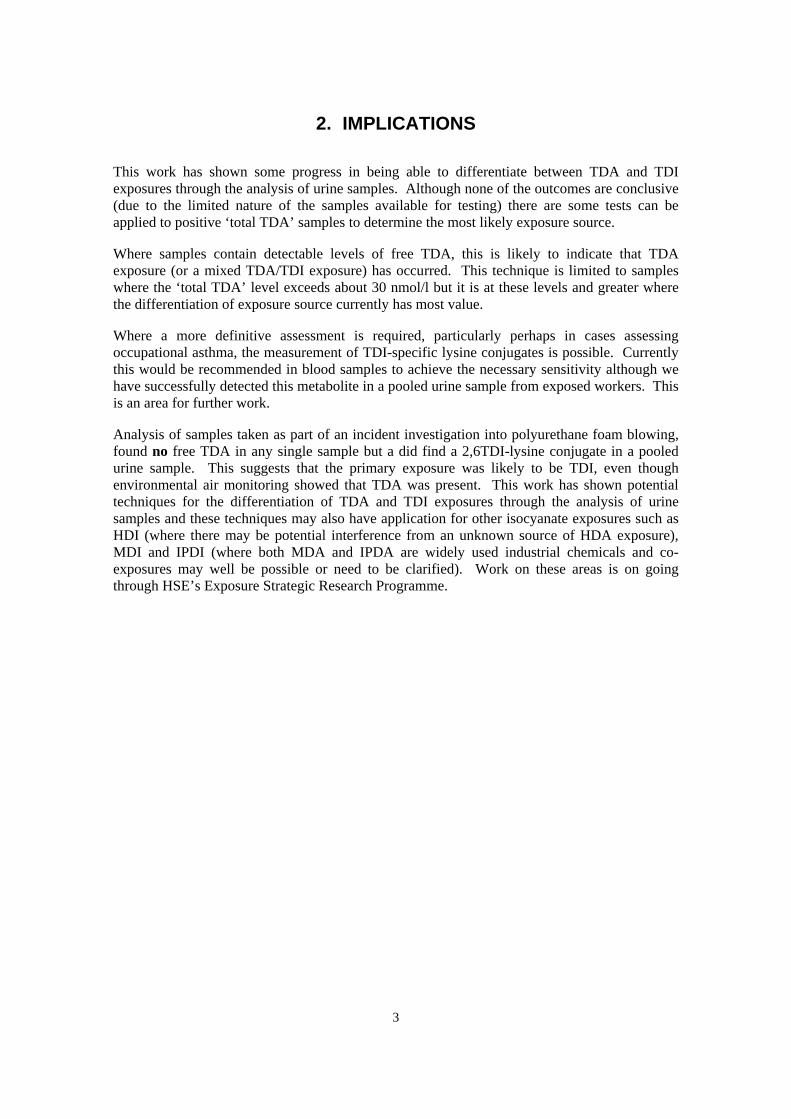
2. IMPLICATIONS
This work has shown some progress in being able to differentiate between TDA and TDI exposures through the analysis of urine samples. Although none of the outcomes are conclusive (due to the limited nature of the samples available for testing) there are some tests can be applied to positive ‘total TDA’ samples to determine the most likely exposure source.
Where samples contain detectable levels of free TDA, this is likely to indicate that TDA exposure (or a mixed TDA/TDI exposure) has occurred. This technique is limited to samples where the ‘total TDA’ level exceeds about 30 nmol/l but it is at these levels and greater where the differentiation of exposure source currently has most value.
Where a more definitive assessment is required, particularly perhaps in cases assessing occupational asthma, the measurement of TDI-specific lysine conjugates is possible. Currently this would be recommended in blood samples to achieve the necessary sensitivity although we have successfully detected this metabolite in a pooled urine sample from exposed workers. This is an area for further work.
Analysis of samples taken as part of an incident investigation into polyurethane foam blowing, found no free TDA in any single sample but a did find a 2,6TDI-lysine conjugate in a pooled urine sample. This suggests that the primary exposure was likely to be TDI, even though environmental air monitoring showed that TDA was present. This work has shown potential techniques for the differentiation of TDA and TDI exposures through the analysis of urine samples and these techniques may also have application for other isocyanate exposures such as HDI (where there may be potential interference from an unknown source of HDA exposure), MDI and IPDI (where both MDA and IPDA are widely used industrial chemicals and coexposures may well be possible or need to be clarified). Work on these areas is on going through HSE’s Exposure Strategic Research Programme.
3

3.1
3. METHODS AND RESULTS
INVESTIGATION OF FREE DIAMINE METABOLITES
The current HSL methodology for isocyanates metabolites in urine involves a vigorous acid hydrolysis to convert all conjugated diamine metabolites to free TDA. Analysis of urine samples without acid hydrolysis would allow determination of free TDA only. Animal studies have shown that exposure to TDA only is likely to result in some free TDA excretion whereas TDI exposure has not been shown to produce any free TDA. Table 1 shows the free and total TDA results from a number of routine samples received by HSL that have tested positive for total TDA in recent months.
Table 1. Free and total TDA results from routine samples received by HSL.
Product/process Urinary TDA results (nM) Likely exposure?
Total TDA Free TDA % Free
“Blocked” TDI, also Diethyl TDA
143 37 26% Amine or mixed?
“Blocked” TDI, also Diethyl TDA
122 21 17% Amine or mixed?
“Blocked” TDI, also Diethyl TDA
85 22 26% Amine or mixed?
Not known 112 ND 0% Isocyanate?
Polyurethane – Company A 9 6.5 72% Suggests amine although concentrations low
Polyurethane – Company A 12 ND 0% Isocyanate?
Polyurethane – Company B 37 ND 0%
Isocyanate?
Polyurethane – Company B 54 ND 0%
Isocyanate?
The datasheet for the “blocked” TDI says that the isocyanate is blocked with methyl ethyl ketoxime and that this is released on heating. It may therefore be possible that the isocyanate may also be released on heating. Tests by HSL on the diethyl TDA also used in the process showed that a very small amount of TDA could be released by our analytical method but it is not possible to say whether human metabolism could produce more TDA from exposure to this amine. Tests on workers urine showed significant excretion of diethyl TDA in urine.
The samples from polyurethane production indicate, mostly, that the exposure is likely to be TDI. The one result from company A may not be reliable due to the low levels of TDA detected. The quantification limit is 5 nmol/l and there is a background population level of about 2 nmol/l (thought to be from environmental exposure to polyurethane foams etc.).
The minimum ‘total TDA’ result that could be clarified by measuring free TDA is probably about 30 nmol/l, based on animal studies indicating that >15% of total TDA in urine is excreted
4

3.2
as free TDA following an exposure to TDA and a quantification limit of 5 nmol/l or three times background levels.
All samples analysed as part of an incident investigation into polyurethane foam blowing showed no free TDA, even though individual total TDA results up to 245 nmol/l were detected.
IDENTIFICATION OF ACETYLATED METABOLITES
3.2.1 Development of an LC-MS-MS method
An LC-MS-MS method was developed for the analysis of 2,4-TDA, 2,6-TDA and 2,4Diacetylaminotoluene (2,4-DAAT). 2,4-TDA and 2,6-TDA were purchased from Qmx Laboratories and 2,4-DAAT was purchased from VitasMLAb Ltd. Although there are published methods for the synthesis of a number of other acetyl TDA metabolites (Glinsukon, Weisburger et al. 1975), it was decided not to pursue these in the first instance as synthesis and purification of 8 putative metabolites would be very resource intensive.
Figure 1. Structures of 2,4-TDA, 2,6-TDA and 2,4-DAAT, respectively.
Compounds optimised in positive ion electrospray MRM on an API3200 AB Sciex tandem mass spectrometer (Tables 2 and 3).
Table 2. Optimised parameters.
Transition Declustering potential (V)
Entrance potential (V)
Cell exit potential (V)
Collision Energy
2,4-TDA 123.1/108.0
123.1/106.0 46 10.5 4
23
23
2,6-TDA 123.1/106.0
123.1/77.1 46 10.5 4
21
37
2,4-DAAT 207.1/122.9
207.1/108.0 71 12 4
31
45
5

Table 3. Source/Gas parameters.
Curtain Gas (psi) 50
Collision cell gas (psi) 10
Ion Spray Voltage (V) 5500
Temperature (oC) 500
Source gas 1 (psi) 30
Source gas 2 (psi) 40
A chromatographic method was set up using a Phenomenex Synergi Hydro-RP column (150 x 2 mm) using a mobile phase of 20 mM ammonium acetate (0.1% acetic acid) and methanol with a flowrate of 0.2 ml/minute. A gradient elution was run from 20% methanol (held for 3 minutes) ramping to 80% methanol by 6 minutes, then immediately reverting to 20% methanol and equilibrating with a total run time of 10 minutes. Chromatography is illustrated in Figure 2.
Figure 2. Chromatogram of 2,4-TDA, 2,6-TDA and 2,4-DAAT standards. The retention times for 2,6-TDA, 2,4-TDA and 2,4-Diacetyl TDA are 3.43, 4.21 and 5.33 minutes respectively.
6

3.2.2 Analysis of samples from exposed workers
Two pooled samples were made – one from samples collected as part of an incident investigation into polyurethane foam blowing where exposure may be to a mixture of both TDI and TDA and one from workers presumed to have exposure to only TDI. It should be noted that these samples were from workers with real workplace exposures and therefore, for example, the possibility that the TDI sample may have also had exposure to TDA cannot be ruled out.
Aliquots (5 ml) of each pooled sample were extracted into diethyl ether (neutral conditions) and the organic layer was removed. The urine was then made alkaline by the addition of 2 ml 10M NaOH and re-extracted into diethyl ether (alkaline conditions) and the organic layer removed. Each organic layer was separately evaporated under a nitrogen stream and reconstituted in mobile phase (100 µl). A ‘blank’ urine was extracted the same way.
3.2.2.1 Neutral extraction
Figure 3. Spiked urine sample, neutral extraction.
7

Figure 4. Neutral extractions of samples from TDI exposure (blue) and samples from mixed TDI and TDA exposure (red) samples overlaid after ‘blank’ urine correction.
No 2,4-TDA or 2,6-TDA (3.5 and 4.4 minutes, respectively) is present in either sample however both samples show a large peak at 4.1 minutes on the 123.1/77.1 transition. Neither sample shows evidence of 2,4-DAAT (207.1/122.9, 5.5 minutes) however both samples show at peak at 2.0 minutes on this transition.
All peaks found are mirrored in both samples so are probably not useful as discriminatory markers between TDA and TDI exposures.
8

3.2.2.2 Alkaline extraction
Figure 5. Spiked urine sample alkaline extraction.
9

Figure 6. Alkaline extractions of samples from TDI exposure (blue) and samples from mixed TDI and TDA exposure (red) samples overlaid after ‘blank’ urine correction.
Again, no evidence of 2,6-TDA or 2,4-TDA (3.5 and 4.4 minutes) in figure 6 but both samples showed a peak at 5.9 minutes on the 123.1/106.0 transition. The chromatogram from the mixed TDI/TDA sample (red) also shows another peak at 5.7 minutes on this transition. No 2,4DAAT (5.6 minutes) is seen in either sample but there is a peak at 5.3 minutes in the chromatogram from the sample with TDI only exposure (blue), which could conceivably be 2,6DAAT (no standard available), as it would be expected to elute slightly earlier than 2,4-DAAT (by analogy with 2,6-TDA eluting before 2,4-TDA).
Attempts to identify these two peaks (207.1/122.9, at 5.3 minutes, blue and 123.4/106.0, at 5.7 minutes, red) by using precursor scanning was unsuccessful – presumably due to the low concentrations present in the samples.
10

3.3 MOLECULAR WEIGHT FRACTIONATION
Initial experiments focussed on using Vivaspin molecular weight cut off (MWCO) centrifuge tubes however these were shown to be prone to contaminating the sample with TDA, even when using water. Because of this, further work proceeded using a Superose 12 gel permeation column (MW 1000 to 300 000) with a fractionator to collect different molecular weight fractions.
Figure 7. Chromatogram showing albumin (main elution in fraction A7) separated on gel permeation column.
An aliquot of each of the pooled samples was fraction ated using the gel permeation column and the first 44 fractions were analysed by HSL’s standard GC-MS method, which includes a strong acid hydrolysis to break down any conjugated metabolites to free TDA. The sample from the mixed TDA/TDI exposure showed the majority of TDA was in fraction 3 (82%) whereas the TDA in fractions from the sample with TDI only exposure was found in at least 4 fractions with 36% in fraction 30, 19% in fraction 2 and 10% each in fractions 12 and 13. Neither sample showed any TDA in fraction 7 which would be expected if the metabolite(s) was associated with albumin. No further sample from the TDI only exposure remained after this experiment so further work was not possible but the sample from mixed TDI/TDA exposure was re-fractionated and analysed by LC/MS/MS (see 3.4).
1111
3.4 ISOCYANATE-SPECIFIC LYSINE CONJUGATE
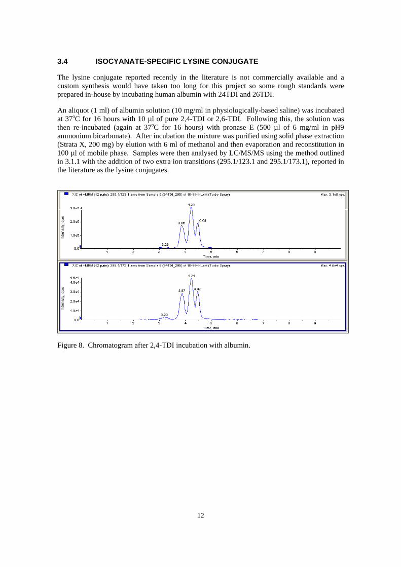
The lysine conjugate reported recently in the literature is not commercially available and a custom synthesis would have taken too long for this project so some rough standards were prepared in-house by incubating human albumin with 24TDI and 26TDI.
An aliquot (1 ml) of albumin solution (10 mg/ml in physiologically-based saline) was incubated at 37oC for 16 hours with 10 µl of pure 2,4-TDI or 2,6-TDI. Following this, the solution was then re-incubated (again at 37oC for 16 hours) with pronase E (500 µl of 6 mg/ml in pH9 ammonium bicarbonate). After incubation the mixture was purified using solid phase extraction (Strata X, 200 mg) by elution with 6 ml of methanol and then evaporation and reconstitution in 100 µl of mobile phase. Samples were then analysed by LC/MS/MS using the method outlined in 3.1.1 with the addition of two extra ion transitions (295.1/123.1 and 295.1/173.1), reported in the literature as the lysine conjugates.
Figure 8. Chromatogram after 2,4-TDI incubation with albumin.
12

Figure 9. Chromatogram after 2,6-TDI incubation with albumin.
As shown in figures 8 and 9, 2,4-TDI results in multiple peaks for these ion transitions whereas 2,6-TDI shows one main peak, with a shoulder. Albumin alone showed no peaks. Repeating these incubations with 2,4-TDA and 2,6-TDA produced no peaks at these ion transitions, indicating that the peaks are specific for TDI.
The literature has reported these lysine conjugate metabolites as being found at low levels in plasma samples of TDI exposed workers (Sabbioni, Gu et al. 2012). The level of albumin in plasma is 100-1000 times greater than that found in urine so this methodology may not be applicable to urine samples (which are the only samples we currently have). However the methodology appears to have been successful and could therefore potentially be used in plasma samples to confirm particular cases where there is the need to determine isocyanate exposure specifically.
Although there were potential sensitivity issues with urine samples, the method was applied to an aliquot of the sample from mixed TDI/TDA exposure.
13

Figure 10. Urine samples incubated with pronase E and analysed for lysine conjugates – potential mixed TDI/TDA exposure (top) and 2,6-TDI incubated with albumin (bottom).
As can be seen from figure 10, there is a peak at 3.4 in the chromatogram of a urine sample with potential mixed TDI/TDA exposure, which appears to correspond to the dominant peak in the 2,6-TDI incubation. This would be consistent with the total TDA analysis of the urine sample, which determined that there was ten times more 2,6-TDA than 2,4-TDA in the pooled sample (41 and 5 nmol/l, respectively). This is in line with literature on TDI exposure and TDA in urine. There was no corresponding peak in the 295.1/173.1 transition but this might be due to sensitivity (this transition is about 10x less sensitive).
Analysis of fractions of the sample with potential mixed TDI/TDA exposure (see 3.2) using this method showed a peak at 3.4 minutes for 295.1/123.1 in the 15th and 17th fractions. These, again, would not correspond to albumin but may respond to a fragment of albumin (see figure 7,
14

fractions A15, B2). Conjugation to fragments of albumin or lysine is more likely in urine than conjugates to albumin itself.
15

4. DISCUSSION
Various techniques have been investigated to try and differentiate between TDA and TDI exposures from the analysis of urine samples. The work was restricted to some extent by the small number and low volume of samples available for analysis. A handful of routine samples that tested positive for total TDA were available as well as a larger pooled sample made from samples from an incident investigation. Due to the nature of these samples there was no definitive confirmation of specifically TDI or TDA exposures.
4.1 INVESTIGATION OF FREE DIAMINE METABOLITES
There was some consensus that where TDI was a likely exposure (polyurethane production), samples tended to show no free TDA whereas where ‘blocked’ TDI was used (in association with a diethyl TDA component), results were more indicative of an amine exposure (more than 15% free TDA detected). This technique looks to be useful where ‘total TDA’ results exceed 30 nmol/l.
4.2 IDENTIFICATION OF ACETYLATED METABOLITES
No 2,4-diacetylaminotoluene (2,4-DAAT) was detected in the pooled sample although it was possible that some 2,6-DAAT might be present (no standard available to confirm) and some other TDA-related metabolites may exist. However concentrations of these metabolites were too low to investigate further and as both TDA and TDI give rise to some acetylated metabolites, quantification of these other metabolites would probably not give definitive separation of TDA and TDI exposures (unlike the use of free TDA as a biomarker (see 4.1) because no free TDA is expected from TDI exposure).
4.3 MOLECULAR WEIGHT FRACTIONATION
Work on molecular weight fractionation resulted in little helpful information. Standard protein concentrators appeared to contaminate samples with TDA. Gel permeation chromatography with fractionation indicated that TDA in fractions was not associated with albumin but may indicate other metabolites or conjugates.
4.4 ISOCYANATE-SPECIFIC LYSINE CONJUGATE
An in-house preparation of TDI-lysine was made and analysis of the pooled urine sample indicated that there was potentially some of this lysine conjugate present although the confirmation transition was not detected (presumably because of sensitivity). Analysis of gel permeation fractions indicated that this lysine conjugate was linked with fragments of albumin.
Analysis of the samples taken as part of an incident investigation into polyurethane foam blowing, found no free TDA in any single sample but a did find a 2,6-TDI-lysine conjugate in a pooled urine sample. This suggests that the primary exposure was likely to be TDI, even though environmental air monitoring showed that TDA was present. It is possible that this TDA exposure produced metabolites that were below the detection limit of the methods used in this study.
In conclusion, this work has demonstrated techniques for the differentiation of TDA and TDI exposures through the analysis of urine samples and these techniques may also have application for other isocyanate exposures such as HDI (where there may be potential interference from an unknown source of HDA exposure), MDI and IPDI (where both MDA and IPDA are widely used industrial chemicals and co-exposures may well be possible or need to be clarified). Work
16

on these areas, particularly the deter mination of isocyanate-spe cific lysine conjugates in urine, is on going through HSE’s s trategic research programme lookin g at current and future exposure issues. The successful development of this isocyanate-specific method would enable conclusive demonstration of actual isocyanate exposure, resulting in absorption into the body and therefore the potential to cause health effects. Such a method would not be confounded by potential coexposure to diamines.
17
5. REFERENCES
Brorson, T., G. Skarping, et al. (1991). "Biological Monitoring of Isocyanates and Related Amines .4. 2,4-Toluenediamine and 2,6-Toluenediamine in Hydrolyzed Plasma and Urine after T est-Chamber Exposure of Humans to 2,4-Toluene and 2,6-Toluene Diisocyanate." International Archives of Occupational and Environmental Health 63(4): 253-259.
Glinsukon, T., E. K. Weisburger, et al. (1975). "Preparation and spectra of some acetyl derivatives of 2,4-toluenediamine." Journal of Chemical & Engineering Data 20(2): 207-209.
Hettick, J. M., P. D. Siegel, et al. (2012). "Vapor conjugation of toluene diisocyanate to specific lysines of human albumin." Analytical biochemistry 421(2): 706-711.
Sabbioni, G., Q. Gu, et al. (2012). "Determination of isocyanate specific albumin-adducts in workers exposed to toluene diisocyanates." Biomarkers 17(2): 150-159.
Sayama, M., T. Kondo, et al. (2002). "Cytosolic acetyl transfer and N-deacetylation associated with the metabolism of carcinogenic 2,4-diaminotoluene in rat liver." Journal of Health Science 48(6): 485-492.
Timchalk, C., F. A. Smith, et al. (1994). "Route-Dependent Comparative Metabolism of [C-14] Toluene 2,4-Diisocyanate and [C-14] Toluene 2,4-Diamine in Fischer-344 Rats." Toxicology and Applied Pharmacology 124(2): 181-190.
18

19

Published by the Health and Safety Executive 09/12

Health and Safety Executive
Investigation of techniques to discriminate between TDI and TDA exposures in biological samples Isocyanates continue to be one of the leading causes of occupational asthma in the UK and biological monitoring (BM) is a valuable tool in assessing exposure to them. BM for toluene diisocyanate (TDI) is based on the measurement of toluene diamine (TDA) in urine after acid hydrolysis of any conjugates (‘total TDA’). The work described here has shown some progress in being able to differentiate between TDA and TDI exposures through the analysis of urine samples. Although none of the outcomes are conclusive, there are some tests that can be applied to positive ‘total TDA’ samples to determine the most likely exposure source. Detection of free TDA is likely to indicate TDA exposure rather than TDI exposure. This technique is limited to samples where the ‘total TDA’ level exceeds about 30 nmol/l. Where a more definitive assessment of exposure to TDI is required, the measurement of TDI-specific lysine conjugates is possible. Currently this would require blood samples to achieve the necessary sensitivity although we have detected this metabolite in urine. This is an area for further work. These techniques also have application for other isocyanate exposures.
This report and the work it describes were funded by the Health and Safety Executive (HSE). Its contents, including any opinions and/or conclusions expressed, are those of the author alone and do not necessarily reflect HSE policy.
RR947
www.hse.gov.uk