role of the anterior cingulate cortex in the control over...
TRANSCRIPT
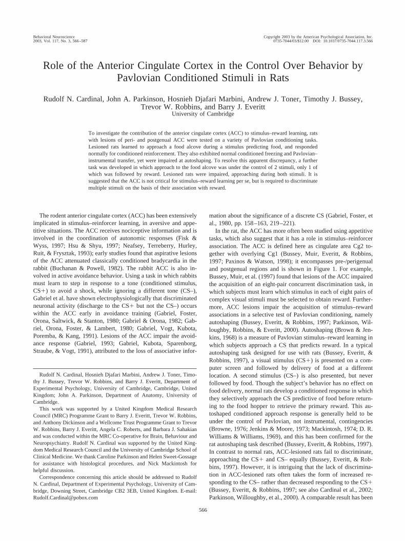
Role of the Anterior Cingulate Cortex in the Control Over Behavior byPavlovian Conditioned Stimuli in Rats
Rudolf N. Cardinal, John A. Parkinson, Hosnieh Djafari Marbini, Andrew J. Toner, Timothy J. Bussey,Trevor W. Robbins, and Barry J. Everitt
University of Cambridge
To investigate the contribution of the anterior cingulate cortex (ACC) to stimulus–reward learning, ratswith lesions of peri- and postgenual ACC were tested on a variety of Pavlovian conditioning tasks.Lesioned rats learned to approach a food alcove during a stimulus predicting food, and respondednormally for conditioned reinforcement. They also exhibited normal conditioned freezing and Pavlovian–instrumental transfer, yet were impaired at autoshaping. To resolve this apparent discrepancy, a furthertask was developed in which approach to the food alcove was under the control of 2 stimuli, only 1 ofwhich was followed by reward. Lesioned rats were impaired, approaching during both stimuli. It issuggested that the ACC is not critical for stimulus–reward learning per se, but is required to discriminatemultiple stimuli on the basis of their association with reward.
The rodent anterior cingulate cortex (ACC) has been extensivelyimplicated in stimulus–reinforcer learning, in aversive and appe-titive situations. The ACC receives nociceptive information and isinvolved in the coordination of autonomic responses (Fisk &Wyss, 1997; Hsu & Shyu, 1997; Neafsey, Terreberry, Hurley,Ruit, & Frysztak, 1993); early studies found that aspirative lesionsof the ACC attenuated classically conditioned bradycardia in therabbit (Buchanan & Powell, 1982). The rabbit ACC is also in-volved in active avoidance behavior. Using a task in which rabbitsmust learn to step in response to a tone (conditioned stimulus,CS�) to avoid a shock, while ignoring a different tone (CS–),Gabriel et al. have shown electrophysiologically that discriminatedneuronal activity (discharge to the CS� but not the CS–) occurswithin the ACC early in avoidance training (Gabriel, Foster,Orona, Saltwick, & Stanton, 1980; Gabriel & Orona, 1982; Gab-riel, Orona, Foster, & Lambert, 1980; Gabriel, Vogt, Kubota,Poremba, & Kang, 1991). Lesions of the ACC impair the avoid-ance response (Gabriel, 1993; Gabriel, Kubota, Sparenborg,Straube, & Vogt, 1991), attributed to the loss of associative infor-
mation about the significance of a discrete CS (Gabriel, Foster, etal., 1980, pp. 158–163, 219–221).
In the rat, the ACC has more often been studied using appetitivetasks, which also suggest that it has a role in stimulus–reinforcerassociation. The ACC is defined here as cingulate area Cg2 to-gether with overlying Cg1 (Bussey, Muir, Everitt, & Robbins,1997; Paxinos & Watson, 1998); it encompasses pre-/perigenualand postgenual regions and is shown in Figure 1. For example,Bussey, Muir, et al. (1997) found that lesions of the ACC impairedthe acquisition of an eight-pair concurrent discrimination task, inwhich subjects must learn which stimulus in each of eight pairs ofcomplex visual stimuli must be selected to obtain reward. Further-more, ACC lesions impair the acquisition of stimulus–rewardassociations in a selective test of Pavlovian conditioning, namelyautoshaping (Bussey, Everitt, & Robbins, 1997; Parkinson, Wil-loughby, Robbins, & Everitt, 2000). Autoshaping (Brown & Jen-kins, 1968) is a measure of Pavlovian stimulus–reward learning inwhich subjects approach a CS that predicts reward. In a typicalautoshaping task designed for use with rats (Bussey, Everitt, &Robbins, 1997), a visual stimulus (CS�) is presented on a com-puter screen and followed by delivery of food at a differentlocation. A second stimulus (CS–) is also presented, but neverfollowed by food. Though the subject’s behavior has no effect onfood delivery, normal rats develop a conditioned response in whichthey selectively approach the CS predictive of food before return-ing to the food hopper to retrieve the primary reward. This au-toshaped conditioned approach response is generally held to beunder the control of Pavlovian, not instrumental, contingencies(Browne, 1976; Jenkins & Moore, 1973; Mackintosh, 1974; D. R.Williams & Williams, 1969), and this has been confirmed for therat autoshaping task described (Bussey, Everitt, & Robbins, 1997).In contrast to normal rats, ACC-lesioned rats fail to discriminate,approaching the CS� and CS– equally (Bussey, Everitt, & Rob-bins, 1997). However, it is intriguing that the lack of discrimina-tion in ACC-lesioned rats often takes the form of increased re-sponding to the CS– rather than decreased responding to the CS�(Bussey, Everitt, & Robbins, 1997; see also Cardinal et al., 2002;Parkinson, Willoughby, et al., 2000). A comparable result has been
Rudolf N. Cardinal, Hosnieh Djafari Marbini, Andrew J. Toner, Timo-thy J. Bussey, Trevor W. Robbins, and Barry J. Everitt, Department ofExperimental Psychology, University of Cambridge, Cambridge, UnitedKingdom; John A. Parkinson, Department of Anatomy, University ofCambridge.
This work was supported by a United Kingdom Medical ResearchCouncil (MRC) Programme Grant to Barry J. Everitt, Trevor W. Robbins,and Anthony Dickinson and a Wellcome Trust Programme Grant to TrevorW. Robbins, Barry J. Everitt, Angela C. Roberts, and Barbara J. Sahakianand was conducted within the MRC Co-operative for Brain, Behaviour andNeuropsychiatry. Rudolf N. Cardinal was supported by the United King-dom Medical Research Council and the University of Cambridge School ofClinical Medicine. We thank Caroline Parkinson and Helen Sweet-Gossagefor assistance with histological procedures, and Nick Mackintosh forhelpful discussion.
Correspondence concerning this article should be addressed to RudolfN. Cardinal, Department of Experimental Psychology, University of Cam-bridge, Downing Street, Cambridge CB2 3EB, United Kingdom. E-mail:[email protected]
Behavioral Neuroscience Copyright 2003 by the American Psychological Association, Inc.2003, Vol. 117, No. 3, 566–587 0735-7044/03/$12.00 DOI: 10.1037/0735-7044.117.3.566
566

observed in a different task, which is nevertheless theoreticallysimilar: Powell, Watson, and Maxwell (1994) found that ACClesions do not prevent conditioned bradycardia to a CS predictiveof shock, but impair discrimination between a CS� and a CS–. AsACC-lesioned rats have been shown to be somewhat disinhibited,reflected in their tendency to make inappropriate premature re-sponses in a test of sustained attention (Muir, Everitt, & Robbins,1996), it is unclear whether their impairment in the autoshapingtask was due to a failure to learn CS–unconditioned stimulus (US)associations entirely (coupled with a tendency to overrespond toboth the CS� and the CS–) or a specific failure to inhibit respond-ing to unrewarded stimuli.
The ACC, as defined above, projects to the nucleus accumbenscore (AcbC; Brog, Salyapongse, Deutch, & Zahm, 1993; Heimer,Zahm, & Alheid, 1995, pp. 600–601; McGeorge & Faull, 1989;Parkinson, 1998; Zahm & Brog, 1992), primarily from the peri-genual ACC (Parkinson & Everitt, 1998). This projection, and theAcbC itself, is also critical for the development of autoshaping(Parkinson, Robbins, & Everitt, 1996), suggesting that informationstored in or retrieved by the ACC gains access to locomotorresponse systems via the AcbC (Cardinal et al., 2002; Parkinson,Cardinal, & Everitt, 2000; Parkinson et al., 1996; Parkinson,Willoughby, et al., 2000). In addition, the nucleus accumbens(Acb) is involved in another aspect of Pavlovian conditioning:conditioned reinforcement, in which subjects make an instrumentalresponse to gain access to a CS. Following the discovery thatintra-Acb injection of the psychostimulant d-amphetamine selec-tively enhances responding for conditioned reinforcement in adose-dependent manner (Taylor & Robbins, 1984), attention fo-cused on the neural structures that convey information regardingthe value of conditioned reinforcers to the Acb. The major corticalinputs to Acb are the basolateral amygdala (BLA), the entorhinalcortex and hippocampus (largely via the ventral subiculum), themedial prefrontal cortex (mPFC), and the ACC (Brog et al., 1993;Parkinson, 1998; Zahm & Brog, 1992). Whereas lesions of theventral subiculum and mPFC do not impair responding for condi-tioned reinforcement (Burns, Robbins, & Everitt, 1993), lesions of
the BLA do so dramatically (Burns et al., 1993; Cador, Robbins, &Everitt, 1989). Though the ACC projects to both the BLA and theAcb and has been implicated in stimulus–reward association, it isnot presently known whether the ACC plays a role in the ability ofneutral stimuli to gain conditioned reinforcing properties.
To address these questions, Experiments 1 and 2 investigatedthe effects of excitotoxic lesions of the ACC (see Figures 1 and 2)on a number of tasks requiring subjects to form stimulus–reinforcer associations. In the first such task, a simple, temporallydiscriminated approach task, a single stimulus predicted the deliv-ery of food at the same location, and approach to this stimulus wasmeasured. Following establishment of the stimulus as an appetitiveCS, the subjects were allowed to respond for the same stimulus inthe absence of any primary reward, the CS now acting as aconditioned reinforcer (CRf). At the same time, the effects ofintra-Acb amphetamine injections were examined in sham-andACC-lesioned subjects; in addition to promoting responding inextinction (Robbins, 1976), this technique allowed the establish-ment of the amphetamine dose–response curve for comparisonwith previous lesion studies. Although the ability of a stimulus toact as a CRf indicates that it has entered into a Pavlovian associ-ation with its US (see Mackintosh, 1983, p. 15), the temporallydiscriminated approach task used to establish this association wasnot itself a pure measure of Pavlovian conditioning. Although theCS predicted the arrival of food, allowing approach behavior to beclassically conditioned to the CS, the CS might also have served asa discriminative stimulus (SD), signaling that an instrumentalcontingency existed between approach behavior and food acquisi-tion. Therefore, the effects of ACC lesions were also tested usinga number of purer measures of appetitive and aversive Pavlovianconditioning: autoshaping, Pavlovian–instrumental transfer (Estes,1948; Lovibond, 1983), and conditioned freezing. Primary con-summatory behavior was also assessed. As Experiments 1 and 2revealed a dissociation between simple measures of Pavlovianconditioning, which were intact in ACC-lesioned rats, and au-toshaping, which was impaired, Experiment 3 used a hybrid task toestablish which psychological differences between the two tasks
Figure 1. Sagittal paramedian view of the rat brain illustrating the definition of the anterior cingulate cortexused here and the region targeted in the present experiments (gray shading). Cg1, Cg2 � cingulate areas 1 and2; PrL � prelimbic cortex; IL � infralimbic cortex; M2 � secondary motor cortex; RSA � retrosplenialagranular cortex; RSGb � retrosplenial granular b cortex. Reprinted from The Rat Brain in StereotaxicCoordinates, 4th ed., G. Paxinos and C. Watson, Copyright 1998, with permission from Elsevier Science.
567ANTERIOR CINGULATE CORTEX IN PAVLOVIAN CONDITIONING

accounted for this dissociation. Preliminary reports of this workhave appeared in abstract form (Cardinal, Lachenal, Parkinson,Robbins, & Everitt, 2000; Cardinal, Parkinson, et al., 2000).
Experiment 1: Temporally Discriminated Approach,Conditioned Reinforcement, Autoshaping,
Sucrose Consumption, Locomotor Activity,and Conditioned Freezing
Method
Overview
Twenty-two male hooded Lister rats (Harlan-Olac, Ltd, UK) receivedlesions of perigenual ACC (ACCX group, n � 12) or sham lesions (shamgroup, n � 10), with all animals also receiving bilateral cannulas aimed atthe Acb. They weighed 295–390 g at the time of surgery. Following
recovery, they were maintained at 85% of their free-feeding mass andunderwent the following behavioral procedures, in this order: (a) tempo-rally discriminated approach to a stimulus predictive of sucrose; (b) ac-quisition of a new response with conditioned reinforcement, with intra-Acbamphetamine injections; (c) autoshaping; (d) a sucrose consumption test inthe home cages; (e) locomotor activity testing in a novel environment; and(f) acquisition of freezing to a stimulus predictive of footshock. During theconditioned freezing test they were allowed free access to food. After thisthey were killed and perfused for histology.
Subjects and Housing Conditions
Subjects were housed in a temperature-controlled room (minimum 22°C) under a 12-hr reversed light–dark cycle. Subjects were approxi-mately 15 weeks old on arrival at the laboratory and were given a minimumof a week to acclimatize, with free access to food, before experimentsbegan. Experiments took place between 0900 and 2300, with individual
Figure 2. Schematics of lesions and cannula locations. Black shading indicates the extent of neuronal losscommon to all subjects; gray shading indicates the area lesioned in at least 1 subject. There are two columns forExperiment 1; the first shows lesion schematics, and the second shows the location of the tips of injectioncannulas within the nucleus accumbens (triangles indicate subjects with lesions of the anterior cingulate cortex[ACC]; crosses indicate sham-operated control subjects). There are also two columns for Experiment 3, showingsubjects whose lesions included or excluded the ventral perigenual region. Subjects were classified as havingwhole or partial ACC lesions on the basis of whether the ventral portion of Cg2 in the “cup” of the genu waslesioned (seen in sections �1.6 and �1.7 mm from bregma). Reprinted from The Rat Brain in StereotaxicCoordinates, 4th ed., G. Paxinos and C. Watson, Figures 9–13, 15, 17, 19, 21, and 23, Copyright 1998, withpermission from Elsevier Science.
568 CARDINAL ET AL.

subjects being tested at a consistent time of day. Unless otherwise stated,subjects were experimentally naive, housed in pairs, provided with freeaccess to water, and maintained throughout the experiment at 85–90% oftheir free-feeding mass using a restricted feeding regimen. Feeding oc-curred in the home cages at the end of the experimental day. All experi-mental procedures were subject to United Kingdom Home Office approval(Project Licenses PPL 80/00684 and PPL 80/1324).
Surgery
Animals were anesthetized with Avertin (2% wt/vol 2,2,2-tribromoeth-anol, 1% wt/vol 2-methylbutan-2-ol, and 8% vol/vol ethanol in phosphate-buffered saline [PBS], 10 ml/kg intraperitoneally) and placed in a stereo-taxic frame (David Kopf Instruments, Tujunga, CA). The skull wasexposed and a dental drill was used to remove the bone directly above theinjection and cannulation sites. The dura mater was broken with the tip ofa needle, avoiding damage to the superior sagittal sinus. Lesions andcannulation were accomplished according to the atlas of Paxinos andWatson (1998), using bregma as the origin and with the incisor bar setat 3.3 mm below the interaural line.
Fiber-sparing excitotoxic lesions were made with 0.09 M quinolinic acid(Sigma, UK) dissolved in 0.1 M phosphate buffer (final pH 7.2–7.4). Toxinwas infused through a 28-gauge stainless steel cannula (Semat TechnicalLtd, St Albans, UK) attached via polyethylene tubing to a 10-�l syringe(Hamilton Bonaduz AG, Bonaduz, Switzerland) mounted on a HarvardApparatus (Edenbridge, UK) infusion pump. Lesion coordinates (in milli-meters from the skull surface at bregma) were AP �1.2, ML �0.5, DV–3.0 and –2.2; AP �0.5, ML �0.5, DV –2.8 and –2.0; AP –0.2, ML �0.5,DV –2.5 and –2.0. At each site, 0.5 �l was infused over 1 min, afterwhich 1 min (lower sites) or 2 min (upper sites) was allowed for diffusionbefore the injector was removed. Sham lesions were made in the samemanner except that the vehicle was infused.
Intracranial cannulas were implanted by drilling holes in the skull asdescribed above. Four stainless steel screws were placed on each sidearound the burr holes, and a pair of 22-gauge, beveled stainless steel guidecannulas (13.0 mm long; Coopers Needle Works, Birmingham, UK) weresimultaneously lowered to the target position (coordinates AP �1.6, ML�1.5, DV –5.0 from the dural surface, so that the injectors, cut to pro-trude 2 mm beyond the cannulas, would be at the final target duringexperimentation, DV –7.0 from dura). The cannulas were cemented to thescrews with dental cement; the inserters were then removed and the guidecannulas were closed with stainless steel wire occluders (diameter 0.36mm). Postoperatively, animals were given 15 ml/kg of sterile 5% (wt/vol)glucose, 0.9% (wt/vol) sodium chloride intraperitoneally. They were thenleft to recover for 7 days, with free access to food. At the end of this period,food restriction was resumed.
Histological Assessment
At the end of the experiment, animals were deeply anesthetized withEuthatal (pentobarbital sodium) and perfused transcardially with 0.01 MPBS followed by 4% paraformaldehyde in PBS. Their brains were removedand postfixed in paraformaldehyde before being dehydrated in 20% su-crose for cryoprotection. The brains were sectioned coronally at 60 �mthickness on a freezing microtome and every third section mounted onchrome alum (chromium potassium sulfate)/gelatin-coated glass micro-scope slides and allowed to dry. Sections were passed through a series ofethanol solutions of descending concentration (3 min in each of 100%,95%, and 70% vol/vol ethanol in water) and were stained for approxi-mately 5 min with Cresyl violet (0.05% wt/vol aqueous Cresyl violet, 2mM acetic acid, and 5 mM formic acid in water). Following staining,sections were rinsed in water and 70% ethanol before being differentiatedin 95% ethanol. Finally, they were dehydrated and delipidated in 100%ethanol and Histoclear (National Diagnostics, UK) before being cover-slipped using DePeX mounting medium (BDH, UK) and allowed to dry.
The sections were used to verify cannula and lesion placement. Lesionswere detectable as the absence of visible neurons, often associated with adegree of tissue collapse and gliosis.
Behavioral Apparatus
Unless otherwise specified, behavioral testing was conducted in eightidentical operant chambers (30 � 24 � 30 cm; MED Instruments Inc.,Georgia, VT; Modular Test Cage model ENV-007CT). Each chamber wasfitted with a 2.8-W overhead houselight and two retractable levers, 16 cmapart and 7 cm above the grid floor, with a 2.8-W stimulus light above eachlever and one located centrally (all 15 cm above the floor). The leversmeasured 4.5 cm wide � 1.5 cm deep and required a force of approxi-mately 0.3 N to operate. In between the two levers was an alcove fittedwith a 2.8-W lightbulb (“traylight,” replaced in some experiments by a60-mcd diffused green LED; RS Components Ltd, UK), an infrared pho-todiode, a dipper that delivered 0.04 ml when elevated through a hole in themagazine floor, and a tray into which food pellets could be delivered. Thechambers were enclosed within sound-attenuating boxes fitted with fans toprovide air circulation. The apparatus was controlled by software writtenby Rudolf N. Cardinal in Arachnid (Paul Fray Ltd, Cambridge, UK), areal-time extension to BBC BASIC V running on an Acorn Archimedesseries computer.
Data Analysis
Data collected by the chamber control programs were imported into arelational database (Microsoft Access 97) and analyzed with SPSS 8.01(SPSS, Chicago, IL) using principles based on Howell (1997). Graphicaloutput was provided by SigmaPlot 5.0. All graphs show group means, anderror bars are �1 SEM unless otherwise stated.
Skewed data, which violate the distribution requirement of analysis ofvariance (ANOVA), were subjected to appropriate transformations (How-ell, 1997, section 11.9). Count data (lever presses and locomotor activitycounts), for which variance increases with the mean, were subjected to asquare-root transformation. Homogeneity of variance was verified usingLevene’s test.
Behavioral data were subjected to ANOVA with a general linear model.Missing values were not estimated but excluded from analysis. All tests ofsignificance were performed at � � .05; full factorial models were usedunless otherwise stated. ANOVA models are described using a form ofKeppel’s (1982) notation; that is, dependent variable � A � (B � S),where A is a between-subjects factor and B is a within-subjects factor; Sdenotes subjects. For repeated measures analyses, Mauchly’s test of sphe-ricity of the covariance matrix was applied and the degrees of freedomcorrected to more conservative values using the Huynh–Feldt epsilon, �
(Huynh & Feldt, 1970), for any terms involving factors in which thesphericity assumption was violated. Corrected degrees of freedom arereported to 1 decimal place.
Significant main effects of interest were investigated using post hocpairwise comparisons with a Sidak correction. Where main effects werefound for between-subjects factors with three or more levels, post hoccomparisons were performed with the REGWQ range test (familywise � �.05), or Dunnett’s test in situations in which several experimental treat-ments were compared with a single control group. These tests do notrequire the overall F for groups to be significant, as they control thefamilywise error rate independently and test different hypotheses from theoverall ANOVA, with different power (Howell, 1997, p. 351).
Where significant interactions were found following factorial analysis ofvariance, simple effects of a priori interest were calculated by one-wayANOVA and tested by hand against the pooled error term. Multiplecomparisons for simple effects were performed as described above butusing the pooled error term. Where significant interactions were foundfollowing repeated measures analysis, a pooled error term was used to testbetween-subjects simple effects of a priori interest, but separate error terms
569ANTERIOR CINGULATE CORTEX IN PAVLOVIAN CONDITIONING

(i.e., plain one-way ANOVA) were used for within-subjects factors, assphericity corrections are inadequate if a pooled error term is used (Howell,1997, p. 468).
Temporally Discriminated Approach
Four of the operant chambers were used for the acquisition of discrim-inated approach behavior and conditioned reinforcement tests; for thesetasks they were fitted with a 2.8-W bulb traylight and the pellet tray wasnot present.
No levers were extended during this task. At the start of any session, thehouselight was on, the traylight was off, and the dipper was not raised. Thisphase lasted for a variable interval (VI) of 30–90 s, randomly chosen foreach cycle of CS–US presentation. This was followed by a CS: Thehouselight was switched off and the traylight was switched on for a periodof 5 s. The CS was immediately followed by the US: The traylight wasswitched off, the houselight was switched back on, and the dipper wasraised for 5 s to deliver 10% (wt/vol) sucrose solution. The dipper was thenlowered to return the chamber to the starting state and the next VI began.
Animals were trained for 11 sessions with 1 session per day. In eachsession, the subjects received 30 presentations of the CS and US. For eachperiod (VI, CS, US), the number of entries into the food alcove and thetime spent in the alcove were recorded. The proportions of the CS and VIperiods that the subject spent in the alcove were combined to calculate anapproach ratio equal to [CS proportion � (CS proportion � VI propor-tion)], used as a measure of conditioning to the CS.
Acquisition of a New Response With ConditionedReinforcement
This task was conducted in the same apparatus. Test sessions wereconducted in extinction, and immediately followed bilateral administrationof one of four doses of intra-Acb D-amphetamine sulfate (Sigma, UK; 0,3, 10, and 20 �g in 1 �l of 0.1 M sterile phosphate buffer, pH 7.4). Doseswere counterbalanced in a Latin square design to eliminate differentialcarryover effects and were separated by 24 hr. The Latin square was of adigram-balanced design (Keppel, 1991, p. 339), in which each conditionimmediately precedes and follows the other conditions once (e.g., 1234,3142, 2413, 4321). Sensitization to amphetamine does not occur withrepeated administration into the Acb (Cador, Bjijou, & Stinus, 1995), sofurther spacing of doses was not required.
A session began when the subject nosepoked in the central alcove andlasted 30 min. Initially, the houselight was switched on, the traylight wasoff, and both levers were extended. Responding on one of the levers, theCRf lever, resulted in the presentation of an abbreviated version of theprevious CS with a probability of 0.5 (a random-ratio-2 schedule). Toproduce this stimulus, the houselight was switched off and the traylight wasswitched on for 0.5 s, after which the lights were returned to the initial stateand the empty dipper was raised for 0.3 s; this stimulus is known tofunction well as a CRf (Burns et al., 1993). Responding on the other(NCRf) lever had no programmed consequence. The lever assignment (leftor right) was counterbalanced across rats. Alcove approach frequency andduration were recorded, together with all lever-pressing activity. All mea-sures of behavior were recorded in six 5-min bins.
Intracranial Infusion During Conditioned ReinforcementTest
Before the 1st test day, all rats were given a preliminary infusion ofvehicle and returned to the home cage to familiarize them with the
hand-held infusion procedure and to minimize nonspecific effects of in-serting the infusion cannulas during subsequent test sessions. Intra-Acbinfusions were performed by inserting two 28-gauge infusion cannulas(diameter 0.36 mm external, 0.18 mm internal; Model C313I, Plastics One,Roanoke, VA; supplied by Semat Technical Ltd, St Albans, UK) throughthe chronically implanted 22-gauge guide cannulas of gently hand-restrained subjects. The infusion cannulas were 15.0 mm long so as toallow them to protrude 2.0 mm beyond the tips of the guide cannulas; theywere connected by polyethylene (PE50) tubing to two 5-�l syringes (SGELtd, Milton Keynes, UK) mounted on a Harvard Apparatus (Edenbridge,UK) infusion pump. Amphetamine was infused in a volume of 1 �l per sideover a 2-min period. After this, 2 min were allowed for diffusion awayfrom the site of the cannulas to occur, before the cannulas were removedand replaced by occluders and behavioral testing began. Animals were heldduring the infusion but otherwise allowed to move freely.
Autoshaping
The apparatus used for autoshaping was described fully in Bussey,Everitt, and Robbins (1997). Briefly, the apparatus consisted of a 48 �30 � 30 cm testing chamber with a display screen on one wall and a pelletdispenser located centrally in front of the display. Pressure-sensitive areasof floor (each 14 � 10 cm) were located directly in front of the display, tothe left and right of the dispenser, and also centrally at the rear of thechamber. The apparatus was controlled by software written in BBC BASICby Timothy J. Bussey, running on a BBC Master series computer.
Pretraining. Rats were first given one session in order to habituate tothe test chamber and to collect 45-mg food pellets (Rodent Diet Formula P,Noyes, Lancaster, NH) from the food receptacle. The houselight wasilluminated, and subjects were placed in the chamber for 5 min with fourto five pellets placed in and around the dispenser. After this, pellets weredelivered on a variable-time (VT) 0–40-s schedule for 15 min.
Acquisition. On the next day, rats were trained to associate stimuli withthe delivery of pellets. Stimuli consisted of 8 � 18-cm white verticalrectangles displayed on the left and right of the screen for 10 s. One wasdesignated the CS� and the other the CS–, counterbalanced betweensubjects. A trial consisted of presentation of both the CS� and CS– in arandomized order. Following a VI of 10–40 s, the program waited for therat to be located centrally at the rear of the chamber; this eliminated chanceapproach to the stimuli, ensured equal stimulus sampling, and allowedaccurate measurement of approach latency. One stimulus was then pre-sented for 10 s. The CS� was always followed immediately by the deliveryof food; the CS– was never followed by food. After this, another VIfollowed, the program waited for the rat to return to the rear of thechamber, and the other stimulus was presented. This procedure ensuredthat the minimum time between CS� and CS– presentation was 10 s andthat there were never more than two consecutive presentations of either theCS� or the CS–.
When a stimulus was presented, activation of one of the two floor panelsin front of the screen was scored as an approach, and no further approacheswere scored during that stimulus presentation. The subject could thereforemake four kinds of active response: approach to the CS�, approach to theCS–, approach to the location of the CS� during CS– presentation, andapproach to the location of the CS– during CS� presentation; the latter twowere not analyzed. Rats were trained for a total of 100 trials (2 days with 50trials per day). Approaches to the CS� and the CS– were scored in blocksof 10 trials, and mean approach latency was calculated over 100 trials(Bussey, Everitt, & Robbins, 1997). Data were analyzed as CS�/CS–approach scores; as difference scores (CS� approaches minus CS–approaches, Bussey, Everitt, & Robbins, 1997); and as the ratio (CS�approaches) � (CS� approaches plus CS– approaches), a measure ofstimulus discrimination that is relatively independent of absolute approachactivity (Cardinal et al., 2002).
Probe trials. After acquisition, a probe test was performed, consistingof 20 trials in which the CS� and CS– were presented simultaneously andapproaches were measured. Food was not delivered, so this test constituted
570 CARDINAL ET AL.

an extinction trial to the CS�, whereas the CS– was still a perfect predictorof food absence. The probe test was intended to be a more sensitive testthan the acquisition task (in which the subject might form CS–US associ-ations perfectly and yet approach all stimuli), as it forced the subject tomake a choice between the CS� and the CS–.
Sucrose Consumption
To assess primary motivation, all animals were given a sucrose con-sumption test while food deprived. Intake of 10% sucrose solution wasmeasured during 1 hr of free access in the home cages with a single subjectpresent.
Locomotor Activity in a Novel Environment
Locomotor activity was measured in wire mesh cages, 25 cm wide � 40cm deep � 18 cm high, equipped with two horizontal photocell beamssituated 1 cm from the floor that enabled movements along the long axis ofthe cage to be registered. Subjects were placed in these cages, which wereinitially unfamiliar to them, and their activity was recorded for 2 hr. Allanimals were tested in the food-deprived state.
Fear Conditioning to a Discrete Cue
Fear conditioning was carried out using two distinctive experimentalcontexts, termed light and dark (after Hall, Thomas, & Everitt, 2001). Thelight context consisted of a 20 cm wide � 21 cm deep � 21 cm highchamber fitted with white and steel walls on three sides and a fourthtransparent Perspex wall that also served as a door. The floor consisted ofa steel grid (bars 0.75 cm apart) on top of which was placed a transparentPerspex sheet; under the grid was a tray of sawdust. There was awhite 2.5-W houselight in the center of the chamber’s ceiling. In front ofthe transparent wall was a Sony VHS-C video camera on a tripod; the roomwas illuminated by a white fluorescent ceiling lamp at moderate intensity.The dark context consisted of a 35 cm wide � 25 cm deep � 40 cm highchamber in a room illuminated only by a 40-W red incandescent lamp. Thechamber had four black Perspex walls and a transparent ceiling; it had ared 2.5-W houselight and a steel grid floor (bars 1 cm apart), 3 cm abovea steel tray scented with a small quantity of apricot-scented oil (Crabtreeand Evelyn, UK). A shock scrambler (Model 521C, Campden Instruments,Loughborough, UK) could deliver brief electric shock to the grid floor.Both contexts were equipped with identical 80-dB clicker relays. Contextswere made more discriminable by ensuring a unique time of day was pairedwith each environment (counterbalanced across rats); for example, half ofthe rats only ever experienced the light context in the morning and the darkcontext in the afternoon.
On Days 1–3 of the experiment, subjects were preexposed by beingplaced for 25 min in each context. On Day 4, they were placed in the darkcontext, in which they received five presentations of a 10-s, 10-Hz clickerCS terminating in a shock of 0.5 mA lasting 0.5 s. The interval betweenpresentations was 4 � 1 min and the animals were in the context for 30min. On Day 5, subjects were placed in the light context and their behaviorwas videotaped. After 5 min of CS absence, the clicker CS was playedcontinuously for 10 min. Freezing activity was assessed by an observerscoring the tapes in 5-s activity bins, using a stringent criterion: If and onlyif the animal was motionless apart from respiratory movements for the full5 s, the bin was scored as freezing. The calculated measure was thepercentage of bins spent freezing; the 2 min preceding CS onset werecompared with the 8 min following CS onset.
Results
One subject in the ACCX group lost its cannulas and was killed.There were 3 other postoperative deaths. After histological anal-ysis, all lesions were found to be complete, leaving 8 animals in
the ACCX group and 10 in the sham group, of which, respec-tively, 6 and 10 also had injection sites correctly located within theAcb. Data from all animals with valid lesions were analyzed,except for the conditioned reinforcement test, for which only datafrom animals with valid lesions and valid cannula placements wereused.
Histology
Neuronal loss and associated gliosis extended from approxi-mately 2.5 mm anterior to bregma to approximately 0.3 mmposterior to bregma, destroying perigenual Cg1 and Cg2; there wasminimal damage to prelimbic cortex (PrL; a few subjects exhibiteda small degree of neuronal loss in the most dorsal aspect of PrL).Infralimbic cortex (IL) and posterior cingulate cortex (PCC) wereundamaged, as was the corpus callosum. Figure 2 presents sche-matics showing the largest and smallest extent of the lesions andthe location of the cannula tips; photomicrographs of such lesionshave been presented previously (Bussey, Muir, et al., 1997; Bus-sey, Muir, Everitt, & Robbins, 1996; Parkinson, Willoughby, et al.,2000).
Temporally Discriminated Approach
All animals learned to approach the alcove during the CSselectively; the lesioned and sham groups did not differ in anyrespect, as shown in Figure 3. All dependent variables wereanalyzed using the model Group � (Session � S). Analysis of theapproach ratios revealed a main effect of session, F(6.9,110.2) � 92.8, � � .689, p � .001, reflecting a selective increasein approach during the CS; but there was no effect of group, F � 1,ns, and no Group � Session interaction, F(6.9, 110.2) � 1.25, � �.689, ns. A similar pattern was observed for the proportion of theCS spent nosepoking: session, F(6.8, 108.5) � 42.1, � � .678, p �.001; group, F(1, 16) � 1.29, ns; Group � Session, F � 1, ns; forthe percentage of trials on which the CS was approached at leastonce: session, F(10, 160) � 76.9, p � .001; group, F � 1, ns;Group � Session, F � 1, ns; and for the time spent approachingthe food alcove during the VI: session, F(6.0, 96.7) � 6.56, � �.604, p � .001; group, F(1, 16) � 1.70, ns; Group � Session,F � 1, ns. It was clear that the learning resulted in dramaticallyimproved access to the US (see Figure 3E), and again there was noeffect of the lesion on this measure: session, F(6.2, 98.8) � 90.7,� � .618, p � .001; group, F � 1, ns; Group � Session, F � 1, ns.
Responding for Conditioned Reinforcement
Animals responded more on the lever producing the CRf (CRflever) than the control (NCRf) lever, and responding for the CRfwas dose-dependently and selectively potentiated by intra-Acbamphetamine, but lesioned and sham groups did not differ (seeFigure 4A). Lever-press data were subjected to a square-roottransformation and analyzed using the model Group � (Lever �Dose � S). Subjects responded more on the CRf than the NCRflever: effect of lever, F(1, 14) � 29.4, p � .001. Amphetamineselectively potentiated responding on the CRf lever: Lever �Dose, F(3, 42) � 2.84, p � .049; there was also a main effect ofdose, F(3, 42) � 13.5, p � .001. ACC-lesioned animals were notdifferent from controls in any respect—group, F(1, 14) � 1.66,
571ANTERIOR CINGULATE CORTEX IN PAVLOVIAN CONDITIONING

p � .218; Lever � Group, F � 1, ns; Dose � Group, F(3,42) � 2.04, p � .122; Lever � Dose � Group, F(3, 42) � 1.2,ns—even when the saline dose was considered on its own: lever,F(1, 14) � 5.71, p � .032; group, F(1, 14) � 1.59, ns; Lever �Group, F � 1, ns.
Nosepoking in the food alcove was dose-dependently reducedby intra-Acb amphetamine, but this effect did not differ betweengroups (see Figure 4B). An analysis by Group � (Dose � S)showed an effect of dose, F(2.563, 35.886) � 9.571, � � .854, p �.001, but no effect of group and no interaction (Fs � 1, ns).
Figure 3. Temporally discriminated approach behavior was unaffected by lesions of the anterior cingulatecortex (ACC). A: Approach ratio. This ratio is calculated as the proportion of the conditioned stimulus (CS) timespent nosepoking divided by the sum of the proportions of CS and variable interval (VI) time spent poking; thismeasure is therefore independent of CS and VI durations. A measure of 0.5 indicates that nosepoking was evenlydistributed between the CS and VI, whereas a ratio of 1.0 indicates that responding occurred solely during theCS. B: Approach during the CS: the proportion of time spent nosepoking during CS presentation. C: Percentageof trials on which the CS was approached at least once. D: Approach during the VI, as a proportion of VIduration. E: Approach during the unconditioned stimulus (US), as a proportion of US duration. sham �sham-operated controls; ACCX � ACC-lesioned group. Error bars represent �1 SEM.
572 CARDINAL ET AL.

Autoshaping
Data from 1 subject in the ACCX group were lost because of amalfunction, leaving 7 lesioned subjects and 10 sham-operatedcontrols.
Acquisition. Lesioned animals were impaired at the acquisi-tion of autoshaping (see Figure 5). An analysis of difference scoresrevealed a significant impairment in the ACCX group—maineffect of group, F(1, 15) � 6.61, p � .021—together with an effectof trial block, F(5.4, 81.5) � 2.42, � � .604, p � .038. Theinteraction was not significant (F � 1, ns). Analysis of ratio scoresalso demonstrated a significant impairment: group, F(1, 15) �8.97, p � .009; trial block, F(5.1, 76.0) � 1.48, � � .563, ns;Group � Trial Block, F � 1, ns.
Although sham subjects approached the CS� faster than theCS–, lesioned rats approached the CS– faster than the CS� (seeFigure 5D). Mean latencies to approach each stimulus were cal-culated across all trial blocks and were analyzed using the modelGroup � (Stimulus � S), revealing a Stimulus � Group interac-tion, F(1, 15) � 7.30, p � .016.
Probe test. In the probe test (see Figure 5E), there was anonsignificant trend toward an impairment in the ACCX group. Adiscrimination ratio was calculated as the number of trials onwhich the CS� was approached divided by the number of trials onwhich either stimulus was approached. This measure was analyzedby one-way ANOVA, revealing no effect of group—F(1,15) � 3.93, p � .066—even though the sham group discriminatedbetween the stimuli: sham group compared to 50% discriminationratio using a one-sample t test, t(9) � 5.67, p � .001, and theACCX group did not, t(6) � 1.69, p � .142.
Sucrose Consumption
Primary consummatory behavior was unaffected by the lesion, withboth groups consuming the same amount of sucrose (mean � SEM:ACCX 25.3 � 2.1 ml, sham 27.7 � 1.1 ml), F(1, 16) � 1.06, ns.
Locomotor Activity in a Novel Environment
There was a trend toward hypoactivity in the ACC-lesionedgroup, but this failed to reach significance (see Figure 6). Follow-ing square-root transformation, an analysis of beam breaks byGroup � (Bin � S) revealed an effect of group that was close tosignificance, F(1, 16) � 4.28, p � .055, together with an effect oftime bin, F(9.0, 144.6) � 15.7, � � .822, p � .001, reflectinghabituation to the novel environment, with no interaction (F � 1, ns).
Freezing to an Aversive CS
ACC-lesioned subjects did not differ from controls in theirability to freeze to a discrete CS predictive of footshock (seeFigure 7). An analysis of the percentage of time spent freezing,using the model Group � (Stimulus Presence � S), showed noeffect of group and no Group � Stimulus interaction (Fs � 1, ns),despite a robust effect of the stimulus, F(1, 12) � 430.00, p �.001.
Summary
Lesions of the ACC did not affect subjects’ ability to showtemporally discriminated approach to a CS for food reward. ThisCS functioned successfully as a CRf in ACC-lesioned rats, andthey showed normal potentiation of responding for conditionedreinforcement when given intra-Acb amphetamine. They were notdifferent from shams in measures of food consumption or loco-motor activity and were also capable of exhibiting conditionedfreezing to an aversive CS. However, the same subjects wereimpaired at autoshaping.
Discussion
The present results establish that a substantial degree of Pav-lovian conditioning can occur in rats with lesions of the ACC,
Figure 4. Responding for conditioned reinforcement, with intra-accumbens amphetamine. Lesions of the anterior cingulate cortex (ACC)had no effect on this task. A: Lever pressing (square-root transformednumber of lever presses). B: Proportion of time spent nosepoking. Nose-pokes during presentation of a conditioned reinforcer were very few andwere not included. SED � 1 SE of the difference between means for theLever � Dose � Group term; CRf � responses on the lever producing theconditioned reinforcer; NCRf � responses on the control lever; sham �sham-operated controls; ACCX � ACC-lesioned group; Acb � nucleusaccumbens. Error bars in Panel B represent �1 SEM.
573ANTERIOR CINGULATE CORTEX IN PAVLOVIAN CONDITIONING

although an autoshaping deficit was observed in the same animals(as observed by Bussey, Everitt, & Robbins, 1997; Parkinson,Willoughby, et al., 2000). The implications are discussed for eachtask used.
Temporally Discriminated ApproachACC-lesioned animals were no different from sham-operated
controls on any measure of temporally discriminated approach.This implies that, at the least, such animals can either form a
Figure 5. Autoshaping was impaired by lesions of the anterior cingulate cortex (ACC). A: Approaches to theconditioned stimuli (CS� and CS–) for each group. B: Approach data expressed as a difference score (CS�approaches � CS– approaches). C: Approach data expressed as a discrimination ratio (CS� approaches � [CS�approaches � CS– approaches]). D: Latencies to approach each stimulus, calculated across all trial blocks. E:Autoshaping probe test. Sham-operated controls (sham) approached the CS� more than the CS– (as the numberof approaches to the two stimuli are not independent, the proportion of trials on which the CS� was approachedwas compared to 50%). Though no such discrimination was detectable in the ACC-lesioned animals (ACCX),the difference between groups did not reach significance ( p � .066). Error bars represent �1 SEM. * p � .05;** p � .01; *** p � .001.
574 CARDINAL ET AL.

Pavlovian association between the CS and the delivery of sucroseand use this representation to approach the CS, or can use the CSas a discriminative stimulus for the performance of an instrumentalapproach response (for there is an ambiguity as to whether this taskmeasures Pavlovian or instrumental behavior, as discussed in theintroduction). Figure 3 shows that the degree to which animalssucceeded in approaching during the US directly paralleled theacquisition of responding to the CS. As the sucrose reward wasonly available for a brief time (5 s) in this task, it was beneficialfor the subjects to be nosepoking when the US began; this illus-trates the unavoidable discriminative stimulus role of the CS.
Conditioned Reinforcement
ACC-lesioned rats acquired an instrumental response with con-ditioned reinforcement to the same level as controls. In this task,the response being tested had never had an instrumental relation-ship to food, so acquisition of discriminated lever pressing dem-onstrates that the animals had acquired a Pavlovian associationbetween the CS and some aspect of the food (Mackintosh, 1974).In addition to leaving the efficacy of the CRf intact, the lesion didnot impair the ability of intra-Acb amphetamine to potentiateresponding on the CRf lever, dose-dependently and selectively.Amphetamine also dose-dependently reduced the proportion oftime the subjects spent nosepoking in the food or CS alcove (asobserved by Parkinson, Olmstead, Burns, Robbins, & Everitt,1999), perhaps because it potentiated the competing response oflever pressing.
Strictly, the present result is also explicable by a novelty-seeking argument, also known as sensory reinforcement (Kish,1966)—the suggestion that animals work for the CS simply be-cause it is interesting. However, this question has long since beenaddressed: Robbins and Koob (1978) demonstrated that a systemicdopamine indirect agonist, pipradrol, potentiated responding onlyfor a CS explicitly paired with a primary reinforcer; this behavioralspecificity has also been demonstrated for intra-Acb amphetamine(Taylor & Robbins, 1984) and dopamine (Cador, Taylor, & Rob-bins, 1991).
As discussed in the introduction, one suggested function of theACC is to inhibit unrewarded responding (Muir et al., 1996). In thepresent study, ACC lesions did not increase approach during theunrewarded (VI) phase of the temporally discriminated approachtask or increase responding on the unrewarded (NCRf) lever in theconditioned reinforcement test. These data are therefore not com-patible with the simple view that the ACC continuously suppressesresponding that (on some occasions) leads to reward, although arole in inhibiting responding to unrewarded stimuli is not ruled out.
Autoshaping
The level of stimulus discrimination exhibited by ACC-lesionedanimals in acquisition of the autoshaping task was significantlybelow that of control subjects, despite normal food consumptionand locomotor behavior in these animals. Thus, the autoshapingdeficit cannot be attributed to differences in general activity levels;furthermore, a deficit was apparent even when considering CS�approach as a proportion of those trials on which some stimuluswas approached (the approach ratio score), and despite absolutelevels of responding in ACC-lesioned animals being comparable tothose of sham-operated controls in the autoshaping apparatus (seeFigure 5A).
This result is especially noteworthy as the same animals werefound to be unimpaired in the temporally discriminated approachtask. At first glance, these tasks are extremely similar: Bothinvolve discriminated approach to a CS predictive of food reward.Two procedural variables seem most likely to account for thedifference: the location of the reward relative to that of the CS(these were in the same location for the temporally discriminatedapproach task but in a different location for autoshaping) and thenumber of CSs used (one vs. two). Therefore, it is possible eitherthat the ACC is critical for conditioned approach to a stimuluswhen that stimulus is not located at a source of reward or that theACC is necessary for discriminating between multiple CSs that aredifferentially associated with reward. (These two possibilities areexamined directly in Experiment 3.)
ACC-lesioned subjects also showed abnormal latencies to re-spond to the stimuli (as found by Bussey, Everitt, & Robbins,1997) and reduced discrimination in a probe test (though this
Figure 7. Freezing to an aversive conditioned stimulus (CS�) was notaffected by lesions of the anterior cingulate cortex (ACC). The dependentvariable is the percentage of time spent freezing, judged from video footagein 5-s bins. The 2 min preceding CS onset are compared with the 8 minfollowing CS onset. sham � sham-operated controls; ACCX � ACC-lesioned animals. Error bars represent �1 SEM.
Figure 6. Locomotor response to novelty in sham-operated controls(sham) and anterior cingulate cortex-lesioned rats (ACCX), shown assquare-root transformed number of beam breaks. Error bars represent�1 SE.
575ANTERIOR CINGULATE CORTEX IN PAVLOVIAN CONDITIONING

difference was not significant). Though CS�/CS– discriminationwas reduced in ACC-lesioned rats throughout training, the deficitwas not precisely characterizable as an increase in CS– approachesor a decrease in CS� approaches; the former effect predominatedearly in training and the latter predominated later on (see Figure 5).Though an impairment was clearly demonstrated, the present studymeasured autoshaping in rats that already had experience of CS–food pairings and of lateralized responding (in the conditionedreinforcement test); for defining the autoshaping impairment moreaccurately, previous studies using naive rats (Bussey, Everitt, &Robbins, 1997; Parkinson, Willoughby, et al., 2000) may be morereliable.
Unconditioned Measures of Behavior
Lesions of the ACC did not affect primary motivation or con-summatory behavior, as assessed by a sucrose consumption test.Similarly, the lesions did not significantly affect locomotor activityin a novel environment. There was a trend toward hypoactivity inthe ACCX group, however, which is surprising given that Weis-senborn, Robbins, and Everitt (1997) found a significant increasein the locomotor response to novelty in animals with ACC lesions.It may be that slight differences in lesion sites across the twoexperiments account for the difference (Weissenborn et al., 1997,used a postgenual lesion).
Freezing to an Aversive CS
ACC-lesioned rats exhibited normal conditioned freezing be-havior. The criterion used to judge freezing was strict, and it wasapparent that following five CS–shock pairings, all animals wereimmobile for virtually the entire 8-min CS. In this experimentthere were no unpaired controls, so it might be suggested that thefreezing was an unconditioned response to the clicker CS; how-ever, previous studies using exactly the same apparatus, stimuli,and assessment criterion as the present experiment have shownthat freezing occurs at a level of approximately 20% when theclicker has been presented unpaired with shock and 80% or greaterwhen paired (Hall, 1999; Hall et al., 2001).
These results may be contrasted to the demonstrations byBuchanan and Powell (1982) and Gabriel et al. (Gabriel, 1993;Gabriel, Kubota, et al., 1991) that—in the rabbit—ACC lesionsimpair aversive Pavlovian conditioning and avoidance learning.Rather than appeal to procedural differences (the species differ-ence, or the use of an aspirative lesion by Buchanan & Powell,1982), the discrepancy may be explained through differences in thetasks used. First, Buchanan and Powell observed normal eyeblinkconditioning in their subjects, though heart rate conditioning wasimpaired. Aversive eyeblink conditioning is dependent on thecerebellum (see Steinmetz, 2000; Thompson, Swain, Clark, &Shinkman, 2000); as Buchanan and Powell pointed out, evencomplete decortication does not prevent the acquisition of thisconditioned response (Oakley & Russell, 1972, 1975, 1976), andGabriel et al. have shown a double dissociation between avoidancelearning, which involves the ACC, and eyeblink conditioning,which does not (Gabriel et al., 1996; Steinmetz, Sears, Gabriel,Kubota, & Poremba, 1991). It may be that freezing is anotherresponse that the ACC does not govern. Second, Buchanan andPowell (1982) found at least some heart rate conditioning inACC-lesioned rabbits, though the magnitude of cardiac decelera-
tion was reduced compared with controls; Gabriel et al. have alsoreported acquisition of avoidance responding in rabbits with ACClesions, though acquisition was retarded (Gabriel, Kubota, et al.,1991). Powell et al. (1994) found that although lesions of the ACCprevented rabbits from discriminating between a CS� and a CS–,they did not abolish the conditioned bradycardic response itself.Given the interesting dissociation in the present series of experi-ments between autoshaping and temporally discriminated ap-proach tasks, discussed above, the necessity to discriminate be-tween multiple stimuli may be a key factor in determining whetherACC lesions produce observable impairments in Pavlovian con-ditioning.
Summary
These data suggest that it is incorrect to characterize ACC-lesioned rats as being unable to form stimulus–reward associa-tions. At some level, they are capable of Pavlovian conditioning,both appetitive and aversive. Nevertheless, lesions of the rat ACCclearly cause impairments in several appetitive tasks that dependon stimulus–reward associations (Bussey, Everitt, & Robbins,1997; Bussey, Muir, et al., 1997; Cardinal et al., 2002; Parkinson,Willoughby, et al., 2000).
Experiment 2: Instrumental Conditioning andPavlovian–Instrumental Transfer
Pavlovian CSs may elicit autonomic and skeletomotor condi-tioned responses and serve as behavioral goals (as CRfs), but theymay also elicit conditioned motivation. A good example isPavlovian–instrumental transfer (PIT), in which an appetitive Pav-lovian CS potentiates ongoing instrumental responding (Estes,1948; Lovibond, 1983). In the simplest version of this task, aPavlovian association is first established between a CS and reward.Subjects are then trained to respond instrumentally for the samereward (with no CS present), and in an extinction test, respondingis assessed in the presence and absence of the CS. In the presentexperiment, ACC-lesioned rats were tested on such a task.
One auditory and one visual stimulus were used, for a numberof reasons. First, demonstration of PIT requires that conditionedand unconditioned effects on instrumental responding be distin-guished; therefore, responses to a CS and a neutral (unpaired)stimulus must be compared. So that any potential deficits instimulus discrimination in ACC-lesioned subjects did not mask thedetection of a PIT effect, the CS and neutral stimulus were madeas discriminable as possible by choosing two stimuli from differentsensory modalities. Second, when a well-localized visual stimulusserves as a Pavlovian CS, it can engender autoshaped approach aswell as PIT; if the stimulus is located near or at the instrumentalmanipulandum, autoshaping and PIT can be confounded. In con-trast, a poorly localizable sound (such as the clicker used in thepresent experiment) cannot easily support autoshaping and cantherefore provide a very clear demonstration of PIT. In addition,however, these design constraints allowed the assessment of (a)whether the two stimuli were equally effective at supporting PIT incontrol animals and, if so, (b) whether ACC-lesioned subjectsdiffered in their basic ability to condition to visual and auditorystimuli.
576 CARDINAL ET AL.

Method
Subjects
The subjects had previously served in an autoshaping performance studybut were naive to the apparatus and stimuli used in the present experiment.Group numbers were 6 (sham) and 9 (ACCX).
Simple PIT
The method was based on that of Balleine (1994). The task was con-ducted in the operant chambers; a 2.8-W houselight was illuminatedthroughout. Throughout the experiment, the reinforcer used was one 45-mgsucrose pellet (Rodent Diet Formula P, Noyes, Lancaster, NH). The taskused two stimuli. Stimulus 1 consisted of the left and right stimulus lights(2.8-W bulbs) flashed at 3 Hz. Stimulus 2 was a clicker relay operated at 10Hz. These stimuli were designated as the CS and a neutral stimulus(NEUT) in counterbalanced fashion.
Pavlovian training. Eight training sessions were given. Each sessioncontained six 2-min presentations of the CS, during which reinforcementwas delivered on a random time 30-s schedule. Stimulus presentations wereseparated by an interstimulus interval (ISI) of 2–4 min, during which noreinforcement was given. In the final session, two 2-min presentations ofthe NEUT stimulus were also given, unreinforced, to reduce unconditionedsuppression when this stimulus was subsequently presented during the testphase.
Instrumental training. Instrumental training was conducted in eight30-min sessions with a single lever present. Responding was reinforced ona random interval schedule, in which the parameter in subsequent sessionswas 2, 15, 30, and thereafter 60 s.
Instrumental extinction. A single 30-min session was given in whichthe lever was available but unreinforced, following the observation that PITis best observed when the response has been partially extinguished (Dick-inson, Smith, & Mirenowicz, 2000, p. 473). No further Pavlovian sessionswere given after instrumental training.
Transfer test. The transfer test was conducted over two sessions withthe lever present but never reinforced. In each session, the CS, NEUT, andISI were presented four times each; the stimuli (including the ISI) alllasted 2 min and were randomized in triplets, with the constraint that thesame stimulus was never presented in two consecutive 2-min periods.
Results
Histology
Neuronal loss and associated gliosis extended from approxi-mately 2.5 mm anterior to bregma to approximately 0.3 mmposterior to bregma, destroying perigenual Cg1 and Cg2; as be-fore, there was very slight damage to dorsal PrL in a few subjectsand no damage to IL or PCC. All lesions were correctly sited, sothe final group sizes were 9 (ACCX) and 6 (sham). Figure 2 showsthe largest and smallest extent of the lesions.
Pavlovian Training
The sham and ACCX groups did not differ in their stimulus-related behavior during Pavlovian training (see Figure 8A). Theapproach ratio during Pavlovian sessions was calculated from theproportion of the CS spent nosepoking (%CS) and the proportionof the ISI spent nosepoking (%ISI) as follows: approach ratio �(%CS) � (%CS � %ISI). As pellets were being delivered duringCS presentation, this measure is not a pure measure of conditionedresponding, being contaminated by unconditioned approach to thefood. However, the two groups did not differ: An analysis using
Figure 8. Pavlovian–instrumental transfer. A: Pavlovian training.The approach ratio is the proportion of total nosepoking behavioroccurring at times when the conditioned stimulus (CS) was pre-sented (see text). Both groups approached the alcove more duringthe CS than during the interstimulus interval (ISI), with no groupdifferences. As food was delivered during the CS, the approach be-havior partly reflects unconditioned responding. B: Anterior cingu-late cortex (ACC) lesions did not impair the acquisition of a free-operant instrumental response or affect responding in extinction.C: Transfer test. ACC lesions did not affect Pavlovian–instrumen-tal transfer; the CS elevated responding relative to the ISI and aneutral stimulus (NEUT). ext � extinction session; sham � sham-operated controls; ACCX � ACC-lesioned animals. Error bars repre-sent �1 SEM.
577ANTERIOR CINGULATE CORTEX IN PAVLOVIAN CONDITIONING
the model Group � Counterbalancing � (Session � S) revealedno effect of group, F(1, 11) � 2.023, ns, and no Group � Sessioninteraction, F(7, 77) � 1.05, ns, with the main effect of sessionapproaching significance, F(7, 77) � 1.98, p � .068. Subjectsnosepoked more during the clicker than the light CS (mean ap-proach ratios were 0.681 and 0.603, respectively): main effect ofcounterbalancing, F(1, 11) � 6.56, p � .027, but there were noother effects of the counterbalancing condition (Fs � 1, ns).
Instrumental Training
Both groups acquired the instrumental response at the same rate(see Figure 8B). Lever-press data from instrumental acquisitionsessions were subjected to a square-root transformation and ana-lyzed using the model Group � (Session � S). There was no effectof group and no Group � Session interaction (Fs � 1, ns), thoughthere was a main effect of session, F(4.3, 56.1) � 11.5, � � .617,p � .001. Similarly, responding did not differ between the groupsduring the extinction session (univariate ANOVA, F � 1, ns).
Transfer Test
The CS reliably elevated responding relative to the ISI and theneutral stimulus, and this effect did not differ between groups (seeFigure 8C). Response rates for the two test sessions were square-root transformed and analyzed using the model Group � Coun-terbalancing � (Session � Stimulus � S), in which stimulus hadthree levels (CS, ISI, and NEUT) and counterbalancing had two(light or clicker CS). Predictably, subjects responded more on thefirst test session than on the second—effect of session, F(1,11) � 75.0, p � .001—but there were no other effects of the testsession. Similarly, the counterbalancing condition had no effect onresponding; thus, the light and clicker were equally effective asCSs. The CS significantly affected behavior: stimulus, F(2,22) � 72.8, p � .001. Pairwise comparisons using a Sidak cor-rection showed that responding during the CS was greater thanduring the ISI or the NEUT stimulus ( p � .001), which did notdiffer from each other ( p � .966). The sham and ACCX groupsdid not differ in any respect, maximum F(2, 22) � 1.55, ns.
Discussion
These results provide a further demonstration of normal Pav-lovian conditioning in ACC-lesioned rats, who exhibited normalPIT, indicating that the conditioned motivational impact of theappetitive CS (see Dickinson, 1994) was intact and able to mod-ulate instrumental behavior. In addition, ACC-lesioned rats exhib-ited normal free-operant instrumental acquisition.
Experiment 3: Two-Stimulus Temporally DiscriminatedApproach and Conditioned Reinforcement Tasks
Experiment 1 demonstrated a striking dissociation in whichACC-lesioned rats successfully learned to approach a single ap-petitive CS in a temporally discriminated approach task but wereimpaired at autoshaping. Indeed, a neural dissociation of thesetasks is not unprecedented (Parkinson, Robbins, & Everitt, 2000;Robledo, Robbins, & Everitt, 1996). Therefore, a further experi-ment was designed to explore the difference between the two tasks.As discussed earlier, these two tasks differ in two main ways.
The first is the location of the CS relative to the US. In thetemporally discriminated approach task, the CS is presented in thesame spatial location as the food, whereas in the autoshaping task,approach to the CS takes the subject away from the food source. Itmay be that the ACC is critical for appetitive approach to a CS butnot for approach to a US (literally, sign tracking vs. goal tracking,or preparatory vs. consummatory behavior).
This might also reflect the differential contribution of Pavlovianand instrumental responding. Autoshaping is most probably aPavlovian response (Browne, 1976; Jenkins & Moore, 1973;Mackintosh, 1974; D. R. Williams & Williams, 1969)—an alter-native explanation, that it reflects instrumental approach to aconditioned reinforcer (B. A. Williams, 1994), cannot easily ex-plain the impairment observed in ACC-lesioned rats, as Experi-ment 1 showed that ACC-lesioned animals work normally for aCRf. However, in the temporally discriminated approach task,there is an unavoidable instrumental contingency between ap-proach to the site of the CS and food acquisition: The CS mightserve as a discriminative stimulus for instrumental approach.
In summary, this difference between the two tasks leads to thehypothesis (Hypothesis 1) that the rat ACC is critical for Pavlovianconditioned approach, not instrumental or consummatory approachbehavior and not other simple forms of Pavlovian conditioning(such as conditioned freezing or PIT).
The second difference is the number of stimuli used. In theautoshaping task, the subject is required to discriminate two stim-uli that are identical except for their location. In the simplediscriminated approach task, the discrimination is temporal: Thesubject is merely required to discriminate the presence of a singlestimulus from its absence. The hypothesis that follows from this(Hypothesis 2) is that the rat ACC is necessary for discriminatingsimilar stimuli on the basis of their association with reward.
To distinguish these two possibilities, a task was designed thathad features of both the temporally discriminated approach andautoshaping tasks. Approach was to the food source, as in thetemporally discriminated approach task, but two similar stimuligoverned approach, as in autoshaping. One stimulus (CS�) sig-naled the imminent delivery of sucrose solution to a food alcove,whereas the other (CS–) did not. Essentially, this task was identicalto autoshaping except that approach was measured to the foodalcove rather than to the stimuli. Finding an impairment in ACC-lesioned rats with this task would support Hypothesis 2, andnormal performance would support Hypothesis 1. In addition, aconditioned reinforcement test was given using the two stimuli.
Method
Overview
Naive subjects received lesions of the ACC (n � 12) or sham lesions(n � 12); their body mass at the time of surgery was 333–379 g. Followingrecovery, they were maintained at 85% of their free-feeding mass. Thesubjects were subsequently trained for 12 sessions on a two-stimulusdiscriminated approach task (described below), as pilot studies had deter-mined that significant CS�/CS– discrimination emerged in normal animalswithin this time. A conditioned reinforcement test was then conducted for 2sessions.
Two-Stimulus Temporally Discriminated Approach Task
This task was conducted in the operant chambers. The levers were notextended during training. The stimulus lights located above the levers were
578 CARDINAL ET AL.

designated the CS� and CS–, counterbalanced left or right across rats. Atthe start of every session, the houselight was on and the dipper waslowered. This phase lasted for a VI of 30–90 s. Next, the houselight wasextinguished and one of the stimulus lights was illuminated for 5 s.Following presentation of the CS�, the houselight was illuminated and thedipper raised for 5 s to deliver 10% sucrose solution; this constituted theUS. Following presentation of the CS–, the houselight was similarlyilluminated, but the dipper was not raised, and a brief click was generatedin order that both stimuli had an auditory and a visual component. Regard-less of the stimulus, the chamber was then in the starting state and the nextVI began.
One trial consisted of a presentation of the CS� and a presentation of theCS–; the order of the stimuli was randomized within each trial. A sessionconsisted of 15 trials, after which the houselight was extinguished. Subjectsreceived one session per day. For each period (VI, CS�/CS–, US or anotional 5-s equivalent following the CS–), the number of alcove entriesand the time spent nosepoking in the alcove were recorded.
Two-Stimulus Test Of Conditioned Reinforcement
This task was conducted in the same apparatus. Two 30-min sessionswere given on consecutive days, during which the houselight was illumi-nated and two levers were available, designated the CRf and NCRf levers.Responding on the CRf lever produced an abbreviated version of the CS�with probability 0.5, whereas responding on the NCRf lever produced anabbreviated version of the CS– with probability 0.5. The abbreviated CS�was produced by extinguishing the houselight and illuminating the CS�stimulus light for 0.5 s, after which the houselight was reilluminated, thestimulus light was switched off, and the empty dipper was raised for 0.3 s.The corresponding CS– stimulus was identical except that the other stim-ulus light was used, and a click replaced elevation of the dipper. The leverswere assigned so that the CRf lever was located underneath the CS�stimulus light, and the NCRf lever was located under the CS– stimulus.Lever pressing and nosepoking were recorded in 5-min bins.
Results
Histology
Histological analysis determined that two of the lesions in theACCX group were incomplete, and these subjects were excluded.Neuronal loss and associated gliosis extended from approxi-mately 2.7 mm anterior to bregma to approximately 0.3 mmposterior to bregma. However, the ACCX group was somewhatheterogeneous; 4 animals had lesions including the ventral peri-genual portion of Cg2 at 1.6–1.7 mm anterior to bregma,whereas 6 animals had lesions that did not extend this far ventrally.As retrograde tracing studies (Parkinson, 1998; Parkinson & Ever-itt, 1998) have indicated that this region of the ACC projects moststrongly to the AcbC, strongly implicated in appetitive approachbehavior (Parkinson, 1998; Parkinson, Robbins, & Everitt, 1999;Parkinson, Willoughby, et al., 2000), a priori analyses were con-ducted using both the complete lesion group (ACCX group, n �10) and the subgroup with ventral perigenual lesions (designatedthe ACCX-whole group, n � 4). Figure 2 shows the largest andsmallest extent of the lesions for the two subgroups. No shamanimal was excluded.
Two-Stimulus Discriminated Approach Task
As this task was designed to be comparable to the autoshapingtask used previously, but also to the temporally discriminatedapproach task, two primary measures of performance were used.
First, for direct comparison with autoshaping, the number oftrials was calculated in which at least one nosepoke occurredduring stimulus presentation, for both the CS� and the CS�.From these, difference and ratio scores were calculated, as for theautoshaping task. (If no approach occurred to either stimulusduring a session, a ratio score of 0.5 was assigned, though this wasa very rare occurrence.)
Second, for comparison with previous temporally discriminatedapproach tasks, an approach discrimination ratio was calculated:The proportion of each stimulus period spent nosepoking (%stim-ulus) was compared to the proportion of the ISI spent nosepoking(%ISI) using the following formula: discrimination ratio � %stim-ulus � (%stimulus � %ISI). This ratio was calculated for both theCS� and CS–, and ISI responding was calculated over both ISIperiods in the corresponding trial (including the ISI preceding theCS� and that preceding the CS–). Therefore, the ratios for CS�and CS– are directly comparable, as both are calculated relative tothe same %ISI.
Analyses based on the number of trials in which approachoccurred. The ACCX group were impaired in their ability todiscriminate between the two stimuli (see Figure 9A–9C). Analy-sis of absolute approach scores using the model Group � (Stim-ulus � Session � S) demonstrated that the ACCX group madefewer approaches overall: main effect of group, F(1, 20) � 7.48,p � .013. There was a main effect of stimulus, F(1, 20) � 57.6,p � .001; of session, F(5.5, 110.3) � 53.5, � � .501, p � .001; anda Stimulus � Session interaction, F(11, 220) � 14.4, p � .001. Inaddition, there were Stimulus � Group, F(1, 20) � 6.83, p � .017,and Stimulus � Session � Group, F(11, 220) � 2.18, p � .017,interactions. The Session � Group interaction was not significant(F � 1, ns).
This complex pattern of results was investigated using simpleeffects analyses. First, the CS� and CS– were considered sepa-rately. The ACCX group responded less to the CS� than did thesham group—group, F(1, 20) � 9.57, p � .006—across allsessions—session, F(8.0, 159.0) � 60.1, � � .723, p � .001;Session � Group, F(8.0, 159.0) � 1.024, �� .723, p � .42. TheACCX group also responded less to the CS– than did shams:group, F(1, 20) � 4.46, p � .048, again in a session-independentmanner: session, F(6.7, 133.2) � 24.5, � � .605, p � .001;Session � Group, F � 1, ns. Second, the ACCX and sham groupswere considered separately. The sham group learned to discrimi-nate between the stimuli: stimulus, F(1, 11) � 56.9, p � .001;session, F(5.0, 54.8) � 31.4, � � .453, p � .001; Stimulus �Session, F(10.6, 111.1) � 7.08, � � .96, p � .001. The ACCXgroup also learned to discriminate eventually: stimulus, F(1,9) � 11.5, p � .008; session, F(5.1, 46.2) � 23.1, � � .467, p �.001; Stimulus � Session, F(11, 99) � 11.1, p � .001. Third, thegroups’ performance was considered for each session. The ACCXgroup showed discrimination between CS� and CS– ( p � .05)from Session 9 on, whereas the sham group first showed discrim-ination on Session 4 (and subsequently on Sessions 6 and 8–12).
These analyses indicate that both groups acquired discrimina-tion, with the shams acquiring faster, but do not answer thequestion of whether the degree of discrimination differed betweengroups. For this, direct measures of discriminative ability wereused.
Analysis of difference scores (approaches during the CS� mi-nus approaches during the CS–) using the model Group � (Ses-sion � S) revealed a significant main effect of group, F(1,
579ANTERIOR CINGULATE CORTEX IN PAVLOVIAN CONDITIONING

Figure 9. Two-stimulus discriminated approach task (sham-operated controls [sham], n � 12; anteriorcingulate cortex-lesioned animals [ACCX], n � 10) showing impaired discrimination in the ACC-lesioned rats.Left-hand panels are based on the number of stimulus presentations during which a subject approached the foodalcove, for comparison with the autoshaping task (see Figure 5). A: Raw approach scores, as a percentage of thetotal number of trials (which was 15). B: Difference scores (CS� approaches � CS– approaches). The maximumpossible difference score is 15 (# p � .05, Group � Session interaction). C: Discrimination ratio scores (CS�approaches � [CS� approaches � CS– approaches]); * p � .05, group difference. Right-hand panels are basedon the proportion of time spent nosepoking during each stimulus, relative to the interstimulus interval, for directcomparison with the single-stimulus temporally discriminated approach task (see Figure 3). D: Performance ofsham-operated controls, which successfully discriminated between the CS� and the CS– (** p � .01). E:Performance of animals with lesions of the ACC (ACCX group, n � 10), which did not discriminate betweenCS� and CS–. F: Performance of that subset of animals with ACC lesions encompassing the ventral perigenualregion (ACCX-whole group, n � 4), which did not discriminate. Error bars represent �1 SEM.
20) � 6.83, p � .017. In addition, there was a main effect ofsession, F(11, 220) � 14.4, p � .001, reflecting learning, and aGroup � Session interaction, F(11, 220) � 2.18, p � .017. Thisinteraction was due to slower learning in the ACCX group; theywere impaired at the early stages of learning (the simple effect ofgroup was significant for Sessions 6, 8, and 9 at p � .01) butreached the same difference score as shams by the end ofSession 12.
The impairment did not depend on the use of a difference scoreas the dependent measure but was apparent when ratio scores(which are relatively independent of general activity levels) wereanalyzed. Again, the ACCX group showed significantly poorerdiscrimination: effect of group, F(1, 20) � 7.00, p � .016. As-sessed by this measure, the discrimination was poorer across allsessions (Group � Session: F � 1, ns), though ratio scoresincreased during training: session, F(7.5, 150.6) � 2.71, � � .685,p � .009.
Analyses based on the proportion of time spent nosepoking toeach stimulus. This approach score measures approach to a stim-ulus relative to that occurring during the VI. It proved less sensi-tive than the number of trials on which approach occurred. By thismeasure, the ACCX group did not discriminate between CS� andCS–, and the sham group did; however, this between-group dif-ference did not reach significance. Those animals with ACC le-sions encompassing the ventral perigenual region were signifi-cantly impaired compared with shams (Figure 9D–9F).
The approach scores from all subjects were analyzed using themodel Group � (Session � Stimulus � S). This showed a non-significant trend toward lower levels of stimulus-directed approachin the ACCX group: effect of group, F(1, 20) � 3.57, p � .073.There were main effects of stimulus, F(1, 20) � 7.01, p � .015,and of session, F(5.9, 117.5) � 48.9, � � .534, p � .001; there wasalso a Stimulus � Session interaction, F(7.2, 114.7) � 2.78, � �.658, p � .009, reflecting the acquisition of differential approachto the two stimuli. The Stimulus � Group interaction did not reachsignificance, F(1, 11) � 3.18, p � .09, and no other termsinvolving group were significant (Fs � 1, ns). However, it isinteresting that analysis of the sham and ACCX groups separatelydemonstrated significant stimulus discrimination in the shams:stimulus, F(1, 11) � 13.5, p � .004; session, F(4.2, 45.9) � 24.8,� � .546, p � .051. There was no evidence of discrimination in theACCX group: stimulus, F � 1, ns; session, F(7.0, 63.0) � 25.4, �� .636, p � .001; Stimulus � Session, F(6.3, 56.8) � 1.44, � �.574, ns, despite similar group sizes (and therefore statisticalpower).
However, when the ACCX-whole subgroup was compared toshams, they were found to be significantly impaired. Despite thesmaller number of animals, there was a Stimulus � Group inter-action, F(1, 14) � 7.28, p � .017, in addition to a main effect ofsession, F(5.4, 75.6) � 30.6, � � .491, p � .001, and a Stimulus �Session interaction, F(7.2, 100.2) � 2.11, � � .65, p � .048. Noother terms were significant (Fs � 1.38, ns). To explore the natureof the Stimulus � Group interaction, data from each group wereanalyzed using the model (Session � Stimulus � S). This dem-onstrated significant discrimination in the sham group, whichapproached more during the CS� than during the CS–: stimulus,F(1, 11) � 13.5, p � .004; session, F(4.2, 45.9) � 24.8, � � .38,p � .001; Stimulus � Session, F(6.0, 66.1) � 2.23, � �.546, p �.051. No such discrimination was demonstrated in the ACCX-whole group: stimulus, F(1, 3) � 1.31, ns; session, F(3.6, 10.8) �
12.5, � � .327, p � .001; Stimulus � Session, F(11, 33) �1.13, ns.
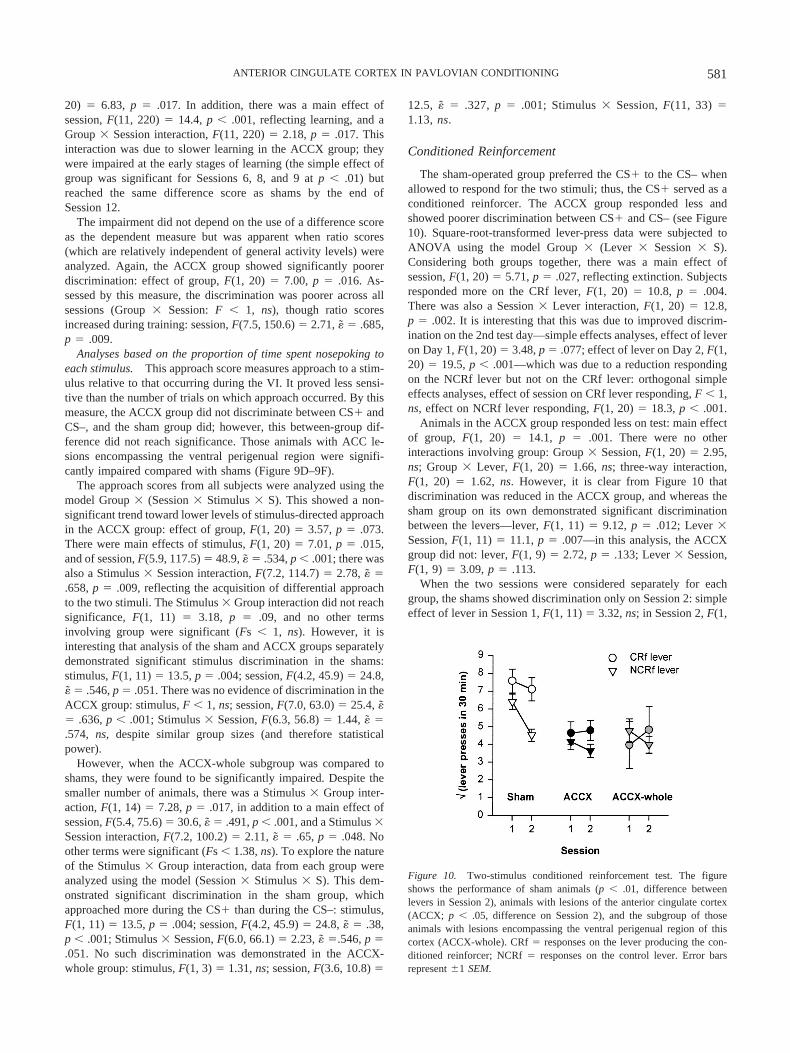
Conditioned Reinforcement
The sham-operated group preferred the CS� to the CS– whenallowed to respond for the two stimuli; thus, the CS� served as aconditioned reinforcer. The ACCX group responded less andshowed poorer discrimination between CS� and CS– (see Figure10). Square-root-transformed lever-press data were subjected toANOVA using the model Group � (Lever � Session � S).Considering both groups together, there was a main effect ofsession, F(1, 20) � 5.71, p � .027, reflecting extinction. Subjectsresponded more on the CRf lever, F(1, 20) � 10.8, p � .004.There was also a Session � Lever interaction, F(1, 20) � 12.8,p � .002. It is interesting that this was due to improved discrim-ination on the 2nd test day—simple effects analyses, effect of leveron Day 1, F(1, 20) � 3.48, p � .077; effect of lever on Day 2, F(1,20) � 19.5, p � .001—which was due to a reduction respondingon the NCRf lever but not on the CRf lever: orthogonal simpleeffects analyses, effect of session on CRf lever responding, F � 1,ns, effect on NCRf lever responding, F(1, 20) � 18.3, p � .001.
Animals in the ACCX group responded less on test: main effectof group, F(1, 20) � 14.1, p � .001. There were no otherinteractions involving group: Group � Session, F(1, 20) � 2.95,ns; Group � Lever, F(1, 20) � 1.66, ns; three-way interaction,F(1, 20) � 1.62, ns. However, it is clear from Figure 10 thatdiscrimination was reduced in the ACCX group, and whereas thesham group on its own demonstrated significant discriminationbetween the levers—lever, F(1, 11) � 9.12, p � .012; Lever �Session, F(1, 11) � 11.1, p � .007—in this analysis, the ACCXgroup did not: lever, F(1, 9) � 2.72, p � .133; Lever � Session,F(1, 9) � 3.09, p � .113.
When the two sessions were considered separately for eachgroup, the shams showed discrimination only on Session 2: simpleeffect of lever in Session 1, F(1, 11) � 3.32, ns; in Session 2, F(1,
Figure 10. Two-stimulus conditioned reinforcement test. The figureshows the performance of sham animals (p � .01, difference betweenlevers in Session 2), animals with lesions of the anterior cingulate cortex(ACCX; p � .05, difference on Session 2), and the subgroup of thoseanimals with lesions encompassing the ventral perigenual region of thiscortex (ACCX-whole). CRf � responses on the lever producing the con-ditioned reinforcer; NCRf � responses on the control lever. Error barsrepresent �1 SEM.
581ANTERIOR CINGULATE CORTEX IN PAVLOVIAN CONDITIONING

11) � 15.0, p � .003. The ACCX group showed a similar pattern:simple effect of lever in Session 1, F � 1, ns; in Session 2, F(1,9) � 6.18, p � .035. Thus, some discrimination was apparent inACC-lesioned subjects, but it was much poorer than in sham-operated controls.
These conclusions were not materially altered by considerationof the ACCX-whole subgroup alone, except that these subjectsshowed a significant Lever � Session interaction, F(1, 3) � 10.7,p � .047. Although this might be interpreted as evidence of leverdiscrimination, Figure 10 shows that this was not the case: Theinteraction was due to a crossover, with the ACCX-whole sub-group responding more on the NCRf lever in Session 2. Consid-ering each session separately, the ACCX-whole subgroup nevershowed discrimination (simple effect of lever in Session 1, F � 1,ns; in Session 2, F � 1, ns).
Summary
ACC-lesioned rats were significantly impaired at acquiring adiscriminated approach response governed by two similar stimuli,only one of which was followed by reward. Like shams, theylearned to approach during the CS�, but they also approachedduring the CS– and exhibited much poorer CS�/CS–discrimination during acquisition. On at least some measures, theyeventually acquired the discrimination but took longer to learn itthan shams. Whereas the sham group responded more for the CS�than the CS– in a test of conditioned reinforcement, the ACCXgroup responded less and did not discriminate to the same degreeas shams.
Discussion
The results of this experiment provide support for Hypothesis 2:that the rat ACC contributes to discriminating similar stimuli onthe basis of their association with reward, though it is not neces-sary for stimulus–reward associations per se. It is highly unlikelythat the lesioned subjects were simply poorer at discriminatingbetween the sensory stimuli (in a manner irrespective of theirassociation with reward): ACC-lesioned rats have been shown tobe normal (Bussey, Muir, et al., 1997) or even improved (Busseyet al., 1996) at tasks requiring left–right discrimination, and thestimuli used in the present task (and in the autoshaping experi-ments) differed in no way except in their location. Similarly,ACC-lesioned rats have previously been shown to succeed inlearning a conditional visual discrimination using stimuli withwhich they failed to learn an eight-pair concurrent discrimination(Bussey, Muir, et al., 1997), again making a perceptual deficit anunlikely explanation. Nor is it plausible that a failure of responsediscrimination can account for the present results, as no responsediscrimination was required in the approach task—the responsesmeasured following the CS� and CS– were identical.
Finally, it is not plausible that the ACC is required simply whentasks become difficult in some general sense. In the context of alesion study, task difficulty can only be defined empirically on thebasis of sham performance (a more difficult condition being one inwhich shams perform worse). Manipulations expected to reduceACC function have been shown to impair the more difficultcondition of a number of tasks, often when multiple stimuli areintroduced to a task (discussed in detail below), but sometimes inother conditions, such as when stimulus unpredictability increases
in a detection task (impaired by infusion of a muscarinic acetyl-choline receptor antagonist into the ACC; J. M. Williams, Mohler,& Givens, 1999). However, ACC lesions do not impair perfor-mance on all tasks when they are made more difficult. For exam-ple, when subjects must choose between a small, immediate re-ward and a large, delayed reward, an increase in the delay to thelarge reward produces a progressive decline in normal subjects’success at obtaining food—that is, the task becomes more difficult.ACC lesions do not impair performance on this task (Cardinal,Pennicott, Sugathapala, Robbins, & Everitt, 2001) and, in sometasks, ACC lesions improve performance (e.g., Bussey et al.,1996). Therefore, task difficulty cannot fully explain the ACC’scontribution to behavior, and a more specific explanation of its rolemust be sought.
In the approach task used here, the CS� and CS– may haveserved as instrumental discriminative stimuli, just as in the one-stimulus version of the task used in Experiment 1. Indeed, it is notobvious that approach to a food alcove located away from thestimulus is in any sense a Pavlovian conditional response; thus,performance on the approach task may have been instrumental.Nevertheless, the CS� predicted food delivery, so it was expectedto enter into Pavlovian association with reward; in confirmation ofthis, the CS� served as a CRf for both sham- and ACC-lesionedrats. (These results demonstrate in a within-subjects design that theCS� was a more effective reinforcer than the CS– in sham-operated animals, eliminating a stimulus-seeking explanation oftheir preference for the CRf lever in this task.) However, discrim-ination was much reduced in ACC-lesioned animals. Their poordiscrimination was not simply attributable to generally low levelsof operant responding (as ACC-lesioned rats acquired a free-operant response normally in Experiment 2), or to failure torespond for conditioned reinforcement (given that they respondednormally for the CRf in Experiment 1). Thus, the specific failureof discrimination affected two kinds of behavior, locomotor ap-proach and instrumental responding.
General Discussion
Contribution of the ACC to Instrumentaland Pavlovian Behavior
Lesions of the ACC have been shown to impair discriminationof reward- or punishment-associated stimuli in Pavlovian tasks(including autoshaping and autonomic conditioning: presentexperiments; Bussey, Everitt, & Robbins, 1997; Parkinson,Willoughby, et al., 2000; Powell et al., 1994), in tasks whosePavlovian or instrumental status is ambiguous (two-stimulus dis-criminated approach task, above; eight-pair concurrent discrimi-nation, Bussey, Muir, et al., 1997; two-way active avoidance,Gabriel, 1993) and in instrumental tasks that depend on Pavlovianassociations (responding for conditioned reinforcement, Experi-ment 3). In tasks where stimulus–reinforcer learning is a disad-vantageous strategy, ACC lesions can improve performance (Bus-sey et al., 1996). At present, the most parsimonious explanation isthat the ACC forms or retrieves stimulus–outcome (Pavlovian)associations that may then influence instrumental behavior, con-sistent with previous suggestions (Bussey et al., 1996; Bussey,Muir, et al., 1997; Gabriel, Foster, et al., 1980).
582 CARDINAL ET AL.

Synthesis: A Suggested Role for the ACC inDisambiguating Stimuli for Its Corticostriatal Circuit
The ACC has been shown to be critical in a wide range ofappetitive and aversive tasks in which two or more similar stimulimust be discriminated on the basis of their association with rein-forcement (in the autoshaping, two-stimulus discriminated ap-proach, and two-stimulus conditioned reinforcement tasks pre-sented here and by Bussey, Everitt, & Robbins, 1997; Bussey,Muir, et al., 1997; Gabriel, Kubota, et al., 1991; Parkinson, Wil-loughby, et al., 2000; Powell et al., 1994). It is unlikely that theseresults reflect an attentional deficit (Muir et al., 1996) or a failureof spatial discrimination (Bussey, Muir, et al., 1996, 1997; Gabriel,Vogt, et al., 1991; Powell et al., 1994). Specifically, ACC-lesionedrats have been shown to be capable of making left–right spatialdiscriminations (of stimuli and responses) in a number of para-digms. Though Ragozzino and Kesner (2001) found a deficit inworking memory for egocentric spatial responses (left or rightturns) following excitotoxic lesions of PrL and pregenual ACC,their subjects were normal at discriminating these responses atzero and short delays. Furthermore, a number of studies haveshown that spatial stimulus discrimination, other forms of spatialworking memory, and rats’ ability to discriminate left and rightresponses are normal following ACC lesions (Aggleton, Neave,Nagle, & Sahgal, 1995; Bussey, Muir, et al., 1996, 1997; Cardinalet al., 2001; Ragozzino, Adams, & Kesner, 1998; Ragozzino &Kesner, 1998).
In addition, the deficit observed in the present study is unlikelyto be perceptual: ACC-lesioned rats can discriminate between twostimuli of different modalities (Experiment 2) and between twovisual stimuli differing in a primary submodality such as color(Bussey, Muir, et al., 1997, Experiment 3). In at least some studies,ACC-lesioned animals have exhibited an early failure to discrim-inate between two CSs but eventually improved or succeededcompletely, implying that the early failure to discriminate was notdue to a primary perceptual deficit (Experiment 3; Gabriel, 1990,1993; Gabriel, Kubota, et al., 1991; Parkinson, Willoughby, et al.,2000). However, the present results demonstrate that ACC-lesioned rats are capable of Pavlovian conditioning in many forms.No deficits are apparent when lesioned subjects are requiredmerely to discriminate between the presence and absence of asingle CS (be it appetitive or aversive), as judged by a wide varietyof response systems (Experiments 1 and 2). Thus, ACC-lesionedrats were unimpaired at a single-stimulus discriminated approachtask, responding for conditioned reinforcement, conditioned freez-ing, and PIT.
On the basis of these data, it is suggested that the ACC contrib-utes to a sensorimotor aspect of conditioning (Parkinson, Cardinal,& Everitt, 2000). Without the ACC, animals can learn a motiva-tional conditioned response to CSs; thus, they perform normally inthe single-stimulus discriminated approach task and exhibit PIT.They can also call up a motivational representation of the uncon-ditioned stimulus (a role attributed to the BLA; Everitt, Cardinal,Hall, Parkinson, & Robbins, 2000); thus, they can learn a newinstrumental response for conditioned reinforcement and acquireconditioned freezing. However, CS specificity of these represen-tations is impaired in ACC-lesioned rats; as a result, tasks thatdepend on stimulus–reinforcer associations when similar stimulimust be discriminated require the ACC. Such tasks include au-toshaping (Bussey, Everitt, & Robbins, 1997; Parkinson, Wil-
loughby, et al., 2000), eight-pair concurrent visual discrimination(Bussey, Muir, et al., 1997), the two-stimulus temporally discrim-inated approach task used in Experiment 3, the active avoidancetask of Gabriel et al. (Gabriel, 1993; Gabriel, Kubota, et al., 1991),and CS discrimination in conditioned bradycardia (Powell et al.,1994). According to this hypothesis, the ACC disambiguates CSsfor the rest of the limbic circuit of which it is part (see Figure 11).
In Experiment 2 (PIT), there was no evidence that ACC-lesioned rats generalized from the CS to the neutral stimulus. Atfirst glance, this appears to be evidence against the discriminationhypothesis. However, the two stimuli were of different sensorymodalities. Generalization between stimuli requires that stimulishare common elements (e.g., Mackintosh, Kaye, & Bennett,1991). In Experiment 2, the stimuli had no elements in common,generalization would not be expected in the first place, and there-fore the loss of a disambiguating structure would not be expectedto impair performance. We hypothesize, based on the presentresults, that ACC lesions would reduce CS�/CS– discriminationin PIT tasks if the CS� and CS– (or neutral stimulus) sharedcommon elements (i.e., were in the same sensory modality andwere similar). Similarly, if the stimuli used for autoshaping werehighly discriminable (did not share common elements), we wouldhypothesize that ACC lesions would not impair autoshaping.
For at least one class of response—locomotor approach—itseems very likely that the ACC influences behavior through theAcb. The ACC projects to the AcbC, which in turn projects tolocomotor control regions of the ventral pallidum; lesions of theAcbC impair both autoshaping (Parkinson, Willoughby, et al.,2000) and single-stimulus discriminated approach (Parkinson,Olmstead, et al., 1999), and a functional connection between theACC and the AcbC is necessary for autoshaping to develop (Par-kinson, Willoughby, et al., 2000). The effects of ACC and AcbClesions on autoshaping differ, however; whereas ACC lesionstypically result in “disinhibited” responding to the CS– (Bussey,Everitt, & Robbins, 1997; Parkinson, Willoughby, et al., 2000),AcbC lesions impair the conditioned approach response itself(Parkinson, Willoughby, et al., 2000), just as they prevent condi-tioned approach to a single CS (Parkinson, Olmstead, et al., 1999).
For tasks in which the ventral striatum is the output structure forbehavior, such as locomotor approach (Parkinson, Cardinal, &Everitt, 2000; Parkinson, Olmstead, et al., 1999; Parkinson, Wil-loughby, et al., 2000), the extra conditioning circuitry that theACC is suggested to embody (see Figure 11) may be a necessaryrefinement, as the striatum is itself anatomically capable only ofdiscriminating among linearly separable cortical inputs (Wickens& Kotter, 1995, p. 206); on its own, the striatum should thereforebe unable to perform an exclusive-or (XOR) discrimination (A�,B�, AB–) and may need cortical assistance for this. Furthermore,discrimination of two linearly separable input patterns A and AB,where A3reward and AB30, requires an inhibitory projectionfrom Unit B. As the direct cortical inputs to the striatum are allglutamatergic (excitatory), the striatum might be thought unable tosolve even this discrimination. However, the different corticalafferents to the Acb have been shown to gate each other’s gluta-matergic inputs (Cools, van den Bos, Ploeger, & Ellenbroek, 1991;Floresco, Yang, Phillips, & Blaha, 1998; Pennartz & Kitai, 1991);thus, the ACC may operate to control the input of affectiveinformation to the striatum from other structures, in order to directmotivational responses toward appropriate environmental stimuli.This hypothesis therefore predicts that ACC-lesioned rats would
583ANTERIOR CINGULATE CORTEX IN PAVLOVIAN CONDITIONING

be impaired at stimulus–reinforcer association tasks involvingconfigural or XOR discriminations.
Note that this account of ACC function does not suggest aprimary sensory or perceptual role (ACC-lesioned rats may makethe sensory discrimination) but a more specific function—theretrieval of affective information associated with specific stimuli,allowing the generation of appropriate affective responses to stim-uli (see also Turken & Swick, 1999). The concept that even earlysensory representations may be neurally dissociated on the basis ofthe response for which the representation is used is not new(Goodale & Milner, 1992); from this perspective, the ACC may becritical for discriminating stimuli that share common elements forthe purposes of stimulus–reinforcer associations but not for otherperceptual processes. ACC-lesioned animals would be able todiscriminate a CS� from a CS– perceptually but would be un-aware as to the correct stimulus toward which appropriate affectiveresponses should be made.
Comparison With Other Interventional Studies in Rodents
The hypothesis of ACC function developed above may berelated to studies using very different paradigms. For example,Meunier, Jaffard, and Destrade (1991) have shown that mice withACC lesions are able to learn a spatial T maze discriminationnormally but are impaired when the discrimination is repeatedlyreversed. More specifically, Meunier et al. found that ACC lesionsdid not impair performance on the first, slowly learned reversal.However, sham performance improved over the course of subse-quent reversal training, whereas that of ACC-lesioned mice didnot. Thus, the presence of the ACC may confer on normal mice anability to respond rapidly and flexibly to an environment withchanging stimulus–reinforcement relationships, withholding re-sponses to unrewarded stimuli. This is consistent with electrophys-
iological evidence that ACC exhibits reversal of discriminatedneuronal activity following behavioral reversal training in rabbits(see Gabriel, 1990, p. 475) and primates (Nishijo et al., 1997).Directly comparable studies using rats are not available. AlthoughBussey, Muir, et al. (1997) found that ACC-lesioned rats werecapable of learning a one-pair visual discrimination and wereunimpaired in reversal testing, Bussey, Muir, et al. (1997) did notobserve a serial reversal learning effect (an improvement in rever-sal speed) in shams over the course of three reversals. As shamperformance did not improve in this paradigm, the lack of an effectof ACC lesions is consistent with Meunier et al. (1991). Interven-tional studies using rats have revealed other features of the phe-notype of ACC lesions that are not all easy to encompass withinthe hypothesis outlined above. In particular, they emphasize dis-inhibition and overresponding in ACC-lesioned rats. Weissenbornet al. (1997) studied the acquisition of responding for intravenouscocaine under second-order schedules of reinforcement. ACC-lesioned rats exhibited greater locomotor activity (both spontane-ous and cocaine-induced), they were more likely to self-administerexcessive amounts of cocaine during acquisition, and while theirdose–response curve was normal on a fixed-ratio 1 schedule, theyresponded at high rates throughout the fixed-interval phase of thesecond-order schedule, exhibiting an attenuated fixed-interval“scallop.” Weissenborn et al. related this to Bussey et al.’s (1997)stimulus–reinforcer hypothesis of ACC function by suggesting thatthe rats had failed to learn the significance of the cocaine-associated stimulus that normally maintains responding on thisschedule. Such hyperactivity was not found in the present series ofexperiments; as discussed above, this may have resulted fromdifferences in lesion site, as the most anterior injection in thepresent experiments was 0.5 mm anterior to that of Weissenborn etal. Another factor to be considered in Weissenborn et al.’s exper-
Figure 11. Disambiguation of stimuli, applied to autoshaping. In this example, the CS� is a white rectangleon the left and the CS– is an identical stimulus on the right. Autoshaping requires the central nucleus of theamygdala, nucleus accumbens core, and nucleus accumbens dopamine (see Cardinal et al., 2002; Parkinson,Cardinal, & Everitt, 2000). In the absence of discriminated activity in the ACC, animals generalize from the CS�to the CS–, impairing their behavioral discrimination in a disinhibited fashion. However, the animals stilldiscriminate between the presence and the absence of the CS�. From “Limbic Cortical-Ventral Striatal SystemsUnderlying Appetitive Conditioning,” by J. A. Parkinson, R. N. Cardinal, and B. J. Everitt, 2000, Progress inBrain Research, 126, p. 272. Copyright 2000 by Elsevier Science. Reprinted with permission.
584 CARDINAL ET AL.

iments was chronic cocaine experience, which might interact withthe effects of ACC lesions.
Muir et al. (1996) studied a five-choice serial reaction time task(5CSRTT) in which rats must wait for the presentation of one offive brief visual stimuli and then respond at the location of thestimulus to gain reward. Muir et al. found that ACC lesions had noeffect on the accuracy of visual attentional performance, either atbaseline or with superimposed attentional manipulations (varyingthe stimulus duration or the ITI, or interpolating bursts of whitenoise). However, the lesions increased the number of premature,anticipatory responses (in which the animal responds before astimulus has been presented), increased the number of persevera-tive responses (in which the animal responds several times to thelocation where a stimulus was recently presented), and decreasedthe number of errors of omission. The same animals performednormally on a test of passive avoidance, in which electric shock isdelivered in one half of a two-chamber apparatus and the subjectsubsequently avoids the dangerous chamber. Clearly, these resultsmay be explained in terms of disinhibited or impulsive motorresponding. The results of Muir et al. suggest that the ACC-lesioned rats were unable to withhold responding to locationswhere rewarded stimuli were intermittently presented. However,there was no evidence of such a deficit in the present series ofexperiments; locomotor hyperactivity was not apparent, no test offree-operant responding demonstrated hyperactivity, and ACC-lesioned rats did not overrespond to the location of a rewarded CSwhen that CS was not present. Differences in lesion site may partlybe responsible for these discrepancies—the present lesions weremore anterior than those used by Muir et al., and recent resultssuggest that ACC lesions centered on the perigenual region, sim-ilar to those used in the present experiments, do not producedeficits on the 5CSRTT (Christakou, 2001). It should be noted thatthe psychological basis of premature responding in the 5CSRTT isnot well understood; however, it is not clear that these results canbe reconciled in terms of a single deficit. At present, the effects ofACC lesions on explicit tests of motor impulsivity (see Evenden,1999) are not known.
Other studies of the rat ACC have frequently concentrated onthe region directly superior to PrL, an area that was not the focusof the present experiments (see Figure 1). For example, Delatourand Gisquet-Verrier (2001) emphasized the role of this moreanterior region of pregenual Cg1 (sometimes called dorsal medialprefrontal cortex) in behavioral sequencing. Despite the differ-ences in location, there are some commonalities among findings.For example, such lesions have minimal effects on rats’ spatialdiscrimination or working memory (Neave, Lloyd, Sahgal, &Aggleton, 1994; Ragozzino et al., 1998) or their ability to switchstrategies between the use of visual and spatial cues (Ragozzino,Wilcox, Raso, & Kesner, 1999), yet they produce severe impair-ments in a number of radial maze tasks (Seamans, Floresco, &Phillips, 1995): Rats with reversible (lidocaine) lesions of the ACCpreferentially revisit previously baited arms. This last deficit hasclear analogies with the disinhibited, perseverative behavior ob-served in the 5CSRTT by Muir et al. (1996) but might also beexplicable in terms of a failure to inhibit responding to unrewardedstimuli (maze arms) in a situation in which there are many stimuli,differentially associated with reward, and in which the rewardedstimulus changes rapidly. Seamans et al. (1995, p. 1071) describedthe ACC as providing response flexibility by suppressing the effect
of simple stimulus–reward associations on behavior, an interpre-tation clearly compatible with the present results.
Conclusions
The results presented here provide support for the view that theACC contributes to Pavlovian conditioning. However, it makes aspecific contribution to this process: The ACC appears to discrim-inate similar stimuli (stimuli that share common elements) on thebasis of their differential association with reinforcement, providingstimulus specificity to more basic Pavlovian conditioning pro-cesses that do not require the ACC. These suggestions are consis-tent with modern theoretical analyses that view Pavlovian condi-tioning as resulting from multiple interacting but dissociableprocesses of learning and associative memory.
References
Aggleton, J. P., Neave, N., Nagle, S., & Sahgal, A. (1995). A comparisonof the effects of medial prefrontal, cingulate cortex, and cingulumbundle lesions on tests of spatial memory: Evidence of a double disso-ciation between frontal and cingulum bundle contributions. Journal ofNeuroscience, 15, 7270–7281.
Balleine, B. (1994). Asymmetrical interactions between thirst and hungerin Pavlovian–instrumental transfer. Quarterly Journal of ExperimentalPsychology: Comparative and Physiological Psychology, 47(B), 211–231.
Brog, J. S., Salyapongse, A., Deutch, A. Y., & Zahm, D. S. (1993). Thepatterns of afferent innervation of the core and shell in the “accumbens”part of the rat ventral striatum: Immunohistochemical detection of ret-rogradely transported Fluoro-Gold. Journal of Comparative Neurology,338, 255–78.
Brown, P. L., & Jenkins, H. M. (1968). Auto-shaping of the pigeon’skeypeck. Journal of the Experimental Analysis of Behavior, 11, 1–8.
Browne, M. P. (1976). The role of primary reinforcement and overtmovements in auto-shaping in the pigeon. Animal Learning & Behav-ior, 4, 287–292.
Buchanan, S. L., & Powell, D. A. (1982). Cingulate cortex: Its role inPavlovian conditioning. Journal of Comparative and Physiological Psy-chology, 96, 755–774.
Burns, L. H., Robbins, T. W., & Everitt, B. J. (1993). Differential effectsof excitotoxic lesions of the basolateral amygdala, ventral subiculum andmedial prefrontal cortex on responding with conditioned reinforcementand locomotor activity potentiated by intra-accumbens infusions ofD-amphetamine. Behavioural Brain Research, 55, 167–183.
Bussey, T. J., Everitt, B. J., & Robbins, T. W. (1997). Dissociable effectsof cingulate and medial frontal cortex lesions on stimulus–reward learn-ing using a novel Pavlovian autoshaping procedure for the rat: Implica-tions for the neurobiology of emotion. Behavioral Neuroscience, 111,908–919.
Bussey, T. J., Muir, J. L., Everitt, B. J., & Robbins, T. W. (1996).Dissociable effects of anterior and posterior cingulate cortex lesions onthe acquisition of a conditional visual discrimination: Facilitation ofearly learning vs. impairment of late learning. Behavioural Brain Re-search, 82, 45–56.
Bussey, T. J., Muir, J. L., Everitt, B. J., & Robbins, T. W. (1997). Tripledissociation of anterior cingulate, posterior cingulate, and medial frontalcortices on visual discrimination tasks using a touchscreen testing pro-cedure for the rat. Behavioral Neuroscience, 111, 920–936.
Cador, M., Bjijou, Y., & Stinus, L. (1995). Evidence of a completeindependence of the neurobiological substrates for the induction andexpression of behavioral sensitization to amphetamine. Neuro-science, 65, 385–395.
Cador, M., Robbins, T. W., & Everitt, B. J. (1989). Involvement of theamygdala in stimulus–reward associations: Interaction with the ventralstriatum. Neuroscience, 30, 77–86.
Cador, M., Taylor, J. R., & Robbins, T. W. (1991). Potentiation of the
585ANTERIOR CINGULATE CORTEX IN PAVLOVIAN CONDITIONING

effects of reward-related stimuli by dopaminergic-dependent mecha-nisms in the nucleus accumbens. Psychopharmacology, 104, 377–385.
Cardinal, R. N., Lachenal, G., Parkinson, J. A., Robbins, T. W., & Everitt,B. J. (2000). Effects of anterior cingulate cortex lesions on respondingfor conditioned reinforcement, discrete fear conditioning, autoshapingperformance and Pavlovian–instrumental transfer [Abstract]. EuropeanJournal of Neuroscience, 12(Suppl. 11), 88.
Cardinal, R. N., Parkinson, J. A., Djafari Marbini, H., Toner, A. J.,Robbins, T. W., & Everitt, B. J. (2000). Role of the anterior cingulatecortex in the control over behaviour by Pavlovian conditioned stimuli inrats [Abstract]. Society for Neuroscience Abstracts, 26, 980.
Cardinal, R. N., Parkinson, J. A., Lachenal, G., Halkerston, K. M., Ru-darakanchana, N., Hall, J., et al. (2002). Effects of lesions of the nucleusaccumbens core, anterior cingulate cortex, and central nucleus of theamygdala on autoshaping performance in rats. Behavioral Neuroscience,116, 553–567.
Cardinal, R. N., Pennicott, D. R., Sugathapala, C. L., Robbins, T. W., &Everitt, B. J. (2001, June 29). Impulsive choice induced in rats by lesionsof the nucleus accumbens core. Science, 292, 2499–2501.
Christakou, A. (2001). Prefrontal cortico-striatal systems underlying ex-ecutive attention. Unpublished doctoral dissertation, University of Cam-bridge, Cambridge, United Kingdom.
Cools, A. R., van den Bos, R., Ploeger, G., & Ellenbroek, B. A. (1991).Gating function of noradrenaline in the ventral striatum: Its role inbehavioural responses to environmental and pharmacological chal-lenges. In P. Willner & J. Scheel-Kruger (Eds.), The mesolimbic dopa-mine system: From motivation to action (pp. 139–171). Chichester,England: Wiley.
Delatour, B., & Gisquet-Verrier, P. (2001). Involvement of the dorsalanterior cingulate cortex in temporal behavioral sequencing: Subregionalanalysis of the medial prefrontal cortex in rat. Behavioural Brain Re-search, 126, 105–114.
Dickinson, A. (1994). Instrumental conditioning. In N. J. Mackintosh(Ed.), Animal learning and cognition (pp. 45–79). San Diego, CA:Academic Press.
Dickinson, A., Smith, J., & Mirenowicz, J. (2000). Dissociation of Pav-lovian and instrumental incentive learning under dopamine antagonists.Behavioral Neuroscience, 114, 468–483.
Estes, W. K. (1948). Discriminative conditioning: II. Effects of a Pavlovianconditioned stimulus upon a subsequently established operant response.Journal of Experimental Psychology, 38, 173–177.
Evenden, J. L. (1999). Varieties of impulsivity. Psychopharmacology, 146,348–361.
Everitt, B. J., Cardinal, R. N., Hall, J., Parkinson, J. A., & Robbins, T. W.(2000). Differential involvement of amygdala subsystems in appetitiveconditioning and drug addiction. In J. P. Aggleton (Ed.), The amygdala:A functional analysis (2nd ed., pp. 353–390). New York: Oxford Uni-versity Press.
Fisk, G. D., & Wyss, J. M. (1997). Pressor and depressor sites areintermingled in the cingulate cortex of the rat. Brain Research, 754(1–2),204–212.
Floresco, S. B., Yang, C. R., Phillips, A. G., & Blaha, C. D. (1998).Basolateral amygdala stimulation evokes glutamate receptor-dependentdopamine efflux in the nucleus accumbens of the anaesthetized rat.European Journal of Neuroscience, 10, 1241–1251.
Gabriel, M. (1990). Functions of anterior and posterior cingulate cortexduring avoidance learning in rabbits. Progress in Brain Research, 85,467–482.
Gabriel, M. (1993). Discriminative avoidance learning: A model system. InB. A. Vogt & M. Gabriel (Eds.), Neurobiology of cingulate cortex andlimbic thalamus: A comprehensive handbook (pp. 478–523). Boston:Birkhauser.
Gabriel, M., Foster, K., Orona, E., Saltwick, S., & Stanton, M. (1980).Neuronal activity of cingulate cortex, anteroventral thalamus, and hip-pocampal formation in discriminative conditioning: Encoding and ex-
traction of the significance of conditional stimuli. In J. M. Sprague &A. N. Epstein (Eds.), Progress in psychobiology and physiologicalpsychology (Vol. 9, pp. 125–231). New York: Academic Press.
Gabriel, M., Kang, E., Poremba, A., Kubota, Y., Allen, M. T., Miller, D. P.,& Steinmetz, J. E. (1996). Neural substrates of discriminative avoidancelearning and classical eyeblink conditioning in rabbits: A double disso-ciation. Behavioural Brain Research, 82, 23–30.
Gabriel, M., Kubota, Y., Sparenborg, S., Straube, K., & Vogt, B. A. (1991).Effects of cingulate cortical lesions on avoidance learning and training-induced unit activity in rabbits. Experimental Brain Research, 86, 585–600.
Gabriel, M., & Orona, E. (1982). Parallel and serial processes of theprefrontal and cingulate cortical systems during behavioral learning.Brain Research Bulletin, 8, 781–785.
Gabriel, M., Orona, E., Foster, K., & Lambert, R. W. (1980). Cingulatecortical and anterior thalamic neuronal correlates of reversal learning inrabbits. Journal of Comparative and Physiological Psychology, 94,1087–1110.
Gabriel, M., Vogt, B. A., Kubota, Y., Poremba, A., & Kang, E. (1991).Training-stage related neuronal plasticity in limbic thalamus and cingu-late cortex during learning: A possible key to mnemonic retrieval.Behavioural Brain Research, 46, 175–185.
Goodale, M. A., & Milner, A. D. (1992). Separate visual pathways forperception and action. Trends in Neurosciences, 15, 20–25.
Hall, J. (1999). The roles of the amygdala and hippocampus in Pavlovianconditioning. Unpublished doctoral dissertation, University of Cam-bridge, Cambridge, United Kingdom.
Hall, J., Thomas, K. L., & Everitt, B. J. (2001). Fear memory retrievalinduces CREB phosphorylation and Fos expression within the amyg-dala. European Journal of Neuroscience, 13(7), 1453–1458.
Heimer, L., Zahm, D. S., & Alheid, G. F. (1995). Basal ganglia. In G.Paxinos (Ed.), The rat nervous system (pp. 579–628). London: Aca-demic Press.
Howell, D. C. (1997). Statistical methods for psychology (4th ed.). Bel-mont, CA: Wadsworth.
Hsu, M. M., & Shyu, B. C. (1997). Electrophysiological study of theconnection between medial thalamus and anterior cingulate cortex in therat. NeuroReport, 8, 2701–2707.
Huynh, H., & Feldt, L. S. (1970). Conditions under which mean squareratios in repeated measures designs have exact F-distributions. Journalof the American Statistical Association, 65, 1582–1589.
Jenkins, H. M., & Moore, B. R. (1973). The form of the auto-shapedresponse with food or water reinforcers. Journal of the ExperimentalAnalysis of Behavior, 20, 163–181.
Keppel, G. (1982). Design and analysis: A researcher’s handbook (2nded.). London: Prentice Hall.
Keppel, G. (1991). Design and analysis: A researcher’s handbook (3rded.). London: Prentice Hall.
Kish, G. B. (1966). Studies of sensory reinforcement. In W. K. Honig(Ed.), Operant behavior: Areas of research and application (pp. 109–159). New York: Appleton-Century-Crofts.
Lovibond, P. F. (1983). Facilitation of instrumental behavior by a Pavlov-ian appetitive conditioned stimulus. Journal of Experimental Psychol-ogy: Animal Behavior Processes, 9, 225–247.
Mackintosh, N. J. (1974). The psychology of animal learning. London:Academic Press.
Mackintosh, N. J. (1983). Conditioning and associative learning. Oxford,UK: Oxford University Press.
Mackintosh, N. J., Kaye, H., & Bennett, C. H. (1991). Perceptual learningin flavour aversion conditioning. Quarterly Journal of ExperimentalPsychology: Comparative and Physiological Psychology, 43(B), 297–322.
McGeorge, A. J., & Faull, R. L. M. (1989). The organization of theprojection from the cerebral cortex to the striatum in the rat. Neuro-science, 29, 503–537.
586 CARDINAL ET AL.

Meunier, M., Jaffard, R., & Destrade, C. (1991). Differential involvementof anterior and posterior cingulate cortices in spatial discriminativelearning in a T-maze in mice. Behavioural Brain Research, 44, 133–143.
Muir, J. L., Everitt, B. J., & Robbins, T. W. (1996). The cerebral cortex ofthe rat and visual attentional function: Dissociable effects of mediofron-tal, cingulate, anterior dorsolateral, and parietal cortex lesions on afive-choice serial reaction time task. Cerebral Cortex, 6, 470–481.
Neafsey, E. J., Terreberry, K. M., Hurley, K. M., Ruit, K. G., & Frysztak,R. J. (1993). Anterior cingulate cortex in rodents: Connections, visceralcontrol functions, and implications for emotion. In B. A. Vogt & M.Gabriel (Eds.), Neurobiology of cingulate cortex and limbic thalamus: Acomprehensive handbook (pp. 206–233). Boston: Birkhauser.
Neave, N., Lloyd, S., Sahgal, A., & Aggleton, J. P. (1994). Lack of effectof lesions in the anterior cingulate cortex and retrosplenial cortex oncertain tests of spatial memory in the rat. Behavioural Brain Re-search, 65, 89–101.
Nishijo, H., Yamamoto, Y., Ono, T., Uwano, T., Yamashita, J., & Ya-mashima, T. (1997). Single neuron responses in the monkey anteriorcingulate cortex during visual discrimination. Neuroscience Letters,227(2), 79–82.
Oakley, D. A., & Russell, I. S. (1972). Neocortical lesions and Pavlovianconditioning. Physiology & Behavior, 8, 915–922.
Oakley, D. A., & Russell, I. S. (1975). Role of cortex on Pavloviandiscrimination learning. Physiology & Behavior, 15, 315–321.
Oakley, D. A., & Russell, I. S. (1976). Subcortical nature of Pavloviandifferentiation in the rabbit. Physiology & Behavior, 17, 947–954.
Parkinson, J. A. (1998). Limbic corticostriatal circuitry underlying Pav-lovian associative learning. Unpublished doctoral dissertation, Univer-sity of Cambridge, Cambridge, United Kingdom.
Parkinson, J. A., Cardinal, R. N., & Everitt, B. J. (2000). Limbic cortical-ventral striatal systems underlying appetitive conditioning. Progress inBrain Research, 126, 263–285.
Parkinson, J. A., & Everitt, B. J. (1998). [Projections from anterior cingu-late cortex.] Unpublished raw data.
Parkinson, J. A., Olmstead, M. C., Burns, L. H., Robbins, T. W., & Everitt,B. J. (1999). Dissociation in effects of lesions of the nucleus accumbenscore and shell on appetitive Pavlovian approach behavior and the po-tentiation of conditioned reinforcement and locomotor activity byd-amphetamine. Journal of Neuroscience, 19, 2401–2411.
Parkinson, J. A., Robbins, T. W., & Everitt, B. J. (1996). Lesions of thenucleus accumbens core, but not basolateral amygdala or subiculum,disrupt stimulus–reward learning in a novel autoshaping procedure [Ab-stract]. Society for Neuroscience Abstracts, 22, 1118.
Parkinson, J. A., Robbins, T. W., & Everitt, B. J. (1999). Selectiveexcitotoxic lesions of the nucleus accumbens core and shell differen-tially affect aversive Pavlovian conditioning to discrete and contextualcues. Psychobiology, 27, 256–266.
Parkinson, J. A., Robbins, T. W., & Everitt, B. J. (2000). Dissociable rolesof the central and basolateral amygdala in appetitive emotional learning.European Journal of Neuroscience, 12(1), 405–413.
Parkinson, J. A., Willoughby, P. J., Robbins, T. W., & Everitt, B. J. (2000).Disconnection of the anterior cingulate cortex and nucleus accumbenscore impairs Pavlovian approach behavior: Further evidence for limbiccortical–ventral striatopallidal systems. Behavioral Neuroscience, 114,42–63.
Paxinos, G., & Watson, C. (1998). The rat brain in stereotaxic coordinates(4th ed.). San Diego, CA: Academic Press.
Pennartz, C. M., & Kitai, S. T. (1991). Hippocampal inputs to identifiedneurons in an in vitro slice preparation of the rat nucleus accumbens:Evidence for feed-forward inhibition. Journal of Neuroscience, 11,2838–2847.
Powell, D. A., Watson, K., & Maxwell, B. (1994). Involvement of subdi-visions of the medial prefrontal cortex in learned cardiac adjustments inrabbits. Behavioral Neuroscience, 108, 294–307.
Ragozzino, M. E., Adams, S., & Kesner, R. P. (1998). Differential involve-
ment of the dorsal anterior cingulate and prelimbic–infralimbic areas ofthe rodent prefrontal cortex in spatial working memory. BehavioralNeuroscience, 112, 293–303.
Ragozzino, M. E., & Kesner, R. P. (1998). The effects of muscariniccholinergic receptor blockade in the rat anterior cingulate and prelimbic/infralimbic cortices on spatial working memory. Neurobiology of Learn-ing and Memory, 69, 241–257.
Ragozzino, M. E., & Kesner, R. P. (2001). The role of rat dorsomedialprefrontal cortex in working memory for egocentric responses. Neuro-science Letters, 308, 145–148.
Ragozzino, M. E., Wilcox, C., Raso, M., & Kesner, R. P. (1999). Involve-ment of rodent prefrontal cortex subregions in strategy switching. Be-havioral Neuroscience, 113, 32–41.
Robbins, T. W. (1976, November 4). Relationship between reward-enhancing and stereotypical effects of psychomotor stimulant drugs.Nature, 264, 57–59.
Robbins, T. W., & Koob, G. F. (1978). Pipradrol enhances reinforcingproperties of stimuli paired with brain stimulation. Pharmacology Bio-chemistry and Behavior, 8, 219–222.
Robledo, P., Robbins, T. W., & Everitt, B. J. (1996). Effects of excitotoxiclesions of the central amygdaloid nucleus on the potentiation of reward-related stimuli by intra-accumbens amphetamine. Behavioral Neuro-science, 110, 981–990.
Seamans, J. K., Floresco, S. B., & Phillips, A. G. (1995). Functionaldifferences between the prelimbic and anterior cingulate regions of therat prefrontal cortex. Behavioral Neuroscience, 109, 1063–1073.
Steinmetz, J. E. (2000). Brain substrates of classical eyeblink conditioning:A highly localized but also distributed system. Behavioural Brain Re-search, 110, 13–24.
Steinmetz, J. E., Sears, L. L., Gabriel, M., Kubota, Y., & Poremba, A.(1991). Cerebellar interpositus nucleus lesions disrupt classicalnictitating-membrane conditioning but not discriminative avoidancelearning in rabbits. Behavioural Brain Research, 45, 71–80.
Taylor, J. R., & Robbins, T. W. (1984). Enhanced behavioural control byconditioned reinforcers following microinjections of d-amphetamineinto the nucleus accumbens. Psychopharmacology, 84, 405–412.
Thompson, R. F., Swain, R., Clark, R., & Shinkman, P. (2000). Intracer-ebellar conditioning: Brogden and Gantt revisited. Behavioural BrainResearch, 110, 3–11.
Turken, A. U., & Swick, D. (1999). Response selection in the humananterior cingulate cortex. Nature Neuroscience, 2, 920–924.
Weissenborn, R., Robbins, T. W., & Everitt, B. J. (1997). Effects of medialprefrontal or anterior cingulate cortex lesions on responding for cocaineunder fixed-ratio and second-order schedules of reinforcement in rats.Psychopharmacology, 134, 242–257.
Wickens, J., & Kotter, R. (1995). Cellular models of reinforcement. In J. C.Houk, J. L. Davis, & D. G. Beiser (Eds.), Models of informationprocessing in the basal ganglia (pp. 187–214). Cambridge, MA: MITPress.
Williams, B. A. (1994). Conditioned reinforcement: Neglected or out-moded explanatory construct? Psychonomic Bulletin & Review, 1, 457–475.
Williams, D. R., & Williams, H. (1969). Auto-maintenance in the pigeon:Sustained pecking despite contingent nonreinforcement. Journal of theExperimental Analysis of Behavior, 12, 511–520.
Williams, J. M., Mohler, E. G., & Givens, B. (1999). The role of the medialprefrontal cortex in attention: Altering predictability of task difficulty.Psychobiology, 27, 462–469.
Zahm, D. S., & Brog, J. S. (1992). On the significance of subterritories inthe “accumbens” part of the rat ventral striatum. Neuroscience, 50,751–767.
Received August 6, 2002Revision received November 7, 2002
Accepted December 11, 2002 �
587ANTERIOR CINGULATE CORTEX IN PAVLOVIAN CONDITIONING