role of cenh3/cenp-a in the regulation of centromere...
TRANSCRIPT

UNIVERSIDADE DE LISBOA
FACULDADE DE CIÊNCIAS
DEPARTAMENTO DE BIOLOGIA VEGETAL
Role of CenH3/CENP-A in the regulation of
centromere function and chromosome segregation
Márcia Sofia Ribeiro Lamy
Mestrado em Biologia Molecular e Genética
2010
2
UNIVERSIDADE DE LISBOA
FACULDADE DE CIÊNCIAS
DEPARTAMENTO DE BIOLOGIA VEGETAL
Role of CenH3/CENP-A in the regulation of
centromere function and chromosome segregation
Márcia Sofia Ribeiro Lamy
Dissertação de mestrado orientada por:
Doutor Ferran Azorin – Institute for Research in Biomedicine (IRB) and Institute of
molecular Biology of Barcelona (CSIC), Barcelona
Doutor Júlio Duarte – Faculdade de Ciências da Universidade de Lisboa, Lisboa
Mestrado em Biologia Molecular e Genética
2010
3
Acknowledgments
I start this by thanking to all the persons that somehow contribute to my stay in
Barcelona. It´s been without doubts the best period of my life not only by the persons
that I meet but also by the things that i learn. It has been a great journey where I really
know myself which enable me to grown.
I thanks in first place to Mr. Ferran Azorín that accept me to do part of this
project in his laboratory of Development biology and by it´s always disponibility to take
doubts and for always have some time to talk and discuss in an informal way with all of
his students . Second but not least important a big thank you to the person that has been
directly working with me and that teach me everything since the simple places of the
things (that at the beginning I think that the poor one have to had a big patience ;) )
until the more complex parts of the work, thanks a lot Olga Moreno.
After I want to thanks of course to my ―company de poiata‖ Marta Orcheta not
only a super funny person help me a lot to understand more and more the Català
(because for the ones who know her knows that it´s a person that likes a lot of talking)
and for the good talks that we had. Thanks to Sònia Medina my company of running
and a person always with a smile on is face I always remember it´s joy in living. To
Olivera for the kindness, help and teaching Salsa in the free time, to Gaylord company
of work, football, beach for his cooking‘s and of course for his precious help in the
confocal. I would like to thank of course to Sergi the capitan of our football team not
only for it´s support but for the first day take me has one more of the team. To Salva the
one who like´s karaoke has much as I am. To Martí although for few months but it´s a
very nice person always with a smile in is face. To Joan and Esther for teach me to
―jirar‖ larvas very difficult in the beginning but with some help it become more easier.
Also of course to Roman to share the same interest in running like me, to Tomás and
Katrin that arrived when few months left me to stay but persons that I like to much not
only by their great amiability to help in any time, for the important conversations and
for being always honest (the verdito some times to honest ;) ). To all the Martas, Carles,
Anne, Alicia and to Gemma (our ―mama‖ in the lab but with an enviable youth). Also to
Jordi (our marker in the football team), Lluïsa, Mònica y Josep for always being nice
and friendly persons!! And in general a BIG THANK for all the persons that I met in

4
the Parc Científic always helpful and available to make that every day could be more
funny and interesting then the other.
A big thanks also to my family, Mom, Dad, Brother ;) for the support and to my
cousin Denise for receiving me and take care of me in this year!
Thanks to Stephanie and Maria João for also sharing with me this experience of
doing the last year of master in strange country!
Of course a big THANKS to all the wonderful persons that I met in this beautiful
city and that always will remember as a magic year!

5
Resumo
Os centrómeros são estruturas especializadas do cromossoma, com uma
composição de cromatina única, que desempenham um papel chave nos processos de
segregação dos cromossomas durante a mitose e meiose para além de determinarem o
local de formação do cinetócoro. A formação e propagação do centrómero é um
exemplo claro de um processo epigenético, isto é, estruturas proteicas são formadas ou
associadas ao DNA e depois estavelmente propagadas através de numerosas divisões
celulares numa maneira que é independente da sequência de DNA. Mais
especificamente o cinetócoro é uma estrutura fundamental formada no final da prófase
na região centromérica que é responsável por mediar a ligação dos cromossomas as
fibras do fuso, monitorizar a ligação bipolar e puxar os cromossomas para os pólos
durante a anáfase. O centrómero apesar de já ter sido identificado há muito tempo como
a primeira constrição dos cromossomas metafásicos, a sua caracterização molecular
ainda permanece um pouco inconclusiva devido ao seu invulgar enriquecimento em
sequencias DNA satélite (a excepção dos centrómeros das leveduras gemulação). Para
além do seu valor biológico intrínseco, o estudo e a análise do centrómero é também
importante sob uma perspectiva biomédica uma vez que alterações na função
centromérica levam frequentemente às aneuploidias (perda ou ganho de cromossomas)
que muitas vezes levam a letalidade.
Os centrómeros Eucarióticos podem ser classificados como monocêntricos ou
holocêntricos. Os monocêntricos caracterizam-se por serem localizados, isto é, a sua
formação é restrita a um específico loccus cromossomal, já nos holocêntricos,o
centrómero forma-se difusamente ao longo de todo o cromossoma. Neste trabalho,
pretendeu-se aprofundar o conhecimento acerca da estrutura e função da cromatina
centromérica uma vez que tem um papel fundamental na segregação cromossomica,
usando para este estudo como organismo modelo a mosca Drosophila melanogaster.
Uma das características da cromatina centromérica é a presença de uma específica
variante da histona H3, genericamente designada por CenH3, que substitui a histona
canónica H3.1 nos nucleossomas quer in vivo quer in vitro. O que se verifica é que na
ausência de CenH3, a função centromérica e a localização de outros componentes do
cinetócoro é abolida e pelo contrário experiências em que se procedia a sobrexpressão
da CenH3 humana (CENP-A) isso levava a sua mislocalization para os braços

6
cromossomicos onde recrutava componentes do cinetócoro levando muitas vezes a
formação ectópica de cinetócoros, afectando a progressão no ciclo celular. Em
Drosophila a CenH3 designa-se por Cid (centromere identifier) e é extremamente
divergente da H3 canónica uma vez que apresenta um domínio N-terminal altamente
variável, que pode ir desde os 20 aos 200 aminoácidos, não apresentando quase
nenhuma homologia. O domínio C-terminal de Cid, mais especificamente o HFD, já
apresenta uma significante homologia com a H3 mas apenas 48% de identidade dentro
da filogenia. E portanto é fácil de perceber que estas diferenças tenham levado a uma
rápida evolução das CenH3 o que reflecte a sua especialização funcional. Assim se
identifica esta CenH3 como sendo a marca epigenética que nos permite identificar o
centrómero, uma vez que existe uma falta de conservação no que diz respeito a
sequência de DNA, isto é, não existe uma especifica sequência de DNA que seja
necessária e suficiente para levar a formação do centrómero (excepção nas leveduras de
gemulação). No entanto o exacto mecanismo molecular pelo qual a específica deposição
de CenH3 ocorre nos centrómeros ainda não é bem conhecida, o que se sabe é que no
caso da Drosophila melanogaster, e com base nestas observações enunciadas
anteriormente, a CenH3 funciona ou é fundamental quer estrutural quer funcionalmente
para o cinetócoro mais especificamente para a sua formação o que lhe confere a
capacidade de ser a proteína identificadora da função centromérica e daí o interesse do
estudo dos mecanismos que levam a sua deposição e regulação durante o ciclo celular.
A analise das fibras de cromatina de Drosophila revelaram que a região
centromérica em realidade não esta composta exclusivamente por nucleossomas que
contem a cenH3 senão que há uma distribuição em blocos de nucleossomas com a H3
intercalados com nucleossomas com cenH3 (Blower et al. 2002). Assim os
nucleossomas que contem a CenH3 são a base para uma correcta organização do
cinetócoro e a sua formação é independente da replicação (Henikoff et al. 2001). Pensa-
se que este processo possa iniciar-se com interacções entre o DNA e o Loop1 da região
C-terminal da CenH3-H4 e quando a totalidade do nucleossoma ((H2A-H2B)2(H3-
H4)2) é formado, a região N-terminal da CenH3 interactuara com o DNA de ligação.
A nível do ciclo celular sabe-se que a expressão de CenH3CENP-A
surge no início
da fase S mas os seus níveis só atingem um máximo em G2 sendo que a sua deposição
ocorre no final da mitose (final da telófase) inicio da G1. Sabe-se no entanto que

7
mecanismos adicionais tem de existir para evitar quer a deposição de nucleossomas
contendo CENP-A em locais não centroméricos durante a replicação do DNA e/ou para
remove-los após a replicação do DNA. No caso de Drosophila em embriões não são
observadas as fases gap (G1/G2) mas a deposição de CenH3Cid
também tem lugar
durante a mitose mais concretamente em Anáfase.
Em suma CenH3 é essencial para a função centromérica uma vez que em todos
os eucariotas analisados até a data, a depleção de CenH3 é letal quer a nível da célula
quer a nível do organismo. Para aprofundar o conhecimento acerca desta proteína
vamos verificar qual o mecanismo que regula os seus níveis na célula, através do uso de
mutantes para os genes β2 e/ou β6 do proteossoma 26S (via de degradação mais comum
dos eucariotas) investigou-se o seu papel na possível regulação de Cid via proteossoma.
A caracterização fenotípica sugere que estes genes têm papéis importantes uma vez que
mutantes nestes genes actuam de uma maneira dominante-negativa que interfere com as
funções do proteossoma, assim usando linhas UAS-GAL4 com mutantes nestes genes e
verificando qual a localização de Cid nas células de discos imaginais (pequeno grupo de
células progenitoras tecido-específicas que se mantêm quiescentes durante a
embriogénese e que proliferam durante o desenvolvimento larvar afim de originarem a
maioria das futuras estruturas do individuo adulto) será um dos objectivos a que nos
propomos.
Numa segunda parte do trabalho estamos interessados em perceber que
complexos do ciclo celular estão envolvidos na regulação de Cid, pela adição de uma
cauda de ubiquitina a esta proteína que depois pode então ser reconhecida pelo
proteossoma. Iremos analisar em específico o complexo SCF (Skip, Cullin, F-box), um
complexo multi-proteico E3 ubiquitina ligase que catalisa a ubiquitinação de proteínas
destinadas a degradação proteossomal, visto que já foi testado que a F-box deste
complexo interactua directamente com Cid in vitro. Pela utilização de linhas RNAi
contra as subunidades Skp1 (16983R-III e CG16983) e Cul1 (1877-III e CG1877) deste
complexo iremos analisar a localização de Cid nestes mutantes em neuroblastos (células
progenitoras que dão origem ao sistema nervoso) de larvas de Drosophila.
Também foi realizado um pequeno ensaio experimental de expressão transitória
em células Kc de Drosophila com o objectivo de provar se o CATD (CENP-A targeting

8
domain) de Cid é necessário e suficiente, como nos mamíferos, para determinar a sua
localização centromérica. Com isto podemos aprofundar o conhecimento a cerca das
vias reguladoras de deposição e eliminação desta proteína chave centromérica e
perceber como isso afecta o recrutamento de outras proteínas importantes para a
formação do cinetócoro, para a função do fuso mitótico entre outros.
Palavras – chave: Centrómero, Variante Centromérica da Histona 3 (CenH3),
Cinetócoro, Nucleossomas, Proteossoma

9
Abstract
The centromere is a specialized structure of the chromosome that plays a key role in the
process of chromosome segregation during mitosis and meiosis. Surprisingly, the
centromere appears to represent the most stable form of epigenetic inheritance known to
date since it is maintained not only through mitotic divisions but also through the germ
line. Although the histones are one of the most conserved proteins, one of the
characteristics of the centromeric chromatin is the presence of a specific histone H3
variant (CenH3), which replaces canonical H3.1 in all eukaryotic centromeres. CenH3
appears to dictate centromere identity, being required for the kinetochore formation and
for a correct centromere function. However, the precise molecular mechanisms
accounting for its specific deposition at centromeres are not well understood. In a
previous work had been showed that transiently expressed Cid, the CenH3 of
Drosophila melanogaster, is incorporated throughout chromatin and that this
mislocalization affects cell cycle progression. Therefore, expression of CenH3 must be
tightly regulated during cell cycle to prevent deposition at non centromeric sites and
additional mechanisms must exist to either avoid deposition of CenH3 containing
nucleosomes at non centromeric sites and/or to remove them afterwards. Previously had
been showed how a process of proteasome degradation contributes to the centromeric
localization of Cid by eliminating mislocalized Cid as well as by regulating available
Cid levels. To be recognised by the proteasome Cid requires to be polyubiquitinated , a
function that it´s performed by a sequential action of 3 enzimes : E1, E2 and E3. The
activity of E3 it´s essential since it´s involved in substract recognition, where upon they
become substrates for the proteolytic machine, the proteasome. One of the major E3 are
the SCF (Skp1, Cul1, F-box) complex that will be analysed since one of the F-box
protein of this complex Ppa (partener of paired) interacts directly in vitro with Cid and
this interaction could contribute to the proteolytic degradation of CID.
Key words: Centromere, Centromeric histone H3 variant (CenH3), Kinetochore,
Nucleosome, Proteasome
10
Abbreviations acronyms and symbols
BSA- Bovine Serum Albumin
CaCl2- Calcium chloride
CAF- chromatin assembly factor
CATD – CENP-A targeting domain
CenH3- Centromeric histone H3 variant
CID- Centromer Identifier
C-terminal- carboxyl terminal
DAPI - 4', 6-diamidino-2-phenylindole
DMSO- Dimethyl Sulfoxide
DTS - dominant temperature-sensitive
HFD- Histone Fold Domain
FBS- Fetal Bovine Serum
LRR´s – Leucine Rich repeats
LN2- Liquid nitrogen
H3 Lys4-diMe- dimetylacion of Lysin 4from Histone 3
N-terminal- Amino terminal
PBS- Phosphate buffered saline
Ppa- Partner of Paired
o/n- over night
Rpm- rotations per minute
UAS- Upstream Activating Sequence
YFP- Yellow Fluorescent Protein

11
APPENDIX OF THE FIGURES
Index
Figure I.1 - Types of Eukaryotic centromers Adptaded from Epigenetic Control 14
of Centromere Behavior Karl Ekwall (2007)
Figure I.2 - Schematic view of centromeric DNA in Drosophila 15
Figure I.3 - Representation of the sequence of Cid 15
Figure I.4 - Structure of some of the centromeric variants of H3 (CenH3) 16
Figure I.5 - Schematic view of the 26S proteasome and it´s subunits 18
Figure I.6 - Representation of the SCF complex 18
Figure I.7 - Representation of the interaction saw in vitro between 19
our study protein,Cid, and the F-box protein Ppa.
Fig M.1 - The GAL4 system in Drosophila 21
Figure M.2- UAS constructs driving expression of dominant temperature- sensitive 23
proteasome mutant genes
Fig R.1- Images of immunostaining in neuroblasts from third instar larvae of the 30
effects that RNAi against sub-units of SCF complex induce in the Cid localization.
Figure R.2 Graphics with the result of the qRT-PCR experiments 32
Figure R.3 - Analysis of the effect of mutations in the proteasome in Cid 33
localization in imaginal discs.
Figure R.4 – Analysis of the effect of mutations in the proteasome in Cid 34
localization in imaginal
Figure R.5 – Transitory transfection of Kc cell with four different plasmideos 35
by the method of Effectene:
Figure R.6 - Adapted from Centromere identity maintained by nucleosomes 36
assembled with histone H3 containig the CENP-A Targeting Domain (Black et al 2007)
Figure A.1- Images of immunostaining in neuroblasts from third instar 45
larvae of the effects that RNAi against sub-units of SCF complex induce in the Cid
localization

12
Index
ACKNOWLEDGMENTS 4
RESUMO 5
ABSTRACT 9
ABBREVIATIONS ACRONYMS AND SYMBOLS 10
APPENDIX OF THE FIGURES 11
I- INTRODUCTION
I.1. The centromeric chromatin 13
I.2. The centromeric histone H3 variants 15
I.3. The proteasome pathway 17
II- OBJECTIVES/GOALS 20
III- MATERIALS AND METHODS
III.1.1. Materials
III.1.1.1. Genetic Tools 21
III.1.1.2. Drosophila Strains 22
III.1.1.2.1. RNAi Lines against sub-units of the SCF
III.1.1.2.2. UAS mutant lines against sub-units of proteasome
III.1.1.3 Gal4 drivers 23
III.1.2. Methods
III.1.2.1 Work with Neuroblasts of Drosophila 24
III.1.2.1. Work with Imaginal Discs of Drosophila 25
III.1.2.2. Work with Kc cells of Drosophila 26
III.1.2.3. qRT-PCR 28
IV- RESULTS 29
V- DISCUSSION 37
VI- CONCLUDING REMARKS 40
VII- REFERENCES 41
VIII- ANNEXES 44

13
I- INTRODUCTION
One of the subjects very important nowadays and that should be understood is the
chromosome inheritance since it´s a process that affect the lives of all cell and
organisms. If an incorrect chromosome segregation occurs not only functional germ
cells will be damaged but also the somatic tissues and organs will face problems in
terms of development and diferenciacion (Sullivan et al. 2001). Eukaryotic genomes
are packaged into a nucleoprotein complex known as chromatin, a DNA-protein
complex whose basic repeating unit is the nucleosome, which affects most processes
that occur on DNA (Chang et al. 2009). The nucleosome is composed of 147 base pairs
of DNA wrapped 1.7 times around an octamer of histone proteins (two histones of each
H2A, H2B, H3, and H4) (Vermaak et al. 2002).
I.1. The centromeric chromatin
The centromers are chromosomal regions responsible for many functions in the cell
like the poleward movement at meiosis and mitosis, are essential to the faithful
segregation of the chromosomes and are the place of kinetochore assembly (Ekwall
2007, Howman et al. 2000, Henikoff 2002). Because multiple spindle attachment points
(kinetochores) per chromatid result in chromosome missegregation in most organisms,
this complex machinery needs to be assembled at a single site on each chromosome
(Karpen et al. 2008). The centromers are characterized by a unique chromatin structure
that contains a histone H3 variant (CenH3) which is considered the epigenetic mark of
centromere function being this epigenetic nature of centromeres manifest in the fact
that—although there are different requirements for centromere establishment—a
functional centromere is transmitted epigenetically to daughter cells. The Centromeric
(CEN) chromatin is embedded in heterochromatin and contains blocks of histone H3
nucleosomes interspersed with blocks of CenH3 nucleosome. Have been demonstrated
also that some characteristics of this chromatin is present some specific post-
translational modifications. The H3 subdomains present within CEN chromatin it´s
enriched for H3 Lys4-diMe, a modification associated with open but not active
euchromatin. These same H3 subdomains within CEN chromatin did not contain the H3
Lys9 di- or trimethylation associated with heterochromatin, and lacked acetylations at
H3 Lys9. The knowledge about the specific pattern of post-translational modifications
14
that the CenH3 is subjected is low but it´s anticipated that could give un important
contribution to understand better the regulation of chromatin functions in the chromatin
assembly/disassembly processes (Torras-Llort et al. 2009).
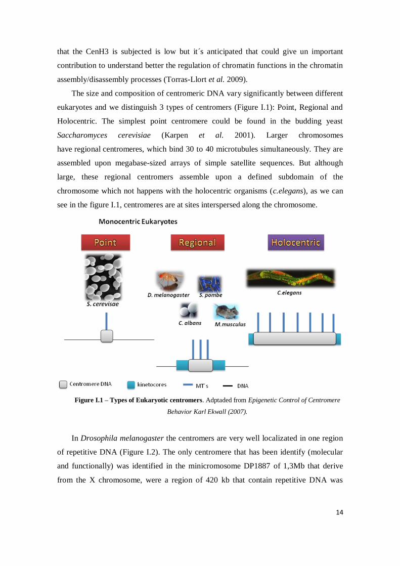
The size and composition of centromeric DNA vary significantly between different
eukaryotes and we distinguish 3 types of centromers (Figure I.1): Point, Regional and
Holocentric. The simplest point centromere could be found in the budding yeast
Saccharomyces cerevisiae (Karpen et al. 2001). Larger chromosomes
have regional centromeres, which bind 30 to 40 microtubules simultaneously. They are
assembled upon megabase-sized arrays of simple satellite sequences. But although
large, these regional centromers assemble upon a defined subdomain of the
chromosome which not happens with the holocentric organisms (c.elegans), as we can
see in the figure I.1, centromeres are at sites interspersed along the chromosome.
Figure I.1 – Types of Eukaryotic centromers. Adptaded from Epigenetic Control of Centromere
Behavior Karl Ekwall (2007).
In Drosophila melanogaster the centromers are very well localizated in one region
of repetitive DNA (Figure I.2). The only centromere that has been identify (molecular
and functionally) was identified in the minicromosome DP1887 of 1,3Mb that derive
from the X chromosome, were a region of 420 kb that contain repetitive DNA was

15
sufficient and able to perform a normal chromosomal segregation (Murphy and Karpen
1995).
Figure I.2 - Schematic view of centromeric DNA in Drosophila. This region present 2 blocks of DNA
satellite AATAT and CTCTT , and in the first block exist the insertion of 5 transposons. Also exist 2
domains in this centromers: one responsible for the assemble of the kinetochore were localize the
centromeric variant of H3, Cid, and another domain implicated in the attachment of the sister chromatids.
Adapted from Sullivan et al. 2001).
I.2. The centromeric histone H3 variants
The histones are the major structural components of chromatin and are expressed as a
family of sequence variants encoded by multiple genes being the scaffold in which the
eukaryotic genome is packaged and subject to a bewildering array of covalent
modifications (Brown 2001).The canonical histones (H2A, H2B, H3 and H4), that are
part of the nucleosome, are small and basic proteins composed of a globular domain
(carboxil terminal
domain) and a
flexible domain
(amino terminal
domain). In addition
to the canonical
histones, histone
variants such as
HAX, H2AZ and H3.3 are associated with distinct regions of the genome and substitute
one of the canonical histones. Another issue is the fact that the histone pass to some
modifications that are indicators of active or repressed chromatin, and the proposed
'histone code' hypothesis suggests that combinations of specific histone modifications
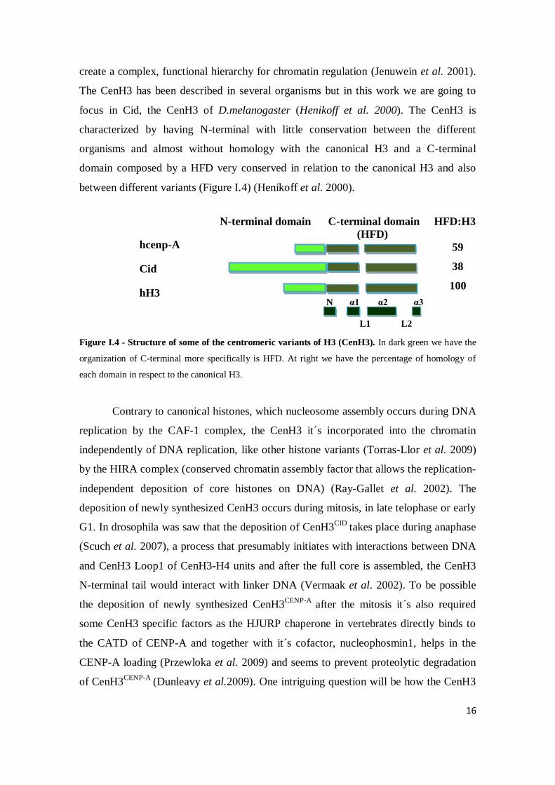
Figure I.3 - Representation of the sequence of Cid
16
create a complex, functional hierarchy for chromatin regulation (Jenuwein et al. 2001).
The CenH3 has been described in several organisms but in this work we are going to
focus in Cid, the CenH3 of D.melanogaster (Henikoff et al. 2000). The CenH3 is
characterized by having N-terminal with little conservation between the different
organisms and almost without homology with the canonical H3 and a C-terminal
domain composed by a HFD very conserved in relation to the canonical H3 and also
between different variants (Figure I.4) (Henikoff et al. 2000).
N-terminal domain C-terminal domain HFD:H3
(HFD)
59
38
100
Figure I.4 - Structure of some of the centromeric variants of H3 (CenH3). In dark green we have the
organization of C-terminal more specifically is HFD. At right we have the percentage of homology of
each domain in respect to the canonical H3.
Contrary to canonical histones, which nucleosome assembly occurs during DNA
replication by the CAF-1 complex, the CenH3 it´s incorporated into the chromatin
independently of DNA replication, like other histone variants (Torras-Llor et al. 2009)
by the HIRA complex (conserved chromatin assembly factor that allows the replication-
independent deposition of core histones on DNA) (Ray-Gallet et al. 2002). The
deposition of newly synthesized CenH3 occurs during mitosis, in late telophase or early
G1. In drosophila was saw that the deposition of CenH3CID
takes place during anaphase
(Scuch et al. 2007), a process that presumably initiates with interactions between DNA
and CenH3 Loop1 of CenH3-H4 units and after the full core is assembled, the CenH3
N-terminal tail would interact with linker DNA (Vermaak et al. 2002). To be possible
the deposition of newly synthesized CenH3CENP-A
after the mitosis it´s also required
some CenH3 specific factors as the HJURP chaperone in vertebrates directly binds to
the CATD of CENP-A and together with it´s cofactor, nucleophosmin1, helps in the
CENP-A loading (Przewloka et al. 2009) and seems to prevent proteolytic degradation
of CenH3CENP-A
(Dunleavy et al.2009). One intriguing question will be how the CenH3
hcenp-A
Cid
hH3
L1 L2
N α1 α2 α3
17
assembly it´s maintained at sites pre-existing CenH3 in the absence of DNA sequence
determinants.
I.3. The proteasome pathway
Once the CenH3 can form a dimer with the H4 it´s reasonable to think that in
principle CenH3 could be located everywhere on chromosome, but what happens is that
normally CenH3 is restrict to a particular region. Therefore mechanisms must exist in
the cells that lead to a selective localization of CenH3 in centromeric regions (Ekwall et
al. 2007). So we could have two hypotheses or the CenH3 is deposited everywhere and
actively removed from noncentromeric regions or CenH3 could be selectively deposited
only at centromers. In some assays was prove that the proteolysis contribute to CenH3
(Cse4) restricted localization to centromers in budding yeast (Collins et al. 2004).
The proteasome represents the major nonlysosomal proteolytic system in eukaryotes
(De Mot et al. 1999). In eukaryotic cells, degradation of many proteins involves their
covalent modification by conjugation with ubiquitin and requires the sequential action
of three enzymes: an ubiquitin activator enzyme (E1) an ubiquitin-conjugating enzyme
(E2) and ubiquitin-protein ligase (E3). The specificity of this destruction system is mainly
governed by the E3 and therefore play a pivotal role in this degradation pathway,
enabling the quickly degradation of proteins by a large multisubunit complex called the
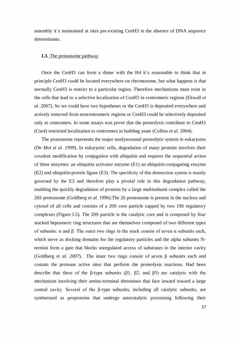
26S proteasome (Goldberg et al. 1996).The 26 proteasome is present in the nucleus and
cytosol of all cells and consists of a 20S core particle capped by two 19S regulatory
complexes (Figure I.5). The 20S particle is the catalytic core and is composed by four
stacked heptameric ring structures that are themselves composed of two different types
of subunits: α and β. The outer two rings in the stack consist of seven α subunits each,
which serve as docking domains for the regulatory particles and the alpha subunits N-
termini form a gate that blocks unregulated access of substrates to the interior cavity
(Goldberg et al. 2007). The inner two rings consist of seven β subunits each and
contain the protease active sites that perform the proteolysis reactions. Had been
describe that three of the β-type subunits (β1, β2, and β5) are catalytic with the
mechanism involving their amino-terminal threonines that face inward toward a large
central cavity. Several of the β-type subunits, including all catalytic subunits, are
synthesized as proproteins that undergo autocatalytic processing following their

18
Figure I.6 - Representation of the SCF complex
assembly into the 20S complex (Schmidtke et al. 1996, Neuburger et al. 2006). The 19S
particle, which is the regulatory component with ATPase activity, is responsible for
stimulating the 20S to degrade proteins and it´s primary function is to open the gate in
the 20S that blocks the entry of substrates into the degradation chamber.
The most known E3 enzymes are the APC
and the SCF complexes. Many diverse cellular
processes are regulated by the SCF family of
ubiquitin ligases that have the function to target
specific proteins for proteolysis (Deshaies, 1999).
The SCF complex exists and is evolutionarily
conserved among all the eukaryotes. It´s a
multimeric complex that recognize
specific subtracts and promote the
transition G1-S. Typically it´s composed by 4 subunits (Figure I.6): an invariant core
containing Skp1, Cul1, and Rbx1(RING finger protein) complexed with one member
of a large family of F-box proteins (Murphy 2003).The Skp1 is the bridging protein
essential in the recognition and binding to the F-box. The Cul1 is the major structural
scaffold of the SCF complex, and the Rbx1 is the protein to which the E2-ubiquitin
conjugate binds. The F-box proteins are a family of specificity factors, each of which
direct a set of substrates for ubiquitin-mediated degradation (Patton et al. 1998) and
could be split in two groups with base in it´s protein –protein interaction domain that
recognizes the substrate: WD40 or LRR.
Figure I.5 - Schematic view of the 26S proteasome and it´s subunits.

19
Figure I.7 - Representation of the interaction saw in vitro between our study protein, Cid, and the F-box
protein Ppa.
Previous experiments in Azorin's lab have shown that Cid interacts with Ppa
(Moreno-Moreno O. et al, unpublished results). Ppa is an F-box protein that is involved
in the degradation of the transcription factor Paired (Raj et al. 2000) and the first
example of an F-box protein that is integrated into the Drosophila segmentation cascade
(Das et al. 2001). Have a Sequence of 538 a.a and contain 3 types of domains: one
Domain rich in Ala/ His/Pro, a Domain F-box and 11 domains LRR (leucine-rich
repeats) (Figure I.7). Like other F-box proteins, it can have multiple substrates, what
could explain the phenotype of its null mutant. Recently it´s also been prove that Ppa
not only are evolved in the degradation of the transcription factor Paired but also in the
degradation of proteins related with the regulation of the cellular cycle (Raj et al.2000)

20
II- Objectives/goals
As I have explained in the introduction, the precise molecular mechanisms
accounting for the specific deposition of CenH3 at centromeres are still poorly
understood so, our main objective has been try to improve our knowledge about this
process. For this we have focus our work on:
1. - Analyze the role of the SCF complex in the centromeric localization of Cid
2. - Analyze if the mechanism of proteasome-mediated degradation also contributes
to the centromeric localization of the Cid endogenous protein
3. - Analyze which domain of Cid is involved in this specific localization

21
III- Materials and Methods
III. 1.1.1 Genetic Tools
III.1.1.1.1 UAS-Gal4 System
The UAS-GAL4 system was developed by Brand and Perrimon and is a
powerful biochemical method used to study gene expression and function (Brand and
Perrimon, 1993). This is a systems that enable the geneticists to create a great variety of
Gal4 lines, each one expressing the Gal4 in some subset of the fly's tissues and reporter
lines UAS promoter sequences have been introduced in a number of eukaryotic
expression vectors, allowing the production of transgenic constructs that are
conditionally expressed upon the availability of GAL4, enabling the expression of lethal
products (Duffy, 2002). Thus, in Drosophila, a parental stock expressing GAL4 can be
crossed to another carrying a UAS-construct, therefore driving expression of the latter
in the F1 progeny (Fig. M.1). Furthermore, to yield spatio-temporal specific drivers of
gene expression, several well-characterized enhancer sequences have been trapped by
the GAL4 coding gene (Brand and Perrimon, 1993).
Fig M.1 - The GAL4 system in Drosophila. One of the parental stocks carries the GAL4 gene in close
proximity to a known enhancer of gene expression. This stock can be crossed to another containing a
UAS-transgene, and the resulting progeny will express this transgene in the pattern of expression of the
GAL4 factor, which is specified by the associated enhancer region.
22
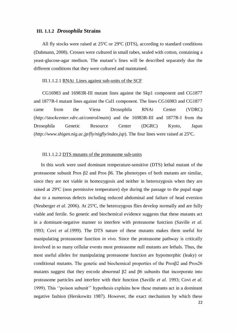
III. 1.1.2 Drosophila Strains
All fly stocks were raised at 25ºC or 29ºC (DTS), according to standard conditions
(Dahmann, 2008). Crosses were cultured in small tubes, sealed with cotton, containing a
yeast-glucose-agar medium. The mutant‘s lines will be described separately due the
different conditions that they were cultured and maintained.
III.1.1.2.1 RNAi Lines against sub-units of the SCF
CG16983 and 16983R-III mutant lines against the Skp1 component and CG1877
and 1877R-I mutant lines against the Cul1 component. The lines CG16983 and CG1877
came from the Viena Drosophila RNAi Center (VDRC)
(http://stockcenter.vdrc.at/control/main) and the 16983R-III and 1877R-I from the
Drosophila Genetic Resource Center (DGRC) Kyoto, Japan
(http://www.shigen.nig.ac.jp/fly/nigfly/index.jsp). The four lines were raised at 25ºC.
III.1.1.2.2 DTS mutants of the proteasome sub-units
In this work were used dominant temperature-sensitive (DTS) lethal mutant of the
proteasome subunit Pros β2 and Pros β6. The phenotypes of both mutants are similar,
since they are not viable in homozygosis and neither in heterozygosis when they are
raised at 29ºC (non permissive temperature) dye during the passage to the pupal stage
due to a numerous defects including reduced abdominal and failure of head eversion
(Neuberger et al. 2006). At 25ºC, the heterozygous flies develop normally and are fully
viable and fertile. So genetic and biochemical evidence suggests that these mutants act
in a dominant-negative manner to interfere with proteasome function (Saville et al.
1993; Covi et al.1999). The DTS nature of these mutants makes them useful for
manipulating proteasome function in vivo. Since the proteasome pathway is critically
involved in so many cellular events most proteasome null mutants are lethals. Thus, the
most useful alleles for manipulating proteasome function are hypomorphic (leaky) or
conditional mutants. The genetic and biochemical properties of the Prosβ2 and Pros26
mutants suggest that they encode abnormal β2 and β6 subunits that incorporate into
proteasome particles and interfere with their function (Saville et al. 1993; Covi et al.
1999). This ‗‗poison subunit‘‘ hypothesis explains how these mutants act in a dominant
negative fashion (Herskowitz 1987). However, the exact mechanism by which these

23
abnormal subunits are interfering with proteasome activity is not known. So the
expression of these mutants forms under the control of the UAS-Gal4 system allow us
to express them only in an specific tissue (not in all the fly) and then avoid the lethality
effect. In Drosophila melanogaster, two mutants that enable us to test if the proteasome
are involved in the degradation of our study protein are the UAS-Pros26 and UAS-
Prosbβ2 (Neuburger et al. 2006).
A
Figure M.2- UAS constructs driving expression of dominant temperature- sensitive
proteasome mutant genes. A- DTS7 (Pros β2) B-DTS5 (Pros26)
III. 1.1.3 Gal4 drivers
Table 1 Description of all gal4 drivers used in this work
Gal4 Drivers Domain of activity
Patched –Gal4
(Ptcgal4)
Patched is express in imaginal discs ( in the posterior
compartment) from early embryogenesis until end of larval stage
Engrailed-Gal4
(Engal4)
Express the gal4 in the engrailed domain (Antero-Posterior
boundary) from end of embryogenesis until end of larval stage.
Aptereou-Gal4
(Apgal4)
dorsal selector gene required for patterning and growth of the
wing
Scabrous-Gal4 (Scagal4) Scabrous gene expressed in the neurons
Elav-gal4 (Embryonic lethal abnormal vision) express in the neurons
B

24
The drivers used in this work are depicted in Table1 and the pictures that show is
domain of expression were kindly provided by Gaylord Darras.
III.1.2. Methods of work with drosophila
III.1.2.2 Work with neuroblasts of Drosophila
III 1.2.2.1 RNA Extraction from third instar larvae brains
Previously, to isolate the brains is necessary dissect them in PBS 1X , transfer to an
1,5mL eppendorf with 50 µl of PBS 1X and maintained on ice. After that centrifuge at
100 rpm and eliminate the supernatant. To finish we freeze the brains in LN2 and store
at -80ºC until they are going to be used. We for each cross collect between 80 and 150
brains and follow the protocol in annexes III.1.2.2.1.
III 1.2.2.2 Protocol of dissection of neuroblasts
To analyze if the E3-Ubiquitin ligase SCF complex has a role in the specific
localization of Cid we have done some imunolocalizations in neuroblasts to see if this
protein appear delocalized to all the chromatin. The neuroblasts were obtained from
Drosophila lines expressing RNAi against one of the subunits of the SCF complex:
Skp1 (16983R-III and CG16983) or Cul1 (1877R-I and CG1877). We cross males from
this RNAi lines with females Scagal4 or Elavgal4 (the drivers that are expressed in the
neuroblasts) at 25ºC and wait more or less 6 days until the first progeny arrives to third
instard larvae. In this moment we can dissect the brains of the larvae to obtain the
neuroblasts.
So we dissect the brains of L3 larvae in 50µL of saline medium 0.7% Nacl (0.07g
Nacl in 10ml of H20) and left in this medium between 2 to 5minutes. After that and to
be able to observe metaphases we treat for 1h30 minutes with 5 µg/µL colcemid (at the
concentration 10 µg/µL) in one dark and humid box at room temperature. After we

25
perform un hypotonic shock with sodium citrate 0.5% medium at room temperature for
5-10minutes (0.05g citrate de sodium in 10mL of H20) and proceed to the fixation of the
brains in formaldehyde 3,7% for 30minutes. Next we transfer the brains to a siliconized
coverslip that already has 16 µL of acetic acid 60% (1200 µL acetic acid in 800 µL of
H20) and do a squash. To finish this first part we submerge the slides in LN2 and stored
the preparations in PBS-Triton X-100 (100 µL Triton X-100; 100mL PBS 10X).
III.1.2.2.3 Imunostaining of neuroblasts
After we prepare the neuroblasts like it´s explain in III 1.2.2.2 we start the
imunolocalization by blocking with PBS-Triton-Milk for 1hour (100mL PBS-
0,1%Triton X-100 + 1g powder milk) and after we incubate with 25 µL of first
antibody in a humid box for 1hour at room temperature and let o/n at for 4ºC (we have
used a 1:300 dilution of rabbit in PBS-0,1%Triton-1%Milk).
In the next day we wash twice for 10 minutes with PBS-0,1%Triton-1%Milk and
incubate with 25 µL of secondary antibody (from Jackson ImunoReserch) in one dark
and humid box for 45 minutes at room temperature (diluted 1: 400 in PBS-0,1%Triton-
1%Milk). As we did before with the primary antibody we wash twice, for 10 minutes,
with PBS-0,1%Triton-1%Milk. To finish we mounted in Mowiol+Dapi (50 µL DAPI
2ng/µL in 500 µL Mowiol), let it dry and storage at 4ºC until visualization at the
inverted fluorescence microscopy NikonEclipse E-1000. The images were collected
with a colour view 12 camera using a 40X objective lens. Acquisition parameters were
controlled by Metamorph Software and images were analyzed with the program Adobe
Photoshop.
III.1.2.3 Work with imaginal discs of Drosophila
III 1.2.3.1 Protocol of dissection and Imunostaining of imaginal discs of third
instar larvae
To see if in vivo the levels of Cid are affected or regulated by the biggest mechanism
of degradation in Eukaryotes, the proteasome, we will do some imunolocalizations in
imaginal discs. We start by dissecting the brains and discs (all together) of third instar
larvae in could PBS and fixed the tissues in PBS-4% formaldehyde for 15 to 20 minutes
at room temperature. Next the larvae were washed and permeabilized in PBT (PBS-

26
0.3%Triton X-100) 3 times for 10 minutes each. Afterwards they were blocked in fresh
PBS-0.3% Triton X-100-2% BSA 3 times also 10 minutes each and incubate at 4ºC o/n
with the primary antibody rabbit anti-CidNt (diluted 1:300 in PBS-0,3% Triton X-100-
2% BSA) on agitation.
In the next day we wash in agitation, first with fresh PBS-0.3% Triton X-100 and
then four times with PBS-0.3% Triton X-100-2%BSA . After these washes we incubate
for 2h at room temperature with the secondary antibody anti-rabbit cy3 (from Jackson
ImunoReserch) diluted 1:400 in PBS-0,3% TritonX-100-2%BSA in agitation covered
by aluminum paper. Next, they were washed 4 times, 10 minutes each, in PBT and then
twice in PBS for 10 minutes. Finally they were incubated with Dapi (600 µL PBS+ 6
µL DAPI 2ng/ µL) for 5 minutes at room temperature and washed for 5 minutes in
PBS-0,3%Triton X-100. To finish we dissect and mount the discs in Mowiol and
visualized at the confocal microscopy. The images were analyzed with the program
ImageJ.
III.1.2.4 Work with Kc cells of Drosophila
The Kc cell line (or Kc 167) is one of the firsts lines of D.melanogaster established
for the in vitro culture (Echelier and Ohanessian 1969). Derived from 8 or 12h
drosophila embryos, more specifically from embryonic hemocytes (blood cells that
differentiate into macrophages). Have a spherical aspect, growth in suspension and form
grumps. It´s cellular cycle is about 20h. One of it´s characteristic is since they are
solved don´t attaches one to each others like other lines (S2 line).
III.1.2.4.1 Mantencien of the cells
a) Growth – The cells growth at 25ºC without CO2 in Schneider medium (Sigma)
and 100µg/mL of Streptomycin (Gibco), 100U/mL of penincilin (Gibco) and 10%
FBS (Gibco) that will be inactivate during 30minutes at 56ºC. We maintained this
medium in flasks of 75cm2
at one density of 1-8x10⁶
cells/mL in a volume of 5-6 mL,
doing dilutions of 1/6 or 1/8 every 3-4days. To have great amounts of cells to transfect
we going to prepare flasks of 175 cm2
doing dilutions of 1/6 in a final volume of 30
mL.

27
b) Freezing – To freeze the cells we let them growth in 75 cm2
flasks until they
reach a density of 5x10⁶ cell/mL. To recover the cells we going to transfect the
medium to a falcon tube of 15 mL and centrifuge 2 minutes at 1300 rpm at room
temperature. Afterwards, we eliminate the supernatant and resuspend the cellular
sediment with 900 µL FBS. We going to pass to cryotubs and add drop by drop 100µL
of DMSO. The process of freezing have to be slow so first we storage the cryotube
during 2 houres at -20ºC after o/n at -80ºC and finally storage in LN2.
c) Defrost - We use one of the cryotubes of Kc cells storage in LN2 and defrost
quickly in our hands. After we pass to a 15 mL falcon with 5 mL of medium and
centrifuge 2 minutes at 1300 rpm at room temperature and resuspend the cellular
sediment with 5mL of medium. Finally, we pass this medium with cells to 75 cm2
flask and let it growth during 3-4 days.
III.1.2.4.2 Transfection of the Kc cells by Effecten method
To transitory transfections of the DNA plasmids we used the Effectene method.
To obtain a sufficient number of cells to transfect we let the cells growth until they
reach a density of more or less 6x10⁶ cell/mL in a volume of 30 mL in 175cm
2 flasks.
Once quantificated the exact concentration of cells by counting with the Neubauer´s
chamber we inoculate 2x10⁶ cells in harvest plats of 60mm
2 in one final volume of 5mL
and incubate 24h at 25ºC. For each plate we transfect 10 µg of DNA (obtained from
maxipreps with Quiagen columns) in a final volume of 50 µL. For every transfection we
going to made duplicates so we going to prepare for 2,5X transfections. In the next day
after we plate the cells we prepare in eppendorfs the necessary quantity of DNA in one
final volume 125µL.
III.1.2.4.3 Analyze of the transfection by fluorescence microscopy
To determine the transfection efficiency and the localization of the proteins of interested
in fusion with YFP we use the fluorescence microscopy. 48 hours after the transfection,
we impact 200 µL the cells in one porta slides by centrifugation at 500 rpm for 10
minutes. First we had to collect 300 µL the cells from the plate and to a tube that
28
already contain 1mL of MAC hypotonic medium (50 mM glycerol; 5mM KCl; 10 mM
Nacl; 0,8 mM CaCl2; 10 mM Sucrose).Afterwards we incubate for 5 minutes to the
cells incorporate water, but with the careful of not pass this time because they can end
up blowing. Quickly we put 200 µL of this solution in the deposits of the centrifuge in
the porta slides, and centrifuge for 10 minutes at 500 rpm in the ThermoShadon
Cytospin4. To acoplate the embuts and the porta slides we use a clip and between them
put a filter. In this way the cells will be impacted in one small circle in the slides that
coincides with the exit of the embut. Afterwards we let dry the slides for 1hour at room
temperature and fixe the cells with 50µL paraformaldehid 3,7% (diluted in PBS 1X)
directly above the cells for 10 minutes at room temperature. Next step was wash the
slides with PBS 1X for 15 minutes and after that let it dry and storage dry at 4ºC
involved in aluminum paper until we observe in fluorescent microscopy.
III 1.2.3 qRT-PCR
The qRT-PCR was done using the RNA obtained by the protocol in annex of
RNA extraction (III 1.2.2.1) using as a internal control actin gene and the primers used
for the qRT-PCR are described in Annexes (table A.1). After the extraction of total
RNA, it´s integrity was tested. The RNA with good integrity was subjected to a RT-
PCR using the Qiagen Kit under the manufacter´s instructions and the termocycler
LightCycler480. The PCR conditions used was: 1 minute to 94ºC; 1 minute to 55ºC, 1
minute to 72ºC all for 30 cycles and 1 minute to 72ºC.
29
IV- RESULTS
As I have explained before the goal of this work has been to deep the understanding
of the mechanism(s) that are involved in the centromeric localization of Cid. To achieve
this we have focused our work in the study of how some proteasome mutants and RNAi
lines against some proteins involved in the proteasome pathway affect Cid's
localization. And moreover, we have tried to found if any specific domain of Cid is
required to be degradated.
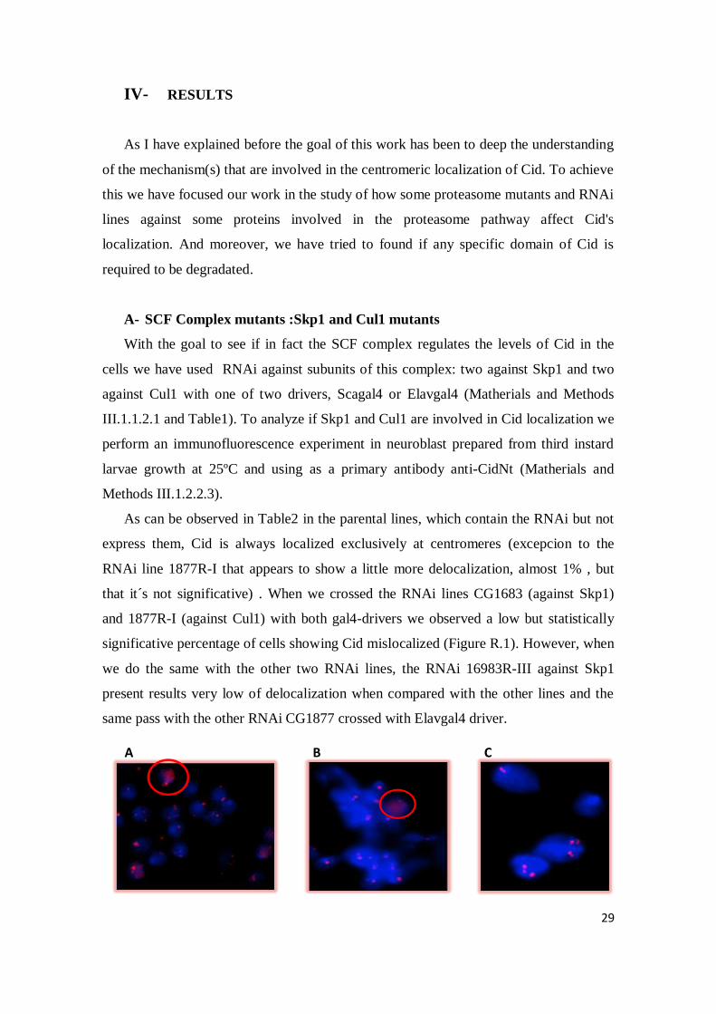
A- SCF Complex mutants :Skp1 and Cul1 mutants
With the goal to see if in fact the SCF complex regulates the levels of Cid in the
cells we have used RNAi against subunits of this complex: two against Skp1 and two
against Cul1 with one of two drivers, Scagal4 or Elavgal4 (Matherials and Methods
III.1.1.2.1 and Table1). To analyze if Skp1 and Cul1 are involved in Cid localization we
perform an immunofluorescence experiment in neuroblast prepared from third instard
larvae growth at 25ºC and using as a primary antibody anti-CidNt (Matherials and
Methods III.1.2.2.3).
As can be observed in Table2 in the parental lines, which contain the RNAi but not
express them, Cid is always localized exclusively at centromeres (excepcion to the
RNAi line 1877R-I that appears to show a little more delocalization, almost 1% , but
that it´s not significative) . When we crossed the RNAi lines CG1683 (against Skp1)
and 1877R-I (against Cul1) with both gal4-drivers we observed a low but statistically
significative percentage of cells showing Cid mislocalized (Figure R.1). However, when
we do the same with the other two RNAi lines, the RNAi 16983R-III against Skp1
present results very low of delocalization when compared with the other lines and the
same pass with the other RNAi CG1877 crossed with Elavgal4 driver.
A B C

30
Elavgal4x1887R-I
DAPI DAPI + αNCid αNCid
Fig R.1- Images of immunostaining in neuroblasts from third instar larvae of the effects that
RNAi against sub-units of SCF complex induce in the Cid localization. A,B- Scagal4 x 16983R-
III (Skp1) 25ºC where we see that some cells shows Cid delocalized (red circule) and C- Scagal4 x
CG16983 (Skp1) 25ºC in which Cid appears localized. In blue we have the DNA stained with DAPI
an in red the signal of the Anti-CidNt.. D- Elavgal4x CG1877(Cul1) 25ºC the DNA of the cells
stained with DAPI, E- signal of the Anti-CidNt, F- Overlay were we see the cells with Cid localize,
G- Elavgal4x1877R-I (Cul1) 25ºC where we also see the cells with Cid localize(H) but a fews with
Cid desocalized (I).
D E F
G
H
I

31
Table.2- Percentage of neuroblasts from third instar larvae showing Cid delocalized in the Skp1 or Cul1
subunits. D-number of cells with Cid delocalized, Total nº-total number of cells count, %D-percentage of
delocalization, * the total number of cells count in this crosses was less then in the others.
The differences in the percentage of cells with Cid mislocalized may could be explain
with a difference in the RNAi efficiency. So, to analyze this we have performed a qRT-
PCR analysis with the RNAs from 3 crosses: Elavgal4xCG16983, Scagal4x1877R-I
(Figure R.2) and Elavgal4x 16983R-III (not show).
Figure R.2 Graphics with the result of the qRT-PCR experiments. Analysis of the efficiency of our RNAi
lines against the subunits of the SCF complex. A- dark green the mRNA levels of the RNAi lines against
Skp1( CG16983) component and lighter green the mRNA from Actin our internal control of the samples
that we used to normalized all the data ; Axis yy- relative levels of mRNA- B- dark blue green the
mRNA levels of the RNAi lines against Cul1(1877R-I ) component and lighter blue the mRNA from our
normalization control Actin.; Axis yy- relative levels of mRNA.
A B
32
In theory the actin mRNA doesn't change between samples so we used actin as a
internal control gene to correct the possible errors due to differences in the total RNA
used in every well (errors in the determination of RNA concentrations, different
efficiency of the RT, errors in loading). Once we normalized all data we compared our
mRNA levels (Skp1 or Cul1) in the parental line (which contains but not express the
RNAi) and our mRNA levels when we have crossed with the drivers (Scagal4 or
Elavgal4). We observe that in both crosses CG16983x Elavgal4 and 1877R-Ix Scagal4 a
significative decrease in these mRNA levels are achieved, a decrease of 40% and 60%
respectively.
B- Proteasome mutants
In the proteasome mutants, as I have explained in the introduction, it is
reasonable to expect that mutations in genes encoding proteasome subunits would
exhibit lethal phenotypes. The lethal effects of the mutations reported here indicate that
in flies, as has been previously shown in yeast proteasome function is essential for
viability (Saville et all 1993). So to study the function of this genes it´s better to use the
UAS-Gal4 system since in previews experiments in Ferran´s lab, were they perform
studies of imunostaining with this β2 and β6 proteasome mutants without the UAS-Gal4
system , were observed only an 8% of Cid mislocalized in the neuroblasts. Due to the
fact that with this mutants appear the problem that the mutant effect begin at the third
instar larvae leading to the dead in the final of this instar, we decided to do this
experiments with the UAS-Gal4 system in an temptative to see higher levels of
mislocalization. In our experiments we cross 10 males of one of the two UAS lines that
we have (Table1) with 10 virgin females of the driver lines (Ptcgal4, Engal4 or Apgal4).
The cross were put at 25ºC and 24h late change to 29ºC (restrictive temperature). We
performed an immulocalization assay in wing imaginal disc using anti-CidNt (Materials
and Methods III 1.2.2.4) and the images taken in the confocal microscopy (figure R.3
and R.4) shows an absence of the mislocalization of our study protein, Cid, in the
domains of expression of our drivers in the imaginal discs. We always see the typical
punctuated pattern in the cells. However one interesting phenotype that it´s also saw it´s
an abnormal cellular proliferation (figure R.3).
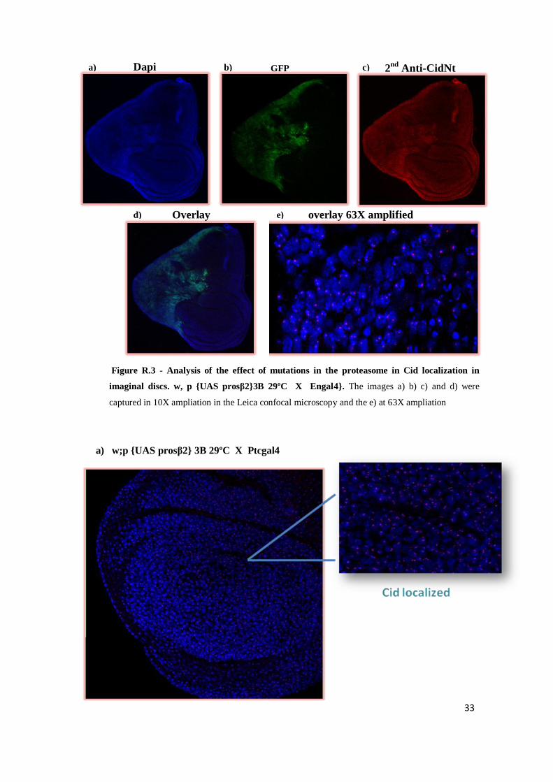
33
a) w;p {UAS prosβ2} 3B 29ºC X Ptcgal4
a) b) c)
d) e)
Dapi
Overlay overlay 63X amplified
GFP 2nd
Anti-CidNt
Figure R.3 - Analysis of the effect of mutations in the proteasome in Cid localization in
imaginal discs. w, p {UAS prosβ2}3B 29ºC X Engal4}. The images a) b) c) and d) were
captured in 10X ampliation in the Leica confocal microscopy and the e) at 63X ampliation

34
b) w, p {UAS pros β2}3B 29ºC X Apgal4
Cid localized
Figure R.4 – Analysis of the effect mutations in the proteasome in Cid localization in imaginal discs.. a)
w, p{UAS prosβ2}3B 29ºC x ptcgal4}, b) w, p {UAS prosβ2}3B 29ºC x Apgal4}. The images a) b) were
captured in 20X , 40X and 63X and ampliation in the Leica confocal microscopy.
35
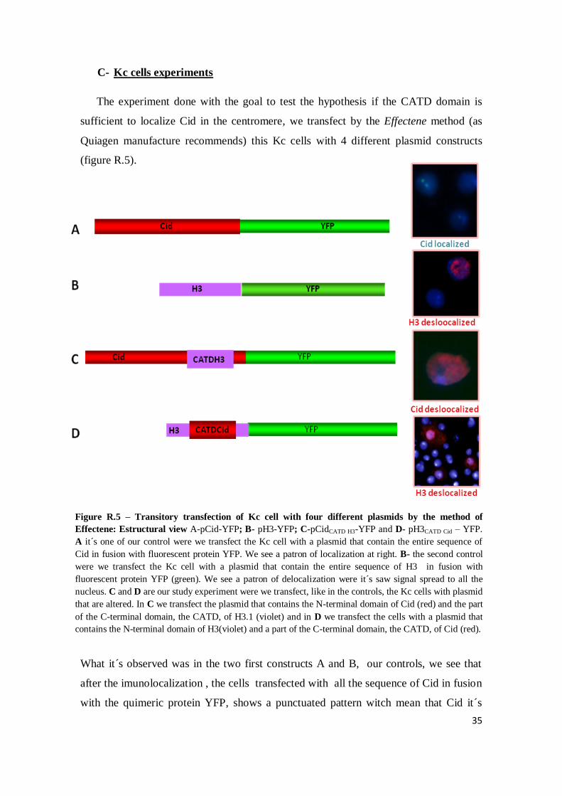
C- Kc cells experiments
The experiment done with the goal to test the hypothesis if the CATD domain is
sufficient to localize Cid in the centromere, we transfect by the Effectene method (as
Quiagen manufacture recommends) this Kc cells with 4 different plasmid constructs
(figure R.5).
What it´s observed was in the two first constructs A and B, our controls, we see that
after the imunolocalization , the cells transfected with all the sequence of Cid in fusion
with the quimeric protein YFP, shows a punctuated pattern witch mean that Cid it´s
Figure R.5 – Transitory transfection of Kc cell with four different plasmids by the method of
Effectene: Estructural view A-pCid-YFP; B- pH3-YFP; C-pCidCATD H3-YFP and D- pH3CATD Cid – YFP.
A it´s one of our control were we transfect the Kc cell with a plasmid that contain the entire sequence of
Cid in fusion with fluorescent protein YFP. We see a patron of localization at right. B- the second control
were we transfect the Kc cell with a plasmid that contain the entire sequence of H3 in fusion with
fluorescent protein YFP (green). We see a patron of delocalization were it´s saw signal spread to all the
nucleus. C and D are our study experiment were we transfect, like in the controls, the Kc cells with plasmid
that are altered. In C we transfect the plasmid that contains the N-terminal domain of Cid (red) and the part
of the C-terminal domain, the CATD, of H3.1 (violet) and in D we transfect the cells with a plasmid that
contains the N-terminal domain of H3(violet) and a part of the C-terminal domain, the CATD, of Cid (red).
36
only localize in the centromers. This was expected since the HFD don´t suffer any
alteration in is sequence and so the CATD, the domain responsible for targeting the
CenH3CENP-A
to the centromers, was also functional and unchangeable. In B we also
have the entire sequence but this time of the canonical H3 in fusion with YFP. In this
case since the CATD domain it´s not the same then in the CenH3 don´t exist the
determination necessary to localize this protein, canonical H3, only in the centromere
and so what it´s saw it´s a typically patron of delocalization were the signal spread to
all the nucleus. In the constructions C and D we want to prove if in fact the CATD of
Cid it´s required and sufficient for it´s centromeric localization like happens with
CENP-A in mammals. So, by fluorescent microscopy we can say that the C-terminal
domain it´s required for the centromeric localization, which is in agree with some
studies performed with quimeric proteins CENP-A and H3 (Sullivan et al 1994),
specifically was described that a specific region of the C-terminal domain, the CATD
domain L1/α2 was the responsible for the centromeric localization of Cid (Vermaak et
al 2002). What is saw with our experiments is that in Drosophila this CATD domain
it´s required but seems it´s not sufficient to target Cid in the centromers because in the
last construct with the sequence of canonical H3 and CATD domain of Cid if in fact this
CATD was sufficient we should see a pattern of localization and not the mislocalization
through all the nucleus that it´s observed in D. This results are also in agreement with a
experiment performed by Black et al 2007 (Figure R.6) in yeast. They saw that the
CATD domain was necessary but never sufficient, seems like it always required some
little sequence, like the α3 region, to target Cid only in the centromers.
Figure R.6 – Adptaded from Centromere identity manteined by nucleosomes assembled with histone H3
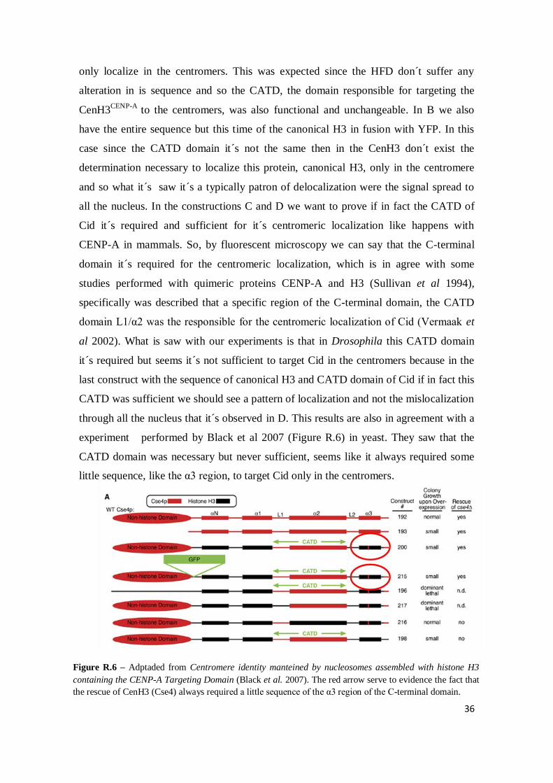
containing the CENP-A Targeting Domain (Black et al. 2007). The red arrow serve to evidence the fact that
the rescue of CenH3 (Cse4) always required a little sequence of the α3 region of the C-terminal domain.

37
V- DISCUSSION
Since the discovery of centromeric variants in last century this area become a very
interesting field to improve the knowledgment about the centromeric chromatin in a
way that this CenH3 have a key role in the formation of the kinetochore and in
chromosome segregation. As I have said before one common characteristic of all the
centromers is the presence of CenH3, which it´s thought to be the epigenetic
determinant of the centromere identity (Howman et al. 2000, Vermaak et al. 2002). The
specific mechanism that determines that this variant only deposit in the centromere and
the factors responsible for that it´s not completely known, so with this work we had the
goal to try to understand a little better some of this doubts.
To the proteins being degradate by the proteasome they previews had to be
poliubiquitinated. First we start to perform some imunolocalization experiments in
neuroblasts of third instar larvae that are mutants in one of the invariable components of
an E3 ubiquitin ligase, the SCF complex: Skp1 or Cu11. We observe a very low
percentage of delocalization in all the RNAi lines (table2) but principal in the lines
16983R-III (Skp1) and CG1877 (Cul1). To have sure that our RNAlines are working
well and the low delocalization is not due to problems with them we perform qRT-PCR
using the RNA from the crosses: Elavgal4xCG16983 (figure R.2 A), Scagal4x1877R-I
(figure R.2 B) and Elavgal4x 16983R-III (not shown) to be sure what are the level of
the decrease that our RNAi pathway promote. We choose the ones that in the beginning
present the higher percentage of delocalization for each component Skp1 or Cul1 with
base in the table 2. With these results we could compare if our levels of delocalization
are significative or no. Based on the graphics we could see that both RNAi line against
the Skp1 and Cul1 component, promotes a significative decrease in the levels of these
proteins in the cells, ~40% and 60% respectively. In the experiment done with the other
RNAi line, CG16983, don´t show a significative decrease in the levels of this protein,
Skp1, in the cells. So these lines are working reasonable well and the low percentage of
mislocalization of Cid in the cells couldn´t be for that reason. All the crosses were
raised at 25ºC and maybe these temperatures were not sufficient enough to induce the
UAS-Gal4 system and few levels of the mRNA Skp1 or Cul1 are silence. The problem
was when we put the crosses at 29ºC we don´t obtain any larvae, probably because at
this temperature the UAS system are more active and we have a massive silence of the
38
mRNA of Skp1 or Cul1 and in this conditions the SCF complex could lose it´s function
which could be unviable to the progression of celular cycle and so the larvae die before
arrive the third instar or don´t pass the embrionary development. In future will be
valuable use a two subunits mutant to see if the percentage of delocalization appears
bigger, or inclusively if it´s viable.
Second in the experiments with mutants of the proteasome and since the
function of the proteasome genes, β2 and β6, are very important to the correct function
of this structure one of the ways of test there function in terms of the involvement in the
localization of Cid, was express this mutants sub-units of the proteasome in one specific
tissue by the using the UAS-Gal4 system (Belote et al 2002). In this way we be able to
compare in the same individual the effects of the presence or absence of proteosomal
activity in the levels of the endogenous Cid.
In imaginal discs we cross every mutant line (III.1.1.2.2) with drivers that
express in that appendage (ptcgal4, Engal4, Apgal4 III.1.1.3) and after we try to
optimize all the conditions in the imunolocalization assay (III.1.2.3.1) not only the type
of first antibody used but also the dilution. The results indicate us a lack of Cid
delocalized in the discs of this mutant larvae. One possibility for this was the fact that
we could not achieve an optimization of the imunostaining conditions because in the
majority of the cases we see a background noise from the second antibody which many
times don´t let us understand if the punctuated pattern that we see it´s due to the
centromeric localization of Cid or it´s due to this not specific stain of the secondary
antibody. Further optimization work will be one of the goal to clearly understand if in
imaginal discs , like was already saw in neuroblasts (Moreno-Moreno et al 2002), the
proteasome is the mechanism of degradation responsible to maintained the Cid
localization in the centromers.
Meanwhile an intriguing phenotype that we see in all the discs was an abnormal
cellular proliferation in this UAS proteasome mutants crossed with the Engal4 driver
(figure R.3) and Apgal4 (not shown). We know that since the proteasome it´s evolved in
the degradation of so many and different protein this phenotype probably could have
nothing to do with the localization of Cid. So in the future one of the goal´s will be
study the phenotype of these proteasome mutants in the wings of the adults flies,

39
possible because we are using UAS-Gal4 lines, to see if this phenotype has something
to do with Cid localization.
Third we also perform some studies in vitro to prove if the CATD, a particular
region of the C-terminal domain, the helix α2 and the loop1 (figure I.2) that connect
with the H4 and that conferring the thickness and compactation of the nucleosome
(black et al.2004) is sufficient to localize Cid in the centromere. The results obtained
by transient transfection of the different construct plasmid that express the YFP proteins
(Figures R.5) we can say that a particular region of the C-terminal domain it´s required
for the centromeric localization because when we transfect the cells with N-terminal
sequence of Cid but with the CATD of canonical H3 we see a mislocalization of this
protein which is in agree with some studies performed with quimeric proteins, CENP-A
and H3 (Sullivan et al 1994), that shows that this domain it´s required and sufficient to
target CENP-A in the cells. However in Drosophila this CidCATD it´s not sufficient to
target Cid in the centromers, because when we transfect the cells with the N-terminal
sequence of the canonical H3 with the CATD of Cid this is not sufficient to localize H3
only in the centromere. Appears that some other region outside the CATD, like the α3
region in the C-terminal region of the CenH3 of yeast (Black et al 2007), could be
required to the targeting of Cid to the centromere. One further work that will be of value
to do was do some constructions with the CATD and also different parts of this α3
region to identify all the regions required to targeting Cid only in the centromers.

40
VI- CONCLUSIONS
1- Appears that the SCF complex regulates the levels of Cid in the cells, but it´s not
essential to it´s degradation. When we mutate Skp1 or Cul1 in the both cases we see
very few delocalization of Cid which could mean that some other subunits are able to
compensate the function of Skp1 or Cul1 or this SCF complex it´s not the only one that
regulated the degradation of this protein.
2- The proteasome mutants don´t show a mislocalization of Cid in the imaginal discs
in our assays. This lack of results could be due to we don´t achieve the correct
optimization in the imunostaining assay. Other possibility it´s the fact that a single
mutation in each one of the two subunits is not strong enough to disrupt the proteasome
function and somehow the function of these subunits could be compensate by other
ones. But it´s clear that the proteasome in somehow have to regulate the levels of this
protein in the cells with base in some assays done before.
3- The function of the CATD seems to be conserved from fungi to mammals and
appears that this domain it´s necessary but not sufficient to the centromeric localization
of CenH3 in Drosophila, as have been previously show for budding yeast (Black et al
2007). It seems that it´s required some other region of the HFD, not only the CATD, to
localize Cid in the centromers.
41
VII- REFERENCES
1. Brown, D. T., (2001) Histone variants: are they functionally heterogeneous?
Genome Biol.; 2(7): reviews
2. Black B.E, Foltz D.R, Chakravarthy S, Luger K, Woods VL Jr, Cleveland DW
(2004) Structural determinants for generating centromeric chromatin Nature.
;430(6999): pp.578-82
3. Cleveland, D. W. (2007) Centromere identity maintained by nucleosomes assembled
with histone H3 containig the CENP-A Targeting Domain; Molecular cell 25, pp.
309-322
4. Collins KA, Furuyama S, Biggins S. (2004) Proteolysis contributes to the exclusive
centromere localization of the yeast Cse4/CENP-A histone H3 variant. Curr. Biol.
14:pp.1968–72
5. Coux, O., Tanaka, K. & Goldberg, A. L. (1996) Structure and functions of the 20S
and 26S proteasomes. Annu. Rev. Biochem. 65,pp. 801–847
6. Covi,J.A; Belote, J.M and Mykles, D.L (1999) Subunit compositions and catalytic
properties of proteasomes from developmental temperature-sensitive mutants of
Drosophila melanogaster. Arch Biochem.Byophy368: pp.85-97.
7. Das, T.;Purkayastha-Mukherjee, C;D´Angelo,J.; Weir,M. (2002) A conserved F-box
gene with unusual transcript localization. Dev Genes Evol 212:pp. 134-140
8. Deshaies, R.J (1999) SCF and Cullin/Ring H2-based ubiquitin ligases. Annu Rev
Cell Dev Biol.;15;pp. 435-67
9. De Mot, R., Nagy I., Walz J. and Baumeister, W. ( 1999) Proteasomes and other self
compartmentalizing proteases in prokaryotes .Trends Microbiol 7(2): pp. 88-92
10. Duffy, J. B., (2002 ) GAL4 System in Drosophila: A Fly Geneticist’s Swiss Army
Knife Genese. 34(1-2):pp 1-15
11. Dunleavy, E.M; Roche, D; Tagami, H.; Lacoste, N; Ray-Gallet, D.; Nakamura, Y.;
Daigo, Y.; Nakatani, Y.; Almouzni-Pettinotti, G. (2009) HJURP is a cell cycle
dependent maintenance and deposition factor of CENP-A at centromeres. Cell 28:
pp. 1029–1044
12. Ekwall, K. (2007) Epigenetic Control of Centromere Behavior.
Annu.Rev.Genetic.2007 41: pp.63-81
13. Goldberg, A.L (2007) Functions of the proteasome: from protein degradation and
immune surveillance to cancer therapy . Biochem Soc Trans. 35(Pt 1): pp.12-7.
14. Jenuwein, T. and Allis, C.D. (2001) Translating the histone code. Science 293,
pp.1074–1080

42
15. Henikoff, S.,; Ahmad, Kami,; Platero, J. Suso and Steensel, Bas van; (2000)
Heterochromatic deposition of centromeric histone H3-like proteins PNAS
16. Henikoff , S. and Dalal ,Y. (2005) Centromeric chromatin: what makes it unique?
Current Opinion in Genetics & Development Vol. 15, Issue 2,pp. 177-184
17. Herskowitz,I,; (1987) Functional inactivation of genes by dominant negative
mutations. Nature 329: pp 219-222.
18. Moreno-Moreno, O. Torras-Llort, M. and Azorín, F. (2006) , Proteolysis restricts
localization of CID, the centromere-specific histone H3 variant of Drosophila, to
centromeres Developmental Cell 10, pp. 303–315
19. Murphy, T.D and Karpen,G.H (1995) Localization of centromere function in a
Drosophila minichromosome Cell 82(4):pp 599-609
20. Neuburger,1 Kenneth J. Saville,2 Jue Zeng,3 Kerrie-Ann Smyth4 and John M.
Belote5 (2006) A Genetic Suppressor of Two Dominant Temperature-Sensitive
Lethal Proteasome Mutants of Drosophila melanogaster Is Itself a Mutated
Proteasome Subunit Gene Genetics 173: 1377–1387
21. Patton E, Willems AR, Tyers M (1998) Combinatorial control in ubiquitin-
dependent proteolysis: don’t Skp the F-box hypothesis.Trends Genet 14:236–243
22. Przewloka, M.R.,Glover, D.M, (2009) The Kinetochore and the Centromere: A
Working Long Distance Relationship. Annu.Rev. Genet. 2009,43 pp 439-465
23. Raj L, Vivekanand P, Das TK, Badam E, Fernandes M, Finley RL, Brent R, Appel
LF, Hanes SD, Weir M. (2000) Targeted localized degradation of Paired protein in
Drosophila development. Curr Biol.19;10(20):pp.1265-72
24. Ray-Gallet D, Quivy JP, Scamps C, Martini EM, Lipinski M, et al. (2002) HIRA is
critical for a nucleosome assembly pathway independent of DNA synthesis. Mol
Cell 9 (5): pp. 1091–1100.
25. Saville, K. J. and Belote, J. M.(1993) Identification of an essential gene, 1(3)73Ai,
with a dominant temperature-sensitive lethal allele, encoding Drosophila
proteasome subunit. Proc. Natl. Acad. Sci. USA Vol. 90, pp. 8842-8846, Genetics
26. Schmidtke, G.;Kraft, R.;Kostka,S.; Henklein,P.; Frõmmel et al (1996) Dominant-
negative mutations in the β2 and β6 proteasome sub-unit genes affect alternative
cell fate decisions in the Drosophila sence organ lineage. Proc. Natl.Acad Sci. USA
96:pp11382-11386.

43
27. Smith DM, Chang SC, Park S, Finley D, Cheng Y, Goldberg AL (2007). Docking of
the proteasomal ATPases' carboxyl termini in the 20S proteasome's alpha ring
opens the gate for substrate entry. Mol. Cell 27 (5), pp. 731–44
28. Sullivan, B. A., Blower, M. D. and Karpen, G. H., (2001) Determining centromere
identity: cyclical stories and forking paths. Nature Review, vol.2
29. Sullivan, B. and Karpen, G.H (2004) Centromeric chromatin exhibits a histone
modification pattern that is distinct from both euchromatin and heterochromatin
Nature Structural & Molecular Biology 11, pp.1076 - 1083
30. Torras-Llort, M., Moreno-Moreno, O and Azorín, F. (2009) Focus on the centre: the
role of chromatin on the regulation of centromere identity and function. The EMBO
Journal 28,pp. 2337-2348
31. Vermaak, D.;Hayden,H.S. and Henikoff, S. (2002) Centromere Targeting Element
within the Histone Fold Domain of Cid. Molecular and Cellular Biology vol.22,
No.21 pp.7553-7561
Books
- Allis, C.D; Jenuwein, T.; Reinberg, D.; (2007). Epigenetics Cold Spring Harbor
Laboratory Press.
- Dahmam, C.(2008) Drosophila: Methods and Protocols, Humana Press
- Sullivan, W.; Ashburner, M., Hawley S. R. (2004) Drosophila Protocols Cold
Spring Harbor Laboratory Press

44
VIII- ANNEXES
A) III 1.2.2.1 RNA Extraction from third instar larvae brains
1. Homogenize brains (~100) in 500 µl of Trizol using the plastic blue homogenizers
2. Incubate at room temperature for 5min
3. Add 100µl of chloroform and shake tubes vigorously for 10s
4. Incubate at room temperature for 5min
5. Centrifuge at maximum speed for 15min at 4ºC
6. Transfer the aqueous phase (~160-200 µl) to a new tube and place it at 4ºC
7. Add 3.5V of RLT buffer (Qiagen) and briefly vortex
8. Add 2.5V of 100% ethanol and briefly vortex
9. Immediately load onto an Rneasy column
10. Centrifuged at maximum speed at Room temperature for 15-30s.
11. o-column dnase i digestion:
a. Add 350 µl of buffer RW1 to the Rneasy spin column and centrifuge for 15s at
maximum speed to wash the spin column membrane. Discard the flow-through.
b. Add 10 µl of Dnase I stock solution to 70 µl of buffer RDD. Afterwards mix
gently inverting the tube, and centrifuge briefly to collect residual liquid from
the sides of the tube
c. Add the Dnase I incubation mix (80 µl) directly to the Rneasy spin column
membrane and place on the benchtop (20-30ºC) for 15minutes.
d. Add 350 µl of Buffer RW1 to the Rneasy spin column and centrifuge at
maximum speed for 15s. Discard the flow-through.
12. Add 500 µl of RPE wash buffer to the columns and spin for 30s.
13. Wash twice more
14. Spin the column dry for 2 minutes and then discard the 2 mL collection tube.
15. Place the column in an RNAse-free water.
16. Incubate again for 2 minutes and then spin at max speed for 30s to elute RNA
17. Load 30 µl more of RNAse free water
18. Incubate again for 2 minutes and then spin at maximum speed for 30s to elute RA
1,5 mL tube as before).Discard the column.
19. Add 30 µl more of RNAse free water to our 60 µl of RNA sample.
45
20. Precipitate it again: add 10µl of NaAc 3M, 300 µl of 100% Ethanol and 2 µl of
Pellet Paint NF(Novagen).
21. Incubate o/n at -20ºC. Centrifuge at maximum speed for 1hour at 4ºC.
22. Remove the supernatant and wash the RNA pellet with 300µl of 75%Ethanol.Mix
the sample by vortexing.
23. Centrifuge at maximum speed for 1hour at 4ºC.Remove the supernatant
immediately, briefly dry the pellet and dissolve in 20 µl of RNAse-free water. Place
RNA on ice and quantify by Nanodrop,
B) Results
SCF Complex mutants : Skp1 and Cul1 mutants
C) III 1.2.3 RT-PCR
a. cDNA Symthesis procedures with sequence specifc primer
1. In a sterile and nuclease free PCR tubes we prepare the template mix for
a 20 µL reaction with the fowling components:
(a) Template mix:
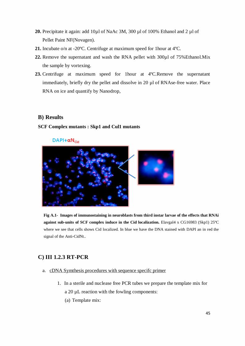
Fig A.1- Images of immunostaining in neuroblasts from third instar larvae of the effects that RNAi
against sub-units of SCF complex induce in the Cid localization. Elavgal4 x CG16983 (Skp1) 25ºC
where we see that cells shows Cid localized. In blue we have the DNA stained with DAPI an in red the
signal of the Anti-CidNt..

46
(i) V RNA
(ii) OligodT 50 pmol/ µL
(iii) H20 fms (final volume 13 µL)
2. Incubate 10 minutes at 65ºC
3. Put on ice
4. Add the components of table in this same order
Table A.1 – Composition of template primer mixture
Component Volume Final concentracion
Transcriptase reversa
Reaction buffer 5X conc.
L 1X (8mM MgCl2)
Protector Rna inhibitor
40U/L
0.5 L 20U
Deoxynucleotidde
(dNTP´s) Mix, 10mM
L 1mM each
Transcriptase reversa
20U/L
0.5L 10 U
Final volume 20L
5. Put in thermocycler Light cycler 480 for 1h at 50ºC and after 5min at
85ºC
6. To stop the reaction put on ice and storage at -20ºC
b. LightCycler® 480 SYBR Green I Master
This is designed for research studies and the LightCycler® 480 System it´s a kit ideally
suited for hot-start PCR applications. In combination with the LightCycler® 480
System and suitable PCR primers, this kit allows very sensitive detection and
quantification of defined DNA sequences.

47
c. Master Mix SyBr Green
Table A.1 – Composition of template primer mixture
Components Volume Concentracion
SyBr Green (2x)
5 µL
Oligo forword 0,2 µL 10 µM
Oligo reverse 0,2 µL 10 µM
H20 ( Vf=7 µL) 1,6 µL
All the information‘s are in www.roche-applied-science.com
Table A.1 primers used in RT-PCR and qRT-PCR
D) Culturing drosophila Kc (167) cells
Medium
1. Measure 800ml of H20 (for 1L of medium)
2. Add the powdered medium in continuous stirring (don´t dissolve completely but can´t
be heating)
3. Full the bottle with a small amount of water and add to the solution in step 2
4. Add 5,3 ml 7,5%(w/v) NaHCO3. Stir until we achieve the dissolution of the
powdered
5. Adjust to the pH 9 with 1M NaOH (approximately 50 ml 1N NaOH for litre). Stir for
10 minutes. Solution will become turbid
6. Add to pH 6.9 with 1M HCl (aprox. 35 ml 1N HCl). The solution will clear
7. Add 50 mL 12g/l CaCl2
8. Adjust the pH with 1N HCl or 1N NaOH to 0.1-0.3 pH units of the desired pH, 6 in
our case
9. Add H20 to bring the solution to 1L
10. Sterilize by filtration using a membrane with a porosity of 0,22µm. Store at 4ºC
(don´t freeze).

48
Growth conditions
- Growth at 23-25ºC or room temperature. Don´t need CO2
- You have to split them every 3-4 days to maintain
- They are density sensitive : Die if the culture is too dense or too dilute
Maintance
- Monitorizing the cells by microscopy before splitting and decide whether to adjust
recommended dilution factor accordingly
Kc 3-4days 1:6/1:8
Density between 1-8.106 cell/ml for Kc cells.
Freezing
1. Growth the cells to a density of 5-6.106 cells/ml in a T25 flask with 5mL of medium
2. Spin the cells in a 15 mL falcon tube for 2 minutes at 1300 rpm
3. Remove the supernatant and resuspend the cells in 900 µl of FBS
4. Transfer the cell to a cryovial and add 100 µl of DMSO
5. Place vial in a -20ºC for 2h
6. Then, place in-80ºC o/n (but if there is more than one night don´t exist any problem.
7. Transfer vial to LN2 for long storage
Thawing
1. Prepare a 15mL falcon tube with 5 mL medium
2. Thaw cells quickly
3. Transfer the cells to the falcon tube
4. Spin at 1300 rpm for 2 minutes
5. Aspirate and resuspend the cells in 5 mL medium (Vf = 15mL in the T25)
6. Transfer to a T25 flask
7. Watch every day. Initially, cells may need to be split at irregular intervals. After
2 weeks cells could be used to transfect.