risk-based scoring and genetic identification for
TRANSCRIPT
RESEARCH ARTICLE Open Access
Risk-based scoring and geneticidentification for anisakids in frozen fishproducts from Atlantic FAO areasGiorgio Smaldone1,2, Elvira Abollo3, Raffaele Marrone4* , Cristian E. M. Bernardi5, Claudia Chirollo4,Aniello Anastasio2,4 and Santiago P. del Hierro6
Abstract
Background: The presence of Anisakis larvae in fish represents a major public health concern. Effective riskmanagement procedures should be applied to prevent heavily infected products from reaching the market. Theaim of the study is to provide preliminary data on parasite exposure and risk classification in frozen fish products byapplying a risk categorization scheme (site, abundance, density and epidemiology – SADE) and Fish Parasite Rating(FPR) method. Fish and cephalopods samples (N = 771) from 5 different FAO Atlantic areas were examined andcategorized after an accurate visual inspection and a chloro-peptic digestion.
Results: In 25 out of 33 fish species parasite larvae were found. 10897 anisakids larvae were collected and identifiedto genus level. Molva dypterygia, Conger conger, Zeus faber and Aphanopus carbo were shown to be the most highlyinfected species. SADE and FPR scores were 1 and poor, respectively, for the referred species, because of thedisseminated Anisakis infection and commercial rejection.
Conclusion: SADE/FPR method showed high specificity and accuracy. The information provided in this work couldbe used in early warning systems for the detection of parasites in fishery products and might help fishing industriesin establishing management strategies for infected stocks in terms of cost saving decisions.
Keywords: Anisakis, food safety, risk ranking tool, zoonotic risk, fish inspection
BackgroundNematodes of the Anisakidae family are fish parasitesthat can be found all around the world. The larvae livein the gut, visceral peritoneum and flesh of many marinefish and cephalopod species and can colonize throughdifferent trophic bridges ensuring and widening theparasite life cycle. Differences in host range, host specifi-city and pathogenetic potential, even among members ofa given sibling species complex, have been historicallysuggested for anisakids [1–3]. Additionally, a positive re-lationship between body size/age of fish and larvalnematode prevalence and/or abundance has been dem-onstrated in several commercially important fish speciesfrom different wild catch sea areas [4–7].
Because no sea fishing grounds can be considered Ani-sakis free and the infection by anisakid larvae in fish is anatural condition and their complete eradication is notfeasible [8], surveillance studies are of great interest todetermine the risk exposure for those hot-spot geo-graphic areas of parasite recruitment to fish productionvalue chains. Moreover, nematodes of Anisakis generaare zoonotic parasites. In humans the ingestion of Anisa-kidae larvae can result in infection with live larvae, an al-lergic reaction to Anisakidae allergens or both [9–12].The increased consumption of raw or undercooked fishconstitutes an underestimated zoonotic potential risk[13–15]. In the last decade, Anisakis have been includedamong the biological hazards reported through theRapid Alert System for Food and Feed (RASFF) of theEuropean Commission, within the European Union(EU).
© The Author(s). 2020 Open Access This article is distributed under the terms of the Creative Commons Attribution 4.0International License (http://creativecommons.org/licenses/by/4.0/), which permits unrestricted use, distribution, andreproduction in any medium, provided you give appropriate credit to the original author(s) and the source, provide a link tothe Creative Commons license, and indicate if changes were made. The Creative Commons Public Domain Dedication waiver(http://creativecommons.org/publicdomain/zero/1.0/) applies to the data made available in this article, unless otherwise stated.
* Correspondence: [email protected] of Veterinary Medicine and Animal Production, Unit of FoodHygiene, University of Naples, Federico II, via Delpino 1, 80137 Naples, ItalyFull list of author information is available at the end of the article
Smaldone et al. BMC Veterinary Research (2020) 16:65 https://doi.org/10.1186/s12917-020-02286-7
European legislation [16] enforces an accurate visualinspection during the official control and in self-monitoring programs to prevent fish borne zoonoses: inthis context Food Business Operators (FBO) must en-sure that no fishery products obviously contaminatedwith visible parasites reach the consumers. According tothe “Guidance document on the implementation of cer-tain provisions of Regulation (EC) No 853/2004 on thehygiene of food of animal origin” [17], a fishery productis considered obviously contaminated if visible parasitesare found in edible portions; however, a maximum num-ber of parasites was not defined. Furthermore, the appli-cation of visual inspection procedure in the fisheryindustry depends on the ability and training of FBO [18].Because the presence of dead visible parasites could onlyrepresent a defect [19, 20] altering the global productsquality and in order to comply with the EU prescrip-tions, in addition to the official control and self-monitoring procedures, the most practical procedurecould be the use of a predicting scheme for evaluation ofnematode larvae in the edible part of the fish batches assuggested by the European Food Safety Authority(EFSA) [21]. The use of the SADE scheme (acronym ofSite of infection; Assurance of quality; Demography -density of parasites; Epidemiology of parasites) proposedby Llarena-reino et al., [22], combined with the FishParasite Rating (FPR) method [23], aimed at preciselyevaluating the likely outcome of infected fish lots, whichcould be useful tools. FPR standard is a certified Com-munity Trade Mark - Register No 012266607 at the Of-fice for Harmonization in the Internal Market (OHIM)and provides the staging of fish lots, helping in planningmanufacture, commercial, and research decisions duringself-management programs. The aim of this research isto provide data on parasite risk exposure in commercialfrozen fishery products collected in Atlantic FAO areasusing the SADE scheme combined with FPR method incomparison with the official visual inspection procedure.
ResultsParasites frequency in fish sub lotsA total of eight fish species (24.24% of sampled fish spe-cies/sub lots) were anisakid-free. Nematode larvae werenot detected in Mallotus villosus, Glyptocephalus cyno-glossus, Dicologlossa cuneata, Galeoides decadactylus,Trachurus trecae, Salilota australis, Atlantoraja castel-naui and Serranus cabrilla. A total of 10897 anisakidlarvae were collected and identified to genus level in theflesh of 25 species. Among these species, M. dypterygia,C. conger, Z. faber and A. carbo, coming from FAO area27 (Northeast Atlantic) were the most highly parasitized(Table 1) showing a total prevalence of infection (P) of100% with a mean abundance (MA ± SD) of 204.52 ±91.14, 115.16 ± 96.77, 44.96 ± 32.66, 74.1 ± 28.55
respectively. In these species, 90.45 % of the total larvaewere detected: in particular M. dypterygia reached thehighest density of parasites (102.26 larvae/kg). As much as46.97 % of total larvae in this species were detected. Thestatistical analyses indicate that there was a correlation be-tween MA and fish sample mean weight (p < 0.001).The hypaxial region was the most infected location. In
fact, in 11 fish species, anisakid larvae were found onlyin this region and in general the 98.53% of larvae (n.10737) were identified in this location. In 50% of thesamples, the epaxial infection took place simultaneouslywith hypaxial location.
Parasite frequency in fishing areasP of infection (± CI 95%) MA and mean intensity (MI)(± SD) in the different fishing grounds of the study arereported in Table 2. No parasites were found in FAOarea 34. Table 2 shows the comparison of P between dif-ferent FAO areas: significative statistical differences (p <0.0001) between FAO areas were found. In this study,regarding MA and MI, significative statistical differencesbetween FAO area 27 and the other sampling areas werefound (p < 0.001).
Parasite identificationThe results show mixed infection in 45.83 % of the fishsub lot examined. All sequences obtained in this studyshared 99-100 % nucleotide identity with other se-quences of anisakid species deposited in the GenBank(accession ID and web links for each identified parasite,linked to fish species and FAO area, are indicated in thesupplementary materials) belonging to Anisakis simplexsensu stricto, Anisakis pegreffii, Anisakis typica, Anisakisberlandi, Pseudoterranova cattani, Pseudoterranova deci-piens s.l., Contracaecum osculatum s.l. and Hysterothyla-cium aduncum.In this study A. simplex was the main parasite isolated
in fishery products from FAO area 21 (100%) and fromFAO area 27 (88.40%), while A. pegreffii was the mainparasite isolated in fishery products from FAO area 41(65.9%) and from FAO area 47 (63.82%). Fish collectedfrom FAO area 41 showed the highest variability interms of different species of parasites found.
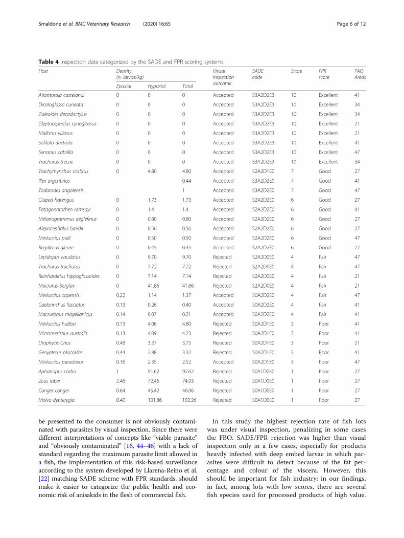
Risk categorisationTable 4 shows inspection data categorized by the SADEand FPR scoring systems. Using visual inspection,36.36% of the lots didn't meet the EU standards [16] andwere rejected. The rejected batches during the naked eyevisual inspection present at least 1 visible larva in the ed-ible portion. Over 66 % of fish sub lots have been ac-cepted as they present less than one parasite in the flesh,expressed as MA [24] (Table 1). MA, due to its correl-ation with P and with the number of samples [25], could
Smaldone et al. BMC Veterinary Research (2020) 16:65 Page 2 of 12
Table
1Infectionvalues
accordingto
Bush
etal.1997
Sublotho
stspecies
Parasites
PREVALEN
CE(%
±CI
95%)
MEANINTENSITY
±SD
MEANAB
UNDAN
CE±SD
Foun
dCo
rrectly
classified
Epaxial
Hypaxial
Total
Epaxial
Hypaxial
Total
Epaxial
Hypaxial
Total
Alepocepha
lusbairdii
145
0.00
16.00±0.14
16.00±0.14
0.00
3.50
±2.38
3.50
±2.38
0.00
0.56
±1.55
0.56
±1.55
Apha
nopuscarbo
741
660.00±0.30
100.00
100.00
1.33
±2.56
73.30±28.46
74.10±28.55
0.8±2.29
73.30±28.46
74.1±28.55
Atlantorajacastelna
ui0
00.00
0.00
0.00
0.00
0.00
0.00
0.00
0.00
0.00
Caelorinchus
fasciatus
33
4.00
±0.07
8.00
±0.10
12.00±0.12
1.00
1.00
1.00
0.04
±0.20
0.08
±0.27
0.12
±0.33
Clupea
hareng
us13
120.00
32.00±0.18
32.00±0.18
0.00
1.62
±0.74
1.62
±0.74
0.00
0.52
±0.87
0.52
±0.87
Cong
ercong
er2879
1264.00±0.18
100.00
100.00
2.50
±2.12
113.56
±96.34
115.16
±96.77
1.6±2.08
113.56
±96.34
115.16
±96.77
Dicologlossacuneata
00
0.00
0.00
0.00
0.00
0.00
0.00
0.00
0.00
0.00
Galeoides
decadactylus
00
0.00
0.00
0.00
0.00
0.00
0.00
0.00
0.00
0.00
Genypterusblacoides
8311
28.00±0.17
64.00±0.18
80.00±0.15
1.57
±0.78
4.50
±4.93
4.15
±4.76
0.44
±0.82
2.88
±4.48
3.32
±4.56
Glyptocepha
luscyno
glossus
00
0.00
0.00
0.00
0.00
0.00
0.00
0.00
0.00
0.00
Illex
argentinus
22
10.00±0.18
2.00
0.20
±0.63
Lepidopuscaudatus
449
0.00
100.00
100.00
0.00
4.88
±3.18
4.88
±3.18
0.00
4.88
±3.18
4.88
±3.18
Macruronu
smagellanicus
32
3.57
±0.06
3.57
±0.06
7.14
±0.10
2.00
1.00
1.50
±0.70
0.07
±0.38
0.03
±0.18
0.11
±0.41
Macrurusberglax
314
130.00
84.00±0.14
84.00±0.14
0.00
14.95±16.92
14.95±16.92
0.00
12.56±16.42
12.56±16.42
Mallotusvillosus
00
0.00
0.00
0.00
0.00
0.00
0.00
0.00
0.00
0.00
Melan
ogrammus
aeglefinus
75
0.00
12.00±0.12
12.00±0.12
0.00
2.33
±0.57
2.33
±0.57
0.00
0.28
±0.79
0.28
±0.79
Merlucciuscapensis
66
4.00
±0.07
16.00±0.14
20.00±0.15
1.00
1.25
±0.50
1.20
±0.50
0.04
±0.20
0.20
±0.50
0.24
±0.52
Merlucciushu
bbsi
216
1436.00±0.18
84.00±0.14
84.00±0.14
3.55
±6.57
8.66
±8.10
10.19±8.79
1.32
±4.20
7.28
±8.08
8.56
±8.90
Merlucciusparadoxus
1514
2.94
±0.05
23.52±0.14
23.52±0.14
1.00
1.75
±0.88
1.875±0.99
0.03
±0.17
0.411±0.85
0.44
±0.92
Merlucciuspolli
33
0.00
10.00±0.10
10.00±0.10
0.00
1.00
1.00
0.00
0.10
±0.30
0.10
±0.30
Micromesistiusaustralis
6112
11.76±0.15
70.58±0.21
70.58±0.21
1.00
4.91
±3.47
5.08
±3.34
0.11
±0.33
3.47
±3.69
3.58
±3.65
Molva
dypterygia
5113
836.00
100.00
100.00
2.22
±1.48
203.72
±90.78
204.52
±91.14
0.80
±1.38
203.72
±90.78
204.52
±91.14
Patagono
tothen
ramsayi
70
0.00
16.00±0.14
16.00±0.14
0.00
1.75
±1.50
1.75
±1.50
0.00
0.28
±0.84
0.28
±0.84
Regalecusglesne
43
0.00
36.00±0.28
36.00±0.28
0.00
1.33
±0.57
1.33
±0.57
0.00
0.36
±0.67
0.36
±0.67
Reinha
rditius
hippoglossoides
125
140.00
44.00±0.19
44.00±0.19
0.00
8.92
±14.2
8.92
±14.2
0.00
5.00
±11.39
5.00
±11.39
Salilotaaustralis
00
0.00
0.00
0.00
0.00
0.00
0.00
0.00
0.00
0.00
Serran
uscabrilla
00
0.00
0.00
0.00
0.00
0.00
0.00
0.00
0.00
0.00
Todarodesan
golensis
33
10.00±0.18
3.00
0.30
±0.94
Trachu
rustrachu
rus
6812
0.00
45.45±0.20
45.45±0.20
0.00
6.80
±14.54
6.80
±14.54
0.00
3.09
±10.13
3.09
±10.13
Trachu
rustrecae
00
0.00
0.00
0.00
0.00
0.00
0.00
0.00
0.00
0.00
Trachyrhynchus
scabrus
186
0.00
32.00±0.18
32.00±0.18
0.00
2.25
±1.83
2.25
±1.83
0.00
0.72
±1.45
0.72
±1.45
Uroph
ycisCh
us31
710.00±0.10
10.00±0.10
20.00±0.14
1.33
±0.57
9.00
±13.85
5.16
±9.72
0.13
±0.43
0.90
±4.55
1.03
±4.52
Zeus
faber
1124
1272.00±0.17
100.00
100.00
2.05
±1.35
43.48±32.11
44.96±32.66
1.48
±1.47
43.48±32.11
44.96±32.66
Smaldone et al. BMC Veterinary Research (2020) 16:65 Page 3 of 12

be used to estimate the degree of infestation [26], parti-curarly in the case of fishery products sold in batches.With regard to the naked eye rejected products, 33.33%of the total rejections belong to FAO 27. Not infectedfish batches in FAO area 34 were found.Using the SADE/FPR schemes, 27.27 % of fish sub lots
(M. hubbsi, M. australis, U. Chus, G. blacoides, M. para-doxus, A. carbo, Z. faber, C. conger, M. dypterygia) wereassigned a low SADE score (from 1 to 3) corresponding toa “poor” FPR standard. Hence, these fish lots must be dis-carded. The lowest score (SADE 1) was assigned to A.carbo, Z. faber, C. conger and M. dypterygia, belonging toFAO 27, corresponding to the 44.44 % of the total rejec-tion. No statistical differences (p = 0.3711) betweenSADE/FPR outcomes and visual inspection were observed.Finally, the non-zoonotic H. aduncum (Raphidascari-
dae) was also detected in I. argentinus: this parasite isgenerally considered not zoonotic, even if a case of inva-sive gastro-allergic infection was recently reported [27].This result did not show differences in the application ofSADE scoring system because a co-infection with thezoonotic A. simplex was noticed.
DiscussionParasites distributionThe high frequency of parasites and MA observed in thisstudy supports that Anisakis has the status of compo-nent parasite of many fish species and FAO fishing areas.Among the different species of Anisakis isolated, A. sim-plex commonly occurred in various ecologically and eco-nomically important fish species from Atlantic FAOareas 21, 27 and 41 (Atlantic Northwest, Northeast andSouthwest) as reported by Mattiucci et al. [28]. A. pegref-fii was found in southern Atlantic Ocean (FAO areas 41and 47) and in FAO area 27 in agreement to previousstudies [29–33]. According to Mattiucci et al. [34], A.typica can occur from 30° S to 35° N in warmer temper-ate and tropical waters and this data were confirmed byour findings (FAO area 47). Unlike Mattiucci [30] whohighlighted a discontinuous range of distribution of thisspecies including Pacific Canada, Chile, New Zealandwaters and the Atlantic South African coast, A. berlandiwas found only in South-Atlantic (FAO area 41 and
FAO area 47). P. decipiens s. l., as reported by Szosta-kowska et al., [35], occurs sporadically and in our workonly in 2 fish species were found (C. conger and Z. faberfrom FAO area 27) confirming that only parasites be-longing to the P. decipiens complex are present in theNE Atlantic Ocean. P. cattani was found in G. blacoidesfrom FAO area 41, in agreement with Timi et al. [36]. H.aduncum and C. osculatum s.l. were found only in FAOarea 41 with low prevalence, in contrast to data reportedby Niklitschek et al. [37] in the same sampling area inN= 41 samples of M. australis. Furthermore, in the samefish species caught in this area were found only parasitesbelonging to Anisakis genera.Anisakis and Pseudoterranova are generally most
abundant in European NE Atlantic waters [8]. These aretraditionally some of the most productive fishing areasin Europe and the abundance of different hosts at alltrophic levels presumably accounts for the overall abun-dance of the parasites. Differences in infection levelscould also be related to the presence of definitive hostsor to host’s feeding habits [1] and to the abundance ofobligate intermediate crustacean and/or cephalopodshosts. M. dypterygia, C. Conger and Z. faber were themost highly infected species (rejected after visual inspec-tion and with the lowest SADE/FPR scores), probablybecause of their relatively high trophic level in FAO area27 ecosystems, their size (p < 0.05) and high quantity offood intake confirm that this fishing area had the stron-gest effect on larvae infection [9, 38].Worst results corresponded to this fishing grounds with
significative differences in P, MA and MI match this areaand the others (p < 0.0001). No statistical differences (p >0.05) between FAO areas with low MA and MI (FAO 21,FAO 34, FAO 41 and FAO 47, Table 3) were observed.Moreover, the different spatial distribution in fish body
of Anisakis infecting the same fish species could be in-fluenced by Anisakis species. Cipriani et al. [7] notedthat in M. merluccius from FAO area 27, A. simplex lar-vae outnumbers A. pegreffii larvae in the flesh of thesame fish host; on the other hand, in the viscera themean abundance of two larvae species was superimpos-able. This phenomenon could be the result of differentresource utilization or linked to the different migrating
Table 2 FAO areas infection values according to Bush et al. 1997. Comparison of prevalence (χ2 ) of infection between different FAOareas
FAO areas Individuals Infected P (%) IC (±) 95 % χ2 p MA ± SD MI ± SD
Total sampling areas 771 251 32.55 3.30 106.25 p < 0.0001 14.13 77.18 43.41 48.46
21 130 41 31.53 7.98 3.61 10.07 11.46 15.33
27 196 111 56.63 6.93 50.57 85.89 89.30 97.91
34 100 0 0.00 0.00 0.00 0.00 0.00 0.00
41 190 63 33.15 6.69 1.97 4.75 5.95 6.68
47 155 36 23.22 6.64 0.89 4.10 3.86 7.89
Smaldone et al. BMC Veterinary Research (2020) 16:65 Page 4 of 12
ability of the Anisakis species because of different abil-ities of the two species to respond to the fish host’s im-mune system [39].
Safety and quality considerationsOur study confirms the presence of anisakid species withpublic health implications in lots of fishery products fromdifferent FAO areas. Although freezing condition andother treatments as salting and spicing assure no viablelarvae in the fish products [40–43], the risk of allergens inthe edible part of fish for hypersensitive individuals shouldbe highlighted. EU legislation [44] recognizes that any par-asitized fish under a visual inspection scheme should beunfit for human consumption. Comparing predictiveschemes and visual inspection, in general the highest
scores were associated with the acceptance of the fishbatches as stated by the EU legislation. A different situ-ation was found in the case of some batches: 7 fish sub-lots reached SADE score 4, corresponding to a “fair” FPRstandard. “Fair” fish batches have neither pathological norcommercial problems (A2 SADE code – Table 4) andFBO have the possibility to give different final destinationsto these fish lots, as processing, assuring safety and costsaving. Under visual inspection 4 “fair” fish batches wererejected because of the number of parasites detected (MAover 3, high parasite density – D0 SADE code) despite theabsence of flesh alterations. This approach matches theprecautionary principle set by Reg. EU 178/02 [20] butwas restrictive in terms of economics gain. As stated byEU Reg. 853/04, FBO have to ensure that the product to
Table 3 Number and percentage of parasites well sequenced collected in fishery products from different Atlantic areas
A. simplex(n./%)
A. pegreffii(n./%)
P. decipiens s. l.(n./%)
H. aduncum(n./%)
A. berlandi(n./%)
A. typica(n./%)
C. osculatum(n./%)
P. cattani(n./%)
FAO 21 34/100% 0% 0% 0% 0% 0% 0% 0%
Urophycis Chus 7/100% 0% 0% 0% 0% 0% 0% 0%
Reinharditiushippoglossoides
14/100% 0% 0% 0% 0% 0% 0% 0%
Macrurus berglax 13/100% 0% 0% 0% 0% 0% 0% 0%
FAO 27 61/88.40% 2/2.89 6/8.69 0% 0% 0% 0% 0%
Alepocephalus bairdii 5/100% 0% 0% 0% 0% 0% 0% 0%
Conger conger 6/50% 1/8.33% 5/41.66% 0% 0% 0% 0% 0%
Molva dypterygia 8/100% 0% 0% 0% 0% 0% 0% 0%
Zeus faber 11/91.66% 0% 1/8.33% 0% 0% 0% 0% 0%
Aphanopus carbo 6/100% 0% 0% 0% 0% 0% 0% 0%
Trachyrhynchus scabrus 6/100% 0% 0% 0% 0% 0% 0% 0%
Clupea harengus 12/100% 0% 0% 0% 0% 0% 0% 0%
Melanogrammusaeglefinus
5/100% 0% 0% 0% 0% 0% 0% 0%
Regalecus glesne 2/66.66% 1/33.33% 0% 0% 0% 0% 0% 0%
FAO 41 1/2.27% 29/65.9% 0% 1/2.27% 8/18.18% 0% 4/9.09% 1/2.27%
Micromesistius australis 0% 8/66.66% 0% 0% 3/25% 0 1/8.33% 0%
Genypterus blacoides 0% 7/63.63% 0% 0% 3/27.27% 0% 0% 1/9.09%
Merluccius hubbsi 0% 10/71.42% 0% 0% 1/7.14% 0% 3/21.42% 0%
Caelorinchus fasciatus 0% 2/66.66% 0% 0% 1/33.33% 0% 0% 0%
Macruronusmagellanicus
0% 2/100% 0% 0% 0% 0% 0% 0%
Illex argentinus 1/50% 0% 0% 1/50% 0% 0% 0% 0%
FAO 47 0% 30/63.82% 0% 0% 5/10.63% 12/25.53% 0% 0%
Trachurus trachurus 0% 12/100% 0% 0% 0% 0% 0% 0%
Lepidopus caudatus 0% 0% 0% 0% 0% 9/100% 0% 0%
Merluccius paradoxus 0% 13/92.86% 0% 0% 0% 1/7.14% 0% 0%
Merluccius capensis 0% 5/83.33% 0% 0% 1/16.66% 0% 0% 0%
Todarodes angolensis 0% 0% 0% 0% 3/100% 0% 0% 0%
Merluccius polli 0% 0% 0% 0% 1/33% 2/66.66% 0% 0%
Smaldone et al. BMC Veterinary Research (2020) 16:65 Page 5 of 12
be presented to the consumer is not obviously contami-nated with parasites by visual inspection. Since there weredifferent interpretations of concepts like “viable parasite”and “obviously contaminated” [16, 44–46] with a lack ofstandard regarding the maximum parasite limit allowed ina fish, the implementation of this risk-based surveillanceaccording to the system developed by Llarena-Reino et al.[22] matching SADE scheme with FPR standards, shouldmake it easier to categorize the public health and eco-nomic risk of anisakids in the flesh of commercial fish.
In this study the highest rejection rate of fish lotswas under visual inspection, penalizing in some casesthe FBO. SADE/FPR rejection was higher than visualinspection only in a few cases, especially for productsheavily infected with deep embed larvae in which par-asites were difficult to detect because of the fat per-centage and colour of the viscera. However, thisshould be important for fish industry: in our findings,in fact, among lots with low scores, there are severalfish species used for processed products of high value.
Table 4 Inspection data categorized by the SADE and FPR scoring systems
Host Density(n. larvae/kg)
Visualinspectionoutcome
SADEcode
Score FPRscore
FAOAreas
Epaxial Hypaxial Total
Atlantoraja castelanui 0 0 0 Accepted S3A2D2E3 10 Excellent 41
Dicologlossa cuneata 0 0 0 Accepted S3A2D2E3 10 Excellent 34
Galeoides decadactylus 0 0 0 Accepted S3A2D2E3 10 Excellent 34
Glyptocephalus cynoglossus 0 0 0 Accepted S3A2D2E3 10 Excellent 21
Mallotus villosus 0 0 0 Accepted S3A2D2E3 10 Excellent 21
Salilota australis 0 0 0 Accepted S3A2D2E3 10 Excellent 41
Serranus cabrilla 0 0 0 Accepted S3A2D2E3 10 Excellent 47
Trachurus trecae 0 0 0 Accepted S3A2D2E3 10 Excellent 34
Trachyrhynchus scabrus 0 4.80 4.80 Accepted S2A2D1E0 7 Good 27
Illex argentinus 0.44 Accepted S3A2D2E0 7 Good 41
Todarodes angolensis 1 Accepted S3A2D2E0 7 Good 47
Clupea harengus 0 1.73 1.73 Accepted S2A2D2E0 6 Good 27
Patagonotothen ramsayi 0 1.4 1.4 Accepted S2A2D2E0 6 Good 41
Melanogrammus aeglefinus 0 0.80 0.80 Accepted S2A2D2E0 6 Good 27
Alepocephalus bairdii 0 0.56 0.56 Accepted S2A2D2E0 6 Good 27
Merluccius polli 0 0.50 0.50 Accepted S2A2D2E0 6 Good 47
Regalecus glesne 0 0.45 0.45 Accepted S2A2D2E0 6 Good 27
Lepidopus caudatus 0 9.70 9.70 Rejected S2A2D0E0 4 Fair 47
Trachurus trachurus 0 7.72 7.72 Rejected S2A2D0E0 4 Fair 47
Reinharditius hippoglossoides 0 7.14 7.14 Rejected S2A2D0E0 4 Fair 21
Macrurus berglax 0 41.86 41.86 Rejected S2A2D0E0 4 Fair 21
Merluccius capensis 0.22 1.14 1.37 Accepted S0A2D2E0 4 Fair 47
Caelorinchus fasciatus 0.13 0.26 0.40 Accepted S0A2D2E0 4 Fair 41
Macruronus magellanicus 0.14 0.07 0.21 Accepted S0A2D2E0 4 Fair 41
Merluccius hubbsi 0.73 4.06 4.80 Rejected S0A2D1E0 3 Poor 41
Micromesistius australis 0.13 4.09 4.23 Rejected S0A2D1E0 3 Poor 41
Urophycis Chus 0.48 3.27 3.75 Rejected S0A2D1E0 3 Poor 21
Genypterus blacoides 0.44 2.88 3.32 Rejected S0A2D1E0 3 Poor 41
Merluccius paradoxus 0.16 2.35 2.52 Accepted S0A2D1E0 3 Poor 47
Aphanopus carbo 1 91.62 92.62 Rejected S0A1D0E0 1 Poor 27
Zeus faber 2.46 72.46 74.93 Rejected S0A1D0E0 1 Poor 27
Conger conger 0.64 45.42 46.06 Rejected S0A1D0E0 1 Poor 27
Molva dypterygia 0.40 101.86 102.26 Rejected S0A1D0E0 1 Poor 27
Smaldone et al. BMC Veterinary Research (2020) 16:65 Page 6 of 12
In fact, M. dypterygia is used for deep or light saltedproducts and M. hubbsi, M. capensis and M. para-doxus are the most used species for fish sticks. Ac-cording to EU legislation [16], M. paradoxus wouldnot have been rejected because of the low number ofdeep embed larvae not detectable by naked eye in-spection. These sub lots, according to the schemeadopted, were rejected to prevent food business oper-ator to suffer serious commercial losses.This work aims to present the application of the above
mentioned method on fishery products coming fromseveral Atlantic FAO areas. Recently Rodriguez et al.[23], according to the SADE/FPR scheme, examined fishcaught from 3 different ICES areas (ICES VII – GrandSole, ICES VIII – Galician coast and ICES IX – Portu-guese coast) located in the same FAO area (NE Atlanticareas – FAO 27). These authors gave “poor” FPR scoreto only 2 fish species, M. merluccius (ICES VII and ICESVIII) and Lophius budegassa (ICES VII), of the 9 exam-ined, differently from our results where several fish spe-cies (A. carbo, Z. faber, C. conger, M. dypterygia), caughtin NE Atlantic areas, reached SADE 1 and consequently“poor” FPR score.The combined scoring systems are less restrictive than
visual inspection: results compared between the differentmethods could be helpful to analyse an appropriate bal-ance in terms of consumer’s safety and FBO interests.The SADE/FPR method has an acceptable sensitivity(66.7%; CI95% 34.8 – 90.1%) but a high specificity(95.2%; CI95% 76.2 – 99.9%). The accuracy of 84.85%(CI95% 68.1 – 94.9%) indicates that the SADE/FPRmethod has a high capacity to correctly classify fisheryproducts. This predictive scheme, proposing correctivemeasures within HACCP procedures, proved to be veryuseful for fish lots with the lowest FPR rating particu-larly and offers a crucial food safety device for assessingrisks associated with parasites.
ConclusionSADE score combined with FPR standard may repre-sent a specific low-cost tool in fish inspection, ensur-ing both safety and quality, that could be useful forcompetent authorities and fish industry operators toestablish standard management strategies. Thecategorization of lots in 5 quality batches, allowingthe possibility of calculating accurately both parasiticload and flesh integrity, could give a unique languageand modus operandi during self-control inspections inHACCP procedures and programs addressing fish lotsin different ways depending on the score. The highspecificity and accuracy of the applied predictive testsguarantees its correct applicability during the fish in-spection procedures.
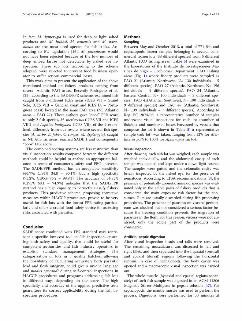
MethodsSamplingBetween May and October 2013, a total of 771 fish andcephalopods frozen samples belonging to several com-mercial frozen lots (33 different species) from 5 differentAtlantic FAO fishing areas (Table 5) were examined inthe laboratories of the Instituto de Investigaciones Ma-rinas de Vigo – Ecobiomar Department. FAO Fishingareas (Fig. 1) where fishery products were sampled asFAO 21 (Atlantic, Northwest, N= 130 individuals – 5different species), FAO 27 (Atlantic, Northeast, N= 196individuals – 9 different species), FAO 34 (Atlantic,Eastern Central, N= 100 individuals – 3 different spe-cies), FAO 41(Atlantic, Southwest, N= 190 individuals –9 different species) and FAO 47 (Atlantic, Southwest,N= 139 individuals – 7 different species). According toReg. EC 2074/05, a representative number of samplesunderwent visual inspection; for each lot (number offish/box and number of boxes harvested by vessels thatcompose the lot is shown in Table 5) a representativesample (sub lot) was taken, ranging from 12% for Mer-luccius polli to 100% for Aphanopus carbo).
Visual inspectionAfter thawing, each sub lot was weighed, each sample wasweighed individually, and the abdominal cavity of eachsample was opened and kept under a down-light source.The samples were gutted and the celomatic cavity wasbriefly inspected by the naked eye, for the presence ofnematodes. According to EFSA recommendations [8], thepresence of potentially zoonotic anisakid species was eval-uated only in the edible parts of fishery products that isconsidered the main exposure risk factor for the con-sumer. Guts are usually discarded during fish-processingprocedures. The presence of parasites on visceral periton-eum was checked but not considered a serious factor be-cause the freezing condition prevents the migration ofparasites in the flesh. For this reason, viscera were not an-alyzed, only the edible part of the products wereconsidered.
Artificial peptic digestionAfter visual inspection heads and tails were removed.The remaining musculature was dissected in left andright fillets and then separated into the hypaxial (ventral)and epaxial (dorsal) regions following the horizontalseptum. In case of cephalopods, the body cavity wasopened and a macroscopic visual inspection was carriedout.The whole muscle (hypaxial and epaxial regions separ-
ately) of each fish sample was digested in an ACM-11806Magnetic Stirrer Multiplate in pepsin solution [47]. Forcephalopods, the mantle muscle was used to perform theprocess. Digestions were performed for 30 minutes at
Smaldone et al. BMC Veterinary Research (2020) 16:65 Page 7 of 12
incubation temperature of 37° C in an acid solution (pH =1.5) with HCl 0.063 M. Assays using liquid pepsin at con-centration of 0.5 % and a ratio 1:20 sample weight/solu-tion volume were used. The digestion solution wasdecanted through a sieve and the rests of digestion andnematodes were inspected under stereomicroscope. Allanisakids were placed in individual eppendorf with ethanol70% for further molecular diagnosis.
Molecular analysisAll anisakid larvae were identified at genus level bymicroscopic examination of diagnostic characters. The
biomolecular identification was performed by randomlychoosing 15 larvae per species; in sub lots/species with anumber of parasites lower than 15, all larvae were ana-lyzed. A total of 275 anisakid larvae, previously identifiedat genus level, were used for molecular identification butonly 194 were correctly classified by biomolecular ana-lysis (Table 1). DNA extractions were performed usingthe commercial kit NucleoSpin®Tissue kit (Macherey-Nagel) following the manufacturer’s recommended pro-tocols. DNA quality and quantity were checked in aspectrophotometer Nanodrop® ND-1000 (Nanodroptechnologies, Inc). The entire ITS (ITS1, 5.8S rDNA
Table 5 Samples collected from Atlantic FAO areas
FAO fishing areas Coordinates Host N. boxes / total fish count Individuals sampled (N)
FAO 21 Atlantic, Northwest Glyptocephalus cynoglossus 1/60 25
FAO 21 Atlantic, Northwest 48°38'N 45°43'W Macrurus berglax 2/50 25
FAO 21 Atlantic, Northwest 46°51'N 47°20'W Mallotus villosus 1/50 25
FAO 21 Atlantic, Northwest 48°33'N 45°45'W Reinharditius hippoglossoides 8/109 25
FAO 21 Atlantic, Northwest 48°38' N 45°42' W Urophycis chus 4/114 30
FAO 27 Atlantic, Northeast 56°13'N 17°34'W Alepocephalus bairdii 7/52 25
FAO 27 Atlantic, Northeast 56°13'N 17°35'W Aphanopus carbo 1/10 10
FAO 27 Atlantic, Northeast FAO 27 IIa Clupea harengus 2/100 25
FAO 27 Atlantic, Northeast FAO 27/ VII Conger conger 6/100 25
FAO 27 Atlantic, Northeast Melanogrammus aeglefinus 1/50 25
FAO 27 Atlantic, Northeast 54°34'N 17°59'W Molva dypterygia 11/71 25
FAO 27 Atlantic, Northeast 58°38'N 15°04'W Regalecus glesne 2/11 11
FAO 27 Atlantic, Northeast FAO 27/XII Trachyrhynchus scabrus 2/100 25
FAO 27 Atlantic, Northeast Zeus faber 1/50 25
FAO 34 Atlantic, Eastern Central Dicologlossa cuneata 2/50 25
FAO 34 Atlantic, Eastern Central 12°50'N17°25'W Galeoides decadactylus 5/50 25
FAO 34 Atlantic, Eastern Central 13°00'N17°15'W Trachurus trecae 2/60 50
FAO 41 Atlantic, Southwest Atlantoraja castelanui 8/50 10
FAO 41 Atlantic, Southwest Caelorinchus fasciatus 1/50 25
FAO 41 Atlantic, Southwest Genypterus blacoides 3/50 25
FAO 41 Atlantic, Southwest Illex argentinus 3/50 10
FAO 41 Atlantic, Southwest Macruronus magellanicus 9/>200 28
FAO 41 Atlantic, Southwest Merluccius hubbsi 6/50 25
FAO 41 Atlantic, Southwest Micromesistius australis 3/50 17
FAO 41 Atlantic, Southwest Patagonotothen ramsayi 1/50 25
FAO 41 Atlantic, Southwest Salilota australis 2/50 25
FAO 47 Atlantic, Southeast 13°37,09S 12°17,38'E Lepidopus caudatus 1/25 9
FAO 47 Atlantic, Southeast 23°21,5' S 13°22,3'E Merluccius capensis 1/36 25
FAO 47 Atlantic, Southeast 27°11,2`' S 14° 22,5'E Merluccius paradoxus 1/50 34
FAO 47 Atlantic, Southwest 11°48,84S 13°22,97'E Merluccius polli 12/>250 30
FAO 47 Atlantic, Southeast 25°53,1' S 13° 41,7W Serranus cabrilla 1/50 25
FAO 47 Atlantic, Southeast 27°03,8S 14°14,7E Todarodes angolensis 3/50 10
FAO 47 Atlantic, Southeast 24°10,9S 13°31,0'E Trachurus trachurus 2/50 22
Smaldone et al. BMC Veterinary Research (2020) 16:65 Page 8 of 12
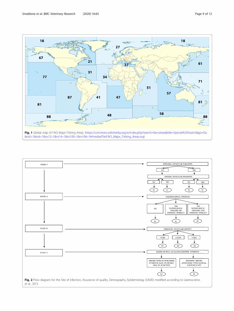
Fig. 2 Flow diagram for the Site of infection, Assurance of quality, Demography, Epidemiology (SADE) modified according to Llarena-reinoet al., 2013
Fig. 1 Global map of FAO Major Fishing Areas. (https://commons.wikimedia.org/w/index.php?search=fao+areas&title=Special%3ASearch&go=Go&ns0=1&ns6=1&ns12=1&ns14=1&ns100=1&ns106=1#/media/File:FAO_Major_Fishing_Areas.svg)
Smaldone et al. BMC Veterinary Research (2020) 16:65 Page 9 of 12
gene and ITS2) was amplified using the forward primerNC5 (5’-GTA GGT GAA CCT GCG GAA GGA TCATT-3’) and the reverse primer NC2 (5’-TTA GTT TCTTTT CCT CCG CT-3’). PCR assays were carried out ina total volume of 25 μl containing 100 ng of genomicDNA, 0.3 μM of each primer, 2.5 μl of 10x buffer, 1.5mM of MgCl2, 0.2 mM of dNTPs and 0.625 U of TaqDNA polymerase (Roche Mannheim, Germany). PCRcycling parameters included denaturation at 94°C for 2min, followed by 35 cycles of 94 °C for 30 s, annealing at55 °C for 30 s, and extension at 72 °C for 75 s, and afinal extension at 72 °C for 7 min. PCR products werepurified for sequencing using ExoSAP-IT © followingrecommended protocol by the manufacturer. Sequencingwas performed by Secugen (Madrid, Spain) and the elec-tropherograms were analysed using the program Chro-masPro version 1.41 Technelysium Pty LtdA. Allsequences were searched for similarity using BLAST(Basic Local Alignment Search Tool) through webservers of the National Center for Biotechnology Infor-mation (USA).
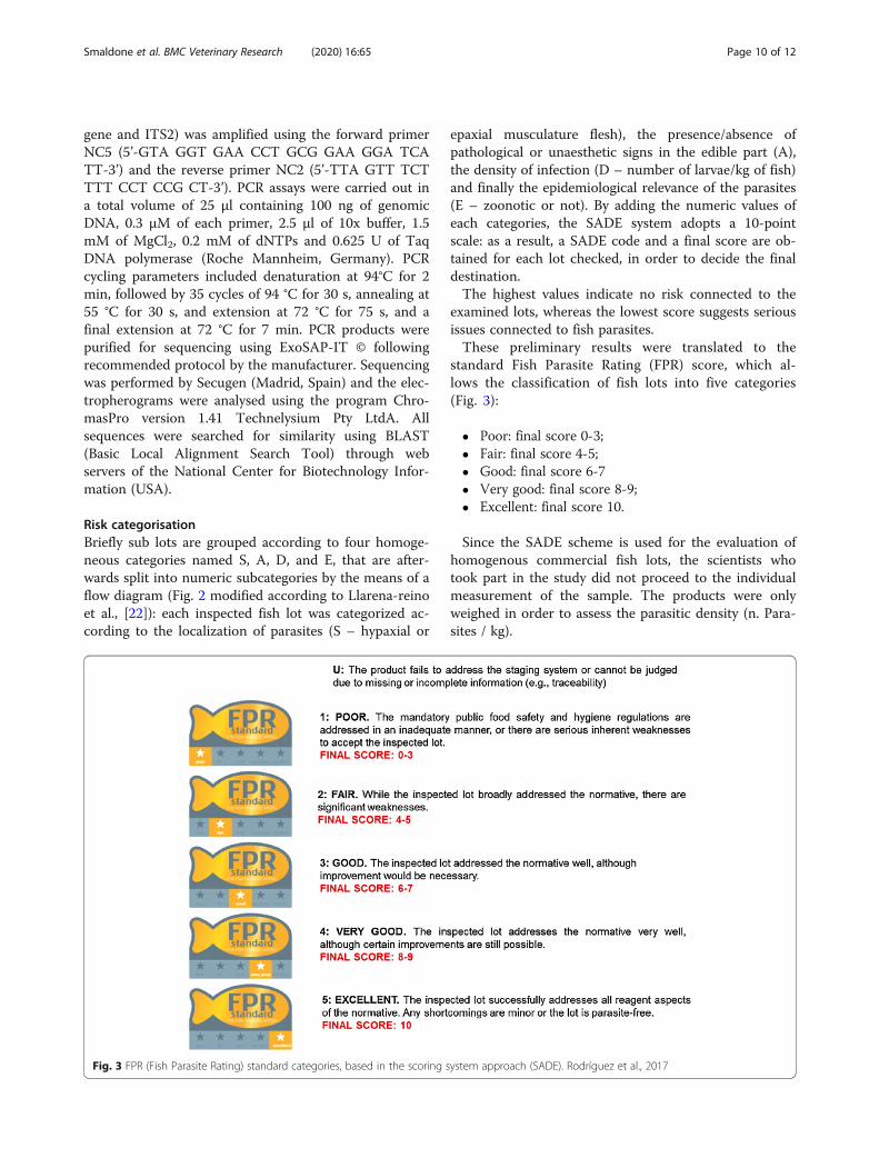
Risk categorisationBriefly sub lots are grouped according to four homoge-neous categories named S, A, D, and E, that are after-wards split into numeric subcategories by the means of aflow diagram (Fig. 2 modified according to Llarena-reinoet al., [22]): each inspected fish lot was categorized ac-cording to the localization of parasites (S – hypaxial or
epaxial musculature flesh), the presence/absence ofpathological or unaesthetic signs in the edible part (A),the density of infection (D – number of larvae/kg of fish)and finally the epidemiological relevance of the parasites(E – zoonotic or not). By adding the numeric values ofeach categories, the SADE system adopts a 10-pointscale: as a result, a SADE code and a final score are ob-tained for each lot checked, in order to decide the finaldestination.The highest values indicate no risk connected to the
examined lots, whereas the lowest score suggests seriousissues connected to fish parasites.These preliminary results were translated to the
standard Fish Parasite Rating (FPR) score, which al-lows the classification of fish lots into five categories(Fig. 3):
� Poor: final score 0-3;� Fair: final score 4-5;� Good: final score 6-7� Very good: final score 8-9;� Excellent: final score 10.
Since the SADE scheme is used for the evaluation ofhomogenous commercial fish lots, the scientists whotook part in the study did not proceed to the individualmeasurement of the sample. The products were onlyweighed in order to assess the parasitic density (n. Para-sites / kg).
Fig. 3 FPR (Fish Parasite Rating) standard categories, based in the scoring system approach (SADE). Rodríguez et al., 2017
Smaldone et al. BMC Veterinary Research (2020) 16:65 Page 10 of 12
Finally, according to the European Hygiene Rules (Reg.EC 853/2004, Section VIII, Chapter V, Pt. D), the per-centage of fishery products that should be unfit for hu-man consumption was calculated and compared withSADE – FPD scores.
Statistical analysisTo describe parasite population the following parameterswere used, according to Bush et al. [24]: P (the ratio be-tween parasitized subjects/sub lots and the total sub-jects/ sub lots analysed), MA (the ratio between thenumber of larvae recovered and the number of all exam-ined subjects of sub lots) and MI (the ratio between thenumber of larvae recovered and the number of exam-ined parasitized subjects of sub lots). Infection indexeswere calculated regardless of parasites’ localisation site(Epaxial/Hypaxial flesh), species/sub lot and for eachFAO areas. The differences in the P between FAO areaswere assessed by the two-sided chi-square test.Statistical significance between MA and MI of differ-
ent FAO Areas was performed using One-way ANOVAwith Bonferroni’s post-test.In order to assess statistical association between MA
and fish sample mean weight a simple regression ana-lysis was used. Finally, statistical comparisons betweenSADE/FPR and Visual Inspection were performed byMcNemar’s chi-square test [48]. Moreover, sensitivity,specificity and accuracy values of the SADE/FPR relativeto Visual Inspection were calculated. Statistical analyseswere performed using GraphPad InStat Version 3.0(GraphPad Software, San Diego California USA) andMedCalc for Windows, version 18.11.3 (MedCalc Soft-ware, Ostend, Belgium); p < 0.05 was considered signifi-cant for all statistical tests.
Supplementary informationSupplementary information accompanies this paper at https://doi.org/10.1186/s12917-020-02286-7.
Additional file 1. Identified parasites in fish species,Accession ID relatedto the aligned sequences and web links (https://www.ncbi.nlm.nih.gov/pubmed/).
AbbreviationsEFSA: European Food Safety Authority; EU: European Union; EU: EuropeanUnion; FAO: Food and Agriculture Organization of the United Nations;FBO: Food Business Operators; FPR: Fish Parasite Rating; HACCP: HazardAnalysis and Critical Control Points; ICES: International Council for theExploration of the Sea; MA: Mean abundance; MI: Mean intensity;OHIM: Office for Harmonization in the Internal Market; P: Prevalence ofinfection; RASFF: Rapid Alert System for Food and Feed; SADE: Site,abundance, density and epidemiology
AcknowledgementsWe would like to thank the Instituto de Investigaciones Marinas de Vigo -Consejo Superior de Investigaciones Científicas (IIM-CSIC) and the all team ofDepartment of Ecology and Marine Biodiversity (ECOBIOMAR) for the humanand technical support during the study.
Authors’ contributionsGS and SP designed the overall study; EA and CC developed the molecularprotocol. GS performed parasites inspection in fishery products andmorphological identification of anisakid samples. AA, RM and SP analysedthe results and drafted the paper. SP, CB and GS contributed to the ideasbehind the study and the writing of the paper. All authors critically reviewedthe paper and agreed the final content of the version to be published. Theauthor(s) read anda pproved the final manuscript
FundingThis work has been partially supported by the Istituto ZooprofilatticoSperimentale della Sicilia – granted project "Indagine epidemiologica sullaprevalenza di allergie da Anisakis nella Regione Sicilia”.
Availability of data and materialsThe datasets used and/or analyzed during the current study are availablefrom the corresponding author on reasonable request. Sequencing wasperformed by Secugen (Madrid, Spain) and the electropherograms wereanalysed using the program ChromasPro version 1.41 Technelysium PtyLtdA. All sequences were searched for similarity using BLAST (Basic LocalAlignment Search Tool) (https://blast.ncbi.nlm.nih.gov/Blast.cgi). Accession IDwere in the supplementary materials.
Ethics approval and consent to participateNot applicable.
Consent for publicationNot applicable.
Competing interestsThe authors declare that they have no competing interests.
Author details1Department of Agricultural Sciences, University of Naples, Federico II, viaUniversità 100, 80055 Naples, Portici (NA), Italy. 2Centro di RiferimentoRegionale per la Sicurezza Sanitaria del Pescato CRiSSaP, Naples, CampaniaRegion, Italy. 3Centro Tecnológico del Mar - Fundación CETMAR, C/EduardoCabello s/n, 36208 (Pontevedra), Vigo, Spain. 4Department of VeterinaryMedicine and Animal Production, Unit of Food Hygiene, University of Naples,Federico II, via Delpino 1, 80137 Naples, Italy. 5Department of VeterinaryScience and Technologies for Food Safety, Laboratory of Food Inspection,Università degli Studi di Milano, Via A. Grasselli, 7-20137, Milano, Italy.6Ecobiomar - Instituto de Investigaciones Marinas de Vigo – CSIC - C/Eduardo Cabello 6, 36208 (Pontevedra),, Vigo, Spain.
Received: 24 January 2019 Accepted: 13 February 2020
References1. Mattiucci S, Cipriani P, Webb SC, Paoletti M, Marcer F, Bellisario B, Gibson DI,
Nascetti G. Genetic and morphological approaches distinguish the threesibling species of the Anisakis simplex species complex, with a speciesdesignation as Anisakis nascettii n. sp. for A. simplex sp. C (Nematoda:Anisakidae). J Parasitol. 2014;100:199–214.
2. Mattiucci S, Nascetti G. Advances and trends in the molecular systematics ofanisakid nematodes, with implications for their evolutionary ecology andhost– parasite co-evolutionary processes. Adv Parasitol. 2008;66:47–148.
3. Arizono N, Yamada M, Tegosh T, Yoshikawa M. Anisakis simplex sensu strictoand Anisakis pegreffii: Biological Characteristics and Pathogenetic Potential inHuman Anisakiasis. Foodborne Pathog Dis. 2012;9(6):517–21.
4. McGladdery SE. Anisakis simplex (Nematoda: Anisakidae) infection of themusculature and body cavity of Atlantic herring (Clupea harengus harengus).Can J Fish Aquat Sci. 1986;43:1312–7.
5. Valero A, Martín-Sánchez J, Reyes-Muelas E, Adroher FJ. Larval anisakidsparasitizing the blue whiting, Micromesistius poutassou, from Motril Bay inthe Mediterranean region of southern Spain. J Helminthol. 2000;74:361–4.
6. Levsen A, Midthun E. Occurrence and spatial distribution of Anisakis sp. inthree commercially important pelagic fish stocks from the NE Atlantic, withcomments on the significance to consumer safety. Parassitologia. 2007;2:402–3.
Smaldone et al. BMC Veterinary Research (2020) 16:65 Page 11 of 12
7. Cipriani P, Smaldone G, Acerra V, D’Angelo L, Anastasio A, Bellisario B, PalmaG, Nascetti G, Mattiucci S. Genetic identification and distribution of theparasitic larvae of Anisakis pegreffii and Anisakis simplex (s. s.) in Europeanhake Merluccius merluccius from the Tyrrhenian Sea and Spanish Atlanticcoast: implications for food safety. Int J Food Microbiol. 2015;198:1–8.
8. European Food Safety Authority, Panel on Biological Hazards (BIOHAZ).Scientific opinion on risk assessment of parasites in fishery products. EFSAJournal. 2010;8:1543.
9. Rello FJ, Adroher FJ, Benítez R, Valero A. The fishing area as a possibleindicator of the infection by anisakids in anchovies (Engraulis encrasicolus)from southwestern Europe. Int J Food Microbiol. 2009;129:277–81.
10. Mattiucci S, Fazii P, De Rosa A, Paoletti M, Salomone MA, Glielmo A, DeAngelis M, Costa A, Meucci C, Calvaruso V, Sorrentini I, Palma G, Bruschi F,Nascetti G. Anisakiasis and gastroallergic reactions associated with Anisakispegreffii infection. Italy. Emerg Infect Dis. 2013;19:496–9.
11. Bao M, Pierce GJ, Pascual S, González-Muñoz M, Mattiucci S, MladineoI, Cipriani P, Bušelić I, Strachan NJC. Assessing the risk of anemerging zoonosis of worldwide concern: anisakiasis. ScientificReports. 2017;7:43699.
12. Moneo I, Carballeda-Sangiao N, González-Muñoz M. New perspectives on thediagnosis of allergy to Anisakis spp. Curr Allergy Asthma Rep. 2017;17(5):27.
13. Daschner A, Cuéllar C, Rodero M. The Anisakis allergy debate: does anevolutionary approach help? Trends Parasitol. 2012;28:9–15.
14. Mazzucco W, Raia DD, Marotta C, Costa A, Ferrantelli V, Vitale F, Casuccio A.Anisakis sensitization in different population groups and public healthimpact: A systematic review. PLoS ONE. 2018;13(9):e0203671.
15. Kruse Fæstea C, Jonscherb KR, Doopera MMWB, Egge-Jacobsenc W, Moenc A,Daschnerd A, Egaasa E, Christiansb U. Characterisation of potential novel allergensin the fish parasite Anisakis simplex. EuPA Open Proteomics. 2014;4:140–55.
16. Regulation (EC) No 2074/2005 (EC 853/2004 rev) of 5 December, 2005laying down implementing measures for certain products under Regulation(EC) No 853/2004, Regulation (EC) No 854/2004 and Regulation (EC) No882/2004, derogating from Regulation (EC) No 852/2004 and amendingRegulations (EC) No 853/ 2004 and (EC) No 854/2004, Official Journal of theEuropean Union.
17. European Commission, 2014. Guidance Document on the Implementationof Certain Provisions of Regulation (EC) No 853/2004 on the Hygiene ofFood of Animal Origin. https://ec.europa.eu/food/sites/food/files/safety/docs/biosafety_fh_legis_guidance_reg-2004-853_en.pdf, .
18. Levsen A, Lunestad B, Berland B. Low detection efficiency of candling as acommonly recommended inspection method for nematode larvae in theflesh of pelagic fish. J. Food Prot. 2005;68(4):828–32.
19. Codex Alimentarius, 2012. Code of Practice for Fish and Fishery Products.World Health Organization and Food and Agriculture Organization of theUnited Nations, Rome. ftp://ftp.fao.org/codex/Publications/Booklets/Practice_code_fish/CCFFP_2012_EN.pdf, .
20. Regulation EC No 178/2002 of the European Parliament and of the Councilof 28 January 2002 Laying Down the General Principles and 1426 FoodAnal. Methods, 2016. 9: 1418–1427 Requirements of Food Law, Establishingthe European Food Safety Authority and Laying Down Procedures inMatters of Food Safety. OJEC L31, 1–24.
21. European Food Safety Authority, Panel on Biological Hazards (BIOHAZ).Scientific Opinion on the development of a risk ranking framework onbiological hazards. EFSA Journal. 2012; 10(6): 2724.
22. Llarena-Reino M, Abollo E, Pascual S. A Scoring System Approach for theParasite Predictive Assessment of Fish Lots: A Proof of Concept withAnisakids. Foodborne Pathog Dis. 2013;10(12):1067–74.
23. Rodríguez H, Abollo E, González ÁF, Pascual S. Scoring the parasite risk in highly-valuable fish species from southern ICES areas. Fish Res. 2017;202:134–9.
24. Bush AO, Lafferty KD, Lotz JM, Shostak AW. Parasitology meets ecology onits own terms: Margolis et al. revisited. J Parasitol. 1997;83:575–83.
25. Rózsa L, Reiczigel J, Majoros G. Quantifying parasites in samples of hosts. J.Parasitol. 2000;86:228–32.
26. Guardone L, Malandra R, Costanzo F, Castigliego L, Tinacci L, Gianfaldoni D,Guidi A, Armani A. Assessment of a sampling plan based on visualinspection for the detection of Anisakid larvae in fresh Anchovies (Engraulisencrasicolus). A first step towards official validation? Food Anal. Methods.2016;1:10.
27. González-Amores Y, Clavijo-Frutos E, Salas-Casanova C, Alcain-Martínez G.Direct parasitological diagnosis of infection with Hysterothylacium aduncumin a patient with epigastralgia. Rev Esp Enf Digest. 2015;107(11):699–700.
28. Mattiucci S, Nascetti G. Molecular systematics, phylogeny and ecology ofanisakid nematodes of the genus Anisakis Dujardin, 1845: an update.Parasite. 2006;13:99–113.
29. Nascetti G, Paggi L, Orecchia P, Smith JW, Mattiucci S, Bullini L. Electrophoreticstudies on the Anisakis simplex complex (Ascaridida: Anisakidae) from theMediterranean and North East Atlantic. Int J Parasitol. 1986;16:633–40.
30. Mattiucci S, Nascetti G, Clanchi R, Paggi L, Arduino P, Margolis L, Brattey J,Webb S, D'Amelio S, Orecchia P, Bullini L. Genetic and ecological data onthe Anisakis simplex complex, with evidence for a new species (Nematoda,Ascaridoidea, Anisakidae). J Parasitol. 1997;83:401–16.
31. Paggi L, Mattiucci S, D’Amelio S, Nascetti G. Nematodi del genere Anisakis inpesci, cefalopodi e cetacei del Mar Mediterraneo e dell’Oceano Atlantico ePacifico. Biologia Marina Mediterranea. 1998;5(3):1585–92.
32. Abollo E, D'Amelio S, Pascual S. Fitness of the marine parasitic nematodeAnisakis simplex s. str. in temperate waters of the NE Atlantic. Dis AquatOrgan. 2001;45:131–9.
33. Mattiucci S, Abaunza P, Ramadori L, Nascetti G. Genetic identification ofAnisakis larvae in European hake from Atlantic and Mediterranean waters forstock recognition. J Fish Biol. 2004;65:495–510.
34. Mattiucci S, Paggi L, Nascetti G, Portes Santos C, Costa G, Di Beneditto AP,Ramos R, Argyrou M, Cianchi R, Bullini L. Genetic markers in the study ofAnisakis typica (Diesing, 1860): larval identification and genetic relationshipswith other species of Anisakis Dujardin, 1845 (Nematoda: Anisakidae). SystParasitol. 2002;51:159–70.
35. Szostakowska B, Myjak P, Wyszynski M, Pietkiewicz H, Rokicki J. Prevalence ofAnisakis nematodes in fish from southern Baltic sea. Pol J Microbiol. 2005;54:41–5.
36. Timi JT, Paoletti M, Cimmaruta R, Lanfranchi AN, Alarcos AJ, Garbin L,George-Nascimento M, Rodrìguez HD, Giardino GV, Mattiucci S. Molecularidentification, morphological characterization and new insights into theecology of larval Pseudoterranova cattani in fishes from the Argentine coastwith its differentiation from the Antarctic species, P. decipiens sp. E(Nematoda: Anisakidae). Vet Parasitol. 2013;199:59–72.
37. Niklitschek E. J, Secor DH, Toledo P, Lafon A, Nascimento MG. Segregationof SE Pacific and SW Atlantic southern blue whiting stocks: integratingevidence from complementary otolith microchemistry and parasiteassemblage approaches. Environ Biol Fish. 2010;89:399–413.
38. Levsen A, Karl H. Anisakis simplex (s. l.) in grey gurnard (Eutrigla gurnardus)from the North Sea: food safety considerations in relation to fishing groundand distribution in the flesh. Food Cont. 2014;36:15–9.
39. Mattiucci S, Cipriani P, Paoletti M, Levsen A, Nascetti G. Reviewingbiodiversity and epidemiological aspects of anisakid nematodes from theNorth-east Atlantic Ocean. J Helminthol. 2017;91(4):422–39.
40. Giarratana F, Panebianco F, Muscolino D, Beninati C, Ziino G, Giuffrida A.Effect of allyl isothiocyanate against Anisakis larvae during the anchovymarinating process. J Food Protect. 2015;78:767–71.
41. Giarratana F, Muscolino D, Panebianco F, Patania A, Benianti C, Ziino G,Giuffrida A. Activity of R(+) limonene against Anisakis larvae. Ita J Food Saf.2015;4(4):5499.
42. Anastasio A, Smaldone G, Cacace D, Marrone R, Lo Voi A, Santoro M,Cringoli G, Pozio E. Inactivation of Anisakis pegreffii larvae in anchovies(Engraulis encrasicolus) by salting and quality assessment of finishedproduct. Food Cont. 2016;64:115–9.
43. Smaldone G, Marrone R, Palma G, Sarnelli P, Anastasio A. Preliminary studyon the inactivation of anisakid larvae in baccalà prepared according totraditional methods. Ita J Food Saf. 2017;6:6964.
44. Regulation (EC) No 853/2004 of the European Parliament and of the Council of29 April 2004, laying down specific hygiene rules for the hygiene of foodstuffs.
45. Regulation (EC) No 852/2004 of the European Parliament and of theCouncil of 29 April 2004, on the hygiene of foodstuffs.
46. Regulation (EC) No 854/2004 of the European Parliament and of the Council of29 April 2004, laying down specific rules for the organization of official controlson products of animal origin intended for human consumption.
47. Llarena-Reino M, Piñeiro C, Antonio J, Outeiriño L, Vello C, González ÁF,Pascual S. Optimization of the pepsin digestion method for anisakidsinspection in the fishing industry. Vet Parasitol. 2013;191:276–83.
48. McNemar Q. Note on the sampling error of the difference betweencorrelated proportions or percentages. Psychometrika. 1947;12(2):153–7.
Publisher’s NoteSpringer Nature remains neutral with regard to jurisdictional claims inpublished maps and institutional affiliations.
Smaldone et al. BMC Veterinary Research (2020) 16:65 Page 12 of 12