identification of genetic and chemical modulators of...
TRANSCRIPT
Identification of Genetic and Chemical Modulators ofZebrafish Mechanosensory Hair Cell DeathKelly N. Owens1,2,3., Felipe Santos2,3., Brock Roberts1, Tor Linbo1, Allison B. Coffin2,3, Anna J. Knisely2,3,
Julian A. Simon4, Edwin W. Rubel2,3,5, David W. Raible1,2*
1 Department of Biological Structure, University of Washington, Seattle, Washington, United States of America, 2 Virginia Merrill Bloedel Hearing Research Center,
University of Washington, Seattle, Washington, United States of America, 3 Department of Otolaryngology—Head and Neck Surgery, University of Washington, Seattle,
Washington, United States of America, 4 Fred Hutchinson Cancer Research Center, Seattle, Washington, United States of America, 5 Department of Physiology and
Biophysics, University of Washington, Seattle, Washington, United States of America
Abstract
Inner ear sensory hair cell death is observed in the majority of hearing and balance disorders, affecting the health of morethan 600 million people worldwide. While normal aging is the single greatest contributor, exposure to environmental toxinsand therapeutic drugs such as aminoglycoside antibiotics and antineoplastic agents are significant contributors. Geneticvariation contributes markedly to differences in normal disease progression during aging and in susceptibility to ototoxicagents. Using the lateral line system of larval zebrafish, we developed an in vivo drug toxicity interaction screen to uncovergenetic modulators of antibiotic-induced hair cell death and to identify compounds that confer protection. We haveidentified 5 mutations that modulate aminoglycoside susceptibility. Further characterization and identification of oneprotective mutant, sentinel (snl), revealed a novel conserved vertebrate gene. A similar screen identified a new class of drug-like small molecules, benzothiophene carboxamides, that prevent aminoglycoside-induced hair cell death in zebrafish andin mammals. Testing for interaction with the sentinel mutation suggests that the gene and compounds may operate indifferent pathways. The combination of chemical screening with traditional genetic approaches is a new strategy foridentifying drugs and drug targets to attenuate hearing and balance disorders.
Citation: Owens KN, Santos F, Roberts B, Linbo T, Coffin AB, et al. (2008) Identification of Genetic and Chemical Modulators of Zebrafish Mechanosensory Hair CellDeath. PLoS Genet 4(2): e1000020. doi:10.1371/journal.pgen.1000020
Editor: James K. Chen, Stanford University School of Medicine, United States of America
Received September 30, 2007; Accepted January 10, 2008; Published February 29, 2008
Copyright: � 2008 Owens et al. This is an open-access article distributed under the terms of the Creative Commons Attribution License, which permitsunrestricted use, distribution, and reproduction in any medium, provided the original author and source are credited.
Funding: This work was funded by NIH NIDCD grants DC0018, DC04661, DC05987, and DC07244, by an NRSA fellowship DC006998 (KNO), the AmericanAcademy of Otolaryngology Head and Neck Surgery Foundation Resident Research Grant (FS), and V. M. Bloedel Hearing Research Center. Funding institutionshad no role in the study design; collection, analysis, and interpretation of data; writing of the paper; or decision to submit it for publication.
Competing Interests: The authors have declared that no competing interests exist.
* E-mail: [email protected]
. These authors contributed equally to this work.
Introduction
Hearing loss and vestibular dysfunction are among the most
common disorders requiring medical attention. Globally, over a
third of older adults suffer from these conditions. Studies of both
laboratory animals and humans reveal tremendous variation in
hearing loss due to ageing as well as exogenous challenges such as
ototoxic drugs and noise exposure, and show that this variability can
be at least partially understood using genetic methods [1–5]. Rapid
progress has been made using genetics to understand the molecular
basis for congenital deafness [6], but adult-onset hearing loss is
poorly understood despite its overwhelming prevalence. There are
several examples where genes underlying familial adult-onset
hearing loss have been identified [7–9], but these are rare diseases
that account for a very small fraction of the enormous variation of
acquired or age-related hearing and balance problems. Understand-
ing how hair cell death is genetically modified by intrinsic and
extrinsic challenges should lead to identification of new therapeutic
targets for prevention of inner ear damage.
The initial cellular basis for most hearing loss and a significant
proportion of balance problems is injury and loss of the
mechanosensory hair cells that reside in the inner ear and transduce
mechanical signals into electrical signals that are sent to the brain via
the VIIIth cranial nerve. Treatments with aminoglycoside antibiotics
or the cancer chemotherapeutics, cisplatin and carboplatin, often
cause irreversible hearing loss [10–12] by killing hair cells. As with
other forms of hearing loss, the effects of aminoglycoside exposure in
humans and other outbred mammalian populations are widely
variable and influenced by genetic factors [13]. For example,
patients with mutations in mitochondrial genes, including mito-
chondrial 12S ribosomal RNA, show greatly enhanced sensitivity to
aminoglycoside exposure [14]. However, these mutations also have
variable penetrance, and are influenced by nuclear genes [15].
Mutations in mitochondrial rRNA are consistent with a model that
aminoglycoside ototoxicity is the result of effects on mitochondrial
translation similar to the antibiotic effects of prokaryotic translation
inhibition [16].
Pharmacological approaches toward the prevention of hearing
loss due to therapeutic drugs or chronic exposure to noise have
centered primarily on antioxidants and cJUN kinase (JunK)
inhibitors. While several studies support the idea that antioxidants
or JunK inhibitors can limit aminoglycoside toxicity and cisplatin
ototoxicity, the literature is complex and often the protection is dose
dependent [11,17]. Target based drug discovery is limited, however,
PLoS Genetics | www.plosgenetics.org 1 2008 | Volume 4 | Issue 2 | e1000020
by our understanding of the cellular pathways contributing to the
inner ear pathology, and by the lack of methods to do broad
screening of potential candidates.
The lateral line system of aquatic vertebrates is composed of
mechanosensory organs on the surface of the head and body, and
is used to detect variations in water pressure. Lateral line hair cells
and their underlying support cells are organized into rosette-like
clusters called neuromasts [18]. Zebrafish lateral line hair cells
show structural, functional and molecular similarities to the
mammalian inner ear hair cells (reviewed in [19,20]). Like
mammalian inner ear hair cells, the lateral line hair cells of
zebrafish are killed by exposure to chemicals including aminogly-
cosides and cisplatin in a dose-dependent manner [21–25]. The
accessibility of lateral line hair cells to visualization and
manipulation, along with the cellular and molecular properties
shared with inner ear hair cells, makes this system a good model
for investigating genetic and pharmacological modulation of hair
cell sensitivity to potentially ototoxic agents [26].
In this report, we describe a new approach for the identification
of genes and pharmacological agents that modulate the sensitivity
of hair cells to ototoxic agents such as aminoglycosides. We use this
approach to identify 2 new pharmacological agents and 5 new
mutations that protect against aminoglycoside-induced hair cell
death. We describe a screen for small drug-like molecules that
protect zebrafish lateral line hair cells and validate effectiveness of
these newly discovered protective compounds in the mammalian
inner ear. We report the initial results of an in vivo genetic screen
for modulators of hair cell susceptibility to ototoxic drug exposure,
including the identification of one such gene. These mutations
provide an entry point for determining which molecular pathways
can be modulated to alter drug response in the hair cells. Variation
in these molecules may underlie differential susceptibility to drugs
clinically and suggest likely points of regulation for prophylactic
treatments in the future.
Results
Hair cells of the lateral line neuromasts in larval zebrafish form
an easily identifiable rosette-like cluster that can be labeled with a
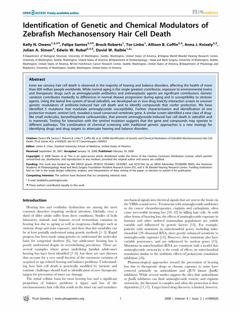
variety of vital dyes and assessed in vivo (Figure 1A). The hair cells
rapidly fragment and die upon treatment with 200 mM neomycin
(Figure 1B). We have developed methods to systematically identify
modulatory pathways altering hair cell response to aminoglycoside
antibiotic exposure by taking advantage of in vivo labeling of
lateral line hair cells with vital dyes. Figure 1C exemplifies this,
showing that lateral line hair cells have a robust, highly
reproducible response to different doses of aminoglycoside
antibiotics [21,23]. We reasoned that by examining animals
treated with concentrations of neomycin at low or high ends of the
dose-response curve, we should be able to identify modifiers that
alter susceptibility to neomycin-induced hair cell death (Figure 1C).
Small Molecule Screening for Protecting CompoundsTo screen for small molecule modifiers, we pretreated 5 day
post-fertilization (dpf) larvae with a chemically diverse library of
10,960 compounds before exposing them to 200 mM neomycin.
Screening was initially carried out by labeling hair cells of 5 dpf
larvae with a combination of a nuclear dye and a cytoplasmic dye
(Yo-Pro-1 and FM 1-43, respectively), then pretreating larvae in
96-well tissue culture plates for 1 hour to a cocktail of five
compounds and then exposing them to 200 mM neomycin. When
protection was observed, the 5 potential contributors were
Figure 1. Screening for modifiers of aminoglycoside toxicity.(A) Neuromast from a control animal pretreated with 0.5% DMSO andstained with rapidly with FM 1-43FX (red) and the nuclear label Yo-Pro-1(green). (B) Negative control pretreated with 0.05% DMSO for 1 hourfollowed by 200 mM neomycin treatment for 30 min. Hair cells arestained with FM 1-43FX (red) and Yo-Pro-1 (green). Hair cell loss, nuclearcondensation and cytoplasmic shrinking are observed. (C) Dose-response function showing decreased hair cell labeling with DASPEI, amitochondrial potentiometric dye, as a function of increasing neomycinconcentration for wildtype zebrafish (N = 25–37 total fish per group,from triplicate experiments). Bars are SEM. Screens for increased ordecreased susceptibility to hair cell loss were performed by treatmentwith either low, 25 mM, or high, 200 mM, neomycin doses, respectively,as highlighted by the orange arrows. (D) Neuromast pretreated withPROTO-2, a compound identified to provide protection against 200 mMneomycin exposure. (E,F) Show the structure for the identifiedcompounds, PROTO-1 (E) and PROTO-2 (F), respectively.doi:10.1371/journal.pgen.1000020.g001
Author Summary
Loss of sensory hair cells in the inner ear is observed in themajority of hearing and balance disorders, affecting thehealth of more than 600 million people worldwide.Exposure to environmental toxins and certain pharmaceu-tical drugs such as aminoglycoside antibiotics and somecancer chemotherapy agents account for many of thesehearing and balance problems. Variation in the geneticmakeup between individuals plays a major role inestablishing differences in susceptibility to environmentalagents that damage the inner ear. Using zebrafish larvae,we developed a screen to uncover genes leading todifferences in antibiotic-induced death of hair cells and toidentify compounds that protect hair cells from damage.The combination of chemical screening with traditionalgenetic approaches offers a new strategy for identifyingdrugs and drug targets to attenuate hearing and balancedisorders.
Modulators of Mechanosensory Hair Cell Death
PLoS Genetics | www.plosgenetics.org 2 2008 | Volume 4 | Issue 2 | e1000020

evaluated singly to determine the active compound. Two
compounds exhibited reliable and robust protection of hair cells
from neomycin. An example of this protection is shown in
Figure 1D, compared to treatment with neomycin alone
(Figure 1B). Both compounds were benzothiophene carboxamides
(Figure 1E and 1F), suggesting specific selection from the diverse
library. We have named these compounds PROTO-1 and
PROTO-2. We next compared the neomycin dose-response
relationship in larvae pretreated with the compounds and controls
(Figure 2). Figure 2A and 2B show that at a concentration of
10 mM both compounds show significant protection of hair cells
over a broad range of neomycin concentrations, from 25 mM to
400 mM (p,0.0001 by two-factorial ANOVA). We also deter-
mined the dose-dependent effects of PROTO-1 and PROTO-2 to
a fixed (200 mM) level of neomycin (Figure 2C and 2D).
Pretreatment with 1 and 10 mM PROTO-1 resulted in significant
protection of hair cells exposed to 200 mM neomycin compared to
neomycin alone (p,0.0001, unpaired t-test). There was no
significant difference in the protection provided by 1 and 10 mM
PROTO-1 (p.0.10). Although exposure to 50 mM and 100 mM
PROTO-1 alone did not alter viability, in combination with
200 mM neomycin these doses were lethal to larvae. Pretreatment
with PROTO-2 provided significant protection of hair cells at all
doses (p,0.0001, unpaired t-tests) with no dose-dependent
difference (p.0.20). PROTO-2 was not lethal at any of the tested
doses with or without neomycin.
Aminoglycosides are used clinically, despite their known
ototoxicity, because of their broad spectrum of antibacterial
actions. Compounds that could be used to limit their ototoxicity
must not limit the intended therapeutic functions. We therefore
had the University of Washington Clinical Microbiology Labora-
tory test the bacteriostatic and bactericidal activity of neomycin in
the presence of PROTO-1 and PROTO-2. The minimum
inhibitory concentration (3.25 mM) and minimum bactericidal
concentration (6.5 mM) for E. coli ATCC 25922 was unchanged
with or without 10 mM of either compound. This indicates that at
Figure 2. Ranges of protection for PROTO-1 and PROTO-2. Hair cells were vitally stained with FM1-43 and Yo-Pro-1, treated with PROTO-1 orPROT0-2 for 1 hour at various concentrations of compounds, then exposed to neomycin for 30 minutes, allowed 1 hr recovery in normal media.Graphs show mean hair cell counts for the SO1, SO2, OC1, and O1 neuromasts (+SEM) as percent of control (mock-treated, no neomycin exposure).Missing error bars indicate that was less than symbol size. (A,B) Neomycin dose-response curve showing effects of 10 mM PROTO-1 ((A), closedsquares) and PROTO-2 ((B), closed squares) pretreatment in comparison to controls (without PROTO-1 or –2). (C,D) Profile of each compound atincreasing doses without aminoglycoside and after 200 mM neomycin exposure. N = 10–20 fish per group.doi:10.1371/journal.pgen.1000020.g002
Modulators of Mechanosensory Hair Cell Death
PLoS Genetics | www.plosgenetics.org 3 2008 | Volume 4 | Issue 2 | e1000020
least under standard in vitro assay conditions benzothiophene
carboxamides do not inhibit aminoglycoside antibacterial activity.
Screening for Genetic ModifiersTo identify genetic modifiers of aminoglycoside-induced hair cell
death, a standard F3 screening paradigm was used. Males were
mutagenized with ethylnitrosourea following standard protocols
[27], then crossed to wildtype females to produce F1 progeny.
Mutagenesis was assessed by specific locus testing against unpig-
mented mitfa mutant animals [28], with a rate of about 1:300. F2
families were produced from F1 individuals, and F3 larvae produced
by pairwise intercrosses within each family. F3 larvae were treated at
5 dpf with either high (200 mM) or low (25 mM) concentrations of
neomycin for 30 minutes to identify mutants that exhibit protection
or heightened susceptibility of hair cells, respectively. Hair cells were
then assessed with the vital dye DASPEI, which is differentially taken
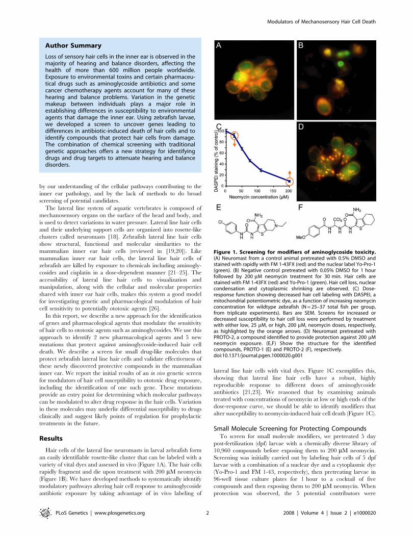
up by neuromast hair cells [29,30]. Figure 3 shows untreated and
neomycin-exposed wildtype animals, and two mutants with altered
susceptibility. In contrast to the wildtype subject (Figure 3B),
persephone mutants (Figure 3C) show robust staining indistinguishable
from an untreated animal (Figure 3A). Animals homozygous for the
sentinel mutation also retain robust staining; in addition they display a
linked morphological phenotype, a variable sinusoidal morphology
that begins to be apparent by 3 dpf (Figure 3D). While persephone
mutants are homozygous viable, the sentinel mutation is lethal at
approximately 10–12 dpf.
To date, we have identified 5 mutations that confer resistance and
behave as simple recessive alleles. Complementation testing
demonstrated that they affect different genes. We identified 5
additional mutations that confer resistance with more complex
genetics, showing semi-dominant effects and/or interactions with
modifying background loci. All mutations were transmitted to the
next generation. We were surprised that all loci identified to date
confer resistance, suggesting that affected genes normally act to
promote cell death. The 5 simple recessive loci can be separated into
two classes, mutations that have no apparent secondary phenotype
(persephone, trainman, bane) and those with additional phenotypes
(sentinel, merovingian). Animals homozygous for the merovingian
mutation show reduced ear size and small otoliths (not shown).
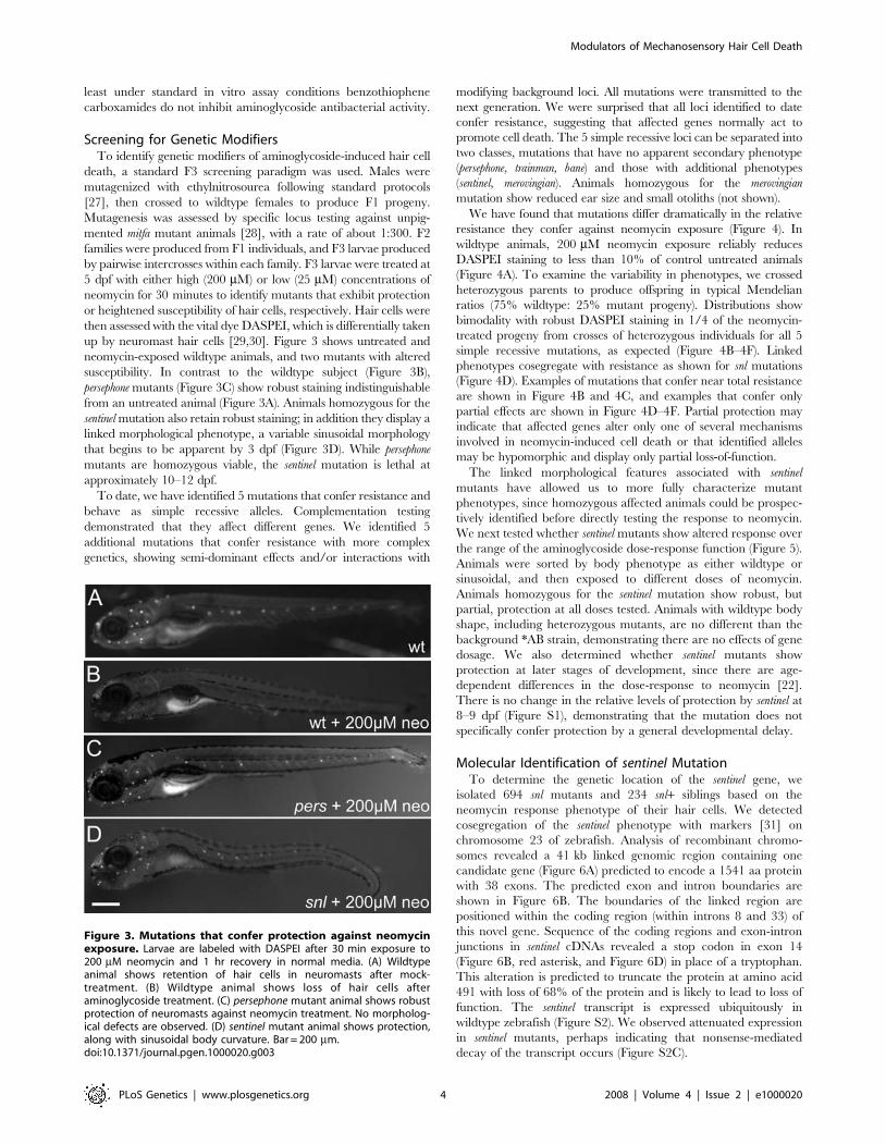
We have found that mutations differ dramatically in the relative
resistance they confer against neomycin exposure (Figure 4). In
wildtype animals, 200 mM neomycin exposure reliably reduces
DASPEI staining to less than 10% of control untreated animals
(Figure 4A). To examine the variability in phenotypes, we crossed
heterozygous parents to produce offspring in typical Mendelian
ratios (75% wildtype: 25% mutant progeny). Distributions show
bimodality with robust DASPEI staining in 1/4 of the neomycin-
treated progeny from crosses of heterozygous individuals for all 5
simple recessive mutations, as expected (Figure 4B–4F). Linked
phenotypes cosegregate with resistance as shown for snl mutations
(Figure 4D). Examples of mutations that confer near total resistance
are shown in Figure 4B and 4C, and examples that confer only
partial effects are shown in Figure 4D–4F. Partial protection may
indicate that affected genes alter only one of several mechanisms
involved in neomycin-induced cell death or that identified alleles
may be hypomorphic and display only partial loss-of-function.
The linked morphological features associated with sentinel
mutants have allowed us to more fully characterize mutant
phenotypes, since homozygous affected animals could be prospec-
tively identified before directly testing the response to neomycin.
We next tested whether sentinel mutants show altered response over
the range of the aminoglycoside dose-response function (Figure 5).
Animals were sorted by body phenotype as either wildtype or
sinusoidal, and then exposed to different doses of neomycin.
Animals homozygous for the sentinel mutation show robust, but
partial, protection at all doses tested. Animals with wildtype body
shape, including heterozygous mutants, are no different than the
background *AB strain, demonstrating there are no effects of gene
dosage. We also determined whether sentinel mutants show
protection at later stages of development, since there are age-
dependent differences in the dose-response to neomycin [22].
There is no change in the relative levels of protection by sentinel at
8–9 dpf (Figure S1), demonstrating that the mutation does not
specifically confer protection by a general developmental delay.
Molecular Identification of sentinel MutationTo determine the genetic location of the sentinel gene, we
isolated 694 snl mutants and 234 snl+ siblings based on the
neomycin response phenotype of their hair cells. We detected
cosegregation of the sentinel phenotype with markers [31] on
chromosome 23 of zebrafish. Analysis of recombinant chromo-
somes revealed a 41 kb linked genomic region containing one
candidate gene (Figure 6A) predicted to encode a 1541 aa protein
with 38 exons. The predicted exon and intron boundaries are
shown in Figure 6B. The boundaries of the linked region are
positioned within the coding region (within introns 8 and 33) of
this novel gene. Sequence of the coding regions and exon-intron
junctions in sentinel cDNAs revealed a stop codon in exon 14
(Figure 6B, red asterisk, and Figure 6D) in place of a tryptophan.
This alteration is predicted to truncate the protein at amino acid
491 with loss of 68% of the protein and is likely to lead to loss of
function. The sentinel transcript is expressed ubiquitously in
wildtype zebrafish (Figure S2). We observed attenuated expression
in sentinel mutants, perhaps indicating that nonsense-mediated
decay of the transcript occurs (Figure S2C).
Figure 3. Mutations that confer protection against neomycinexposure. Larvae are labeled with DASPEI after 30 min exposure to200 mM neomycin and 1 hr recovery in normal media. (A) Wildtypeanimal shows retention of hair cells in neuromasts after mock-treatment. (B) Wildtype animal shows loss of hair cells afteraminoglycoside treatment. (C) persephone mutant animal shows robustprotection of neuromasts against neomycin treatment. No morpholog-ical defects are observed. (D) sentinel mutant animal shows protection,along with sinusoidal body curvature. Bar = 200 mm.doi:10.1371/journal.pgen.1000020.g003
Modulators of Mechanosensory Hair Cell Death
PLoS Genetics | www.plosgenetics.org 4 2008 | Volume 4 | Issue 2 | e1000020
Figure 4. Hair cell retention after neomycin treatment in wildtype and mutant animals. Histograms show the fraction of animals withdifferent levels of DASPEI staining. For each animal, 10 specific neuromasts are evaluated and assigned a score of 2 (normal staining), 1 (reducedstaining), or 0 (no staining) for a maximum total score of 0–20. For each group, the distribution of animals given each DASPEI staining score isdisplayed as a percentage of the total number of animals to illustrate the phenotypic variation within the group; 40–80 animals were tested for eachgroup. (A) Distribution of wildtype fish after mock treatment without neomycin (green bars) or after exposure to 200 mM neomycin (blue bars). (B–F)Distribution of progeny from crosses between heterozygous mutant carriers treated with 200 mM neomycin, showing both wildtype and mutantphenotypes. (B) persephone. (C) merovingian. (D) sentinel. Animals with sinusoidal bodies (later shown to be homozygous mutants) are represented byorange bars, and animals with wildtype body shape (wildtype or heterozygous siblings) are represented by blue bars. (E) bane. (F) trainman.doi:10.1371/journal.pgen.1000020.g004
Modulators of Mechanosensory Hair Cell Death
PLoS Genetics | www.plosgenetics.org 5 2008 | Volume 4 | Issue 2 | e1000020

Alignment of the zebrafish genomic region reveals homology to
human (KIAA1345, 56% identity, 73% similarity) and mouse
(RIKEN 5730509K17, 59% identity, 76% similarity) as well as to
other vertebrates (Figure S3). The intron-exon structure between
the zebrafish and mammalian orthologs is conserved with a few
minor exceptions. We note looser homology to loci in Drosophila
melanogaster, Aedes aegpytii, Caenorhabditis elegans, Trichomo-
nas vaginalis and Paramecium tetraurelia genomes, suggesting that
this is an ancient gene. The phylogenetic relationship between the
predicted proteins is shown in Figure 6C. The Drosophila ortholog
is annotated as two loci (CG18432 and CG18631) corresponding
to the predicted N-terminal and C-terminal end of the zebrafish
protein, indicating that they may encode a single transcript or be
derived from a single ancestral locus. The predicted Sentinel
protein contains a putative C2 domain [32] in the C-terminus
(Figure 6E). The N-terminal third of the Sentinel protein is highly
charged with two glutamine-rich acidic clusters flanking a lysine-
rich basic cluster (Figure 6E). There is a notable absence of other
recognizable domains.
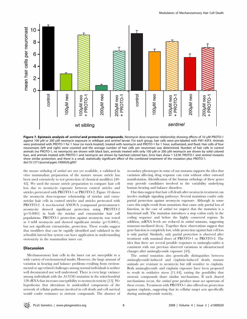
Genetic/Chemical EpistasisTo begin elucidating possible molecular pathways regulating
susceptibility, we tested for an interaction between sentinel mutants
and PROTO-1. Both PROTO-1 treatment and snl loss of function
result in substantial but incomplete protection against neomycin
exposure. We tested whether exposure of PROTO-1 conferred
any additional protection to snl mutants when exposed to 100 mM
or 200 mM neomycin. Figure 7 provides these results for siblings
(left) and sentinel mutants (right), comparing hair cell counts in
control animals and fish exposed to neomycin with or without
pretreatment for 1 hr in 10 mM PROTO-1. At both doses of
neomycin, treatment with PROTO-1 provides a small amount of
additional protection, over and above that provided by the sentinel
mutation. Analyses by one-way ANOVA followed by pair-wise
comparisons (Fisher’s PLSD test) revealed that at both doses the
additional protection provided by PROTO-1 was statistically
reliable (p,0.01), but that even the combined effect did not
provide complete protection (p,0.01).
Determining Cellular Steps in Toxicity Altered byModifiers
Attenuation of drug-induced hair cell death could result from a
number of causes that are not directly linked to the activation of
cell death or cell survival pathways. Some examples include the
well-established link between mechanotransduction-dependent
activity and aminoglycoside uptake and susceptibility [33–35],
the relative resistance seen in young animals [23], and abnormal-
ities of aminoglycoside uptake.
Rapid uptake of the vital dye FM 1–43 is commonly used as an
indicator of sensory hair cell mechanotransduction [36–38]. We
compared the uptake of FM1-43FX in control (wild-type) fish, in
sentinel mutants and in wild-type fish treated with PROTO-1 and
PROTO-2 (Figure 8; Figure S4). Rapid entry of FM1-43FX into
the hair cells of sentinel mutants (Figure 8B and 8D) is comparable
to that of wildtype hair cells (Figure 8A and 8C). Similarly,
PROTO-1 and PROTO-2 did not alter FM1-43FX uptake
(Figure S4A, Figure S4B, Figure S4C), indicating that mechan-
otransduction-associated events appear intact with these modula-
tors. In addition, examination of the neuromasts in sentinel mutants
by light microscopy (compare Figure 8A and 8C to Figure 8B and
8D) reveals that hair cells are organized in the stereotypical rosette
pattern found in wildtype animals. Together these results suggest
that these modifiers do not act by blocking hair cell transduction or
slowing development.
To test whether these modifiers alter drug entry, we evaluated
whether fluorescently-tagged aminoglycosides [39] enter hair cells
in the presence of modifiers. Both the aminoglycosides gentamicin
(Figure 8E and 8F) and neomycin (not shown) tagged with Texas
Red fluorophore enter sentinel hair cells with a rapid, 45-second,
exposure. Similarly, PROTO-1 and PROTO-2 did not alter
labeled gentamicin uptake (Figure S4D, Figure S4E, and Figure
S4F). While these results do not rule out subtle changes in
aminoglycoside uptake, they do show that there are no dramatic
differences that might account for the broad range of protection
seen. Hence, it appears most likely that modifiers affect steps in
toxicity that occur after aminoglycoside entry.
Although the initial mechanism of hair cell death induced by
aminoglycosides and cisplatin may be quite different, the later
general cell death events are thought to be similar. To test whether
these modulators alter cisplatin toxicity, we tested the effects of a
range of cisplatin doses on sentinel mutants and on animals treated
with PROTO-1. The response of sinusoidal sentinel mutants to
cisplatin mirrored wildtype strains and siblings with wildtype body
shape (Figure 9A). Thus, sentinel mutants are not protected against
cisplatin-induced hair cell toxicity. Similarly, PROTO-1 did not
protect against cisplatin-induced cell death (Figure 9B). The
observation that sentinel mutants and fish exposed to PROTO-1
are relatively resistant to aminoglycoside-induced cell death but
remain normally sensitive to cisplatin-induced cell death suggests
that general cell death mechanisms are intact. We hypothesize that
the sentinel mutation and PROTO-1 may abrogate aminoglycoside
targets or early events in aminoglycoside-induced cell death that
are not shared by cisplatin-induced cell death.
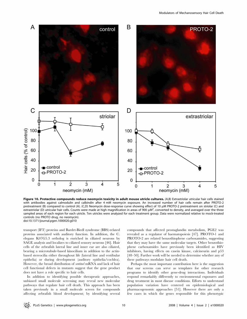
Modifier Test in Adult Mammalian UtriclesFinally, we sought to determine whether modifiers we
discovered in the zebrafish lateral line hair cell assay also confer
protection to hair cells in the murine inner ear. While mutants for
Figure 5. Dose dependent protection of sentinel mutants toneomycin. Hair cell loss as determined by DASPEI staining of progenyof sentinel heterozygous parents with wildtype body shape (blue) orsinusoidal body shape (red) are compared to wildtype *AB fish (green).Error bars are 61 S.D. Mutants show robust, but partial, protectionfollowing 30 min neomycin exposure and one hour recovery.doi:10.1371/journal.pgen.1000020.g005
Modulators of Mechanosensory Hair Cell Death
PLoS Genetics | www.plosgenetics.org 6 2008 | Volume 4 | Issue 2 | e1000020
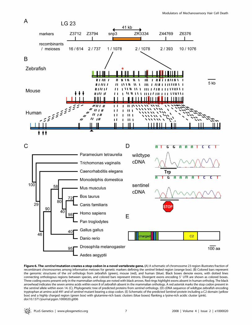
Figure 6. The sentinel mutation creates a stop codon in a novel vertebrate gene. (A) A schematic of chromosome 23 region illustrates fraction ofrecombinant chromosomes among informative meioses for genetic markers defining the sentinel linked region (orange box). (B) Colored bars representthe genomic structures of the snl orthologs from zebrafish (green), mouse (red), and human (blue). Black boxes denote exons, with dotted linesconnecting orthologous regions between species, and colored bars represent introns. Divergent exons encoding 59 UTR are shown as colored boxes.Three coding exons present only in the mammalian orthologs are noted with black arrows. Red rings highlight exons absent in human ortholog. The blackarrowhead indicates the seven amino acids within exon 8 of zebrafish absent in the mammalian orthologs. A red asterisk marks the stop codon present inthe sentinel allele within exon 14. (C), Phylogenetic tree of predicted proteins from sentinel orthologs. (D) cDNA sequence of wildtype zebrafish encodingtryptophan at amino acid 491 and of sentinel mutant bearing a stop codon. (E) Schematic of the predicted Sentinel protein including a C2 domain (yellowbox) and a highly charged region (green box) with glutamine-rich basic clusters (blue boxes) flanking a lysine-rich acidic cluster (pink).doi:10.1371/journal.pgen.1000020.g006
Modulators of Mechanosensory Hair Cell Death
PLoS Genetics | www.plosgenetics.org 7 2008 | Volume 4 | Issue 2 | e1000020
the mouse ortholog of sentinel are not yet available, a validated in
vitro mammalian preparation of the mature mouse utricle has
been used extensively to test protection of chemical modifiers [40-
42]. We used the mouse utricle preparation to compare hair cell
loss due to neomycin exposure between control utricles and
utricles pretreated with PROTO-1 or PROTO-2. Figure 10 shows
the neomycin dose-response relationship of striolar and extra-
striolar hair cells in control utricles and utricles pretreated with
PROTO-2. A two-factorial ANOVA (compound pretreatment6neomycin) showed significant protection using PROTO-2
(p,0.0001) in both the striolar and extrastriolar hair cell
populations. PROTO-1 protection against neomycin was tested
at 4 mM neomycin and showed significant striolar (p,0.0001),
but not significant extrastriolar, protection. These results suggest
that modifiers that can be rapidly identified and validated in the
zebrafish lateral line system can have application in understanding
ototoxicity in the mammalian inner ear.
Discussion
Mechanosensory hair cells in the inner ear are susceptible to a
wide variety of environmental insults. However, the large amount of
variation in hearing and balance problems resulting from environ-
mental or age-related challenges among normal individuals is neither
well documented nor well understood. There is even large variance
among individuals with the A1555G mutation in the mitochondrial
12S rRNA that increases susceptibility to neomycin toxicity [15]. We
hypothesize that alterations in unidentified components of the
network of cellular pathways involved in cell death and cell survival
would confer resistance to ototoxic compounds. The absence of
secondary phenotypes in some of our mutants supports the idea that
variation affecting drug response can exist without other outward
manifestation. Identification of the human orthologs of these genes
may provide candidates involved in the variability underlying
human hearing and balance disorders.
Our data suggest that hair cell death after neomycin treatment can
involve multiple signaling pathways. Several mutations confer only
partial protection against neomycin exposure. Although in some
cases this might result from mutations that cause only partial loss of
function, in the case of sentinel we suspect that the mutation is a
functional null. The mutation introduces a stop codon early in the
coding sequence and before the highly conserved regions. In
addition, mRNA levels are reduced in sentinel mutants, suggesting
nonsense-mediated decay. Together these observations suggest that
gene function is completely lost, while protection against hair cell loss
is only partial. Similarly, only partial protection is observed after
treatment with maximal doses of PROTO-1 or PROTO-2. The
idea that there are several possible responses to aminoglycosides is
consistent with our previous observed variations in ultrastructural
changes after aminoglycoside exposure [43].
The sentinel mutation also genetically distinguishes between
aminoglycoside-induced and cisplatin-induced death; mutant
animals are resistant to neomycin but still sensitive to cisplatin.
Both aminoglycoside and cisplatin exposure have been proposed
to result in oxidative stress [11,44], raising the possibility that
ototoxic compounds share similar mechanisms. If such shared
mechanisms occur, the sentinel gene product must act upstream of
these events. Treatment with PROTO-1 also offered no protection
against cisplatin, suggesting that its cellular target acts specifically
during aminoglycoside toxicity.
Figure 7. Epistasis analysis of sentinel and protective compounds. Neomycin dose-response relationship showing effects of 10 mM PROTO-1against 100 mM or 200 mM neomycin exposure in wildtype and sentinel larvae. For each group, hair cells were pre-labeled with FM1-43FX. Animalswere pretreated with PROTO-1 for 1 hour (or mock-treated), treated with neomycin and PROTO-1 for 1 hour, euthanized, and fixed. Hair cells of fourneuromasts (left and right) were counted and the average number of hair cells per neuromast was determined. Number of hair cells in controlanimals (no PROTO-1, no neomycin) are shown with black bars, animals treated with only 100 mM or 200 mM neomycin are shown by solid coloredbars, and animals treated with PROTO-1 and neomycin are shown by hatched colored bars. Error bars show 1 S.E.M. PROTO-1 and sentinel mutantsshow similar protection, and there is a small, statistically significant effect of the combined treatment of the mutation plus PROTO-1.doi:10.1371/journal.pgen.1000020.g007
Modulators of Mechanosensory Hair Cell Death
PLoS Genetics | www.plosgenetics.org 8 2008 | Volume 4 | Issue 2 | e1000020

Inactivation of sentinel and treatment with PROTO-1 similarly
alter the response of hair cells to neomycin treatment. Both
modulators offer only partial protection against neomycin, offer no
protection against cisplatin, and do not affect entry of FM1-43 or
labeled aminoglycoside. Together these results suggest they work
in common pathways. To test this idea, we performed epistasis
experiments treating wildtype and mutant animals. While the
effects of sentinel and PROTO-1 are not additive, there is a small
but significant increase in protection when combined, suggesting
that they may be accessing different cellular pathways to promote
cell survival. Understanding similarities and differences among
possible pathways will await the identification of the cellular
targets of PROTO-1.
The identification of the sentinel gene highlights one strength of
forward genetic screening, as it would be difficult or impossible to
choose this gene a priori as a candidate regulator of mechanosen-
sory hair cell death. No functional information is known about any
of the sentinel orthologs. The only functional domain of note, the
C2 domain, has been associated with calcium regulation and
interaction with phospholipid membranes in signaling proteins
such as protein kinase C or membrane trafficking proteins like
Synaptotagmin [32]. However, the function of this domain has
been demonstrated in only a few of the many proteins that contain
it. Intriguingly, the D. melanogaster ortholog CG18631 was
identified in a comparative bioinformatics screen as being
associated with compartmentalized cilia-bearing organisms sug-
gesting it may have a role in regulation of cilia [45]. Other
members of this group include molecules related to intraflagellar
Figure 8. sentinel mutation does not affect transduction-dependent dye or aminoglycoside uptake. (A–D) Uptake ofFM1-43FX after 45 sec exposure in wildtype (A,C) and sentinel mutants(B,D). Nuclei are labeled with Yo-Pro-1 (A-D). Confocal images of apical(A,B) and basal (C,D) optical sections through the hair cells. (E,F)Gentamicin-conjugated Texas Red uptake in wildtype (E) and sentinelmutant (F) animals after rapid 45 sec exposure.doi:10.1371/journal.pgen.1000020.g008
Figure 9. sentinel mutation and PROTO-1 do not protectagainst cisplatin. Hair cell survival was quantified using the vitaldye DASPEI, and in each case DASPEI scores were normalized to thosefrom wildtype, untreated fish. Fish (n$12 fish per treatment group)were treated in cisplatin for 4 hours, then allowed to recover for3 hours prior to DASPEI assessment. (A) Hair cell responses in wild-typeversus sentinel mutants. No difference in the dose-response relationshipwas observed between wildtype fish (green), homozygous sentinelmutants (red, sinusoidal body), and sentinel siblings (blue, includingheterozygous and homozygous wildtype sibling, straight body). (B)Response of cisplatin-treated hair cells from wildtype fish in thepresence of the potentially protective compound PROTO-1. There is nodifference between dose-response curves with (red) and without(green) PROTO-1. Error bars represent 61 S.D.doi:10.1371/journal.pgen.1000020.g009
Modulators of Mechanosensory Hair Cell Death
PLoS Genetics | www.plosgenetics.org 9 2008 | Volume 4 | Issue 2 | e1000020
transport (IFT) proteins and Bardet-Biedl syndrome (BBS)-related
proteins associated with auditory function. In addition, the C.
elegans K07G5.3 ortholog is enriched in ciliated neurons by
SAGE analysis and localizes to ciliated sensory neurons [46]. Hair
cells of the zebrafish lateral line and inner ear are also ciliated,
bearing a microtubule-based kinocilium in addition to the actin-
based stereocilia either throughout life (lateral line and vestibular
epithelia) or during development (auditory epithelia/cochlea).
However, the broad distribution of sentinel mRNA and lack of hair
cell functional defects in mutants suggest that the gene product
does not have a role specific to hair cells.
In addition to identifying possible therapeutic approaches,
unbiased small molecule screening may reveal new molecular
pathways that regulate hair cell death. This approach has been
taken previously in a small molecule screen for compounds
affecting zebrafish blood development; by identifying several
compounds that affected prostaglandin metabolism, PGE2 was
revealed as a regulator of haematopoiesis [47]. PROTO-1 and
PROTO-2 are related benzothiophene carboxamides, suggesting
that they may have the same molecular targets. Other benzothio-
phene carboxamides have previously been identified as HIV
inhibitors, having effects on casein kinase, calcineurin and p53
[48–50]. Further work will be needed to determine whether any of
these pathways modulate hair cell death.
Perhaps the most important contribution here is the suggestion
that our screens can serve as templates for other research
programs to identify other gene-drug interactions. Individuals
respond remarkably differently to environmental exposures and
drug treatment in most disease conditions. Efforts to understand
population variation have centered on epidemiological and
pharmacogenomic approaches [51]. However there are only a
few cases in which the genes responsible for this phenotypic
Figure 10. Protective compounds reduce neomycin toxicity in adult mouse utricle cultures. (A,B) Extrastriolar utricular hair cells stainedwith antibodies against calmodulin and calbindin after 4 mM neomycin exposure. An increased number of hair cells remain after PROTO-2pretreatment (B) compared to control (A). (C,D) Neomycin dose-response curve showing effect of 10 mM PROTO-2 pretreatment on striolar (C) andextrastriolar (D) utricular hair cells. Counts were made at high magnification in areas of 900 mM2, converted to density, and averaged over the threesampled areas of each region for each utricle. Ten utricles were analyzed for each treatment group. Data were normalized relative to mock-treatedcontrols (no PROTO drug, no neomycin).doi:10.1371/journal.pgen.1000020.g010
Modulators of Mechanosensory Hair Cell Death
PLoS Genetics | www.plosgenetics.org 10 2008 | Volume 4 | Issue 2 | e1000020
variability have been identified, such as for VKORC1-warfarin
response or PON1-organophosphate toxicity [52,53]. Genetic
analysis may provide a systematic method to identify new
molecules involved in cellular responses to drugs or disease.
Materials and Methods
AnimalsZebrafish embryos (Danio rerio) were produced by paired matings
of adult fish in the University of Washington zebrafish facility by
standard methods [54]. The *AB and WIK wildtype strains are
maintained individually as inbred lines. Three to six-week-old
CBA/CaJ mice were obtained from the Jackson Laboratory (Bar
Harbor, ME) and maintained in the University of Washington
Animal Care facility. All animal protocols were approved by the
University of Washington Animal Care Committee.
Vital Dye StainingLarvae were transferred manually to baskets in 6-well culture
plates containing defined E2 embryo medium. Baskets were
constructed from the tops of 50 ml Falcon tubes in which the center
of the lids were replaced with meshing. All treatment and wash
volumes are 6 ml unless otherwise indicated. Hair cells of larvae
were labeled with the following dyes: 1) FM 1-43FX (n-(3,3-
ammoniumpropyl-dimethyl)ammoniumpropyl)-4-(4-(dibutylamino)-
styryl) pyridinium trichloride), an aminated derivative of FM1-43 (n-
(3-triethylyammoniumpropyl)-4-(4-(dibutylamino)-styryl) pyridinium
dibromide Invitrogen Molecular Probes, Eugene , OR) by
immersing free swimming larvae in 3 mM FM 1-43FX in embryo
medium for 30 or 45 s, followed by three successive rinses in embryo
medium; 2) Yo-Pro-1 (Invitrogen Molecular Probes) at 3 mM for 1
hour followed by 3 rinses to selectively stain hair cell nuclei; or 3)
DASPEI (0.005% final concentration, (2-{4-(dimethylamino)styryl}-
N-ethylpyridinium iodide, Invitrogen Molecular Probes) in the final
15 minutes of the recovery period, and rinsed twice to brightly label
mitochondria-rich hair cell cytoplasm. Larvae were anesthetized
with MS222 (3-aminobenzoic acid ethyl ester, methansulfoneate salt,
Sigma-Aldrich, St. Louis, Missouri) at a final concentration of 0.02%
prior to imaging.
Neomycin TreatmentNeomycin (Sigma-Aldrich, catalog no. N1142) was diluted in
defined E2 embryo medium. Animals were treated with drug or
embryo media (mock-treated controls) for times indicated,
subsequently washed rapidly three times in fresh embryo medium
and allowed to recover for one hour.
Cisplatin TreatmentFor cisplatin treatment, zebrafish larvae were exposed to 0–
400 mM cisplatin (Sigma-Aldrich, catalog no. P4394) for 4 hours,
rinsed several times in embryo medium and held 3 hours in the
same media prior to DASPEI staining and visualization.
Compound ScreeningLarvae were stained with Yo-Pro-1 and FM 1–43 and then
dispensed into 96-well glass bottom plates (Nunc, Rochester, New
York) containing embryo medium (1–2 fish per well). Drug-like
compounds from the Diverset E library (ChemBridge,San Diego,
California), dissolved in 0.05% DMSO to a final concentration of
10 mM, were aliquoted into each well. Fish were incubated at
28.5uC for 1 hour. Neomycin was then introduced into each well
at a final concentration of 200 mM and fish were incubated for an
additional hour. Larvae were anesthetized with MS222 for
immobilization. Visual assessment of hair cell integrity was
performed in vivo using an inverted epifluorescent microscope.
This allowed examination of the whole animal on the side of its
body facing the objective and thus rapid evaluation of many
neuromasts (,20). In each row of the 96-well plate both positive
(neomycin treated only) and negative (no neomycin) control
animals were used for comparison to compound treatment. The
entire plate of 96-well plate with 80 test wells and 16 positive or
negative control wells was evaluated within one hour. Although
intermediate responses were observed for some drugs, only those
exhibiting robust protection were pursued for continued evalua-
tion at this time.
To quantify changes in the hair cell response, hair cell survival
was determined by counting the surviving hair cells from four
neuromast, SO1, SO2, OC1 and O1 for 10–20 fish (i.e. 40–80
neuromasts). The percentage of surviving hair cells following
treatment was calculated relative to mock-treated controls (no
drugs or neomycin exposure).
Minimum Inhibitory and Bactericidal ConcentrationAssays
Determination of the minimum inhibitory concentration (MIC)
and the minimal bactericidal concentration (MBC) of neomycin
alone and in the presence of 10 mM PROTO-1 or PROTO-2
were performed at the Clinical Laboratory of Microbiology at the
University of Washington Medical Center as described by the
National Clinical and Laboratory Standards Institute [55,56].
ENU MutagenesisAdult males from the *AB wildtype strain were mutagenized
with 3 mM ethylnitrosourea (ENU) using standard procedures
[27]). To assess the effectiveness of the mutagenesis, we performed
a specific locus test of mutagenized males with homozygous nacre
females; mutation of the nacre (mitfa) gene results in lack of pigment,
which is readily apparent [28]. The ratio of progeny with a nacre-
like pigment phenotype to total progeny was 1/300. Mutagenized
males were then crossed to wildtype *AB females to produce F1
progeny. F2 families were derived from pairwise matings of F1
progeny of different mutagenized males.
Genetic ScreenFor each family screened, three to twelve F2 pairs were crossed
and their progeny were examined for altered aminoglycoside
response. Neomycin doses of 25 mM or 200 mM were used to
screen for heightened susceptibility or protection, respectively. Ten
neuromasts were evaluated on each fish for DASPEI staining and
each neuromast was assigned a score of 0 for no/little staining, 1 for
reduced staining, 2 for full staining [21], resulting in a final score of
0–20 for each fish. Scores were averaged and normalized to mock-
treated controls. For initial analysis, 12–50 fish were assessed for
typical and atypical responders (i.e. 120–500 neuromasts). Results
were tabulated and chi-squared analysis was done to identify
potential mutant strains of interest. Putative mutants were retested to
confirm phenotype, outcrossed to *AB fish and tested again in the
next generation to confirm transmission.
Genetic MappingHeterozygous mutant carriers were outcrossed to the wildtype
zebrafish from the polymorphic WIK strain for mapping. Hybrid
*AB/WIK carriers of the hair cell modulator were then identified
and crossed to produce progeny for marker analysis. At 5 dpf
larvae were exposed to neomycin as described for the initial
screen. To ensure accurate phenotyping, only individuals with the
highest and lowest DASPEI staining scores after 200 mM
Modulators of Mechanosensory Hair Cell Death
PLoS Genetics | www.plosgenetics.org 11 2008 | Volume 4 | Issue 2 | e1000020
neomycin treatment were retained as mutant and wildtype,
respectively. For bulk segregant analysis, DNA was pooled from
20 wildtype or mutant individuals. Distribution of markers was
compared to DNA from fin clips of *AB/WIK parents and
founder grandparents. Microsatellite markers for each chromo-
some [31] were amplified by PCR and evaluated for cosegregation
with mutant phenotypes. Linked markers were further evaluated
with individual DNAs from 694 mutant fish and 234 wildtype fish
(including both heterozygous and homozygous wildtype siblings).
After determining initial linkage to chromosome 23, fine mapping
identified Z3794 and Z44679 as flanking markers. A contiguous
genomic sequence was then assembled using whole genome shotgun
trace sequences produced by the Zebrafish Sequencing Group at the
Sanger Institute (http://www.sanger.ac.uk/Projects/D_rerio/). Ad-
ditional markers were developed to better define the linked region in
sentinel mutants based on genomic sequence. snp3 amplifies a single
nucleotide polymorphism and sat3334 is a sequence length
polymorphism. They are amplified by the primers:
snp3_forward: GGGTGTCGAACTTGCACCTTTAAT
snp3_reverse: GTTGCTTAATTAGGCCTACAGCACT
sat3334_forward: CTTCATTCGCCCTCTGAACC
sat3334_reverse: GTGCACACTGTGATGTCGATAA
cDNA Isolation and Sequencing/Molecular BiologyRNA was isolated from whole embryos at 62 hpf using Trizol
according to manufacturer’s specifications (Invitrogen, Carlsbad,
California). Oligonucleotide primers were designed based on in
silico genomic sequence. cDNA was synthesized using First Strand
cDNA synthesis kit (Invitrogen) using oligo DT primers. The
following primer pairs were used to amplify portion of cDNA
spanning the recombination breakpoints:
pair 1 forward: AGGTTGAGGCTGGTTTGCCGA
pair 1 reverse: CTCTCAGTGCTTTCAGCTCCTTCCA
pair 2 forward: TTGTCAGACACACTCGACAGTTGCG
pair 2 reverse: TTGGGGTCGAGGCGAGATTCTG
pair 3 forward: AGATGGACGCCATCGCTTGCAT
pair 3 reverse: TCGTTCCAGCAGGGGTTTGGAC
Amplified products were cloned into pCR4 vectors using Topo
TA cloning kit (Invitrogen). cDNA and genomic regions were
sequenced from the vector T3 or T7 sites using Big Dye
terminator v3.1 cycle sequencing chemistry (Applied Biosystems,
Foster City, California).
Comparative GenomicsZebrafish cDNAs were aligned to known ESTs, cDNAs and
genomic sequence from this region using Sequencher software (Gene
Codes, Ann Arbor, Michigan). BLAST alignments of our cDNA
sequences align with predicted cDNA (Genbank XM_693709/
gi:125851476) amino acids 75-1040. Orthologs were identified from
Genbank using BLAST and the corresponding predicted protein
sequences were aligned with the Danio rerio predicted protein
(XP_698801/gi:125851477): Mus musculus (NP_758478.1/
gi:26986583), Homo sapiens (NP_001073991/gi:122937494), Pan
troglodytes (XP_001159814 /gi:114593231), Canis familiaris
(XP_536233/gi:73951827), Bos taurus (XP_595408/gi:119894226),
Monodelphis domestica (XP_001369774/gi:126331991), Gallus
gallus (XP_420777/gi:118090694), Caenorhabditis elegans
(NP_492026/gi:U17508151), Drosophila melanogaster (NP_611229
and NP_611230/gi:24654454/28573534), Aedes aegyptii
(EAT41051/gi:108876826), Trichomonas vaginalis (XP_001323414/
gi:123480792), Paramecium tetraurelia (CAK70738/gi:124405296).
ClustalW multiple sequence alignment software was used to align
predicted proteins of orthologous genes using the Gonnet 250 matrix
[57]. We used the Phylip 3.66 phylogeny software [58] to create a
bootstrapped data set from the original alignment using Seqboot,
then Protml to evaluate these datasets using the maximum likelihood
method with a Jones-Taylor-Thorton model of amino acid
substitution. A consensus tree was determined with Consense
software by extended majority rule. Phylogenetic trees were draw
with TreeView software [59]. Protein motif searching was performed
using the Eukaryotic Linear Motif server (elm.eu.org).
Gentamicin–Texas Red Conjugation4.4 ml of gentamicin sulfate (Sigma-Aldrich, 50 mg/ml) and
0.6 ml succinimidyl esters of Texas Red (Molecular Probes, Eugene,
Oregon; 2 mg/ml in dimethyl formamide) were agitated overnight
to produce the conjugate solution [39]. The conjugated solution was
diluted in embryo media to a final concentration of 200 mM
gentamicin. Because neomycin contains six amino side groups,
neomycin conjugation was performed similarly except that the ratio
of neomycin to Texas Red was adjusted to 3:1 to ensure that on
average one molecule of dye or less labeled each aminoglycoside
molecule. To assess aminoglycoside entry, 5 dpf larvae were
immersed in aminoglycoside-Texas Red conjugate for 45 seconds
and rinsed in embryo medium four times before immediate imaging.
Images were collected using Zeiss LSM5 Pascal confocal microscope.
Z-stack images of neuromasts were collected.
Utricle PreparationUtricles were dissected and cultured in basal medium EAGLE
supplemented with Earle’s balanced salt solution and 5% fetal
bovine serum following established procedures [40]. Neomycin
sulfate stock solution (Sigma-Aldrich) prepared in sterile water was
added directly to culture wells at the desired concentrations. The
utricles were incubated for 4 hours in the compounds diluted with
0.05% DMSO or 0.05% DMSO alone for controls followed by a
24 hour incubation with neomycin.
Utricle ImmunohistochemistryUtricles were fixed for 1 hour at 4uC in 4% paraformaldehyde
in phosphate buffer. Following fixation, otoconia were removed by
gently ‘‘washing’’ the surface with buffer through a 26 gauge
syringe needle. Utricles were then incubated in blocking solution
(2% bovine serum albumin, 0.4% normal goat serum, 0.4%
normal horse serum and 0.4% Triton-X in PBS) for 3 hours at
room temperature. Hair cells were double labeled in whole-mount
preparations with a monoclonal antibody against calmodulin
(Sigma-Aldrich) and polyclonal antibody against calbindin (Che-
micon, Temecula, California) at 4uC diluted in blocking solution,
1:250. The utricles were then rinsed and incubated for 2 hours at
room temperature in secondary antibody diluted in blocking
solution with biotinylated horse anti-mouse IgG (1:200) and Alexa
594-conjugated goat anti-rabbit IgG. Utricles were mounted with
Fluoromount-G (EMS, Hatfield, Pennsylvania) and coverslipped.
The density of mouse utricular hair cells was determined by
counting the number of hair cells in three randomly chosen
nonstriolar regions and the number of striolar hair cells in three
randomly chosen striolar regions from each utricle. Counts were
made at high magnification in areas of 900 mM2, converted to
density, and averaged over the three sampled areas of each region
for each utricle. Ten utricles were analyzed in this way for each
treatment group. Data were normalized relative to mock-treated
controls (no PROTO drug, no neomycin).
Modulators of Mechanosensory Hair Cell Death
PLoS Genetics | www.plosgenetics.org 12 2008 | Volume 4 | Issue 2 | e1000020
Supporting Information
Figure S1 Comparison of the dose response curve of 5 dpf
versus 8–9 dpf sentinel mutants. Hair cell staining by DASPEI was
assessed after neomycin exposure among progeny of sentinel
heterozygous parents with wildtype body shape (blue) or sinusoidal
body shape (red). The dose response curves of wildtype *AB
control fish are shown (green). (A) Dose-response at 5 dpf. (B)
Dose-response at 8–9 dpf. Error bars are {plus minus}1 S.D.
Found at: doi:10.1371/journal.pgen.1000020.s001 (0.26 MB AI)
Figure S2 Figure S2. In situ hybridization of biotinylated probes
to the sentinel locus reveals ubiquitous expression. (A) Wildtype *AB
larvae 64 hpf, antisense probe. (B) *AB larvae 64 hpf, sense probe.
(C) sentinel mutants 64 hpf, antisense probe.
Found at: doi:10.1371/journal.pgen.1000020.s002 (1.69 MB AI)
Figure S3 Aligned sequence of Sentinel-related proteins. Danio
rerio (XM_693709/gi:125851477), Mus musculus (NP_758478/
gi:26986583), and Homo sapiens (NP_001073991/gi:122937494).
Found at: doi:10.1371/journal.pgen.1000020.s003 (0.03 MB
DOC)
Figure S4 Protective compounds do not affect transduction-
dependent dye or aminoglycoside uptake. (A-C) Rapid entry
(45 sec) of 3 mM FM 1–43 (red) into untreated (A), PROTO-1 (B),
or PROTO-2 (C) treated hair cells. Nuclei are labeled with Yo-
Pro-1 (green). (D-F) Exposure to gentamicin-conjugated Texas
Red results in rapid labeling of untreated (D), 10 mM PROTO-1
(E), or PROTO-2 (F) pretreated hair cells.
Found at: doi:10.1371/journal.pgen.1000020.s004 (5.40 MB AI)
Acknowledgments
We thank L. Cunningham for early input, A. Nechiporuk for assistance in
mutagenesis, Glen MacDonald, Dale Cunningham, Mae del Puerto,
Tonibelle Gatbonton, J. Itani, M. Nasry, and K. Reinhart for technical
assistance, S. McFarlane for EM sectioning, and D. White and T. Huyhn
for animal care.
Author Contributions
Conceived and designed the experiments: KO FS JS ER DR. Performed
the experiments: KO FS BR TL AK AC. Analyzed the data: KO FS BR
AC ER DR. Contributed reagents/materials/analysis tools: JS. Wrote the
paper: KO FS ER DR.
References
1. Gates GA, Couropmitree NN, Myers RH (1999) Genetic associations in age-related hearing thresholds. Arch Otolaryngol Head Neck Surg 125: 654–659.
2. Johnson KR, Zheng QY, Noben-Trauth K (2006) Strain background effects and
genetic modifiers of hearing in mice. Brain Res 1091: 79–88.
3. Lanvers-Kaminsky C, Krefeld B, Dinnesen AG, Deuster D, Seifert E, et al.
(2006) Continuous or repeated prolonged cisplatin infusions in children: a
prospective study on ototoxicity, platinum concentrations, and standard serumparameters. Pediatr Blood Cancer 47: 183–193.
4. Nelson EG, Hinojosa R (2006) Presbycusis: a human temporal bone study of
individuals with downward sloping audiometric patterns of hearing loss andreview of the literature. Laryngoscope 116: 1–12.
5. Sill AM, Stick MJ, Prenger VL, Phillips SL, Boughman JA, et al. (1994) Genetic
epidemiologic study of hearing loss in an adult population. Am J Med Genet 54:149–153.
6. Friedman TB, Griffith AJ (2003) Human nonsyndromic sensorineural deafness.
Annu Rev Genomics Hum Genet 4: 341–402.
7. Inoue H, Tanizawa Y, Wasson J, Behn P, Kalidas K, et al. (1998) A geneencoding a transmembrane protein is mutated in patients with diabetes mellitus
and optic atrophy (Wolfram syndrome). Nat Genet 20: 143–148.
8. Lynch ED, Lee MK, Morrow JE, Welcsh PL, Leon PE, et al. (1997)Nonsyndromic deafness DFNA1 associated with mutation of a human homolog
of the Drosophila gene diaphanous. Science 278: 1315–1318.
9. McGuirt WT, Prasad SD, Griffith AJ, Kunst HP, Green GE, et al. (1999)Mutations in COL11A2 cause non-syndromic hearing loss (DFNA13). Nat
Genet 23: 413–419.
10. Forge A, Schacht J (2000) Aminoglycoside antibiotics. Audiol Neurootol 5: 3–22.
11. Rybak LP, Whitworth CA, Mukherjea D, Ramkumar V (2007) Mechanisms ofcisplatin-induced ototoxicity and prevention. Hear Res 226: 157–167.
12. Nakashima T, Teranishi M, Hibi T, Kobayashi M, Umemura M (2000)
Vestibular and cochlear toxicity of aminoglycosides–a review. Acta Otolaryngol120: 904–911.
13. Versnel H, Agterberg MJ, de Groot JC, Smoorenburg GF, Klis SF (2007) Time
course of cochlear electrophysiology and morphology after combined admin-istration of kanamycin and furosemide. Hear Res 231: 1–12.
14. Prezant TR, Agapian JV, Bohlman MC, Bu X, Oztas S, et al. (1993)
Mitochondrial ribosomal RNA mutation associated with both antibiotic-inducedand non-syndromic deafness. Nat Genet 4: 289–294.
15. Guan MX, Fischel-Ghodsian N, Attardi G (1996) Biochemical evidence for
nuclear gene involvement in phenotype of non-syndromic deafness associatedwith mitochondrial 12S rRNA mutation. Hum Mol Genet 5: 963–971.
16. Guan MX, Fischel-Ghodsian N, Attardi G (2000) A biochemical basis for the
inherited susceptibility to aminoglycoside ototoxicity. Hum Mol Genet 9:1787–1793.
17. Cheng AG, Cunningham LL, Rubel EW (2003) Hair cell death in the avian
basilar papilla: characterization of the in vitro model and caspase activation.J Assoc Res Otolaryngol 4: 91–105.
18. Dambly-Chaudiere C, Sapede D, Soubiran F, Decorde K, Gompel N, et al.
(2003) The lateral line of zebrafish: a model system for the analysis of
morphogenesis and neural development in vertebrates. Biol Cell 95: 579–587.
19. Nicolson T (2005) The genetics of hearing and balance in zebrafish. Annu Rev
Genet 39: 9–22.
20. Whitfield TT (2002) Zebrafish as a model for hearing and deafness. J Neurobiol53: 157–171.
21. Harris JA, Cheng AG, Cunningham LL, MacDonald G, Raible DW, et al.(2003) Neomycin-induced hair cell death and rapid regeneration in the lateral
line of zebrafish (Danio rerio). J Assoc Res Otolaryngol 4: 219–234.
22. Murakami SL, Cunningham LL, Werner LA, Bauer E, Pujol R, et al. (2003)Developmental differences in susceptibility to neomycin-induced hair cell
death in the lateral line neuromasts of zebrafish (Danio rerio). Hear Res 186:47–56.
23. Santos F, MacDonald G, Rubel EW, Raible DW (2006) Lateral line hair cell
maturation is a determinant of aminoglycoside susceptibility in zebrafish (Daniorerio). Hear Res 213: 25–33.
24. Williams JA, Holder N (2000) Cell turnover in neuromasts of zebrafish larvae.
Hear Res 143: 171–181.
25. Ou HC, Raible DW, Rubel EW (2007) Cisplatin-induced hair cell loss inzebrafish (Danio rerio) lateral line. Hear Res 233: 46–53.
26. Ton C, Parng C (2005) The use of zebrafish for assessing ototoxic and
otoprotective agents. Hear Res 208: 79–88.
27. Solnica-Krezel L, Schier AF, Driever W (1994) Efficient recovery of ENU-induced mutations from the zebrafish germline. Genetics 136: 1401–1420.
28. Lister JA, Robertson CP, Lepage T, Johnson SL, Raible DW (1999) nacre
encodes a zebrafish microphthalmia-related protein that regulates neural-crest-derived pigment cell fate. Development 126: 3757–3767.
29. Balak KJ, Corwin JT, Jones JE (1990) Regenerated hair cells can originate from
supporting cell progeny: evidence from phototoxicity and laser ablationexperiments in the lateral line system. J Neurosci 10: 2502–2512.
30. Bereiter-Hahn J (1976) Dimethylaminostyrylmethylpyridiniumiodine (daspmi)
as a fluorescent probe for mitochondria in situ. Biochim Biophys Acta 423:1–14.
31. Shimoda N, Knapik EW, Ziniti J, Sim C, Yamada E, et al. (1999) Zebrafish
genetic map with 2000 microsatellite markers. Genomics 58: 219–232.
32. Nalefski EA, Falke JJ (1996) The C2 domain calcium-binding motif: structuraland functional diversity. Protein Sci 5: 2375–2390.
33. Marcotti W, van Netten SM, Kros CJ (2005) The aminoglycoside antibiotic
dihydrostreptomycin rapidly enters mouse outer hair cells through the mechano-electrical transducer channels. J Physiol 567: 505–521.
34. Richardson GP, Forge A, Kros CJ, Fleming J, Brown SD, et al. (1997) Myosin
VIIA is required for aminoglycoside accumulation in cochlear hair cells.J Neurosci 17: 9506–9519.
35. Seiler C, Nicolson T (1999) Defective calmodulin-dependent rapid apical
endocytosis in zebrafish sensory hair cell mutants. J Neurobiol 41: 424–434.
36. Nishikawa S, Sasaki F (1996) Internalization of styryl dye FM1-43 in the haircells of lateral line organs in Xenopus larvae. J Histochem Cytochem 44:
733–741.
37. Gale JE, Marcotti W, Kennedy HJ, Kros CJ, Richardson GP (2001) FM1-43 dyebehaves as a permeant blocker of the hair-cell mechanotransducer channel.
J Neurosci 21: 7013–7025.
38. Meyers JR, MacDonald RB, Duggan A, Lenzi D, Standaert DG, et al. (2003)Lighting up the senses: FM1-43 loading of sensory cells through nonselective ion
channels. J Neurosci 23: 4054–4065.
39. Steyger PS, Peters SL, Rehling J, Hordichok A, Dai CF (2003) Uptake ofgentamicin by bullfrog saccular hair cells in vitro. J Assoc Res Otolaryngol 4:
565–578.
40. Cunningham LL, Cheng AG, Rubel EW (2002) Caspase activation in hair cellsof the mouse utricle exposed to neomycin. J Neurosci 22: 8532–8540.
Modulators of Mechanosensory Hair Cell Death
PLoS Genetics | www.plosgenetics.org 13 2008 | Volume 4 | Issue 2 | e1000020
41. Cunningham LL, Matsui JI, Warchol ME, Rubel EW (2004) Overexpression of
Bcl-2 prevents neomycin-induced hair cell death and caspase-9 activation in theadult mouse utricle in vitro. J Neurobiol 60: 89–100.
42. Sugahara K, Rubel EW, Cunningham LL (2006) JNK signaling in neomycin-
induced vestibular hair cell death. Hear Res 221: 128–135.43. Owens KN, Cunningham DE, Macdonald G, Rubel EW, Raible DW, et al.
(2007) Ultrastructural analysis of aminoglycoside-induced hair cell death in thezebrafish lateral line reveals an early mitochondrial response. J Comp Neurol
502: 522–543.
44. Wu WJ, Sha SH, Schacht J (2002) Recent advances in understandingaminoglycoside ototoxicity and its prevention. Audiol Neurootol 7: 171–174.
45. Avidor-Reiss T, Maer AM, Koundakjian E, Polyanovsky A, Keil T, et al. (2004)Decoding cilia function: defining specialized genes required for compartmen-
talized cilia biogenesis. Cell 117: 527–539.46. Blacque OE, Perens EA, Boroevich KA, Inglis PN, Li C, et al. (2005) Functional
genomics of the cilium, a sensory organelle. Curr Biol 15: 935–941.
47. North TE, Goessling W, Walkley CR, Lengerke C, Kopani KR, et al. (2007)Prostaglandin E2 regulates vertebrate haematopoietic stem cell homeostasis.
Nature 447: 1007–1011.48. Critchfield JW, Coligan JE, Folks TM, Butera ST (1997) Casein kinase II is a
selective target of HIV-1 transcriptional inhibitors. Proc Natl Acad Sci U S A 94:
6110–6115.49. Gualberto A, Marquez G, Carballo M, Youngblood GL, Hunt SW III, et al.
(1998) p53 transactivation of the HIV-1 long terminal repeat is blocked by PD144795, a calcineurin-inhibitor with anti-HIV properties. J Biol Chem 273:
7088–7093.
50. Wang D, Westerheide SD, Hanson JL, Baldwin AS Jr (2000) Tumor necrosis
factor alpha-induced phosphorylation of RelA/p65 on Ser529 is controlled bycasein kinase II. J Biol Chem 275: 32592–32597.
51. Weinshilboum RM, Wang L (2006) Pharmacogenetics and pharmacogenomics:
development, science, and translation. Annu Rev Genomics Hum Genet 7:223–245.
52. Humbert R, Adler DA, Disteche CM, Hassett C, Omiecinski CJ, et al. (1993)The molecular basis of the human serum paraoxonase activity polymorphism.
Nat Genet 3: 73–76.
53. Rieder MJ, Reiner AP, Gage BF, Nickerson DA, Eby CS, et al. (2005) Effect ofVKORC1 haplotypes on transcriptional regulation and warfarin dose.
N Engl J Med 352: 2285–2293.54. Westerfield M (2000) The Zebrafish book. A guide for the laboratory use of
zebrafish (Danio rerio). Eugene (Oregon): University of Oregon Press.55. Wikler MA (2006) Methods for Dilution Antimicrobial Susceptibility Tests for
Bacteria That Grow Aerobically. Clinical and Laboratory Standards Institute.
pp 1–64.56. Wikler MA (2007) Performance Standards for Antimicrobial Susceptibility
Testing. Clinical and Laboratory Standards Institute. pp 1–182.57. Chenna R, Sugawara H, Koike T, Lopez R, Gibson TJ, et al. (2003) Multiple
sequence alignment with the Clustal series of programs. Nucleic Acids Res 31:
3497–3500.58. Felsenstein J (1989) PHLYIP–Phlylogeny Interference Package (version 3.2).
Cladistics 5: 164–166.59. Page RD (1996) TreeView: an application to display phylogenetic trees on
personal computers. Comput Appl Biosci 12: 357–358.
Modulators of Mechanosensory Hair Cell Death
PLoS Genetics | www.plosgenetics.org 14 2008 | Volume 4 | Issue 2 | e1000020