rhoa activation promotes transformation and loss of ...digital.csic.es/bitstream/10261/24536/1/rhoa...
TRANSCRIPT

RhoA Activation Promotes Transformation and Lossof Thyroid Cell Differentiation Interfering withThyroid Transcription Factor-1 Activity
DIEGO L. MEDINA*, MARCOS RIVAS*, PATRICIA CRUZ, ISABEL BARROSO, JAVIER REGADERA,AND PILAR SANTISTEBAN
Instituto de Investigaciones Biomedicas “Alberto Sols” (D.L.M., M.R., P.C., I.B., P.S.), ConsejoSuperior de Investigaciones Cientıficas, Universidad Autonoma de Madrid, and the Departamento deMorfologıa (J.R.), Facultad de Medicina, Universidad Autonoma de Madrid, E-28029 Madrid, Spain
Highly specialized cells, the thyrocytes, express athyroid-specific set of genes for thyroglobulin (Tg),thyroperoxidase, and the transcription factorsTTF-1, TTF-2, and Pax-8. The implication of thesmall GTPase RhoA in TSH-mediated proliferationof FRTL-5 rat thyroid cells has been previouslydemonstrated. To further analyze RhoA function inthyroid cell proliferation and differentiation pat-terns, we combined transient and stable transfec-tion assays to express different mutant RhoAforms in FRTL-5 cells. Constitutively active RhoA(FRTL-5-RhoA QL cells) exhibited a fibroblast-likephenotype with organized actin fibers, whereascells expressing the RhoA negative dominant phe-notype (FRTL-5-RhoA N19 cells) present a roundedmorphology and lose normal cytoskeletal architec-ture. In addition, expression of the constitutivelyactive form of RhoA results in TSH-independentproliferation and anchorage-independent growthand induces tumors when inoculated in nude mice.
Interestingly, FRTL-5-RhoA QL cells express lessTg and TTF-1 than wild-type FRTL-5 (FRTL-5-vector) or FRTL-5-RhoA N19, suggesting a loss atthe differentiation stage. This effect is mediated, atleast in part, by a decrease in TTF-1 activity, sincetransient or stable expression of RhoA QL resultsin a reduction in the activity of the wild-type Tgpromoter as well as an artificial promoter the ac-tivation of which depends exclusively on TTF-1.The similarity between RhoA effects and thyroidtransformation by Ras suggests that RhoA may actas a downstream effector of Ras; in fact, the dom-inant negative RhoA N19 abolished the down-regulatory effect of Ras V12 over the Tg promoter.Taken together, these results show for the firsttime that active RhoA is able to transform FRTL-5cells and that this effect is coupled to a loss ofthyroid differentiation due to impaired TTF-1activity. (Molecular Endocrinology 16: 33–44, 2002)
THE RAS PROTEINS are a group of guanine nucle-otide-binding proteins that function as molecular
switches in many cellular signaling pathways, interact-ing with a wide spectrum of regulators and down-stream effectors; they produce a broad range of cel-lular responses such as proliferation, differentiation, orapoptosis (1, 2). The expression of transforming Rasoncogenes interacts with the establishment and main-tenance of cellular differentiation in different tissues(3–6), including the thyroid. In this tissue, Ras inhibitsthe expression of thyroid-specific genes and confers aproliferative advantage over normal thyroid cells (7–11). Ras transformation in the specialized epithelialthyroid cell line FRTL-5 suppresses the expression ofthyroid differentiation markers such as thyroglobulin(Tg), thyroperoxidase (TPO), and iodine uptake (12). Inparallel, the transcription factors controlling thyroidgene expression, such as TTF-1, TTF-2, or Pax-8, areeither not present or inactive (10, 13, 14). In K-ras-
transformed thyroid cells (FRTL-5-K-ras), both TTF-1and Pax-8 mRNA are undetectable, whereas in H-ras(FRTL-5-H-ras), TTF-1 is present at normal levels andmaintains its DNA binding properties, although thecells lack the ability to express Tg and TPO (11). Sev-eral proteins have been identified as potential effectorsof Ras signaling, including Raf/MAPK kinase/ERK,RalGDS, and PI3K, although the mechanism of Ras-mediated inhibition of thyroid cell differentiation re-mains essentially unclear.
Another Ras protein family that includes RhoA,Rac1, and Cdc42 plays a pivotal role in controllingmany cellular functions, such as cytokinesis, motility,proliferation, and apoptosis (15). These three proteinscooperate with Raf in cell transformation, and thedominant negative forms of RhoA and Rac1 can inhibitRas-induced transformation, indicating an essentialfunction in this process (16–20). Moreover, Rac1 andRhoA have been implicated in the morphogenic andmitogenic responses to transformation by oncogenicRas (21, 22). In the context of thyroid cells, the positiveeffects of RhoA in thyroid cell proliferation have beenrelated to its role in p27Kip1 degradation (23). Amongother functions, p27Kip1 has been implicated in G1
Abbreviations: BrdU, Bromodeoxyuridine; CAT, chloram-phenicol acetyltransferase; CMV-Luc, cytomegalovirus-lucif-erase; Tg, thyroglobulin; TPO, thyroperoxidase; TTF-1, thy-roid transcription factor.
0888-8809/02/$15.00/0 Molecular Endocrinology 16(1):33–44Printed in U.S.A. Copyright © 2002 by The Endocrine Society
33 by on May 20, 2010 mend.endojournals.orgDownloaded from

arrest induced by inhibitors of 3-hydroxy-3-methylglu-taryl-coenzyme-A reductase (23). These inhibitors in-terfere with cell cycle progression by suppressing theisoprenylation of proteins (24), and RhoA is a class ofisoprenylated small GTPases proposed to be involvedin G1/S transition in FRTL-5 cells (23, 24). We recentlyconfirmed these results and demonstrated that over-expression of either the dominant negative form RhoAN19 or the specific inhibitor of RhoA activity, the ex-oenzyme C3, thus inhibits FRTL-5 cell proliferation,causing G1 arrest (25).
In the present study, we combined transient andstable expression of different mutant RhoA forms astools to study the role of this protein in the differenti-ation of FRTL-5 thyroid cells. We found that activationof RhoA induces TSH-independent proliferation, mor-phological transformation, anchorage-independentgrowth, and tumorigenesis when cells are injected intonude mice. These effects could be mediated by theactivation of c-fos and c-jun, the expression of whichis probably central in cell proliferation control and isalso necessary for neoplastic transformation by a va-riety of oncogenes (26, 27). In addition, RhoA activa-tion results in a less differentiated thyroid phenotype,decreasing Tg and TTF-1 gene expression by blockingtransactivation of the Tg promoter through inhibition ofTTF-1 transcriptional activity. These results demon-strate for the first time that RhoA induces transforma-tion of FRTL-5 thyroid cells.
RESULTS
Generation and Analysis of Stable FRTL-5 CellsExpressing Different Mutant Forms ofRhoA Protein
The rat thyroid follicular cell line FRTL-5 provides auseful model with which to study growth and differen-tiation of specialized epithelial cells. To analyze therole of RhoA protein in the differentiation pattern ofthyroid cells, FRTL-5 cells were stably transfected witheither an AU5-tagged dominant positive form of RhoA(RhoA QL) (28), an AU5-tagged dominant negativeform of RhoA (RhoA N19) (29), or with AU5-RhoAQLtogether with an expression vector for C3 toxin whichribosylates and inactivates RhoA (28, 30). TransfectedFRTL-5 clones were tested for exogenous RhoA ex-pression by immunoblotting analysis of total cell ex-tracts using anti-AU5 or -RhoA antibodies. Three rep-resentative neomycin-resistant clones referred asFRTL-5-RhoA QL, RhoA N19, and RhoA QL-C3 wereselected for further study (Fig. 1A). As has been de-scribed extensively, the small GTP-binding proteinRhoA regulates the assembly of focal adhesion andactin stress fibers in response to growth factors (15).We thus analyzed the possible morphological changesdue to overexpression of the different RhoA mutantforms. Phase contrast photomicroscopy of early pas-sage FRTL-5 clones indicated that, in the presence of
6H complete medium, FRTL-5-RhoA QL cells presenta fibroblast-like appearance compared with neomy-cin-resistant FRTL-5 cells carrying control vector(FRTL-5-vector) (Fig. 1B). In addition, the cellular limitswithin the colonies are clearly distinguished. Con-versely, FRTL-5-RhoA N19 cells present a roundedshape and diffuse cell-to-cell limits within colonies.Interestingly, no fibroblast-like morphology was ob-served when the RhoA inhibitor, the exoenzyme C3,was expressed together with RhoA QL. In fact, thesecells show a rounded morphology similar to thoseexpressing the dominant negative form N19 (Fig. 1B).
We support these results by immunofluorescenceusing fluorescein-conjugated phalloidin to visualizethe actin cytoskeleton. FRTL-5-RhoA QL cells presenta more protrusive appearance compared with FRTL-5-vector cells, whereas FRTL-5-RhoA N19 and RhoAQL-C3 cells show a redistribution of the actin fibers,lose their normal cytoskeleton organization and roundup (Fig. 1C).
RhoA Activation Induces a ProliferativeAdvantage and TSH-Independent Growth
In addition to the morphological changes induced byRhoA, results from several laboratories demonstratethat this protein is involved in TSH-mediated thyroidcell proliferation (23–25). Inhibition of RhoA activity bythe C3 exoenzyme or by transient expression of dom-inant negative RhoA N19 thus decreases FRTL-5 cellproliferation, causing G1 arrest in the cell cycle. Toconfirm the role of RhoA in proliferation, growth curveswere performed in stably transfected cells culturedalone or in the presence of TSH (see Materials andMethods).
In the presence of TSH (6H medium), overexpres-sion of constitutively active RhoA (FRTL-5-RhoA QLcells) induces a proliferative advantage compared withcontrols (FRTL-5-vector cells), whereas expression ofthe dominant negative mutant form of RhoA (FRTL-5-RhoA N19 cells) decreases cell proliferation (Fig. 2B).Interestingly, FRTL-5-RhoA QL cells grow in the ab-sence of TSH with a growth rate similar to that seen inthe presence of the hormone, while the FRTL-5-vectoror -RhoA N19 cells barely grow in the absence of TSH(Fig. 2A). These data confirm that RhoA activationinduces TSH-independent growth in FRTL-5 cells. Cy-tometric analysis of FRTL-5-RhoA N19 clones dem-onstrated a decrease in the S phase of the cell cycle asa consequence of G1 arrest (not shown), confirmingprevious results (25).
RhoA Activation Induces Transformation in Vitroand Increases the Tumorigenicity of FRTL-5Thyroid Cells
FRTL-5 cells are considered a normal nontumorigeniccell line, although contradictory results are foundthroughout the bibliography (12, 31). The tumorigenic-ity of FRTL-5 cells is thus conditioned by clonal vari-
34 Mol Endocrinol, January 2002, 16(1):33–44 Medina et al. • RhoA and Thyroid
by on May 20, 2010 mend.endojournals.orgDownloaded from

ability, TSH levels in nude mice, and cell passagenumber (12, 31, 32). Rho proteins have transformingand oncogenic potential in a cell type-specific manner(16–20). Cells expressing constitutively active mutantsof Rac and RhoA display enhanced growth in lowserum, are anchorage independent, and induce tumorformation when inoculated into nude mice (20). In ad-dition, Rac and Rho proteins are essential for trans-formation by Ras (16, 18).
To assess RhoA-dependent FRTL-5 transformation,the ability of each cell line was tested to form coloniesin semisolid medium in the presence or absence ofTSH; only cells expressing the active mutant form ofRhoA (RhoA QL) were able to grow in soft agar (Table1). The number of colonies was similar in the absenceor presence of TSH, again demonstrating the TSH-independent growth of cells expressing RhoA QL.
To test the role of RhoA protein in FRTL-5 tumori-genicity, we inoculated FRTL-5-RhoA QL and FRTL-5-RhoA N19 cells, as well as FRTL-5-vector cells, intonude mice (2 � 106 cells). Four weeks later, we ob-served tumors in the FRTL-5-RhoA QL cell-inoculatedgroup, whereas the groups inoculated with FRTL-5-vector cells or FRTL-5-RhoA N19 cells remained nor-mal (Table 1). These results indicate that RhoA inducesFRTL-5 cell transformation and tumorigenesis. Ex-cised tumors were analyzed histologically as de-scribed in Materials and Methods. FRTL-5-RhoA QL-inoculated mice developed large tumors located in thedermis (Fig. 3A), whereas control or FRTL-5-RhoAN19-inoculated mice remained normal. Tumors werehighly undifferentiated and infiltrative. Tumor cellswere poorly differentiated, showing small cytoplasmand large nuclei with abundant mitosis (Fig. 3B). They
Fig. 1. Expression of RhoA Mutants in Thyroid Follicular CellsA, FRTL-5 cells were stably transfected with vector alone or harboring the AU5-tagged-RhoA QL, AU5-tagged RhoA N19, or
exoenzyme C3 plus the AU5-tagged RhoA QL. Neomycin-resistant colonies were isolated and analyzed by immunoblotting, usinganti-AU5 or anti-RhoA antibodies. A 30-kDa band corresponding to exogenous RhoA protein was detected in several independentclones. FRTL-5 positive clones studied (vector, RhoA QL, RhoA N19, and RhoA QL-C3) were selected for further studies. B,General morphology of the above clones obtained by phase contrast photomicroscopy, of early-passage cells, 2 d after plating.Magnification, �100. C, Cells were stained with fluorescein isothiocyanate-phalloidin to visualize the actin cytoskeleton. Mag-nification, �100.
Medina et al. • RhoA and Thyroid Mol Endocrinol, January 2002, 16(1):33–44 35
by on May 20, 2010 mend.endojournals.orgDownloaded from

grew in a solid, diffuse pattern, with neither follicles norpapillary structures. Infiltration was observed mainlyas satellite tumor nodules (Fig. 3C). Bromodeoxyuri-dine (BrdU) immunostaining showed that proliferativecells were located mainly in peripheral regions of thetumor (Fig. 3D); satellite tumor nodules were highlyproliferative, mainly in the peripheral area (Fig. 3E).Detailed necropsy of the mice revealed no other rele-vant effects.
Molecular mechanisms leading to transformationand tumorigenicity are complex and involve the acti-vation of different signaling pathways, as well as theexpression of a set of genes the induction of whichmay play a role regulating these processes. In anattempt to understand some of the mechanisms re-sponsible for FRTL-5 RhoA QL transformation, we an-alyzed the levels of cAMP as the main signaling path-way controlling thyroid cell growth (33) and because
most thyroid tumors present increased levels of thissecond messenger (34). The results obtained showsimilar levels of cAMP in FRTL-5 vector (370 � 25fmol/well), RhoA QL (374 � 20 fmol/well), and RhoAN19 (365 � 19 fmol/well) cells maintained in 6H me-dium, suggesting that the upstream effector cAMP is
Fig. 2. Proliferative Activity of Thyroid Follicular Cells Carry-ing Constitutively Active RhoA QL or Inactive RhoA N19
FRTL-5 clones were maintained in the absence of TSH (5Hmedium) for 3 d. From then on, cells were cultured either inthe same medium (panel A) or in a medium with TSH (6Hmedium) (panel B). Cell number was monitored every 3 d for12 consecutive days, and viable cell number is represented.The data are the mean � SD of three independent experi-ments.
Table 1. RhoA Activation Induces Transformation in Vitroand Tumorigenesis in Vivo
Cell Line
Colonies in Soft Agar(n)
Tumors in Nude Mice4 Wk after Injection
(n)�TSH �TSH
FRTL-5 vector 0 0 0/12FRTL-5 RhoA QL 129 � 8 140 � 12 12/12FRTL-5 RhoA N19 0 0 0/12
Values for number of colonies are the mean � SD obtainedfrom three independent experiments of each cell type. Tumorfrequency is expressed as number of mice with tumors/totalnumber of mice.
Fig. 3. Histological Analysis of Tumors from FRTL-5-RhoAQL-Inoculated Cells into Nude Mice
A, General view of hematoxylin-eosin-stained large nodu-lar tumor derived from FRTL-5-RhoA QL-inoculated cells (8�magnification). The tumor is well defined, unencapsulated,and localized in the dermis. It presents a solid growth patternwith moderate blood vessel proliferation. B, Hematoxylin-eosin staining of the peripheral area of an undifferentiated,solid tumor derived from FRTL-5-RhoA QL cells, with ana-plastic cells with small cytoplasm (125� magnification). C,Anti-BrdU-hematoxylin staining of the peripheral area of theprevious figure showing abundant highly stained, BrdU-positive cells (intense brown color) (125� magnification). D,Anti-BrdU-hematoxylin staining of an infiltrative, satellitenodule formed in the peripheral area of an undifferentiatedtumor. The arrow indicates the augmented area shown in Fig.3E (50� magnification). E, Anti-BrdU-hematoxylin staining ofthe peripheral area of the infiltrative nodule shown in Fig. 3D.Note that most of the nuclei are highly stained with BrdU(300� magnification).
36 Mol Endocrinol, January 2002, 16(1):33–44 Medina et al. • RhoA and Thyroid
by on May 20, 2010 mend.endojournals.orgDownloaded from

not responsible for either growth independence or celltransformation induced by RhoA activation. To identifydownstream genes that could explain the above phe-nomena, we analyzed the levels of early responsegenes such as c-fos and c-jun. The election of thesegenes was based on previous reports demonstratingtheir involvement in RhoA signaling (35, 36). Westernblot assays using nuclear protein extracts detect in-creased levels of c-Fos and c-Jun in FRTL-5 cellsexpressing the constitutively active form of RhoA (Fig.4); the active phosphorylated form of c-Jun (P-Ser63-c-Jun) is also increased after RhoA QL overexpres-sion. These data suggest that the activation of theseoncogenes is involved in RhoA transformation processin FRTL-5 cells.
Activation of RhoA Is Sufficient to Affect ThyroidCell Differentiation
Cell transformation is usually coupled to a loss ofdifferentiated phenotype. Transformation of FRTL-5cells with two oncogenic Ras forms, Harvey (FRTL-5-H-Ras) and Kirnstein Ras (FRTL-5-K-Ras), has beenstudied extensively. The work of several groups hasshowed that Ras transformation of FRTL-5 cells re-sults in loss of iodine uptake, as well as in Tg and TPOgene expression (7–11). To study further the effect ofRhoA activation in FRTL-5 cells, we measured expres-sion of thyroid-specific mRNA for one of the criticalthyroid-specific markers, Tg, and for its transcriptionfactor TTF-1, in FRTL-5 clones carrying the constitu-tively active RhoA QL or the dominant negative RhoAN19. Tg and TTF-1 mRNA levels were not significantlyaffected by inactivation of RhoA protein (FRTL-5-RhoAN19 cells), whereas activation of RhoA (FRTL-5-RhoAQL cells) clearly decreased both Tg and TTF-1 mRNAlevels (Fig. 5A). To test whether the RhoA-mediateddecrease in Tg and TTF-1 mRNA levels correlates with
a loss of Tg and TTF-1 protein levels, we performedWestern blot assays using total or nuclear proteinextracts to detect Tg or TTF-1 protein levels, respec-tively (Fig. 5B). Total protein extracts from FRTL-5-RhoA QL cells express less Tg than FRTL-5-vectorcells or FRTL-5-RhoA N19 cells. Western blot assaysusing nuclear extracts from FRTL-5 cell clonesshowed a decrease in TTF-1 protein levels in FRTL-5-RhoA QL cells (Fig. 5B). Figure 5 shows representativeNorthern and Western blots. Similar results were ob-tained in all stable clones generated.
Transient Active RhoA Expression Represses theActivity of the Tg Promoter
Several studies have implicated the protein RhoA inthe morphogenic and mitogenic responses to trans-formation by oncogenic Ras (16, 18). In addition, pre-vious studies of Ras-mediated transformation haveshown its negative effect in the activity of thyroid-
Fig. 4. c-Fos and c-Jun Expression in Normal and RhoAExpressing FRTL-5 Cells
Nuclear protein extracts (20 �g) from vector, RhoA QL, orRhoA N19 FRTL-5 clones were analyzed by Western blottingby use of anti-c-Fos, anti-c-Jun, and anti-P-c-Jun antibodies.Anti-Sp1 antibody was used as a loading control.
Fig. 5. Tg and TTF-1 Levels in Normal and RhoA-ExpressingFRTL-5 Cells
A, Total RNA (30 �g) from stable FRTL-5 clones wereblotted onto nytran membranes and sequentially hybridizedwith specific cDNA probes indicated at the right of the panels.As a loading control, membranes were hybridized with anactin-labeled probe. B, Total protein extracts (30 �g) fromstable FRTL-5 clones were blotted onto protran membranesand incubated with specific anti-Tg antibody. Antiactin anti-body was used as a loading control. Nuclear protein extracts(20 �g) from different FRTL-5 clones were immunoblotedwith anti-TTF-1 antibody. Anti-Sp1 antibody was used as aloading control.
Medina et al. • RhoA and Thyroid Mol Endocrinol, January 2002, 16(1):33–44 37
by on May 20, 2010 mend.endojournals.orgDownloaded from
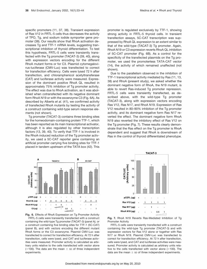
specific promoters (11, 37, 38). Transient expressionof Ras V12 in FRTL-5 cells thus decreases the activityof TPO, Tg, and sodium iodide symporter gene pro-moter (38). Our results show that RhoA activation de-creases Tg and TTF-1 mRNA levels, suggesting tran-scriptional inhibition of thyroid differentiation. To testthis hypothesis, FRTL-5 cells were transiently trans-fected with the Tg promoter (TACAT-3) (39, 40), alongwith expression vectors encoding for the differentRhoA mutant forms or for C3. Plasmid cytomegalovi-rus-luciferase (CMV-Luc) was transfected to correctfor transfection efficiency. Cells were lysed 72 h aftertransfection, and chloramphenicol acetyltransferase(CAT) and luciferase activity were measured. Expres-sion of the dominant positive RhoA QL resulted inapproximately 75% inhibition of Tg promoter activity.The effect was due to RhoA activation, as it was abol-ished when cotransfected with its negative dominantform RhoA N19 or with the exoenzyme C3 (Fig. 6A). Asdescribed by Alberts et al. (41), we confirmed activityof transfected RhoA mutants by testing the activity ofa construct containing wild-type serum response ele-ments (not shown).
Tg promoter (TACAT-3) contains three binding sitesfor the homeodomain-containing protein TTF-1, whichhas been reported as its main transcriptional activator,although it is also regulated by other transcriptionfactors (13, 39, 40). To verify that TTF-1 is involved inthe RhoA-induced reduction of the Tg promoter activ-ity, we used a 5C-CAT reporter gene containing anartificial promoter carrying five binding sites for TTF-1placed in tandem upstream of the TATA box (42). This
promoter is regulated exclusively by TTF-1, showingstrong activity in FRTL-5 thyroid cells. In transienttransfection assays, 5C-CAT transcription was sup-pressed by RhoA QL expression to an extent similar tothat of the wild-type (TACAT-3) Tg promoter. Again,RhoA N19 or C3 expression reverts RhoA QL inhibitionof 5C-CAT promoter (Fig. 6B). As a control for thespecificity of the transfected plasmids on the Tg pro-moter, we used the promoterless TATA-CAT vector(14), the activity of which remained unaffected (notshown).
Due to the parallelism observed in the inhibition ofTTF-1 transcriptional activity mediated by Ras (11, 13,38) and RhoA (present study), we asked whether thedominant negative form of RhoA, the N19 mutant, isable to revert Ras-induced Tg promoter repression.FRTL-5 cells were transiently transfected, as de-scribed above, with the wild-type Tg promoter(TACAT-3), along with expression vectors encodingRas V12, Ras N17, and RhoA N19. Expression of RasV12 resulted in 80–90% inhibition of the Tg promoteractivity, and its dominant negative form Ras N17 re-verted the effect. The dominant negative form RhoAN19 also reverted the inhibitory effect of Ras V12 onthe Tg promoter (Fig. 7). These results clearly demon-strate that the Ras effect on the Tg promoter is RhoAdependent and suggest that RhoA is downstream ofRas in the control of thyroid differentiated phenotype.
Fig. 6. Effects of RhoA Expression on Tg Promoter ActivityFRTL-5 cells were transiently transfected with a construct
containing the wild-type Tg promoter (TACAT-3) (panel A), ora construct containing five binding sites for TTF-1 (5C-CAT)(panel B), and with vectors encoding the different mutantRhoA forms or the C3 exoenzyme. Plasmid CMV-Luc wastransfected to correct for transfection efficiency. At 72 h aftertransfection, cells were lysed, and CAT and luciferase activ-ities were measured. Promoter activity is calculated as arbi-trary units relative to the cells transfected with vector alone(�100). The data are the mean � SD of three independentexperiments.
Fig. 7. RhoA N19 Reverts Ras-Mediated Inhibition of TgPromoter Activity
FRTL-5 cells were transiently transfected with a constructcontaining the wild-type Tg promoter (TACAT-3) and withexpression vectors for Ras V12 alone or together with RasN17 or RhoA N19. Plasmid CMV-Luc was transfected tocorrect for transfection efficiency. At 72 h after transfection,cells were lysed, and CAT and luciferase activities were mea-sured. Promoter activity is calculated as arbitrary units rela-tive to the cells transfected with vector alone (�100). Thedata are the mean � SD of three independent experiments.
38 Mol Endocrinol, January 2002, 16(1):33–44 Medina et al. • RhoA and Thyroid
by on May 20, 2010 mend.endojournals.orgDownloaded from

Stable RhoA QL Expression Represses TTF-1Transcriptional Activity in FRTL-5 Cells
To confirm the RhoA-mediated decrease in TTF-1transcriptional activity in FRTL-5 cells, we performedsimilar assays using FRTL-5 cell clones stably ex-pressing RhoA N19 or RhoA QL. The cells were trans-fected with 5C-CAT and CMV-Luc; after 72 h cellswere lysed and CAT and luciferase activities mea-sured. Expression of the constitutively active RhoA QLclearly inhibits 5C-CAT reporter gene activation,whereas stable expression of the dominant negativeRhoA N19 form had no effect on the TTF-1 transcrip-tional activity (Fig. 8A). As a control, we tested 5C-CATreporter gene activity in two stable FRTL-5 cell linesexpressing the oncogenic Ras forms, H-ras and K-ras(12). As extensively described (13, 14), Ras transfor-
mation inhibits TTF-1 transcriptional activity in thesecell lines (Fig. 8A).
TTF-1 transcriptional activity is regulated mainly byposttranslational modifications such as site-specificphosphorylation and redox mechanisms (43–45). InRas-transformed FRTL-5 cells (FRTL-5-H-ras), Tg ex-pression is absent due to expression of an inactiveform of TTF-1 (11, 14). To understand the mechanismof RhoA-mediated dedifferentiation of thyroid cells, wedetermined whether RhoA activation affects TTF-1transcriptional activity. The 5C-CAT reporter was tran-siently transfected with a vector harboring the TTF-1cDNA into stable FRTL-5 cell clones expressing eitherRhoA QL or RhoA N19. 5C-CAT activity was not af-fected either in FRTL-5-vector cells or in FRTL-5 N19cells when TTF-1 was overexpressed (Fig. 8B). Thismay be due to the existence of saturated levels ofendogenous active TTF-1, as previously reported inFRTL-5 cells (14, 43). Interestingly, overexpression ofthis transcription factor in FRTL-5-RhoA QL had noeffect in reverting RhoA-mediated inhibition of TTF-1transcriptional activity, supporting the idea that RhoAmay decrease Tg expression through some posttrans-lational modifications of TTF-1 protein (Fig. 8B). Asimilar phenomenon has been described in FRTL-5-H-ras cells overexpressing TTF-1 (11, 14).
DISCUSSION
We show that constitutively active RhoA QL trans-forms FRTL-5 thyroid cells, rendering them more pro-liferative, tumorigenic, and undifferentiated. As hasbeen previously demonstrated, RhoA is required forTSH-mediated thyroid cell proliferation (23, 24). Tran-sient expression of dominant negative RhoA N19 orthe inhibition of RhoA activity by choleric exoenzymeC3 decreases thyroid cell proliferation as a conse-quence of G1 arrest (25). Here we present evidencethat stable expression of the dominant positive formRhoA QL induces a proliferative advantage in FRTL-5thyroid cells. Conversely, expression of the dominantnegative RhoA N19 decreases FRTL-5 thyroid cellproliferation.
The Rho protein subfamily, including RhoA, Rac1,and Cdc42, are involved in cell shape regulation andactin filament assembly (46, 47). Lovastatin-inducedinactivation of these proteins causes cell rounding andactin filament disassembly (48). In several cell types,spread cellular morphology allowed DNA synthesis,whereas rounded cells did not proliferate or undergoapoptosis (49). The analysis of FRTL-5-RhoA QLclones with phalloidin shows that these cells spread ina fibroblast-like fashion and display actin stress fibers.This morphology disappears and becomes rounded,with a distinct actin distribution, in clones generatedafter transfection of expression vectors for C3 exoen-zyme and RhoA QL, demonstrating that the changesobserved in the cell phenotype are due to the genes
Fig. 8. Effect of Stable RhoA Expression on TTF-1 Tran-scriptional Activity
A, FRTL-5 cells expressing the different mutant RhoAforms were transiently transfected with the 5C-CAT constructcontaining five specific binding sites for TTF-1. PlasmidCMV-Luc was transfected to correct for transfection effi-ciency. As a control, we transfected the 5C-CAT construct inFRTL-5 cells stably expressing K-ras and H-ras. B, FRTL-5cells expressing the different mutant RhoA forms were tran-siently transfected with 5C-CAT, CMV-Luc, and a TTF-1 ex-pression vector. As a control, we transfected FRTL-5-H-ras.In both panels (A and B), cells were lysed at 72 h aftertransfection, and CAT and luciferase activities were mea-sured. The promoter activity is calculated as arbitrary unitsrelative to the cells transfected with vector alone (�100). Thedata are the mean � SD of three independent experiments.
Medina et al. • RhoA and Thyroid Mol Endocrinol, January 2002, 16(1):33–44 39
by on May 20, 2010 mend.endojournals.orgDownloaded from

transfected and not to an indirect action of the trans-gene used. FRTL-5-RhoA N19 cells also presented arounded cell shape and diffuse cell-to-cell limits. Thisaltered cell morphology was followed by a decrease inFRTL-5-RhoA N19 cell proliferation. In this regard, Zhuet al. (50) reported that for cell cycle progression tooccur, the actin cytoskeleton must be assembled.These clones could thus be useful to study in greaterdetail the links between RhoA-mediated cytoskeletalrearrangement and its positive effects in thyroid cellproliferation.
RhoA activation has transforming and oncogenicpotential in some cell lines (21, 22). Fully differentiatedFRTL-5 thyroid cells are considered nontumorigenic,although they can be rendered so in a single transfor-mation step by expression of retroviral oncogenessuch as H-ras or K-ras (12). As we show here, RhoAQL induces in vitro transformation of FRTL-5 cells,assessed by anchorage- and TSH-independentgrowth and tumors when inoculated into nude mice.Excised tumors revealed highly proliferative infiltratedcarcinomas, suggesting that RhoA induces FRTL-5thyroid transformation in vivo. We show that RhoAtransformation of FRTL-5 cells is not due to increasedcAMP levels, as has been reported in most thyroidtumors (34). The expression of c-Fos and c-Jun isincreased in FRTL-5-RhoA QL cells, suggesting thatthese two oncogenes may be involved in the cell trans-formation described here. As has previously been re-ported, the expression of these two proteins is likely toplay a role in the control of cell proliferation (26, 51), asthey are necessary for cell cycle progression in severalcellular systems and for neoplastic transformation by avariety of oncogenes (27). Interestingly, phospho-c-Jun levels are also increased in FRTL-5-RhoA QLtransformed cells. These results open new questionsabout whether RhoA activation increases JNK activityin FRTL-5 cells as has been demonstrated in othersystems (28). All together, these data concur with theprevious observation of the regulation of early geneexpression and cellular transformation elicited byRhoA.
A general effect in thyroid cell transformation is theloss of cell differentiation. Oncogenic Ras thus sup-presses thyroid differentiation marker expression, andRas-transformed cells do not express Tg or TPO, donot respond to TSH, and do not take up iodine (12). Inparallel, the transcription factors controlling the ex-pression of thyroid-specific genes are either notpresent or inactive (10, 11, 13, 14). In K-ras-trans-formed thyroid cells, both Pax-8 and TTF-1 mRNA areundetectable, whereas in H-ras-transformed cells,TTF-1 is present at normal levels and maintains itsbinding capacity, although the cells lack the ability toexpress Tg and TPO (11, 14). Our results show thatexpression of active RhoA decreases Tg mRNA levels,whereas expression of dominant negative RhoA N19shows Tg mRNA levels comparable to those of con-trols (FRTL-5 cells). RhoA expression also decreasesTg protein levels, confirming that RhoA modifies the
thyroid phenotype affecting Tg expression. The RhoA-mediated decrease in Tg gene expression may be dueto a lack of activity of TTF-1, a specific transcriptionfactor involved in Tg gene activation. Our resultsclearly demonstrate that active RhoA decreases TTF-1expression when compared with FRTL-5-RhoA N19and FRTL-5-vector cells. Moreover, RhoA activationdecreases TTF-1 protein levels in the nucleus, wherethis factor activates thyroid-specific promoters. Thefact that overexpression of exogenous TTF-1 in stableFRTL-5-RhoA QL cells has no effect on Tg promoterconstruct activity, together with the observation thatRhoA QL inhibited the effect of TTF-1 in transienttransfection experiments, suggests that RhoA QL af-fects TTF-1 involving a posttranslational mechanism.This may account not only for the reduction in Tgprotein levels, but also for the reduction in TTF-1, asthis transcription factor is autoregulated (52). Mecha-nisms involved in modulating the transcriptional po-tential of TTF-1 include phosphorylation (43), controlof the redox state (44, 45), and interaction with otherfactors (53). Ras repression of the Tg promoter in-volves changes in TTF-1 phosphorylation (14). Thefact that RhoA N19 reverts the effect of Ras V12 on thewild-type Tg promoter suggests that RhoA is a down-stream effector of Ras in this process. Thus, it wouldbe of interest to test the ability of RhoA to modify thephosphorylation state of TTF-1. Interestingly, the pro-tein kinases PKN, MKK3/6, and ERK6 have been pro-posed as components of a novel signal transductionpathway involved in the regulation of gene expressionand cellular transformation elicited by RhoA (36).TTF-1 contains several minimal consensus sequencesfor ERK phosphorylation and has been reported to bephosphorylated by ERK2 (38). Although ERK2 doesnot mediate the RhoA effect on NIH-3T3 fibroblasts(36), the role of this family of kinases in response toRhoA and its effect in TTF-1 phosphorylation could beaddressed in FRTL-5 cells. However, we cannot ruleout the possibility that TTF-1 may be phosphorylatedby other RhoA effectors.
The new role for RhoA protein in FRTL-5 thyroidcells increases the complexity of the signal transduc-tion pathways implicated in the control of thyroid cellproliferation and function. FRTL-5 thyroid cells dependmainly on TSH for proliferation (33, 54, 55); this hor-mone stimulates thyroid cell proliferation through bothPKA-dependent and -independent pathways (25, 56).After TSH stimulation, downstream effectors such asAkt (57), Rac1 (38), or RhoA (23–25) are involved inthyroid cell growth and function. Activation of Akt,Rac1, or RhoA increases thyroid cell proliferation, al-though Akt or Rac1 expression has no effect on thedifferentiation status of thyroid cells. Here we suggest,for the first time, a role for RhoA in thyroid differenti-ation. The similarity between Ras-mediated thyroidcell transformation, together with our observation thatRhoA N19 reverses the effect of Ras V12 on the Tgpromoter, suggests the existence of cross-talk involv-ing both proteins, as described previously for other
40 Mol Endocrinol, January 2002, 16(1):33–44 Medina et al. • RhoA and Thyroid
by on May 20, 2010 mend.endojournals.orgDownloaded from

cell systems (58). Further studies are needed to ex-plain in detail the role of RhoA in Ras-mediated thyroidtransformation.
MATERIALS AND METHODS
Cell Culture
Rat thyroid follicular FRTL-5 cells (ATCC CRL 8305; AmericanType Culture Collection, Manassas, VA) were kindly providedby Dr. L. D. Kohn (Edison Biotech Institute, Athens, OH). Thecells had the properties previously described (59, 60), werediploid, and their doubling time with TSH was 24–36 h. Cellswere maintained in Coon’s modified Ham’s F-12 medium(Sigma, St. Louis, MO) supplemented with 5% calf serum(Life Technologies, Inc., Gaithersburg, MD) and six-growthfactor (6H complete medium), including TSH (0.5 mU/ml) andinsulin (10 �g/ml) (49). FRTL-5-K-ras and FRTL-5-H-ras cellswere maintained as previously described (11, 12).
Plasmids and Expression Constructs
TACAT-3 corresponds to the wild-type Tg promoter (39, 40).As a negative control, we used the TATA-CAT plasmid con-struct corresponding to the wild-type Tg promoter minus theSal/I-NheI fragment (39), resulting in only the Tg TATA boxlinked to the CAT gene. For detection of TTF-1 transcriptionalactivity, the C5E1b-CAT construct (Ref. 42; referred to hereas 5C-CAT) was transiently transfected into FRTL-5 cells. The5C-CAT construct contains five tandem repeats of the Cbinding site for TTF-1 from the Tg promoter and is exclusivelydependent on TTF-1 for transactivation (42). The CMV-Lucplasmid was used to correct for transfection efficiency (39).For TTF-1 overexpression assays, we used a vector contain-ing wild-type TTF-1 (41). The role of RhoA was analyzed withexpression vectors encoding the dominant positive AU5-tagged-RhoA QL (28), the dominant negative AU5-tagged-RhoA N19 (29), the dominant negative Ras N17 or positiveRas V12 (61), or the botulinum C3 exoenzyme (28).
Transfection Assays
FRTL-5 cells were stably transfected by the calcium phos-phate DNA precipitation method, as described (39, 11).Briefly, calcium phosphate DNA precipitates were preparedwith 10 �g of plasmid DNA containing either constitutivelyactive mutant AU5-tagged RhoA (RhoA QL) (28) or the dom-inant negative form of AU5-tagged RhoA (RhoA N19) (29). Toabolish RhoA activation, a stable cell line was generatedtransfecting together 5 �g of AU5-tagged RhoA QL and 5 �gof the C3 expression vector. In all cases 1 �g of plasmid DNAcontaining the neomycin resistance gene under the control ofviral long terminal repeat promoter, and 40 �g of calf thymusgenomic DNA as carrier (Roche Molecular Biochemicals) wasalso cotransfected. Cells were selected with 300 �g/ml G418(Sigma). After 3 wk, G418-resistant colonies were isolatedand expanded.
For transient transfection assays, cells were plated at adensity of 5 � 105/60 mm diameter tissue culture dish; 48 hlater, TACAT-3, 5C-CAT, TATA-CAT (2.5 �g), or CMV-Luc (1�g) reporter plasmids were transfected with the expressionvectors as indicated in the figure legends. After 72 h, cellextracts were lysed in lysis buffer (10 mM HEPES, pH 7.9, 40mM NaCl, 0.1 mM EGTA, 0.5 mM dithiothreitol, 5% glycerol,and 0.5 mM phenylmethylsulfonyl fluoride). Luciferase (Luc)and chloramphenicol acetyltransferase (CAT) activity weremeasured as described (62, 63).
Immunoblotting and Immunofluorescence
Nuclear or total extracts were prepared in sample buffer, andprotein concentration was determined by the Bradford tech-nique (Bio-Rad Laboratories, Inc., Hercules, CA). Proteinsamples were resolved in SDS-PAGE and transferred to Pro-tran membranes (Schleicher & Schuell, Inc., Keene, NH).Monoclonal anti-AU5 antibody (0.5 �g/ml) used to detectAU5-tag was purchased from Badco. Polyclonal anti-RhoA(1 �g/ml), anti-actin (1 �g/ml), anti-Sp1 (1 �g/ml), anti c-Fos(2 �g/ml), anti-c-Jun (1 �g/ml), and anti-P-Ser 63-c-Jun(2 �g/ml) antibodies were purchased from Santa CruzBiotechnology, Inc. (Santa Cruz, CA). Anti-Tg antibody (0.5�g/ml) was from DAKO Corp. (Carpinteria, CA), and anti-TTF-1 antibody (1 �g/ml) from Biopat Immunotechnologies(Italy). Immune complexes were detected with Luminolreagent as indicated by the manufacturer (Santa Cruz Bio-technology, Inc.).
For immunofluorescence assays, FRTL-5-vector, FRTL-5-RhoA QL, FRTL-5-RhoA N19, and FRTL-5-RhoA QL-C3 cellswere fixed in 4% formaldehyde for 30 min, and then perme-abilized with 0.1% Triton X-100 in PBS. Samples were incu-bated with fluorescein isothiocyanate-phalloidin (Sigma, 1:40dilution in PBS) for 1 h at 37 C. Cells were then washed twicewith PBS and mounted on microscope slides. Fluorescencewas visualized in a photomicroscope (Carl Zeiss, Thornwood,NY) equipped with epifluorescence. Photographs were takenusing Kodak 400 ASA film.
Growth Curve Profiles
To perform growth curves profiles, cells were seeded at aconfluence of 105/100 mm dish and maintained 3 d in theabsence of TSH (5H medium). From then on, cells werecultured either in medium with TSH, for the 6H curve, ormaintained in 5H medium. Fresh medium (6H or 5H, respec-tively) was added every 3 d. The number of viable cells wasdetermined by cell counting every 3 d for 12 consecutivedays. The mean � SD of three independent experiments isrepresented.
Anchorage-Independent Growth
Approximately 9,000 cells were seeded in 60-mm petri dishesin 0.35% noble agar (Sigma) on a 0.5% agar underlayer. Cellswere tested to grow in soft agar containing 6H or 5H medium.Plates were incubated for 3 wk, during which time freshmedium (6H or 5H, respectively) was added to the platesevery 3 or 4 d. After crystal violet staining, colonies largerthan 50 �m diameter were counted. The mean � SD of threeindependent experiments is represented.
Growth of Tumors in Nude Mice
Tumorigenicity was assayed by injecting 2 � 106 cells sc intonude mice, which were palpated weekly for tumor develop-ment. After 4 wk, the animals were killed and tumors excisedfor histological analysis under protocols approved by theHuman Research Committee (Vanderbilt University, Nash-ville, TN). For histopathological analysis (64), tissues werefixed in 10% buffered formaldehyde for 48 h, embedded inparaffin, and cut into 6-�m serial sections that were stainedwith hematoxylin-eosin (Fig. 3), Mason’s trichrome, and pe-riodic acid Schiff methods (not shown). BrdU staining wasperformed as follows: RhoA QL-inoculated mice received 1%5-bromo-2� deoxyuridine (Sigma) ip 2 h before being killed.Tumors were removed and sections prepared as for hema-toxylin-eosin staining. Before immunohistochemistry, sec-tions were deparaffined, hydrated, and washed with PBS.PBS was then removed and endogenous peroxidase inhib-ited using 3% hydrogen peroxidase (10 min, 37 C). Sections
Medina et al. • RhoA and Thyroid Mol Endocrinol, January 2002, 16(1):33–44 41
by on May 20, 2010 mend.endojournals.orgDownloaded from

were blocked in goat serum (Zymed Laboratories, Inc., SouthSan Francisco, CA) and incubated overnight with a mousemonoclonal anti-BrdU antibody (Amersham Pharmacia Bio-tech, Piscataway, NJ). After washing with PBS, sections wereincubated with biotinylated goat antimouse antibody (Biocell)in 20% human serum PBS buffer. After washing, sectionswere incubated with streptavidin-biotin-peroxidase complex(Zymed Laboratories, Inc.) and developed with diaminoben-zidine (Sigma). Sections were counterstained with Harris he-matoxylin, dehydrated in ethanol, and mounted in DePex(Probus, Barcelona, Spain).
RNA Isolation and Analysis
Total RNA was isolated by the guanidinium-isothiocyanate-phenol method (65). Total RNA samples were electropho-resed in 1% agarose gels containing formaldehyde. RNA wastransferred to Nytran membranes (Schleicher & Schuell, Inc.),and RNA integrity was verified by methylene blue staining ofthe blots. Hybridization and washing were performed withprobes specific for Tg (66), TTF-1 (67), or �-actin (68) labeledwith [�32P]-dCTP by random priming.
cAMP Assays
The Biotrak cAMP competitive enzyme immunoassay system(Amersham Pharmacia Biotech) was used following manu-facturer’s instructions for the determination of intracellularcAMP. Briefly, cells were cultured in 24-well plates (105 cellsper dish) and then lysed, moved to a donkey antirabbit Ig-precoated microtiter plate and incubated with anti-cAMPantiserum (2 h, 4 C). Samples were then incubated witha cAMP-peroxidase-conjugated antibody (1 h, 4 C) andwashed four times with washing buffer. The enzyme sub-strate was added immediately afterward to all wells andincubated (1 h, room temperature). Before optical densitydetermination in a plate reader at 450 nm, the reaction wasterminated by adding 0.1 M sulfuric acid to each well. Inparallel, a standard curve with cAMP concentrations from12.5–3,200 fmol/well was prepared. Each value representsthe mean � SD of three independent experiments. As controlfor assay validation, FRTL-5 cells maintained in 5H and 6Hmedium were used, being the cAMP levels 50 � 7 fmol/welland 390 � 30 fmol/well, respectively.
Acknowledgments
We are indebted to Dr. Leonard D. Kohn (Edison BiotechInstitute, Athens, OH) for FRTL-5 cells, to Dr. Silvio Gutkind(National Cancer Institute, NIH, Bethesda, MD) for the RhoAQL, RhoA N19, C3, RasV12, and RasN17 expression vectors,and to Dr. Roberto Di Lauro (Stazione Zoologica, A. Dohrn,Naples, Italy) for cDNA and expression vectors of TTF-1 aswell as the different Tg promoter constructs. We thank Car-men Sanchez-Palomo for help with the immunohistochemicaltechniques and Catherine Mark for her linguistic assistance.
Received December 27, 2000. Accepted August 31, 2001.Address all correspondence and requests for reprints to:
Dr. Pilar Santisteban, Instituto de Investigaciones Biomedi-cas, CSIC/UAM, Arturo Duperier, 4, E-28029 Madrid, Spain.E-mail: [email protected].
This work was supported by DGICYT Grants PM97/0065and BMC2001-2087, and CAM Grant 08.1/0025/99. D.L.M.and M.R. are recipients of a fellowship from the SpanishMinisterio de Educacion y Cultura and Ciencia y Tecnologıa,respectively.
* D.L.M. and M.R. contributed equally to this work andboth should be considered first authors.
REFERENCES
1. Bourne HR, Sanders DA, McCormick F 1990 The GTPasesuperfamily: a conserved switch for diverse cell func-tions. Nature 348:125–132
2. Bos J L 1997 Ras-like GTP-ases. Biochim Biophys Acta1333:M19–M31
3. Beug H, Palmieri S, Freudenstein SC, Zentgraf H, Graf T1992 Hormone-dependent terminal differentiation in vitroof chicken erythroleukemia cells transformed by mutantsof avian erythroblastosis virus. Cell 28:907–919
4. Fiszman MY, Fuchs P 1975 Temperature-sensitive ex-pression of differentiation in transformed myoblasts. Na-ture 254:429–431
5. Olson EN, Spizz G, Tainsky MA 1987 The oncogenicforms of N-ras or H-ras prevent skeletal myoblast differ-entiation. Mol Cell Biol 7:2104–2111
6. Schmidt A, Setoyama C, de Crombrugghe B 1985 Reg-ulation of a collagen gene promoter by the product ofviral mos oncogene. Nature 314:286–289
7. Gallo A, Benusiglio E, Bonapace IM, Feliciello A, CassanoS, Garbi C, Musti AM, Gottesman ME, Avvedimento EV1992 v-ras and protein kinase C dedifferentiate thyroidcells by down-regulating nuclear cAMP-dependent pro-tein kinase A. Genes Dev 6:1621–1630
8. Gallo A, Feliciello A, Varrone A, Cerillo R, Gottesman ME,Avvedimento VE 1995 Ki-ras oncogene interferes withthe expression of cyclic AMP-dependent promoters. CellGrowth Differ 6:91–95
9. Feliciello A, Giuliano P, Porcellini A, Garbi C, Obici S,Mele E, Angotti E, Grieco D, Amabile G, Cassano S, Li Y,Musti AM, Rubin CS, Gottesman ME, Avvedimento EV1996 The v-Ki-Ras oncogene alters cAMP nuclear sig-naling by regulating the location and the expression ofcAMP-dependent protein kinase II�. J Biol Chem 271:25350–25359
10. Avvedimento VE, Musti AM, Ueffing M, Obici S, Gallo A,Sanchez M, DeBrasi D, Gottesman ME 1991 Reversibleinhibition of a thyroid-specific trans-acting factor by Ras.Genes Dev 5:22–28
11. Francis-Lang H, Zannini M, De Felice M, Berlingieri MT,Fusco A, Di Lauro R 1992 Multiple mechanisms of inter-ference between transformation and differentiation inthyroid cells. Mol Cell Biol 12:5793–5800
12. Fusco A, Berlingieri MT, Portella G, Di Fiore PP, GriecoM, Vecchio G 1987 One- and two-step transformation ofrat thyroid epithelial cells by retroviral oncogenes. MolCell Biol 7:3365–3370
13. Missero C, Cobellis G, De Felice M, Di Lauro R 1998Molecular events involved in differentiation of thyroidfollicular cells. Mol Cell Endocrinol 140:37–43
14. Velasco JA, Acebron A, Zannini M, Martın-Perez J, DiLauro R, Santisteban P 1998 Ha-ras interference withthyroid cell differentiation is associated with a down-regulation of thyroid transcription factor-1 phosphoryla-tion. Endocrinology 139:2796–2802
15. Van Aelst L, D’Souza-Schorey C 1997 Rho GTPases andsignaling networks. Genes Dev 11:2295–2322
16. Khosravi FR, Solski PA, Clark GJ, Kinch MS, Der CJ 1995Activation of Rac1, RhoA, and mitogen-activated proteinkinases is required for Ras transformation. Mol Cell Biol15:6443–6453
17. Qiu RG, Chen J, Kirn D, McCormick F, Symons M 1995An essential role for Rac in Ras transformation. Nature374:457–459
18. Qiu RG, Chen J, McCormick F, Symons M 1995 A role forRho in Ras transformation. Proc Natl Acad Sci USA92:11781–11785
19. Qiu RG, Abo A, McCormick F, Symons M 1997 Cdc42regulates anchorage-independent growth and is neces-sary for Ras transformation. Mol Cell Biol 17:3449–3458
42 Mol Endocrinol, January 2002, 16(1):33–44 Medina et al. • RhoA and Thyroid
by on May 20, 2010 mend.endojournals.orgDownloaded from

20. Lin R, Bagrodia S, Cerione R, Manor D 1997 A novelCdc42Hs mutant induces cellular transformation. CurrBiol 7:794–797
21. Ridley AJ, Paterson HF, Johnston CL, Diekmann D, HallA 1992 The small GTP-binding protein rac regulatesgrowth factor-induced membrane ruffling. Cell 70:401–410
22. Ridley AJ, Hall A 1992 The small GTP-binding proteinRho regulates the assembly of focal adhesions and actinstress fibers in response to growth factors. Cell 70:389–399
23. Hirai A, Nakamura S, Noguchi Y, Kitagawa M, Tatsuno I,Oeda T, Hakara, Terano T, Naruyima S, Kohn LD, Saito Y1997 Geranylgeranylated Rho small GTPase(s) are es-sential for the degradation of the p27kip1 and facilitatethe progression from G1 to S phase in growth-stimulatedrat FRTL-5 cells. J Biol Chem 272:13–16
24. Noguchi Y, Nakamura S, Yasuda T, Kitagawa M, KohnLD, Saito Y, Hirai A 1998 Newly synthesized Rho A, notRas, is isoprenylated and translocated to membranescoincident with progression of the G1 to S phase ofgrowth-stimulated rat FRTL-5 cells. J Biol Chem 273:3649–3653
25. Medina DL, Toro MJ, Santisteban P 2000 Somatostatininterferes with thyrotropin-induced G1-S transitionmediated by PKA and PI3-K: involvement of RhoAand cyclin E-CDK2 complexes. J Biol Chem 175:15549–15556
26. Angel P, Karin M 1991 The role of Jun, Fos and the AP-1complex in cell-proliferation and transformation. BiochimBiophys Acta 1072:129–157
27. Suzuki T, Murakami M, Onai N, Fukuda E, Hashimoto Y,Sonobe MH, Kameda T, Ichinose M, Miki K, Iba H 1994Analysis of AP-1 function in cellular transformation path-way. J Virol 68:3527–3535
28. Teramoto H, Crespo P, Coso OA, Igishi T, Xu N, GutkindS 1996 The small GTP-binding protein Rho activatesc-Jun N-terminal kinases/stress-activated protein ki-nases in human kidney 293T cells: evidence for a Pak-independent signaling pathway. J Biol Chem 271:25731–2573430
29. Crespo P, Bustelo XR, Aaronson DS, Coso OA, Lopez-Barahona M, Barbacid M, Gutkind JS 1996 Rac-1 de-pendent stimulation of the JNK/SAPK signaling pathwayby Vav. Oncogene 13:455–460
30. Lerm M, Schmidt G, Aktories K 2000 Bacterial proteintoxins targeting Rho GTPases. FEMS Microbiol Lett188:1–6
31. Ossendorp FA, Bruning PF, Schuuring EM, Van DenBrink JA, van der Heide D, De Vijlder JJ, De Bruin TW1990 Thyrotropin dependent and independent thyroidcell lines selected from FRTL-5 derived tumors grown innude mice. Endocrinology 127:419–430
32. Peter HJ, Gerber H, Studer H, Groscurth P, Zakarija M1991 Comparison of FRTL-5 cell growth in vitro with thatof xenotransplanted cells and the thyroid of the recipientmouse. Endocrinology 128:211–219
33. Medina DL, Santisteban P 2000 Thyrotropin-dependentproliferation of in vitro rat thyroid cell systems. Eur JEndocrinol 143:161–178
34. Farid NR, Shi Y, Zou M 1994 Molecular basis of thyroidcancer. Endocr Rev 15:202–232
35. Hill CS, Wynne J, Treisman R 1995 The rho familyGTPases RhoA, Rac1, and CDC42Hs regulates tran-scriptional activation by SRF. Cell 81:1159–1170
36. Marinissen MJ, Chiariello M, Gutkind JS 2001 Regulationof gene expression by the small GTPase Rho through theERK6 (p38�) MAP kinase pathway. Genes Dev 15:535–553
37. Cobellis G, Missero C, Di Lauro R 1998 Concomitantactivation of MEK-1 and Rac-1 increases the proliferativepotential of thyroid epithelial cells, without affecting theirdifferentiation. Oncogene 17:2047–2057
38. Missero C, Pirro MT, Di Lauro R 2000 Multiple ras down-stream pathways mediate functional repression of thehomeobox gene product TTF-1. Mol Cell Biol 20:2783–2793
39. Sinclair AJ, Lonigro R, Civitareale D, Ghibelli L, Di LauroR 1990 The tissue-specific expression of the thyroglob-ulin gene requires interaction between thyroid-specificand ubiquitous factors. Eur J Biochem 193:311–318
40. Civitareale D, Lonigro R, Sinclair AJ, Di Lauro R 1989 Athyroid specific nuclear protein essential for tissue-specific expression of the thyroglobulin promoter. EMBOJ 8:2537–2542
41. Alberts AS, Geneste O, Treisman R 1998 Activation ofSRF-regulated chromosomal templates by Rho-familyGTPases requires a signal that also induces H4 hyper-acetylation. Cell 92:475–487
42. De Felice M, Damante G, Zannini M, Francis-Lang H, DiLauro R 1995 Redundant domains contribute to the tran-scriptional activity of the thyroid transcription factor-1.J Biol Chem 270:26649–26656
43. Zannini M, Acebron A, De Felice M, Arnone MI, Martın-Perez J, Santisteban P, Di Lauro R 1996 Mapping andfunctional role of phosphorylation sites in the thyroidtranscription factor-1 (TTF-1). J Biol Chem 271:2249–2254
44. Arnone MI, Zannini M, Di Lauro R 1995 The DNA bindingactivity and the dimerization ability of the thyroid tran-scription factor 1 are redox regulated. J Biol Chem 270:12048–12055
45. Kambe F, Nomura Y, Okamoto T, Seo H 1996 Redoxregulation of thyroid-transcription factor, Pax-8 andTTF-1, is involved in their increased DNA-binding activ-ities by thyrotropin in rat FRTL-5 cells. Mol Endocrinol10:801–812
46. Hall A 1998 Rho GTPases and the actin cytoskeleton.Science 279:509–514
47. Takai Y, Sasaki T, Tanaka K, Nakanishi H 1995 Rho as aregulator of the cytoskeleton. Trends Biochem Sci 20:227–231
48. Fenton RG, Kung HF, Longo DL, Smith MR 1992 Regu-lation of intracellular actin polymerization by prenylatedcellular proteins. J Cell Biol 117:347–356
49. Chen CS, Mrksich M, Huang S, Whitesides GM, IngberDE 1997 Geometric controls of cell life and death. Sci-ence 276:1425–1428
50. Zhu X, Ohtsubu M, Bohmer RM, Roberts JM, Assoian RK1996 Adhesion-dependent cell cycle progression linkedto the expression of cyclin D1, activation of cyclin E-cdk2and phosphorylation of the retinoblastoma protein. J CellBiol 133:391–403
51. Kovary K, Bravo R 1991 The jun and fos protein familiesare both required for cell cycle progression in fibroblasts.Mol Cell Biol 11:4466–4472
52. Oguchi H, Kimura S 1998 Multiple transcripts encodedby the thyroid-specific enhancer-binding protein(T/EBP)/thyroid-specific transcription factor-1(TTF-1)gene: evidence of autoregulation. Endocrinology 139:1999–2006
53. Perrone L, Tell G, Di Lauro R 1999 Calreticulin enhancesthe transcriptional activity of thyroid transcription fac-tor-1 by binding to its homeodomain. J Biol Chem 274:4640–4645
54. Dumont JE, Lamy F, Roger P, Maenhaut C 1992 Physi-ological and pathological regulation of thyroid cell prolif-eration and differentiation by thyrotropin and othergrowth factors. Physiol Rev 72:667–698
55. Dremier S, Pohl V, Poteet-Smith C, Roger PP, Corbin J,Doskeland SO, Dumont JE, Maenhaut C 1997 Activationof cyclic AMP-dependent kinase is required but may notbe sufficient to mimic cyclic AMP-dependent DNA syn-thesis and thyroglobulin expression in dog thyroid cells.Mol Cell Biol 17:6717–6726
Medina et al. • RhoA and Thyroid Mol Endocrinol, January 2002, 16(1):33–44 43
by on May 20, 2010 mend.endojournals.orgDownloaded from

56. Cass LA, Summers SA, Prendergast GV, Backer JM,Birnbaum MJ, Meinkoth JL 1999 Protein kinase A-dependent and -independent signaling pathways con-tribute to cyclic AMP-stimulated proliferation. Mol CellBiol 19:5882–5891
57. De Vita G, Berlingieri MT, Visconti R, Castellone MD,Viglieto G, Baldassarre G, Zannini M, Bellacosa A, Tsich-lis PN, Fusco A, Santoro M 2000 Akt/Protein kinase Bpromotes survival and hormone-independent prolifera-tion of thyroid cells in the absence of differentiating andtransforming effects. Cancer Res 60:3916–3920
58. Sahai E, Olson MF, Marshall CJ 2001 Cross-talk betweenRas and Rho signalling pathways in transformationfavours proliferation and increase motility EMBO J 20:755–7666
59. Ambesi-Impiombato FS, Parks LAM, Coon HG 1980 Cul-ture of hormone-dependent functional cells of rat thyroid.Proc Natl Acad Sci USA 77:3455–3459
60. Kohn LD, Valente WA, Grollman EF, Aloj SM, Vitti P 1986Clinical determination and or quantification of thyrotropinand a variety of thyroid stimulatory and inhibitory factorsperformed in vitro with an improved cell line FRTL-5. USPatent 4:609–622
61. Coso OA, Chiariello M, Yu JC, Teramoto H, Crespo P, XuN, Miki T, Gutkind JS 1995 The small GTP-binding pro-
teins Rac1 and Cdc-42 regulate the activity of the JNK/SAP signalling pathway. Cell 81:1137–1146
62. Brasier AR, Tate JE, Habener JF 1989 Optimized use ofthe firefly luciferase assay as a reporter gene in mamma-lian cell lines. Biotechniques 7:1116–1122
63. Gorman CM, Moffat LM, Howard BH 1982 Recombinantgenomes which express chloramphenicol acetyltrans-ferase in mammalian cells. Mol Cell Biol 2:1044–1051
64. Bancroft JD, Cook HC 1994 Manual of histologicaltechniques and their diagnostic application. London:Churchill Livingstone Editorial
65. Chomczynski P, Sacchi N 1987 Single step method ofRNA isolation by acid guanidinium thiocyanate-phenol-chloroform extraction. Anal Biochem 162:156–159
66. Di Lauro R, Obici S, Acquaviva AM, Alvino C 1985 Thesequence of 967 amino acids at the carboxi-end of ratthyroglobulin. Location and surroundings of two thyrox-ine-forming sites. Eur J Biochem 148:7–11
67. Guazzi S, Price M, De Felice M, Damante G, Mattei MG,Di Lauro R 1990 Thyroid nuclear factor 1 (TTF-1) containsa homeodomain and displays a novel DNA binding spec-ificity. EMBO J 9:3631–3639
68. Levi A, Eldridge JD, Paterson BM 1985 Molecular cloningof a gene sequence regulated by nerve growth factor.Science 229:393–395
44 Mol Endocrinol, January 2002, 16(1):33–44 Medina et al. • RhoA and Thyroid
by on May 20, 2010 mend.endojournals.orgDownloaded from