original article the role of rhoa/rock singal pathway in ... · study observed rhoa/rock signal...
TRANSCRIPT

Int J Clin Exp Med 2016;9(7):13642-13649www.ijcem.com /ISSN:1940-5901/IJCEM0027628
Original ArticleThe role of RhoA/ROCK singal pathway in cardiac rupture after infarction
Lina Hao, Chunping Kang
Department of Gerontology, Xi’an Central Hospital, Xi’an 710003, Shaanxi, China
Received March 7, 2016; Accepted June 5, 2016; Epub July 15, 2016; Published July 30, 2016
Abstract: Cardiac rupture is one severe complication of acute myocardial infarction (AMI). The RhoA/ROCK signal protein is up-regulated after AMI, for regulating myocardial injury and fibrosis via inflammation or cytokines. This study observed RhoA/ROCK signal pathway in AMI aged mice, in an attempt to investigate the effect of RhoA/ROCK inhibitor on cardiac rupture after AMI in aged mice, and related mechanisms. Male C57BL/6 mice (18 months old) were assigned into sham, model and inhibitor groups (N=90 each). AMI model was generated by ligation of left coro-nary artery. Hemodynamics and cardiac ultrasound were examined at day 7, to observe left ventricular remodeling and the rate of cardiac rupture. HE staining was employed to observe myocardial morphology. Serum levels of inter-leukin-6 (IL-6), tumor necrosis factor-α (TNF-α) were quantified by ELISA at 3, 7 and 14 days after AMI. Protein levels of ROCK1, RhoA, nuclear factor (NF)-κBp65 and transformation growth factor (TGF)-β1 were measured by Western blotting. AMI model group had contraction dysfunction and ventricle dilation, along with elevated IL-6 and TNF-α levels, as well as higher ROCK1, RhoA, NF-κBp65 and TGF-β1 levels (P<0.05 compared to control group). Inhibitor group had lower rate of cardiac rupture, alleviated contraction disorder, ventricle dilation, or myocardial injury, low-ered IL-6, TNF-α, ROCK1, RhoA, NF-κBp65 and TGF-β1 expression (P<0.05 compared to model group). RhoA/ROCK pathway might be related with cardiac rupture after AMI. Its inhibitor might decrease cardiac rupture via depressing signal activity, and lowering expression of pro-inflammatory and pro-fibrosis factors.
Keywords: Myocardial infarction, ROCK, RhoA, cardiac rupture
Introduction
Acute myocardial infarction (AMI) can cause hypoxia and insufficient perfusion of myocardi-al tissues, leading to secondary responses including oxidative stress, inflammation and cell apoptosis, thus severely affecting left ven-tricular remodeling and cardiac reconstruction. Both ventricular remodeling and cardiac rup-ture are severe complications of AMI. Among these, cardiac rupture occupies about 20%~ 31% of total in-patient death after AMI [1, 2]. The clinical symptom of cardiac rupture de- pends on the site of onset. It usually occurs within 7 days of primary AMI. Independent risk factors of cardiac rupture include aging and gender [3, 4], as it frequently occurs in female aged people of latent myocardial re-perfusion, left ventricular infarction and AMI complicated with hypertension. Currently the mechanism of cardiac rupture after AMI is still unclear [5, 6]. Some studies believed that myocardial remod-eling deficits might lead to cardic rupture. Post-
AMI myocardial repair, inflammatory response, injury of extracellular matrix and cell apoptosis all play important roles in cardiac rupture, which is related with cell apoptosis, genetic suscepti-bility, anti-tension strength of myocardial tis-sues, higher pro-inflammatory factor and medi-ator, and enhanced activation of matrix metalloproteinase [7, 8]. RhoA is one small mol-ecule G protein, and participates in various intracellular signal transduction pathways in conjunction with its downstream effector mole-cule Rho kinase (ROCK). After AMI, RhoA/ROCK proteins in myocardial tissues were elevated and activated, for regulation of inflammatory and cell activity factors to participate in myo-cardial injury, cell apoptosis and myocardial fibrosis [9, 10]. ROCK exists in the form of two homologs, ROCK1 and ROCK2, both of which are expressed in vascular muscle and myocar-dial tissues. These information indicate the important role of RhA/ROCK signal pathway in the prognosis of AMI in aged mice. Fasudil is one RhoA/ROCK inhibitor, and can decrease
Rho kinase in AMI
13643 Int J Clin Exp Med 2016;9(7):13642-13649
the degree of myocardial fibrosis and infarction area in congestive heart failure rats, and allevi-ate inflammatory response after ischemia-reperfusion [11, 12]. This study thus estab-lished an AMI model in age mice, whose dynamic change of RhoA/ROCK signal pathway was observed, in an attempt to investigate the potential effect of RhoA/ROCK pathway inhibi-tor on cardiac rupture of AMI in aged mice and possible mechanism.
Materials and methods
Animals and grouping
Healthy male C57/BL mice (18 months, body weight 25~30 g) were provided by Laboratory Animal Center, Chinese Medicine Academy (Certificate number, SYXK-2013-0025) and were kept in an SPF grade facility with food and water ad libitum. Animals were randomly assigned into sham, AMI model and inhibitor groups (N=90 each). AMI model was estab-lished in mice from the latter two groups by ligating left coronary artery. RhoA/ROCK path-way inhibitor Fasudil (30 mg/kg/d) was applied via intraperitoneal injection. Equal volume of saline was introduced on sham and model groups.
Mice were used for all experiments, and all pro-cedures were approved by the Animal Ethics Committee of Xi’an Central Hospital.
Drugs and reagents
RhoA/ROCK inhibitor Fasudil (30 mg in 2 ml, diluted in saline before use) was provided by Asahi Kasei Pharma Corp. Injection water (Ke-
lun Pharma, China); Pentobarbital and ket-amine (Shanghai Chem, China). ELISA kits for interleukin-6 (IL-6) and tumor necrosis factor-α (TNF-α) were provided by Uscn (China). Rabbit anti-mouse ROCK1, RhoA, nuclear factor (NF)-κBp65 and transformation growth factor (TGF)-β1 were provide by Boster (China). We- stern blotting test kit was provided by Invitrogen (US). Horseradish peroxidase-conjugated goat anti-rabbit secondary antibody was purchased from CST (US).
AMI model
Mouse AMI model was generated by ligating left coronary artery as previously described [13]. Mice were anesthetized (atropine 1.2 mg/kg, xylazine 20 mg/kg and ketamine 100 mg/kg) and were fixed in left side clinostatism posi-tion. Tracheal intubation was performed for connecting ventilator. The chest cavity was opened via left ribs to ligate left coronary artery (using 6-0 suture). AMI model was deduced as whitening of ventricular anterior wall, weak-ened movement. After clearing hemorrhage inside the cavity, excess air was drained for suture. In sham group, coronary artery was sep-arate but not ligated. 24 h after surgery, Fasudil (30 mg/kg/d) was applied via intraperitoneal injection. Equal volumes of saline were applied into the abdominal cavity in sham and model groups.
Drug delivery and observation of cardiac rupture
24 h after AMI model, Fasudil (30 mg/kg) was applied via intraperitoneal injection into inhibi-tor group daily for 14 consecutive days, while the other two groups received equal volume of saline. The survival rate of mice was monitored. 80 mice were chosen from model and inhibitor group for recording the rate of cardiac rupture 7 d after surgery. Those mice died within one week were dissected along with those sacri-ficed mice after 7 d. The cardiac rupture was deduced as abundant blood clot around the heart, or rupture of ventricular wall on infarc-tion side. Cardiac failure was identified as severe heart expansion, large scale of infarc-tion, pericardial effusion and pulmonary con- gestion.
Hemodynamic and cardiac ultrasound
HP 5500 colored Doppler ultrasonic apparatus was used to measure left ventricular end-dia-
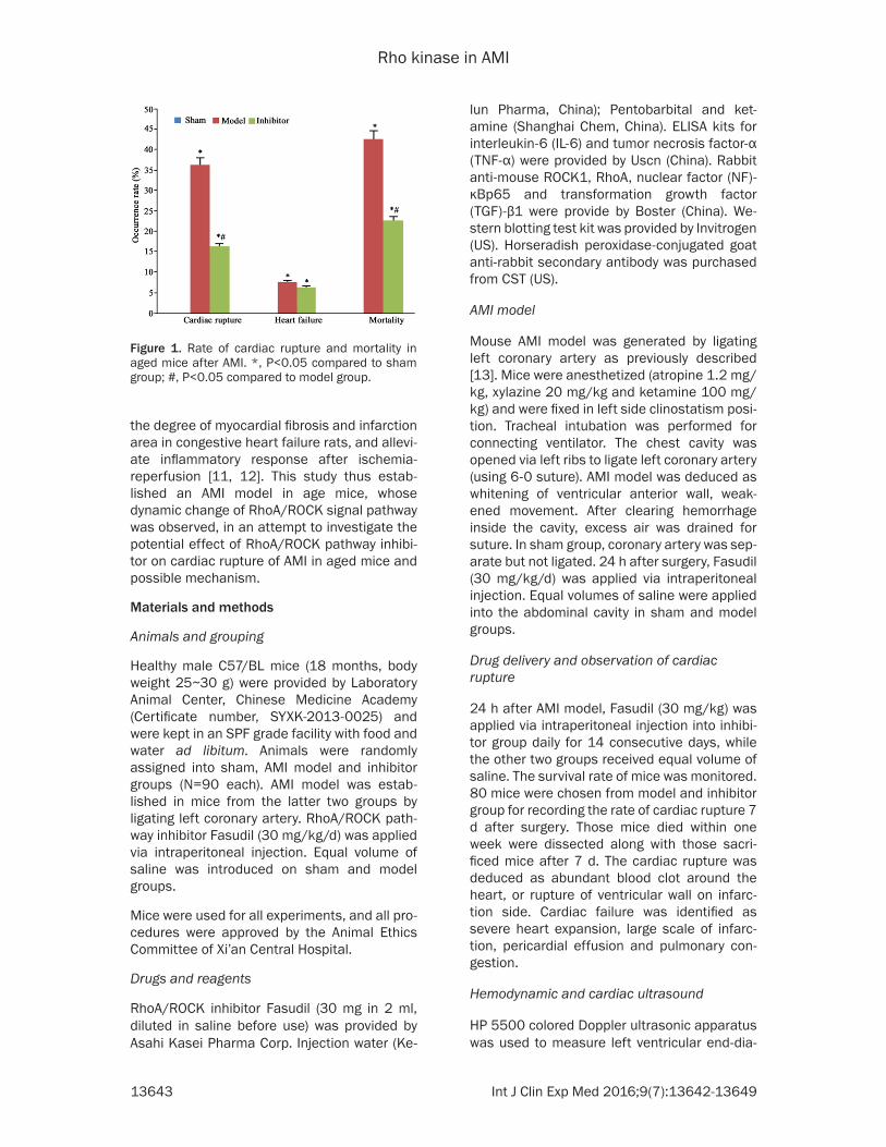
Figure 1. Rate of cardiac rupture and mortality in aged mice after AMI. *, P<0.05 compared to sham group; #, P<0.05 compared to model group.

Rho kinase in AMI
13644 Int J Clin Exp Med 2016;9(7):13642-13649
stolic diameter (LVEDD), left ventricular end-systolic diameter (LVESD), external diameter of left ventricular end-diastolic (EXLVDD) and ven-tricular wall thickness of diastolic/systolic phase (Pwd, Pws). Left ventricular fractional shortening (FS) = (LVEDD-LVESD)/LVEDD × 100%. An 1.4 F micro-cannula was inserted into ascending aorta and left ventricle via right common carotid artery to record blood pres-sure, left ventricular systolic pressure (LVSP), and maximal decreasing/increasing rate of internal pressure of left ventricle (dP/dtmin, dP/dtmax).
HE staining
HE staining was employed to detect the mor-phology of myocardial tissues. Eight animals were drawn from each group at day 7. Animals were sacrificed to extract myocardial tissues. After HE staining, the coverslip was mounted to observe tissue morphology under the light field microscope.
ELISA
ELISA was used to detect serum IL-6 and TNF-α levels from mouse serum samples following manual instruction. Absorbance values at 450 nm were recorded by a microplate reader in triplicates.
Western blotting
Western blotting was used to measure ROCK1, RhoA, NF-κBp65 and TGF-β1 expression in myocardial tissues. In brief, tissue samples were lysed in lysis buffer to collect the superna-tant by centrifugation. The protein content was determined by BCA test kit. After SDS-PAGE separation, proteins were transferred to PVD membrane, which was blocked in buffer, mixed with primary antibody for 4°C overnight incuba-tion. On the next day, the membrane was rinsed in TBST, and incubated in secondary antibody for 1 h. After TBST rinsing for three times, chro-mogenic substrate was added to develop the
Figure 2. Left ventricular remodeling of aged mice after AMI. *, P<0.05 compared to sham group; #, P<0.05 compared to model group.

Rho kinase in AMI
13645 Int J Clin Exp Med 2016;9(7):13642-13649
membrane, which was exposed in a dark room. Quantity One software was used to analyze pro-tein bands, whose optical density was trans-formed into relative expression level, as the ratio between target protein and internal refer-ence protein.
Statistical analysis
SPSS19.0 software was employed for data analysis. Using χ2 test or corrected χ2 test, we compare enumeration data. Those fitted the normal distribution were shown as mean ± standard deviation (SD). One-way analysis of variance (ANOVA) was used for compare means across multiple groups, followed by LSD test. A statistical significance was defined when P<0.05.
Results
Cardiac rupture rate of aged mice after AMI
After 3~6 d of cardiac rupture, inhibitor-treated mice had significantly lowered rate of cardiac
rupture compared to model group (χ2=8.265, P<0.05). The overall mortality rate was also lower in treatment group (χ2=7.293, P<0.05 compared to model group). No significant differ-ence of the heart failure existed between those two groups (P>0.05, Figure 1).
Myocardial remodeling of left ventricle in AMI mice
Compared to sham group, the inner diameter of left ventricle was significantly increased in AMI model group, with expanded ventricular cavity (P<0.05) as well as significantly decreased FS, Pwd and Pws (P<0.05). Inhibitor treatment signifi-cantly decreased inner diameter of left ventri-cle, ventricular cavity, and elevated FS, Pwd and Pws (P<0.05 compared to model group, Figure 2).
Hemodynamic indexes of AMI mice
Compared to sham animals, model mice had significantly lowered heart rate, systemic sys-
Figure 3. Hemodynamics indexes in AMI aged mice. LVSP, left ventricu-lar systolic pressure. dp/dtmin, dp/dtmax, maximal descending/ascend-ing rate of left ventricular internal pressure.

Rho kinase in AMI
13646 Int J Clin Exp Med 2016;9(7):13642-13649
tolic pressure and LVSP (P<0.05). Inhibitor treatment group had significantly elevated heart rate, systemic systolic pressure and LVSP (P<0.05, Figure 3).
Morphology of myocardial tissues after AMI
HE staining results showed no inflammatory infiltration in sham group, with tight and re- gular arrangement of myocardial cells. Model mice had more infiltration of inflammatory cells in myocardial tissues, with lower number of cells within the infarction area, accompanied with disarrangement of myocardial fiber, lysis or breakage. Inhibitor treatment group had alle-viated inflammatory infiltration and regular arrangement of cells (Figure 4).
Serum IL-6 and TNF-α levels after AMI
Compared to sham group, AML model mice had significantly elevated serum IL-6 and TNF-α lev-els (P<0.05). With elongated time, serum IL-6 and TNF-α levels were gradually decreased. Inhibitor treated group had significantly de- pressed IL-6 and TNF-α levels (P<0.05 com-pared to model group, Figure 5).
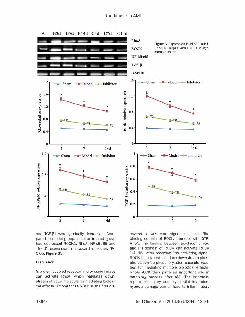
Expression level of ROCK1, RhoA, NF-κBp65 and TGF-β1 proteins
Western blotting results showed elevated ex- pression of ROCK1, RhoA, NF-κBp65 and TGF-β1 expression in AMI model mice (P<0.05 com-pared to sham group). With elongated time, expression levels of ROCK1, RhoA, NF-κBp65
Figure 4. Myocardial morphology of aged mice after AMI (X200, HE staining).
Figure 5. Serum IL-6 and TNF-α levels. *, P<0.05 compared to sham group; #, P<0.05 compared to model group.
Rho kinase in AMI
13647 Int J Clin Exp Med 2016;9(7):13642-13649
and TGF-β1 were gradually decreased. Com- pared to model group, inhibitor treated group had depressed ROCK1, RhoA, NF-κBp65 and TGF-β1 expression in myocardial tissues (P< 0.05, Figure 6).
Discussion
G protein coupled receptor and tyrosine kinase can activate RhoA, which regulates down-stream effector molecule for mediating biologi-cal effects. Among those ROCK is the first dis-
covered downstream signal molecule. Rho binding domain of ROCK interacts with GTP-RhoA. The binding between arachidonic acid and PH domain of ROCK can activate ROCK [14, 15]. After receiving Rho activating signal, ROCK is activated to induce downstream phos-phorylation/de-phosphorylation cascade reac-tion for mediating multiple biological effects. RhoA/ROCK thus plays an important role in pathology process after AMI. The ischemia-reperfusion injury and myocardial infarction-hypoxia damage can all lead to inflammatory
Figure 6. Expression level of ROCK1, RhoA, NF-κBp65 and TGF-β1 in myo-cardial tissues.

Rho kinase in AMI
13648 Int J Clin Exp Med 2016;9(7):13642-13649
response. Previous study showed elevated expression of RhoA and ROCK signal proteins in myocardial tissues after ischemia-reperfusion injury, for mediating the expression of multiple inflammatory factors (interferon, interleukin) and infiltration/adhesion of inflammatory cells. By knocking down ROCK gene or suppressing ROCK activity, leukocyte adhesion can be depressed to decrease expression level of inflammatory factors; while Rho kinase inhibitor could reduce reactive injury of myocarditis [16, 17]. Myocardial fibrosis after infarction is relat-ed with bio-mechanical force or hemodynamic change, elevated extracellular matrix activity, oxidative stress or inflammatory response, which is caused by ischemia/hypoxia. The knockout of ROCK gene could reduce the prolif-eration rate of myocardial fibroblast in hypoxia-reperfusion mice, decrease the number of fibroblast precursor and fibroblast, and sup-press the expression of pro-fibrosis cytokines [18, 19]. In vitro study has demonstrated that RhoA/ROCK signal pathway could facilitate mitochondria-mediated cell apoptosis via regu-lating the expression of apoptotic/anti-apoptot-ic proteins, and initiate exogenous cell apopto-sis via TNF [20, 21].
Age is one risk factor for coronary heart dis-ease. The incidence of cardiac death and AMI is elevated with elder people. Moreover, the prob-ability of cardiac rupture is also higher in aged AMI patients, leaving its pathogenesis mecha-nism unclear [8]. This study observed the effect of RhoA/ROCK signal pathway on the rate of cardiac rupture in aged AMI mice, to investigate related mechanism of post-AMI cardiac rup-ture. Results showed the rupture time between 3 and 6 days post-AMI. Compared to model group, RhoA/ROCK inhibitor group had signifi-cantly lowered rate of cardiac rupture, along with lower overall mortality rate, suggesting that the inhibition of RhoA/ROCK signal path-way could reduce the cardiac rupture of AMI mice. 7 days after AMI, the inner diameter and cavity of left ventricle in model mice were sig-nificantly elevated, accompanied with lower FS%, Pwd and Pws. RhoA/ROCK inhibitor mice had significantly decreased left ventricular inner diameter and cavity volume, along with higher FS%, Pwd and Pws, and alleviated pathol-ogy injury of myocardial tissues, suggesting the important role of RhoA/ROCK signal pathway in improving ventricular remodeling after AMI for the cardiac rupture. After applying RhoA/ROCK signal pathway inhibitor, serum levels of inflam-
matory cytokines including IL-6 and TNF-α were remarkably decreased. Western blotting results showed elevated expression of ROCK1, RhoA, NF-κBp65 and TGF-β1 in myocardial tissues of model mice. With elongated time, expression levels of ROCK1, RhoA, NF-κBp65 and TGF-β1 were gradually decreased. Their expression lev-els were decreased in inhibitor group. Fasudil could block Rho kinase activity via competing for Rho kinase binding sites with ATP, to partici-pate in various cellular pathways, suggesting the correlation between RhoA/ROCK signal pathway inhibitor-induced improvement of ven-tricular remodeling or decreasing cardiac rup-ture incidence with the suppression of RhoA/ROCK activity, lowered pro-inflammatory/pro-fibrotic factors. Such inhibitor could alleviate myocardial fibrosis and infiltration of inflamma-tory cells, to improve ventricular remodeling.
In summary, RhoA/ROCK pathway is probably related with post-AMI cardiac rupture. RhoA/ROCK inhibitor could suppress the activity of RhoA/ROCK pathway, down-regulate pro-inflammatory factor and pro-fibrotic factors, thus decreasing the rate of cardiac rupture.
Disclosure of conflict of interest
None.
Address correspondence to: Drs. Lina Hao and Chunping Kang, Department of Gerontology, Xi’an Central Hospital, No. 161 Xiwu Rd, Xi’an 710003, Shaanxi, China. Tel: +86-029-87268355; Fax: +86-029-87268355; E-mail: [email protected] (LNH); [email protected] (CPK)
References
[1] Bates ER. Reperfusion therapy reduces the risk of myocardial rupture complicating ST-ele-vation myocardial infarction. J Am Heart Assoc 2014; 3: e001368.
[2] Honda S, Asaumi Y, Yamane T, Nagai T, Miyagi T, Noguchi T, Anzai T, Goto Y, Ishihara M, Nishimura K, Ogawa H, Ishibashi-Ueda H, Yas-uda S. Trends in the clinical and pathological characteristics of cardiac rupture in patients with acute myocardial infarction over 35 years. J Am Heart Assoc 2014; 3: e000984.
[3] Serpytis P, Karvelyte N, Serpytis R, Kalinaus-kas G, Rucinskas K, Samalavicius R, Ivaska J, Glaveckaite S, Berukstis E, Tubaro M, Alpert JS, Laucevičius A. Post-infarction ventricular sep-tal defect: risk factors and early outcomes. Hel-lenic J Cardiol 2015; 56: 66-71.

Rho kinase in AMI
13649 Int J Clin Exp Med 2016;9(7):13642-13649
[4] Rizza S, Copetti M, Cardellini M, Menghini R, Pecchioli C, Luzi A, Di Cola G, Porzio O, Ippoliti A, Romeo F, Pellegrini F, Federici M. A score in-cluding ADAM17 substrates correlates to re-curring cardiovascular event in subjects with atherosclerosis. Atherosclerosis 2015; 239: 459-64.
[5] Soeda T, Higuma T, Abe N, Yamada M, Yokoya-ma H, Shibutani S, Ong DS, Vergallo R, Minami Y, Lee H, Okumura K, Jang IK. Morphological predictors for no reflow phenomenon after pri-mary percutaneous coronary intervention in patients with ST-segment elevation myocardial infarction caused by plaque rupture. Eur Heart J Cardiovasc Imaging 2016; [Epub ahead of print].
[6] Hayano S, Takefuji M, Maeda K, Noda T, Ichi-miya H, Kobayashi K, Enomoto A, Asai N, Taka-hashi M, Murohara T. Akt-dependent Girdin phosphorylation regulates repair processes after acute myocardial infarction. J Mol Cell Cardiol 2015; 88: 55-63.
[7] Zhu F, Li Y, Zhang J, Piao C, Liu T, Li HH, Du J. Senescent cardiac fibroblast is critical for car-diac fibrosis after myocardial infarction. PLoS One 2013; 8: e74535.
[8] Anderson DR, Poterucha JT, Mikuls TR, Duryee MJ, Garvin RP, Klassen LW, Shurmur SW, Thiele GM. IL-6 and its receptors in coronary artery disease and acute myocardial infarc-tion. Cytokine 2013; 62: 395-400.
[9] Luo SY, Chen S, Qin YD, Chen ZW. Urotensin-Receptor Antagonist SB-710411 Protects Rat Heart against Ischemia-Reperfusion Injury via RhoA/ROCK Pathway. PLoS One 2016; 11: e0146094.
[10] Chen HC, Chang JP, Chang TH, Lin YS, Huang YK, Pan KL, Fang CY, Chen CJ, Ho WC, Chen MC. Enhanced expression of ROCK in left atrial myocytes of mitral regurgitation: a potential mechanism of myolysis. BMC Cardiovasc Dis-ord 2015; 15: 33.
[11] Mera C, Godoy I, Ramírez R, Moya J, Ocaranza MP, Jalil JE. Mechanisms of favorable effects of Rho kinase inhibition on myocardial remod-eling and systolic function after experimental myocardial infarction in the rat. Ther Adv Car-diovasc Dis 2016; 10: 4-20.
[12] Zhang J, Liu XB, Cheng C, Xu DL, Lu QH, Ji XP. Rho-kinase inhibition is involved in the activa-tion of PI3-kinase/Akt during ischemic-precon-ditioning-induced cardiomyocyte apoptosis. Int J Clin Exp Med 2014; 7: 4107-14.
[13] Liu Z, Chen JM, Huang H, Kuznicki M, Zheng S, Sun W, Quan N, Wang L, Yang H, Guo HM, Li J, Zhuang J, Zhu P. The protective effect of trimetazidine on myocardial ischemia/reperfu-sion injury through activating AMPK and ERK signaling pathway. Metabolism 2016; 65: 122-30.
[14] Lee TM, Lin SZ and Chang NC. Membrane ERalpha attenuates myocardial fibrosis via RhoA/ROCK-mediated actin remodeling in ovariectomized female infarcted rats. J Mol Med (Berl) 2014; 92: 43-51.
[15] James SE, Burden H, Burgess R, Xie Y, Yang T, Massa SM, Longo FM, Lu Q. Anti-cancer drug induced neurotoxicity and identification of Rho pathway signaling modulators as potential neuroprotectants. Neurotoxicology 2008; 29: 605-12.
[16] Gao HC, Zhao H, Zhang WQ, Li YQ, Ren LQ. The role of the Rho/Rock signaling pathway in the pathogenesis of acute ischemic myocardial fi-brosis in rat models. Exp Ther Med 2013; 5: 1123-1128.
[17] Guo J, Wang SB, Yuan TY, Wu YJ, Yan Y, Li L, Xu XN, Gong LL, Qin HL, Fang LH, Du GH. Copti-sine protects rat heart against myocardial isch-emia/reperfusion injury by suppressing myo-cardial apoptosis and inflammation. Athero- sclerosis 2013; 231: 384-91.
[18] Rajapurohitam V, Izaddoustdar F, Martinez-Abundis E, Karmazyn M. Leptin-induced car-diomyocyte hypertrophy reveals both calcium-dependent and calcium-independent/RhoA- dependent calcineurin activation and NFAT nuclear translocation. Cell Signal 2012; 24: 2283-90.
[19] Martinez-Abundis E, Rajapurohitam V, Haist JV, Gan XT, Karmazyn M. The obesity-related pep-tide leptin sensitizes cardiac mitochondria to calcium-induced permeability transition pore opening and apoptosis. PLoS One 2012; 7: e41612.
[20] Moey M, Rajapurohitam V, Zeidan A, Karmazyn M. Ginseng (Panax quinquefolius) attenuates leptin-induced cardiac hypertrophy through in-hibition of p115Rho guanine nucleotide ex-change factor-RhoA/Rho-associated, coiled-coil containing protein kinase-dependent mitogen-activated protein kinase pathway acti-vation. J Pharmacol Exp Ther 2011 339: 746-56.
[21] Soliman H, Nyamandi V, Garcia-Patino M, Va-rela JN, Bankar G, Lin G, Jia Z, MacLeod KM. Partial deletion of ROCK2 protects mice from high-fat diet-induced cardiac insulin resistance and contractile dysfunction. Am J Physiol Heart Circ Physiol 2015; 309: H70-81.