review selenium: from cancer prevention to dna damage et al 2006.pdf · review selenium: from...
TRANSCRIPT

Toxicology 227 (2006) 1–14
Review
Selenium: From cancer prevention to DNA damage
Lucia Letavayova a, Viera Vlckova b, Jela Brozmanova a,∗a Laboratory of Molecular Genetic, Cancer Research Institute, Slovak Academy of Sciences, 833 91 Bratislava, Slovak Republic
b
Department of Genetics, Faculty of Natural Sciences, Comenius University, 842 15 Bratislava, Slovak RepublicReceived 30 March 2006; received in revised form 28 June 2006; accepted 19 July 2006Available online 25 July 2006
Abstract
Selenium (Se) is a dietary essential trace element with important biological roles. Accumulating evidence indicates thatSe compounds possess anticancer properties. Se is specifically incorporated into proteins in the form of selenocysteine andnon-specifically incorporated as selenomethionine in place of methionine. The effects of Se compounds on cells are strictlycompositional and concentration-dependent. At supranutritional dietary levels, Se can prevent the development of many typesof cancer. At higher concentrations, Se compounds can be either cytotoxic or possibly carcinogenic. The cytotoxicity of Seis suggested to be associated with oxidative stress. Accordingly, sodium selenite, an inorganic Se compound, was reported toinduce DNA damage, particularly DNA strand breaks and base damage. In this review we summarize the various activities of Secompounds and focus on their relation to DNA damage and repair. We discuss the use of Saccharomyces cerevisiae for identification
of the genes involved in Se toxicity and resistance. © 2006 Published by Elsevier Ireland Ltd.Keywords: Selenium; Free radicals; Toxicity; DNA damage; DNA repair
Contents
1. Introduction . . . . . . . . . . . . . . . . . . . . . . . . . . . . . . . . . . . . . . . . . . . . . . . . . . . . . . . . . . . . . . . . . . . . . . . . . . . . . . . . . . . . . . . . . . . . . . . . . 22. Overview of Se in human health . . . . . . . . . . . . . . . . . . . . . . . . . . . . . . . . . . . . . . . . . . . . . . . . . . . . . . . . . . . . . . . . . . . . . . . . . . . . . . . 2
2.1. Chemical forms of Se and their metabolism . . . . . . . . . . . . . . . . . . . . . . . . . . . . . . . . . . . . . . . . . . . . . . . . . . . . . . . . . . . . . . 33. Role of Se in cancer prevention . . . . . . . . . . . . . . . . . . . . . . . . . . . . . . . . . . . . . . . . . . . . . . . . . . . . . . . . . . . . . . . . . . . . . . . . . . . . . . . 44. Various mechanisms of Se in cancer prevention . . . . . . . . . . . . . . . . . . . . . . . . . . . . . . . . . . . . . . . . . . . . . . . . . . . . . . . . . . . . . . . . . 5
4.1. The protective role of Se in carcinogenesis . . . . . . . . . . . . . . . . . . . . . . . . . . . . . . . . . . . . . . . . . . . . . . . . . . . . . . . . . . . . . . . 54.2. Antioxidant activities of Se . . . . . . . . . . . . . . . . . . . . . . . . . . . . . . . . . . . . . . . . . . . . . . . . . . . . . . . . . . . . . . . . . . . . . . . . . . . . . 6
5. Prooxidant toxicity of Se . . . . . . . . . . . . . . . . . . . . . . . . . . . . . . . . . . . . . . . . . . . . . . . . . . . . . . . . . . . . . . . . . . . . . . . . . . . . . . . . . . . . . 66. Se and DNA damage . . . . . . . . . . . . . . . . . . . . . . . . . . . . . . . . . . . . . . . . . . . . . . . . . . . . . . . . . . . . . . . . . . . . . . . . . . . . . . . . . . . . . . . . . 87. Se and DNA repair . . . . . . . . . . . . . . . . . . . . . . . . . . . . . . . . . . . . . . . . . . . . . . . . . . . . . . . . . . . . . . . . . . . . . . . . . . . . . . . . . . . . . . . . . . . 9
8. The yeast Saccharomyces cerevisiae as a possible model to test mechanisms of DNA damage and repair induced by Se 99. Conclusions . . . . . . . . . . . . . . . . . . . . . . . . . . . . . . . . . . . . . . . . . . . . . . . . . . . . . . . . . . . . . . . . . . . . . . . . . . . . . . . . . . . . . . . . . . . . . . . . 10Acknowledgements . . . . . . . . . . . . . . . . . . . . . . . . . . . . . . . . . . . . . . . . . . . . . . . . . . . . . . . . . . . . . . . . . . . . . . . . . . . . . . . . . . . . . . . . . 11References . . . . . . . . . . . . . . . . . . . . . . . . . . . . . . . . . . . . . . . . . . . . . . . . . . . . . . . . . . . . . . . . . . . . . . . . . . . . . . . . . . . . . . . . . . . . . . . . . 11
∗ Corresponding author. Tel.: +421 2 59327 333; fax: +421 2 59327 350.E-mail address: [email protected] (J. Brozmanova).
0300-483X/$ – see front matter © 2006 Published by Elsevier Ireland Ltd.doi:10.1016/j.tox.2006.07.017

Toxicol
2 L. Letavayova et al. /1. Introduction
Selenium (Se) is a universal essential trace elementfor mammals which is important for many cellular pro-cesses. In the first half of the 20th century, Se, due toits toxicity was considered an undesirable element forhigher organisms. Toxicity of Se was first confirmed in1933 to occur in livestock that consumed plants of thegenus Astragalus, Xylorrhiza, Oonopsis and Stanleya inthe western regions of the United States. These plantshave the ability to accumulate large quantities of Se fromsoil, and therefore are called Se accumulator plants, orSe indicator plants (Oldfield, 1987).
In the second half of the 20th century, a significantchange in the importance of Se for human nutrition andbiology took place. A new biological perspective of Sewas shown by the pioneering work of Schwarz and Foltz(1957) who reported that Se at very low dietary concen-trations is an essential nutrient. At low concentrationsSe was able to prevent liver necrosis in rats consuming aTorula-yeast and Vitamin E deficient diet. Further sup-port for the benefits of Se came after the discovery of theessential role of Se in the formation of glutathione perox-idase (Rotruck et al., 1973), thioredoxin reductase andother enzymes that provided protection against oxida-tive stress. After 1973 it was confirmed by numerousstudies that selenoproteins and/or selenoenzymes wereinvolved in the metabolism of all higher vertebrates. Theaccumulated evidence showing the role of Se in manyareas important for human health has been reviewed byRayman (2000).
Results obtained from epidemiological studies, lab-oratory bioassays and human clinical intervention sup-ported a protective role(s) of Se against cancer devel-opment (Clark and Marshall, 2001; El-Bayoumy, 2001;Greenwald, 2004; Meuillet et al., 2004). However, thevarious organic and inorganic Se compounds used insuch studies have produced mixed results when tested inanimal models and human subjects. Studies performedin vitro have shown that both the dose and chemical formof Se compounds are critical factors in cellular responses(Ip, 1998). Se compounds at low concentration mayhave protective anticarcinogenic properties, whereas athigher concentration they can be genotoxic and possiblycarcinogenic (Spallholz, 1994). The toxicity of Se com-pounds is now viewed as being caused by the generationof reactive oxygen species (ROS) (Kramer and Ames,1988; Spallholz, 1997; Seko and Imura, 1997; Terada
et al., 1999; Spallholz et al., 2004). In a manner simi-lar to other ROS generating agents, some Se compoundsmay promote DNA oxidation in vivo. In accordance withthe observations that Se generates ROS, sodium seleniteogy 227 (2006) 1–14
(SSe), an inorganic selenium containing compound, hasbeen shown to induce DNA strand breaks in cell culturesystems (Lu et al., 1994, 1995; Zhou et al., 2003).
It is evident that Se has multiple roles in biologicalsystems. Many of them reside in its capability of acting asan antioxidant and disease preventing element. A num-ber of excellent reviews have recently been written onthe chemopreventive effects of Se (Rayman, 2000, 2005;El-Bayoumy, 2001; El-Bayoumy and Sinha, 2004, 2005;Whanger, 2004; Combs, 2005) and thus this topic will beaddressed only very briefly. Se, depending upon chemi-cal form, can be a prooxidant toxic agent that can induceDNA damage and cell death. The mechanisms that deter-mine Se cytotoxicity and the induction of DNA damageare the main subject of present review.
2. Overview of Se in human health
As previously noted, Se is an essential dietary nutri-ent for all mammals. The initial US recommendeddaily allowance in 1989 was 50–70 �g/day (this valuehas recently been lowered to 55 �g/day) for healthyhuman adults (El-Bayoumy, 2001; Whanger, 2004).This amount of Se may fulfill the dietary need forthe 25 known (Stadtman, 2002) selenoproteins aswell as for general human health (Rayman, 2000).Most of the human dietary Se requirement is met bydietary l-selenomethionine and, the lesser amounts of l-selenocysteine, both of which are components of animalproteins (Spallholz, 1994). Both organic selenomethion-ine (as a component of Se-enriched yeast) and inorganicSSe are commercial forms available as supplements(Shen et al., 2001).
Concerning the toxicity, Se has limited doses usedin chemoprevention. Based on human studies, intakesof 400 �g/day were established as the maximum safedietary dose with the no observed adverse effect. Symp-toms of Se toxicity were assessed by patient inter-view with questions regarding breath, hair and nailchanges. The low adverse effect of Se supplementa-tion was calculated to be 1540–1600 �g/day. An intakeof 3200–5000 �g/day resulted in definite occurrence ofselenosis (Reid et al., 2004). On the other hand a level ofabout 40 �g/day was suggested as the minimum require-ment, while an intake of <11 �g/day results in deficiencyproblems (Whanger, 2004).
Se enters the food chain through plants, which is takenup from the soil. The geographic distribution of Se varies
from high concentrations in the soils in certain regionsof the former USSR, Venezuela and the USA to ratherlow Se levels in soils in New Zealand, certain regions inChina, East Siberia, Korea, and to some extent also the
Toxicol
sR
taeoncilsAmcosLnardiSKr
L. Letavayova et al. /
oils in many parts of Europe (Brtkova and Brtko, 1996;ayman, 2000, 2005; Ferguson et al., 2004).
Human Se-deficiency diseases have been first iden-ified in some regions of China; Keshan disease,n endemic cardiomyopathy, and Kashin-Beck dis-ase, a deforming arthritis (Rayman, 2000). Numerousther studies suggest that Se deficiency is accompa-ied by loss of some immunocompetency; with bothell-mediated immunity and humoral immunity beingmpaired (Spallholz et al., 1990). Se deficiency is alsoinked to the occurrence, virulence, and disease progres-ion of some viral infections (e.g. HIV progression toIDS). Low serum Se in women increases the risk ofiscarriages (Barrington et al., 1996) and in men it is
onnected with a decrease in sperm motility and chancesf fertilization (Scott et al., 1998). The turnover rate ofome neurotransmitters is also altered by Se deficiency.ow plasma Se concentrations in the elderly are sig-ificantly associated with senility, Alzheimer’s diseasend depression (Hawkes and Hornbostel, 1996). Se has aole in the thyroid hormone metabolism being part of theeiodinase enzyme (Brtkova and Brtko, 1996). The find-
ngs have been equivocal regarding a correlation betweene and cardiovascular disease risk (Virtamo et al., 1985;ardinaal et al., 1997). The other disorders, such asheumatoid arthritis, pancreatitis and asthma associated
Fig. 1. Schematic representation of the Se metabolic pathway (a
ogy 227 (2006) 1–14 3
with increased oxidative stress or inflammation might beexpected to be influenced by Se levels (McCloy, 1998;Knekt et al., 2000). General oxidative stress response istotally influenced by dietary Se and its enzyme levels(Rayman, 2000).
2.1. Chemical forms of Se and their metabolism
Se exists in mostly organic forms in normaldiets. Organic Se is present in foods mainly in theform of selenomethionine, selenocysteine and Se-methylselenocysteine, whereas inorganic Se either asselenite or selenate occurs much less frequently and invery low amounts. Of the organic forms, selenomethio-nine is the predominant form in most Se rich diets. Bothorganic and inorganic forms of Se appear to be utilizedwith similar efficacy in the body to produce selenopro-teins (Shiobara et al., 1998) but the Se enters at differentpoints in metabolism depending on chemical form. Ametabolic scheme showing Se metabolism is presentedin Fig. 1.
Inorganic forms of Se, selenite and selenate are
reduced (from the valence +4 and +6, respectively) byglutathione (GSH). A number of intermediate metabolicsteps lead to the generation of H2Se, or they directlyenter the metabolic pool (Foster et al., 1986; Lu et al.,dapted from Meuillet et al. (2004) and Lu et al. (1995)).

Toxicol
4 L. Letavayova et al. /1995). SSe produces H2Se and/or elemental Se (Se0) viaselenodiglutathione (GSSeSG) through reduction by thi-ols and NADPH-dependent reductases (Ganther, 1971;Hsieh and Ganther, 1975). This reduction pathway istightly connected to the production of the superoxide(O2
•−) radical (for details, see Section 5).Organic Se, present in the Se-containing amino acids
in foods, is metabolized to the same key intermedi-ate (Esaki et al., 1982; Tanaka et al., 1985). Moreover,H2Se is the intermediate compound between the reduc-tive metabolism of Se and its methylation pathway. H2Seeither serves as a precursor for the synthesis of seleno-proteins, such as glutathione peroxidase, thioredoxinreductase, iodothyronine deiodinases and selenoproteinP, or it undergoes stepwise methylation with the enzy-matic reaction of thiol S-methyltransferases to generatethe mono-, di-, and tri-methylated forms of Se; methylse-lenol, dimethyldiselenide and the trimethylselenoniumion, respectively (Ip et al., 1991; Meuillet et al., 2004).
Selenomethionine, the major dietary form of Se,is subject to several different metabolic fates. Firstly,cells do not distinguish between methionine andselenomethionine during protein synthesis, so this natu-ral selenoamino acid gets incorporated into the generalbody proteins, e.g. albumin, in place of methionine whenmethionine is or is not a limiting factor. This non-specificincorporation of Se into proteins likely accounts forthe observed dose-dependent increase of tissue Se lev-els with selenomethionine supplemented diets comparedwith diets supplemented with other chemical forms of Se(Shiobara et al., 1998). Secondly, selenomethionine getsconverted into selenocysteine by the trans-sulfurationpathway, which is subsequently converted into H2Se(Esaki et al., 1981) and follows the same metabolic fate ofselenocysteine as described above. Lastly, selenomethio-nine may generate methylselenol by the enzymatic reac-tion of methionine �,�-lyase (also known as methioni-nase) (Meuillet et al., 2004).
Selenocysteine, another form of dietary organic Se,either taken from diet or derived from selenomethio-nine, is also converted into H2Se. Selenocysteine isalso synthesized from H2Se after H2Se conversion toselenophosphate by selenophosphate synthetase. Theinsertion of selenocysteine into protein is specified bythe UGA codon in mRNA. Thus, selenocysteine formsthe active catalytic site in all selenoenzymes and is essen-tial for the synthesis of selenoproteins (Hatfield andGladyshev, 2002).
Se-methylselenocysteine, unlike selenomethionine,is not incorporated into proteins but may be converteddirectly to methylselenol by �-lyase (Foster et al.,1986). Like Se-methylselenocysteine, synthetic Se com-
ogy 227 (2006) 1–14
pounds such as selenobetaine, methylseleninic acid andmethylselenocyanate also readily generate methylse-lenol, which is now believed to be the main intermediatemetabolite in the cellular events of Se chemoprevention(Combs and Gray, 1998; Ip et al., 2000; El-Bayoumy andSinha, 2004).
Se compounds that enter either the H2Se pool orthe methylselenol pool undergo methylation by thiolS-methyltransferases and generate different methylatedmetabolic Se forms that sequentially are exhaled inthe breath or excreted in urine contributing to the Sehomeostasis of the body. At low Se doses monomethy-lated forms of Se are excreted into urine as the majorform, while trimethylated forms are being predomi-nant at high doses. When the level of trimethylseleno-nium ion reaches the metabolic plateau, dimethylse-lenide is exhaled into breath (Itoh and Suzuki, 1997).Selenosugars in urine have recently been identified(Kobayashi et al., 2002) and 1�-methylseleno-N-acetyl-d-galactosamine is thought to be a major monomethy-lated urinary metabolite except at extremely high Seintake. Excretion of different Se species in urine at dif-ferent levels of Se intake may thus be useful indicatorsof healthy and toxic doses of Se (Kobayashi et al., 2002).Generally, the methylation pathway is considered to bethe detoxification pathway for all Se in the diet or insupplements (Foster et al., 1986; Lu et al., 1995).
3. Role of Se in cancer prevention
A low dietary Se intake was first noted in asso-ciation with cancer risk approximately 35 years ago(Shamberger and Frost, 1969). Human epidemiologicalstudies conducted over this period of time examined therelationship between dietary intake of Se and total cancerrisk, and have been somewhat controversial (Meuillet etal., 2004). Early epidemiological studies (Schrauzer etal., 1977a,b) showed a geographic correlation betweenlow Se status and a high incidence of certain types ofcancers. Dietary intake of Se in 27 countries found a sig-nificant inverse correlation with age-adjusted mortalityfor cancer of the colon, prostate, breast, ovary and lungas well as with hematopoetic cancers, while only a weakcorrelation was observed for cancers of the pancreas,skin and bladder. Such reports have been made by morethan 100 experimental studies using different forms anddoses of Se (Combs and Gray, 1998). Although not allstudies showed a correlative effect of high plasma Se
level and low cancer incidence (contradictory results arethought to be partially related to the variations in datacollection and methods measuring the Se levels), the epi-demiological data stimulated interest in testing Se as a
Toxicol
ce
Cohrtt3w
itbaeSaa
odwhohm2ocrocMaisediSM2
pssradU
L. Letavayova et al. /
hemopreventive agent in human clinical trials (Meuillett al., 2004).
Chemoprevention trials have been carried out inhina using Se as a single experimental agent. Inne study, 130,000 people from the regions with aigh incidence of hepatocellular carcinoma (HCC) wereecruited. People had their table salt fortified with Se inhe form of SSe (15 mg/kg); the other had their salt unfor-ified. After 6 years, the incidence of HCC decreased by5% in the Se salt supplemented group in comparisonith the unsupplemented control group (Yu et al., 1997).In another study the effect of low Se concentrations
n serum on the incidence of different cancers and onotal death were followed. Significant inverse associationetween base-line serum Se and death from esophagealnd gastric (Wei et al., 2004) as well as lung cancer (Zhuot al., 2004) have been found. These results suggest thate supplementation may have some protective effectsgainst some type(s) of cancer in populations where aver-ge dietary Se levels are low.
The Nutritional Prevention of Cancer Trial carriedut by Clark and co-workers in the USA was the firstouble-blind, placebo-controlled intervention trial in aestern population. The trial was designed to test theypothesis that Se supplementation could reduce the riskf skin cancer (Clark et al., 1996). One thousand threeundred and twelve individuals with a history of non-elanoma skin cancer were randomized to placebo or
00 �g Se per day as selenium yeast. There was no effectn the primary endpoint of non-melanoma skin can-er, however, individuals in the Se-treated group expe-ienced sizable reductions in the risk of primary cancerf colon, rectum, prostate and lung. The prostate can-er incidence decreased by a striking 65% (Clark andarshall, 2001; Combs et al., 2001; Duffield-Lillico et
l., 2002). These findings led to additional clinical stud-es testing the effects of Se on prevention of primary andecondary prostate cancer (Nelson et al., 2002; Brookst al., 2001; Van den Brandt et al., 2003). Newer epi-emiological data supported the hypothesis that theres a significant inverse correlation between total serume level and prostate cancer risk (Vogt et al., 2003;euillet et al., 2004; Waters et al., 2005; Etminan et al.,
005).Clinical cancer prevention trials demonstrate the
otential strategy for reducing the burden of cancer onociety. They suggest that nutritional food components,uch as Se, when supplemented to diets may minimize or
educe cancer risk. The role of Se in human health is stillsubject of intense interest. Large intervention trials withifferent forms of Se are under way in Europe and theSA to assess the effects of Se supplements in the inci-ogy 227 (2006) 1–14 5
dence of cancer and other diseases (Luty-Frackiewicz,2005; Burk et al., 2006; Sabichi et al., 2006; Drake,2006).
4. Various mechanisms of Se in cancerprevention
4.1. The protective role of Se in carcinogenesis
It is generally accepted that carcinogen-inducedgenetic damage via the formation of covalent DNAadducts is necessary, but not sufficient, for the initiationof carcinogenesis. Therefore, several in vitro studies inrodents were conducted to examine the effects of variouslevels and forms of Se on carcinogen DNA adduct for-mation. In most of these studies, Se (as selenite, selenate,1,4-phenylenebis(methylene)selenocyanate (p-XSC) ordiallyl selenide (DASe)) have been shown to inhibit theinitiation phase of carcinogenesis induced by variouscarcinogens in mammary, liver, colon and lung tissuein rats (El-Bayoumy, 2001). In experiments with Se-enriched garlic, Ip and Lisk (1994) reported an inhibitionof both the initiation and post-initiation stages of mam-mary carcinogenesis after dimethylbenz(a)anthracene(DMBA) treatment of rats. The explanation for cancerprevention is that some Se compounds affect procar-cinogen activation and metabolism through inhibitionof phase I and induction of phase II enzymes. PhaseI enzymes are members of the cytochrome P450 sys-tem responsible for converting chemical carcinogens toreactive adducts that can attack to DNA. On the otherhand, defense against carcinogenic injury is providedby phase II detoxifying enzymes. The experiments byIp and Lisk (1997) showed that increased detoxifica-tion of carcinogen via the phase II conjugating enzymesmight represent the mechanism of tumor suppression bySe-enriched garlic. This mechanism has been well docu-mented to be important for the chemopreventive activityof many thiol-reactive chemopreventive agents, mainlyby inhibition of the initiation stage of carcinogenesis(Zhou et al., 2003).
However, suppression of carcinogenesis by Se com-pounds in the post-initiation stages of carcinogen treat-ment in animal models suggests the existence of addi-tional mechanisms for cancer chemoprevention. Theability of Se compounds to inhibit cell growth andto induce tumor cell apoptosis has now been widelydemonstrated and is suggested to be a potential mecha-
nism for cancer chemoprevention (Stewart et al., 1997;Wang et al., 2003; Sinha and El-Bayoumy, 2004). It hasbeen speculated that Se-induced apoptosis can removecarcinogen-initiated transformed cells and suppress the
Toxicol
6 L. Letavayova et al. /clonal expansion of such transformed cell population(Combs, 1997; Zhou et al., 2003).
Many experiments have shown that the chemopreven-tive effect of Se is due in part to its inhibitory effect oncell growth, DNA, RNA and protein synthesis in trans-formed cells (El-Bayoumy, 2001). Although the molec-ular mechanisms of the cancer chemopreventive activityof the Se compounds remains largely unknown, changesin stress-related cellular proteins have been implicatedin explaining the protective effects of Se. Because pro-tein kinases play a central role in the regulation ofcell growth, tumor promotion and differentiation, sev-eral reports have described the inhibitory effect of Seon kinase activity (Gopalakrishna et al., 1997; Sinha etal., 1999). Moreover, cell cycle CDK2 or cell signalingprotein kinases and/or a number of redox-regulated pro-teins, including the critical transcription factors, such asAP-1 and NF-�B, have been proposed as targets againstwhich Se exerts its chemopreventive effects (Zhu et al.,1995; Sinha et al., 1996).
4.2. Antioxidant activities of Se
All living aerobic organisms have developed cer-tain defense mechanisms that provide protection againstoxidative damage induced by ROS (Valko et al., 2004).The best known of these antioxidant enzymes is super-oxide dismutase (Davies, 1994). Se was found to be acomponent of antioxidant defenses either as agent ableto scavenge free radicals, or as a component of a familyselenoenzymes. Experimental evidence suggesting itseffect in eliminating oxidative stress came in 1973 whenSe was found to be incorporated into the first identifiedselenoprotein, glutathione peroxidase (GPx) (Rotruck etal., 1973). Members of the GPx family of enzymes areeffective in catalyzing the reduction of hydrogen per-oxide (H2O2) and lipid peroxides. In view of the roleof GPx in reducing oxidative stress it was proposedthat Se-dependent GPx provided a plausible mechanismfor cancer prevention. However, it was found that GPxactivity was maximized in tissues of animals fed normalamounts of Se and was not elevated as dietary Se wasincreased 10-fold at higher levels necessary to achievechemoprevention. Thus, it seemed that Se supplementsexerted their chemopreventive effect in a manner unre-lated to the saturated levels of GPx (Combs and Gray,1998). Se is found to be a component of several otherselenoproteins, some of which have important enzymatic
functions associated with antioxidant defenses. One ofthese, thioredoxin reductase is considered to be a keyenzyme in Se metabolism, reducing Se compounds andcontrolling the intracellular redox state (Lee et al., 2000;ogy 227 (2006) 1–14
Madeja et al., 2005). Additionally, iodothyronine deiod-inase, produces the active thyroid hormone, T3 from aninactive precursor T4. Another selenoprotein, selenopro-tein P appears to protect endothelial cells against damagefrom free radicals (Allan et al., 1999; Ganther, 1999;Rayman, 2000). Each of these selenoproteins containsSe in the form of selenocysteine, which is incorporatedby the co-translational modification of transfer RNA-bound serine at certain loci encoded by a specific UGAcodon. As noted above, it has generally been thought thatthe selenoproteins fulfill only the nutritional requirementfor dietary Se. There was not unambiguous evidencesupporting the role of any known selenoprotein in can-cer prevention. However, it has recently become clearthat optimal expression of some selenoproteins, notablyselenoprotein P, requires higher amount of dietary Se(Xia et al., 2005) and that a substantial number of indi-viduals may have a higher Se requirement for efficientsynthesis of selenoproteins (Rayman, 2005). This inter-individual variation in selenoprotein expression levelsmay be accounted for single-nucleotide polymorphismsin selenoproteins genes that determine the efficiencywith which individuals can incorporate Se into seleno-proteins (Kumaraswamy et al., 2000; Ichimura et al.,2004; Apostolou et al., 2004).
Whether the selenoproteins are crucial to the anti-cancer effects needs to be elucidated. The functionsof many of the 25 human selenoproteins are as yetunknown, although some of them are involved in antiox-idant and anabolic processes (Hatfield and Gladyshev,2002). Selenoproteins that may be relevant to cancer riskare supposed to be the GPx family, the 15 kDa selenopro-tein (Sep15), selenoprotein P and the thioredoxin reduc-tases, although a beneficial role of thioredoxin reductasein cancer prevention is doubtful (Rayman, 2005).
5. Prooxidant toxicity of Se
The effect of Se upon cells is strictly form andconcentration-dependent. Se compounds induce theexpression of the 25 known selenoproteins, includ-ing those with antioxidant activities (Gladyshev andHatfield, 1999). Many studies have indicated thatthe cancer chemopreventive activity of Se compoundsrequires much higher concentration than that required forthe synthesis of the selenoproteins (Rayman, 2005). Atmoderate, supranutritional doses, Se compounds inhibitcell growth and have a prooxidant activity, generating
superoxide. At higher concentrations of mainly inor-ganic forms of Se compounds, acute toxicity due to DNAstrand breaks occur (Combs and Gray, 1998). Which ofthese effects elicited by the supranutritional and/or toxic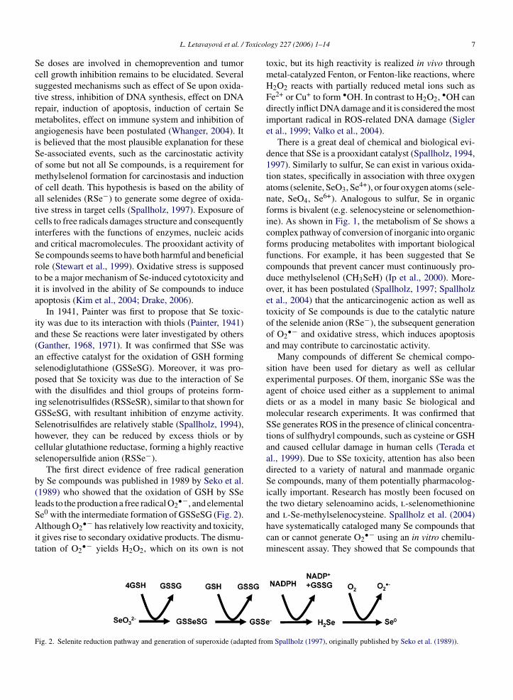
Toxicol
ScstrmaiSomoatciaSrtia
ia(aspwiGShcs
b(lSAit
F
L. Letavayova et al. /
e doses are involved in chemoprevention and tumorell growth inhibition remains to be elucidated. Severaluggested mechanisms such as effect of Se upon oxida-ive stress, inhibition of DNA synthesis, effect on DNAepair, induction of apoptosis, induction of certain Seetabolites, effect on immune system and inhibition of
ngiogenesis have been postulated (Whanger, 2004). Its believed that the most plausible explanation for thesee-associated events, such as the carcinostatic activityf some but not all Se compounds, is a requirement forethylselenol formation for carcinostasis and induction
f cell death. This hypothesis is based on the ability ofll selenides (RSe−) to generate some degree of oxida-ive stress in target cells (Spallholz, 1997). Exposure ofells to free radicals damages structure and consequentlynterferes with the functions of enzymes, nucleic acidsnd critical macromolecules. The prooxidant activity ofe compounds seems to have both harmful and beneficialole (Stewart et al., 1999). Oxidative stress is supposedo be a major mechanism of Se-induced cytotoxicity andt is involved in the ability of Se compounds to inducepoptosis (Kim et al., 2004; Drake, 2006).
In 1941, Painter was first to propose that Se toxic-ty was due to its interaction with thiols (Painter, 1941)nd these Se reactions were later investigated by othersGanther, 1968, 1971). It was confirmed that SSe wasn effective catalyst for the oxidation of GSH formingelenodiglutathione (GSSeSG). Moreover, it was pro-osed that Se toxicity was due to the interaction of Seith the disulfides and thiol groups of proteins form-
ng selenotrisulfides (RSSeSR), similar to that shown forSSeSG, with resultant inhibition of enzyme activity.elenotrisulfides are relatively stable (Spallholz, 1994),owever, they can be reduced by excess thiols or byellular glutathione reductase, forming a highly reactiveelenopersulfide anion (RSSe−).
The first direct evidence of free radical generationy Se compounds was published in 1989 by Seko et al.1989) who showed that the oxidation of GSH by SSeeads to the production a free radical O2
•−, and elemental
e0 with the intermediate formation of GSSeSG (Fig. 2).lthough O2•− has relatively low reactivity and toxicity,t gives rise to secondary oxidative products. The dismu-ation of O2
•− yields H2O2, which on its own is not
ig. 2. Selenite reduction pathway and generation of superoxide (adapted fro
ogy 227 (2006) 1–14 7
toxic, but its high reactivity is realized in vivo throughmetal-catalyzed Fenton, or Fenton-like reactions, whereH2O2 reacts with partially reduced metal ions such asFe2+ or Cu+ to form •OH. In contrast to H2O2, •OH candirectly inflict DNA damage and it is considered the mostimportant radical in ROS-related DNA damage (Sigleret al., 1999; Valko et al., 2004).
There is a great deal of chemical and biological evi-dence that SSe is a prooxidant catalyst (Spallholz, 1994,1997). Similarly to sulfur, Se can exist in various oxida-tion states, specifically in association with three oxygenatoms (selenite, SeO3, Se4+), or four oxygen atoms (sele-nate, SeO4, Se6+). Analogous to sulfur, Se in organicforms is bivalent (e.g. selenocysteine or selenomethion-ine). As shown in Fig. 1, the metabolism of Se shows acomplex pathway of conversion of inorganic into organicforms producing metabolites with important biologicalfunctions. For example, it has been suggested that Secompounds that prevent cancer must continuously pro-duce methylselenol (CH3SeH) (Ip et al., 2000). More-over, it has been postulated (Spallholz, 1997; Spallholzet al., 2004) that the anticarcinogenic action as well astoxicity of Se compounds is due to the catalytic natureof the selenide anion (RSe−), the subsequent generationof O2
•− and oxidative stress, which induces apoptosisand may contribute to carcinostatic activity.
Many compounds of different Se chemical compo-sition have been used for dietary as well as cellularexperimental purposes. Of them, inorganic SSe was theagent of choice used either as a supplement to animaldiets or as a model in many basic Se biological andmolecular research experiments. It was confirmed thatSSe generates ROS in the presence of clinical concentra-tions of sulfhydryl compounds, such as cysteine or GSHand caused cellular damage in human cells (Terada etal., 1999). Due to SSe toxicity, attention has also beendirected to a variety of natural and manmade organicSe compounds, many of them potentially pharmacolog-ically important. Research has mostly been focused onthe two dietary selenoamino acids, l-selenomethionine
and l-Se-methylselenocysteine. Spallholz et al. (2004)have systematically cataloged many Se compounds thatcan or cannot generate O2•− using an in vitro chemilu-minescent assay. They showed that Se compounds that
m Spallholz (1997), originally published by Seko et al. (1989)).

8 L. Letavayova et al. / Toxicol
Fig. 3. Redox cycling of selenides (RSe−) (adapted from Spallholz etal. (2004), originally published by Chaudiere et al. (1992)).
can easily form the selenide anion, RSe−, also gener-ate O2
•− in vitro via oxidation of GSH, or other thiols(RSH). The two selenoamino acids, l-selenomethionineand l-Se-methylselenocysteine, do not redox cycle invitro because they can not directly form methylse-lenol via GSH reduction. The methylselenide anion(CH3Se−) must be formed from these selenoamino acidsin vivo by metabolic enzymes, methioninase (in thecase of l-methionine) or �-lyases (in the case of l-Se-methylselenocysteine) (Fig. 2). Methylselenol can bedirectly formed from methylseleninic acid in vitro byglutathione reduction and it is a potent redox Se species(Fig. 3).
6. Se and DNA damage
It has been known for a long time that some Se com-pounds have the potential to induce DNA damage. Muchof what is known about the DNA damage by Se is derivedfrom bacterial as well as cell culture experiments. In amajority of DNA damaging experiments, SSe has beenchosen as the source of Se. It is now well establishedthat the toxicity of the SSe is strongly influenced by itsmetabolism and production of ROS (Fig. 1, see Section5). In accordance, it was shown (Kramer and Ames,1988) that reactions of SSe with thiols and the con-comitant production of ROS were the primary causalevents of Se mutagenicity and toxicity in Salmonellatyphimurium. Studies of isolated hepatocytes as a modelsystem indicated that SSe-induced DNA fragmentationwas oxygen and redox cycle-dependent (Garberg et al.,1988), thereby confirming the view that DNA damageby SSe in mammalian cells is also mediated by ROS.
Later, it was found that SSe at concentrations of 5–10 �Minduced DNA single-strand breaks (SSB) and double-strand breaks (DSB) in murine leukemic L1210 cellsand other murine mammary carcinoma cell lines. Induc-ogy 227 (2006) 1–14
tion of DNA strand breaks was in all experimental casesassociated with a loss of cell viability (Lu et al., 1994,1995). SSe has also led to chromosomal damage in Swissalbino mice and human peripheral lymphocytes (Biswaset al., 1997, 2000). These data clearly indicated that DNAdamage is implicated in the genotoxicity of SSe and thatSSe-induced toxicity is mediated by its prooxidant activ-ity connected with ROS formation.
The organic Se compounds, selenomethionine andselenocysteine, using the different metabolic steps pro-duce the same metabolite H2Se, as SSe (Fig. 1). Theproduction of the toxic H2Se would suggest that bothinorganic and some natural organic forms of Se can beeffective in induction of DNA damage, although not nec-essarily with similar dose efficiency. Accordingly, DNAdamaging activity of selenomethionine have recentlybeen demonstrated in prostate cells of dogs fed by highdoses (6 �g/kg/day for 7 months) of this dietary aminoacid (Waters et al., 2005). When organic methylatedSe compounds were examined for potency to induceDNA strand breaks, a distinctive cellular response incomparison with SSe was reported (Ip, 1998). In cellculture, SSe at levels of 5–10 �M can induce SSB,cell death by necrosis and acute cell lysis. In con-trast, the methylated Se compounds, such as methylse-lenocyanate or methylselenocysteine, even at levels of10–50 �M induced cell death predominantly by apop-tosis, an event that is characterized by specific mor-phological (condensation of chromatin) and biochem-ical (DNA fragmentation) changes without evidence ofDNA strand breakage (Wilson et al., 1992; Lu et al.,1995). Differences in the DNA damaging activities andin the induction of morphological changes by SSe ver-sus methylselenocyanate indicated induction of differentmetabolic pathways by these two agents leading pre-dominantly to H2Se by SSe and to methylselenol bymethylselenocyanate. Compounds that are directly con-verted to methylselenol, such as methylselenocyanate(Fig. 1), are supposed to be superior to the inorganicSSe as well as to the organic selenomethionine dueto their significant preventive efficacy and the minimalside effects on DNA stability and toxicity (Combs andGray, 1998; El-Bayoumy and Sinha, 2004; Whanger,2004).
Previous studies have suggested that DNA damageis involved in the induction of events leading to apop-totic death in tumor cells (Lu et al., 1994). Recently,it has been demonstrated that SSe-induced apoptosis,
which involved ATM/ATR and TOP II, is likely to becaused by DNA damage (Zhou et al., 2003). Thus, DNAdamage seems to play an important regulatory role inSSe-induced apoptosis.
Toxicol
7
airiaadogtD1aS
onfBtirtuo
rogTbisbhe
drctnsssmise
L. Letavayova et al. /
. Se and DNA repair
Among the essential trace mineral nutrients, Se hasspecial position due to its catalytic activity and toxic-
ty. In comparison to our understanding of the protectiveole(s) of Se in human health and cancer prevention, lessnformation is available about the mechanisms whichre responsible for maintaining of genetic informationnd for repairing DNA lesions induced by Se’s prooxi-ant character. As mentioned above, SSe is an inorganicxidizing agent, which has the capacity to compromiseenetic stability by inducing DNA damage. Althoughhe exact mechanism of SSe-induced modifications toNA remains unknown, DNA strand breaks (Lu et al.,995; Zhou et al., 2003) and base lesions (Wycherly etl., 2004) seem to be the most probable consequence ofSe-induced toxicity.
Data accumulated over many years clearly shows thatxidative DNA damage plays an important role in aumber of disease processes, including malignant trans-ormations (Brozmanova et al., 2001; Evans et al., 2004).ecause there is a relatively narrow range separating
he amount of Se essential for health from that whichs toxic, it is necessary to estimate the possible geneticisk of Se compounds against their beneficial effects tohe humans. This information is also important to eval-ate the safety of possible pharmacological applicationf Se-containing agents such as the anticancer drugs.
To reduce the adverse consequences of many envi-onmental agents, including Se, a complex networkf different repair systems has evolved to maintainenetic integrity (Friedberg, 2000; Hoeijmakers, 2001).he major pathway eliminating DNA base damage,ase free sites and SSB is excision repair, subdividednto nucleotide excision repair (NER) and base exci-ion repair (BER). The replication errors are repairedy the mismatch repair (MMR) pathway and DSB byomologous recombination (HR) or non-homologousnd-joining (NHEJ).
Nevertheless, besides agents that induce exclusivelyifferent types of DNA damage, either directly or indi-ectly via intermediates, there are some agents whichan also interfere with the repair of DNA thus increasinghe adverse consequences of DNA lesions. Well recog-ized examples are the carcinogenic metal compounds,uch as nickel, cobalt, cadmium and arsenic which werehown to inhibit BER and NER repair pathways at low,eemingly non-cytotoxic concentrations. Potential target
olecules for metal ions are the zinc finger structuresn DNA repair proteins, with each zinc finger proteinensitive towards a specific toxic metal ions (Hartwigt al., 2002; Hartwig and Schwerdtle, 2002). Moreover,
ogy 227 (2006) 1–14 9
it was shown that zinc fingers may be sensitive targetsnot only for non-essential toxic metal ions, but also foressential trace elements such as Se. Thus, it was demon-strated in vitro that certain reducible Se compoundscaused a concentration-dependent decrease of activityand released of zinc from the zinc finger motif in thetwo zinc finger repair proteins, bacterial formamidopy-rimidine DNA glycosylase (Fpg) involved in BER andxeroderma pigmentosum group A protein (XPA) essen-tial for NER (Hartwig et al., 2003; Blessing et al., 2004).These results demonstrate that at low concentrations Secompounds that are reducible to selenides may inactivatethe DNA repair processes by the oxidation of zinc fin-gers in DNA repair proteins (Ho, 2004). Moreover, it hasbeen shown that treatment of human lymphocytes andosteosarcoma cells with SSe in the range 0.01–10 �Mled to a decrease of the ability to repair radiation inducedchromosome deletions and chromatid breaks and to reac-tivate the cisplatin-treated reporter system, respectively(Abul-Hassan et al., 2004). These observations suggestthat Se might have a direct inhibitory effect on DSBrepair and transcription-coupled repair (TCR) processes.In opposition to these findings, there are observationsthat selenomethionine increases the DNA repair capac-ity in human fibroblast cells damaged by UV light andH2O2 (Seo et al., 2002) and that SSe and selenomethio-nine protect keratinocytes from UV-induced oxidativeDNA damage (Rafferty et al., 2003). Such contradictorydata might be a consequence of the complexity and diver-sity of events induced by the different forms and dosesof Se compounds. Moreover, it appears that Se statushas multilayered effects on both DNA integrity involv-ing antioxidative defenses, oxidative stress and signalingpathways, and on the integrity and function of DNArepair proteins involving oxidation of SH groups andzinc release from zinc fingers.
8. The yeast Saccharomyces cerevisiae as apossible model to test mechanisms of DNAdamage and repair induced by Se
The eukaryotic budding yeast, S. cerevisiae, hasalready proven to be a powerful model system for DNArepair studies on both cellular and molecular levels. DNArepair studies using S. cerevisiae led to the discoveryof several important repair phenomena and pathwayshighly relevant to areas of investigation in human biol-ogy and diseases (Resnick and Cox, 2000). Systematic
research of mutagen-cell interaction in S. cerevisiae ledto the discovery of a number of genetic loci having func-tions in DNA repair. Yeast mutants originally isolated bya virtue of their radiation sensitivity have been organized
Toxicol
10 L. Letavayova et al. /into three epistasis groups designated RAD3, RAD6 andRAD52, each representing a different DNA repair sys-tem. Phenotypic characterization of these mutants indi-cated that the RAD3 epistasis group comprised genesinvolved in NER. Mutants in the RAD52 epistasis groupwere defective in genetic recombination and DSB repair;and these genes are therefore believed to be requiredfor recombinational DNA repair. Many of the genesin the RAD6 epistasis group were required for sponta-neous and/or damage-induced mutagenesis, suggestingtheir participation in biochemical events associated withaltered replication fidelity. Other genes not designatedas RAD loci are those encoding proteins involved in therepair of lesions induced by mono- and bi-functionalpsoralens (PSO), BER factors, MMR factors and thoseoperating in DSB repair by NHEJ (Brozmanova et al.,2004).
To establish the molecular basis of SSe toxicity andresistance, the yeast mutants compromised in the indi-vidual repair pathways were tested for their sensitivity toSSe (Pinson et al., 2000). Effect of SSe on the doublingtime of various DNA repair deficient mutants grown incomplete medium showed that the rad9 mutant signifi-cantly reduced the cell growth rate after SSe treatment,suggesting that RAD9-dependent mitosis checkpoint iscritical for proper repair of SSe-induced DNA damage.Neither the NER gene, RAD1, nor oxidative DNA dam-age repair genes belonging to the BER pathway, OGG1,NTG1, NTG2 and APN1, play a role in repairing SSe-induced DNA lesions. Therefore, the BER mechanism,which is involved in the repair of oxidative DNA damage,does not seem to be involved in the repair of SSe-inducedDNA damage. Interestingly, mutations in the RAD51and RAD52 genes, which encode components of theDSB repair machinery in yeast (Dudas and Chovanec,2004), reduced the growth rate after SSe treatment onlyvery moderately, indicating that the major effect of SSein yeast probably does not involve DSB. In contrast,more recent data have shown that the recombination-deficient rad52 mutant exhibited extreme sensitivity todiphenyl diselenide (DPDS), a Se compound that wasshown to have a prooxidant activity generated by GSHdepletion in yeast (Rosa et al., 2004; Moreira et al.,2005). The hypersensitivity of the rad52 mutant strainto DPDS suggested that HR is critical for the process-ing of the potentially lethal genetic lesions resultingfrom DPDS-induced DNA damage. This conclusion isin good accordance with our recent, yet unpublished,
data demonstrating that the S. cerevisiae mutant strainaffected in HR is hypersensitive to SSe and unable torepair DSB induced by this agent (Letavayova et al.,in preparation). Thus, the HR pathway seems to be theogy 227 (2006) 1–14
main defense mechanism by which cell deals with DNAdamage induced by Se compounds.
9. Conclusions
Among dietary trace elements, Se has been found tohave special attributes due to its multilayered activities(Combs and Gray, 1998; Whanger, 2004). It is dietaryessential, being specifically incorporated into the activesites of several known proteins or enzymes as an aminoacid, selenocysteine. It is pharmacologically active andat supranutritional dietary levels can prevent the develop-ment of many cancers, thus demonstrating chemopreven-tion and/or carcinostatic activities (Rayman, 2005). Athigher dietary levels, many Se compounds can becometoxic (Spallholz, 1994). All these attributes of Se mainlydepend upon the concentration, the chemical form andmetabolic activity of the compound (El-Bayoumy, 2001;Whanger, 2004). A common specific characteristic ofSe compounds that express the carcinostatic activity andtoxicity in vitro and in vivo is their interaction with thiolsand the generation of free radical species (Kramer andAmes, 1988; Spallholz, 1997). In accordance with theprooxidant activities of Se, the higher doses of some Secompounds have the potential to induce DNA damage(Lu et al., 1994, 1995; Zhou et al., 2003; Wycherly etal., 2004; Waters et al., 2005). Moreover, due to its reac-tivity with thiols, Se can also interfere at the same timewith integrity and/or function of DNA repair proteins,thereby increasing the adverse consequences of inducedDNA lesions (Hartwig et al., 2003; Blessing et al., 2004).
Naturally occurring forms of Se, such as organicselenomethionine and selenocysteine are components ofthe normal diet. Both organic selenomethionine (as aprominent component of Se-enriched yeast) and inor-ganic SSe are commercial forms of Se compounds thatare available to, and taken as supplements by the public(Shen et al., 2001; Whanger, 2002). Several organose-lenium compounds have been shown to have promisingcancer preventing activity and some of them are usedin the synthesis of pharmacologically active drugs (El-Bayoumy and Sinha, 2004). Because the public mayfrequently supplement Se compounds at high doses, thepossible prooxidant effect of Se requires more atten-tion. Taken together, high Se intake either as a result ofdietary or pharmacological supplements could at certaincircumstances expose the body tissues to toxic levelsof these compounds with subsequent negative conse-
quences on DNA integrity and repair. On the other hand,there is no doubt that Se compounds play very impor-tant role in cancer prevention either as components ofantioxidant enzymes, or as anticarcinogenic metabolites.
Toxicol
HehorbcerdsnoDa
A
hDutsRV1
R
A
A
A
B
B
B
B
B
L. Letavayova et al. /
owever, despite the substantial progress in our knowl-dge about the protective role of Se compounds for ourealth and cancer prevention, the molecular mechanismsf Se cytotoxicity in association with DNA damage andepair is still less defined. To evaluate the safety ofoth the dietary and pharmacological applications of Se-ontaining agents, further studies are urgently needed tolucidate effects of Se on DNA damage induction andepair and hence on overall genomic stability. The bud-ing yeast, S. cerevisiae, could be very helpful modelystem in this respect, because of the existence of a largeumber of mutants specifically sensitive to only one typef DNA damage. The use of such mutants can revealNA lesions induced by Se compounds in vivo as well
s pathways responsible for their repair.
cknowledgements
We apologize to all colleagues whose work may notave been cited. The authors express their gratitude tor. Julian Spallholz and Dr. Miroslav Chovanec forseful comments, suggestions and critical reading ofhe manuscript. Work in the LL and JB laboratory isupported by the VEGA Grant Agency of the Slovakepublic (grant no. 2/6082/26). VV is supported by theEGA Grant Agency of the Slovak Republic (grant no./3243/06).
eferences
bul-Hassan, K.S., Lehnert, B.E., Guant, L., Walmsley, R., 2004.Abnormal DNA repair in selenium-treated human cells. Mutat.Res. 565, 45–51.
llan, C.B., Lacourciere, G.M., Stadtman, T.C., 1999. Responsivenessof selenoproteins to dietary selenium. Annu. Rev. Nutr. 19, 1–16.
postolou, S., Klein, J.O., Mitsuuchi, Y., Shetler, J.N., Poulikakos,P.I., Jhanwar, S.C., Kruger, W.D., Testa, J.R., 2004. Growth inhibi-tion and induction of apoptosis in mesothelioma cells by seleniumand dependence on selenoprotein SEP15 genotype. Oncogene 23,5032–5040.
arrington, J.W., Lindsay, P., James, D., Smith, S., Roberts, A., 1996.Selenium deficiency and miscarriage: a possible link? Br. J. Obstet.Gynaecol. 103, 130–132.
iswas, S., Talukder, G., Sharma, A., 1997. Selenium salts and chro-mosome damage. Mutat. Res. 390, 201–205.
iswas, S., Talukder, G., Sharma, A., 2000. Chromosome damageinduced by selenium salts in human peripheral lymphocytes. Tox-icol. In Vitro 14, 405–408.
lessing, H., Kraus, S., Heindl, P., Bal, W., Hartwig, A., 2004. Inter-action of selenium compounds with zinc finger proteins involved
in DNA repair. Eur. J. Biochem. 271, 3190–3199.rooks, J.D., Metter, E.J., Chan, D.W., Sokoll, L.J., Landis, P., Nelson,W.G., Muller, D., Andres, R., Carter, H.B., 2001. Plasma seleniumlevel before diagnosis and the risk of prostate cancer development.J. Urol. 166, 2034–2038.
ogy 227 (2006) 1–14 11
Brozmanova, J., Dudas, A., Henriques, J.A., 2001. Repair of oxidativeDNA damage-an important factor reducing cancer risk. Minire-view. Neoplasma 48, 85–93.
Brozmanova, J., Vlckova, V., Chovanec, M., 2004. How heterolo-gously expressed Escherichia coli genes contribute to understand-ing DNA repair processes in Saccharomyces cerevisiae. Curr.Genet. 46, 317–330.
Brtkova, A., Brtko, J., 1996. Selenium: metabolism and endocrines.Endocr. Regul. 30, 117–128.
Burk, R.F., Norsworthy, B.K., Hill, K.E., Motley, A.K., Byrne, D.W.,2006. Effects of chemical form of selenium on plasma biomarkersin a high-dose human supplementation trial. Cancer Epidemiol.Biomarkers Prev. 15, 804–810.
Chaudiere, J., Courtin, O., Leclaire, J., 1992. Glutathion oxidase activ-ity of selenocystamine: a mechanistic study. Arch. Biochem. Bio-phys. 296, 328–336.
Clark, L.C., Combs Jr., G.F., Turnbull, B.W., Slate, E.H., Chalker,D.K., Chow, J., Davis, L.S., Glover, R.A., Graham, G.F., Gross,E.G., Krongrad, A., Lesher Jr., J.L., Park, H.K., Sanders Jr., B.B.,Smith, C.L., Taylor, J.R., 1996. Effects of selenium supplementa-tion for cancer prevention in patients with carcinoma of the skin.A randomized controlled trial. Nutritional Prevention of CancerStudy Group. JAMA 276, 1957–1963.
Clark, L.C., Marshall, J.R., 2001. Randomized, controlled chemopre-vention trials in populations at very high risk for prostate cancer:elevated prostate-specific antigen and high-grade prostatic intraep-ithelial neoplasia. Urology 57, 185–187.
Combs Jr., G.F., 1997. Dietary selenium allowances and new thresholdintakes with respect to toxicity. Biomed. Environ. Sci. 10, 356–358.
Combs Jr., G.F., 2005. Current evidence and research needs to supporta health claim for selenium and cancer prevention. J. Nutr. 135,343–347.
Combs Jr., G.F., Clark, L.C., Turnbull, B.W., 2001. An analysis ofcancer prevention by selenium. Biofactors 14, 153–159.
Combs Jr., G.F., Gray, W.P., 1998. Chemopreventive agents: selenium.Pharmacol. Ther. 79, 179–192.
Davies, K.J.A., 1994. Oxidative stress: the paradox of aerobic life.Biochem. Soc. Symp. 61, 1–31.
Drake, E.N., 2006. Cancer chemoprevention: selenium as a prooxidant,not an antioxidant. Med. Hypotheses 67, 318–322.
Dudas, A., Chovanec, M., 2004. DNA double-strand break repair byhomologous recombination. Mutat. Res. 566, 131–167.
Duffield-Lillico, A.J., Reid, M.E., Turnbull, B.W., Combs Jr., G.F.,Slate, E.H., Fischbach, L.A., Marshall, J.R., Clark, L.C., 2002.Baseline characteristics and the effect of selenium supplementationon cancer incidence in a randomized clinical trial: a summary reportof the Nutritional Prevention of Cancer Trial. Cancer Epidemiol.Biomarkers Prev. 11, 630–639.
El-Bayoumy, K., 2001. The protective role of selenium on geneticdamage and on cancer. Mutat. Res. 475, 123–139.
El-Bayoumy, K., Sinha, R., 2004. Mechanisms of mammary cancerchemoprevention by organoselenium compounds. Mutat. Res. 551,181–197.
El-Bayoumy, K., Sinha, R., 2005. Molecular chemoprevention by sele-nium: a genomic approach. Mutat. Res. 591, 224–236.
Esaki, N., Nakamura, T., Tanaka, H., Soda, K., 1982. Selenocysteinelyase, a novel enzyme that specifically acts on selenocysteine.
Mammalian distribution and purification and properties of pig liverenzyme. J. Biol. Chem. 257, 4386–4391.Esaki, N., Nakamura, T., Tanaka, H., Suzuki, T., Morino, Y., Soda, K.,1981. Enzymatic synthesis of selenocysteine in rat liver. Biochem-istry 20, 4492–4496.

Toxicol
12 L. Letavayova et al. /Etminan, M., FitzGerald, J.M., Gleave, M., Chambers, K., 2005. Intakeof selenium in the prevention of prostate cancer: a systematicreview and meta-analysis. Cancer Causes Contr. 16, 1125–1131.
Evans, M.D., Dizdaroglu, M., Cooke, M.S., 2004. Oxidative DNAdamage and disease: induction, repair and significance. Mutat. Res.567, 1–61.
Ferguson, L.R., Philpott, M., Karunasinghe, N., 2004. Dietary cancerand prevention using antimutagens. Toxicology 198, 147–159.
Foster, S.J., Kraus, R.J., Ganther, H.E., 1986. The metabolismof selenomethionine, Se-methylselenocysteine, their selenoniumderivatives, and trimethylselenonium in the rat. Arch. Biochem.Biophys. 251, 77–86.
Friedberg, E.C., 2000. Biological responses to DNA damage: a per-spective in the new millennium. Cold Spring Harb. Symp. Quant.Biol. 65, 593–602.
Ganther, H.E., 1968. Selenotrisulfides. Formation by the reaction ofthiols with selenious acid. Biochemistry 7, 2898–2905.
Ganther, H.E., 1971. Reduction of the selenotrisulfide derivative ofglutathione to a persulfide analog by glutathione reductase. Bio-chemistry 10, 4089–4098.
Ganther, H.E., 1999. Selenium metabolism, selenoproteins and mech-anisms of cancer prevention: complexities with thioredoxin reduc-tase. Carcinogenesis 20, 1657–1666.
Garberg, P., Stahl, A., Warholm, M., Hogberg, J., 1988. Studies of therole of DNA fragmentation in selenium toxicity. Biochem. Phar-macol. 37, 3401–3406.
Gladyshev, V.M., Hatfield, D.L., 1999. Selenocysteine-containing pro-teins in mammals. J. Biomed. Sci. 6, 151–160.
Gopalakrishna, R., Chen, Z.H., Gundimeda, U., 1997. Selenocom-pounds induce a redox modulation of protein kinase C in the cell,compartmentally independent from cytosolic glutathione: its rolein inhibition of tumor promotion. Arch. Biochem. Biophys. 348,37–48.
Greenwald, P., 2004. Clinical trials in cancer prevention: current resultsand perspectives for the future. J. Nutr. 134, 3507S–3512S.
Hartwig, A., Asmuss, M., Ehleben, I., Herzer, U., Kostelac, D., Pelzer,A., Schwerdtle, T., Burkle, A., 2002. Interference by toxic metalions with DNA repair processes and cell cycle control: molecularmechanisms. Environ. Health Perspect. 110 (Suppl 5), 797–799.
Hartwig, A., Blessing, H., Schwerdtle, T., Walter, I., 2003. Modula-tion of DNA repair processes by arsenic and selenium compounds.Toxicology 193, 161–169.
Hartwig, A., Schwerdtle, T., 2002. Interactions by carcinogenic metalcompounds with DNA repair processes: toxicological implications.Toxicol. Lett. 127, 47–54.
Hatfield, D.L., Gladyshev, V.N., 2002. How selenium has altered ourunderstanding of the genetic code. Mol. Cell. Biol. 22, 3565–3576.
Hawkes, W.C., Hornbostel, L., 1996. Effects of dietary selenium onmood in healthy men living in a metabolic research unit. Biol.Psychiatry 39, 121–128.
Ho, E., 2004. Zinc deficiency, DNA damage and cancer risk. J. Nutr.Biochem. 15, 572–578.
Hoeijmakers, J.H., 2001. Genome maintenance mechanisms for pre-venting cancer. Nature 411, 366–374.
Hsieh, H.S., Ganther, H.E., 1975. Acid-volatile selenium formationcatalyzed by glutathione reductase. Biochemistry 14, 1632–1636.
Ichimura, Y., Habuchi, T., Tsuchiya, N., Wang, L., Oyama, C., Sato,
K., Nishiyama, H., Ogawa, O., Kato, T., 2004. Increased risk ofbladder cancer associated with a glutathione peroxidase 1 codon198 variant. J. Urol. 172, 728–732.Ip, C., 1998. Lessons from basic research in selenium and cancer pre-vention. J. Nutr. 128, 1845–1854.
ogy 227 (2006) 1–14
Ip, C., Hayes, C., Budnick, R.M., Ganther, H.E., 1991. Chemical formof selenium, critical metabolites, and cancer prevention. CancerRes. 51, 595–600.
Ip, C., Lisk, D.J., 1994. Enrichment of selenium in allium vegetablesfor cancer prevention. Carcinogenesis 15, 1881–1885.
Ip, C., Lisk, D.J., 1997. Modulation of phase I and phase II xenobiotic-metabolizing enzymes by selenium-enriched garlic in rats. Nutr.Cancer 28, 184–188.
Ip, C., Thompson, H.J., Zhu, Z., Ganther, H.E., 2000. In vitro and invivo studies of methylseleninic acid: evidence that a monomethy-lated selenium metabolite is critical for cancer chemoprevention.Cancer Res. 60, 2882–2886.
Itoh, M., Suzuki, K.T., 1997. Effects of dose on the methylation ofselenium to monomethylselenol and trimethylselenonium ion inrats. Arch. Toxicol. 71, 461–466.
Kardinaal, A.F., Kok, F.J., Kohlmeier, L., Martin-Moreno, J.M.,Ringstad, J., Gomez-Aracena, J., Mazaev, V.P., Thamm, M., Mar-tin, B.C., Aro, A., Kark, J.D., Delgado-Rodriguez, M., Riemersma,R.A., van’t Veer, P., Huttunen, J.K., 1997. Association between toe-nail selenium and risk of acute myocardial infarction in Europeanmen. The EURAMIC Study. European Antioxidant MyocardialInfarction and Breast Cancer. Am. J. Epidemiol. 145, 373–473.
Kim, Y.S., Jhon, D.Y., Lee, D.Y., 2004. Involvement of ROS and JNK1in selenite-induced apoptosis in Chang liver cells. Exp. Mol. Med.36, 157–164.
Knekt, P., Heliovaara, M., Aho, K., Alfthan, G., Marniemi, J., Aromna,A., 2000. Serum selenium, serum alpha-tocopherol, and the risk ofrheumatoid arthritis. Epidemiology 11, 402–405.
Kobayashi, Y., Ogra, Y., Ishiwata, K., Takayama, H., Aimi, N., Suzuki,K.T., 2002. Selenosugars are key and urinary metabolites for sele-nium excretion within the required to low-toxic range. Proc. Natl.Acad. Sci. U.S.A. 99, 15932–15936.
Kramer, G.F., Ames, B.N., 1988. Mechanisms of mutagenicity andtoxicity of sodium selenite (Na2SeO3) in Salmonella typhimurium.Mutat. Res. 201, 169–180.
Kumaraswamy, E., Malykh, A., Korotkov, K.V., Kozyavkin, S., Hu,Y., Kwon, S.Y., Moustafa, M.E., Carlson, B.A., Berry, M.J.,Lee, B.J., Hatfield, D.L., Diamond, A.M., Gladyshev, V.N., 2000.Structure–expression relationships of the 15-kDa selenoproteingene. Possible role of the protein in cancer etiology. J. Biol. Chem.275, 35540–35547.
Lee, S.R., Bar-Noy, S., Kwon, J., Levine, R.L., Stadtman, T.C., Rhee,S.G., 2000. Mammalian thioredoxin reductase: oxidation of the C-terminal cysteine/selenocysteine active site forms a thioselenide,and replacement of selenium with sulfur markedly reduces catalyticactivity. Proc. Natl. Acad. Sci. U.S.A. 97, 2521–2526.
Lu, J., Jiang, C., Kaeck, M., Ganther, H., Vadhanavikit, S., Ip, C.,Thompson, H., 1995. Dissociation of the genotoxic and growthinhibitory effects of selenium. Biochem. Pharmacol. 50, 213–219.
Lu, J., Kaeck, M., Jiang, C., Wilson, A.C., Thompson, H.J., 1994.Selenite induction of DNA strand breaks and apoptosis in mouseleukemic L1210 cells. Biochem. Pharmacol. 47, 1531–1535.
Luty-Frackiewicz, A., 2005. The role of selenium in cancer and viralinfection prevention. Int. J. Occup. Med. Environ. Health 18,305–311.
Madeja, Z., Sroka, J., Nystrom, C., Bjorkhem-Bergman, L., Nordman,T., Damdimopoulos, A., Nalvarte, I., Eriksson, L.C., Spyrou, G.,
Olsson, J.M., Bjornstedt, M., 2005. The role of thioredoxin reduc-tase activity in selenium-induced cytotoxicity. Biochem. Pharma-col. 69, 1765–1772.McCloy, R., 1998. Chronic pancreatitis at Manchester, UK. Focus onantioxidant therapy. Digestion 59 (Suppl 4), 36–48.

Toxicol
M
M
N
O
P
P
R
R
R
R
R
R
R
S
S
S
S
S
S
L. Letavayova et al. /
euillet, E., Stratton, S., Prasad, C.D., Goulet, A.C., Kagey, J., Porter-field, B., Nelson, M.A., 2004. Chemoprevention of prostate cancerwith selenium: an update on current clinical trials and preclinicalfindings. J. Cell. Biochem. 91, 443–458.
oreira, R.R., de Oliveira, R.B., Saffi, J., Braga, A.L., Roesler, R., DalPizzol, F., Fonseca Moreira, J.C., Brendel, M., Henriques, J.A.,2005. Pro-oxidant action of diphenyl diselenide in the yeast Sac-charomyces cerevisiae exposed to ROS-generating conditions. LifeSci. 77, 2398–2411.
elson, M.A., Reid, M., Duffield-Lillico, A.J., Marshall, J.R., 2002.Prostate cancer and selenium. Urol. Clin. N. Am. 29, 67–70.
ldfield, J.E., 1987. The two faces of selenium. J. Nutr. 117,2002–2008.
ainter, E.P., 1941. The chemistry and toxicity of selenium compoundswith special reference to the selenium problem. Chem. Rev. 28,179–213.
inson, B., Sagot, I., Daignan-Fornier, B., 2000. Identification of genesaffecting selenite toxicity and resistance in Saccharomyces cere-visiae. Mol. Microbiol. 36, 679–687.
afferty, T.S., Green, M.H., Lowe, J.E., Arlett, C., Hunter, J.A., Beck-ett, G.J., McKenzie, R.C., 2003. Effects of selenium compoundson induction of DNA damage by broadband ultraviolet radiationin human keratinocytes. Br. J. Dermatol. 148, 1001–1009.
ayman, M.P., 2000. The importance of selenium to human health.Lancet 356, 233–241.
ayman, M.P., 2005. Selenium in cancer prevention: a review of theevidence and mechanism of action. Proc. Nutr. Soc. 64, 527–542.
eid, M.E., Stratton, M.S., Lillico, A.J., Fakih, M., Natarajan, R.,Clark, L.C., Marshall, J.R., 2004. A report of high-dose seleniumsupplementation: response and toxicities. J. Trace Elem. Med. Biol.18, 69–74.
esnick, M.A., Cox, B.S., 2000. Yeast as an honorary mammal. Mutat.Res. 451, 1–11.
osa, R.M., Sulzbacher, K., Picada, J.N., Roesler, R., Saffi, J., Brendel,M., Henriques, J.A., 2004. Genotoxicity of diphenyl diselenide inbacteria and yeast. Mutat. Res. 563, 107–115.
otruck, J.T., Pope, A.L., Ganther, H.E., Swanson, A.B., Hafeman,D.G., Hoekstra, W.G., 1973. Selenium: biochemical role as a com-ponent of glutathione peroxidase. Science 179, 588–590.
abichi, A.L., Lee, J.J., Taylor, R.J., Thompson, I.M., Miles, B.J., Tan-gen, C.M., Minasian, L.M., Pister, L.L., Caton, J.R., Basler, J.W.,Lerner, S.P., Menter, D.G., Marshall, J.R., Crawford, E.D., Lipp-man, S.M., 2006. Selenium accumulation in prostate tissue duringa randomized, controlled short-term trial of l-selenomethionine:a Southwest Oncology Group Study. Clin. Cancer Res. 12,2178–2184.
chrauzer, G.N., White, D.A., Schneider, C.J., 1977a. Cancer mor-tality correlation studies-III: statistical associations with dietaryselenium intakes. Bioinorg. Chem. 7, 23–31.
chrauzer, G.N., White, D.A., Schneider, C.J., 1977b. Cancer mortalitycorrelation studies. IV. associations with dietary intakes and bloodlevels of certain trace elements, notably Se-antagonists. Bioinorg.Chem. 7, 35–56.
chwarz, K., Foltz, C.M., 1957. Selenium as integral part of fac-tor 3 against dietary liver degeneration. J. Am. Chem. Soc. 79,3292–3293.
cott, R., Macpherson, A., Yates, R.W., Hussain, B., Dixon, J., 1998.The effect of oral selenium supplementation on human spermmotility. Br. J. Urol. 82, 76–80.
eko, Y., Saito, Y., Kitahara, J., Imura, N., 1989. Active oxygen gen-eration by the reation of selenite with reduced glutathione in vitro.
ogy 227 (2006) 1–14 13
In: Wendel, A. (Ed.), Selenium in Biology and Medicine. SpringerVerlag, Berlin, pp. 70–73.
Seko, Y., Imura, N., 1997. Active oxygen generation as a possi-ble mechanism of selenium toxicity. Biomed. Environ. Sci. 10,333–339.
Seo, Y.R., Sweeney, C., Smith, M.L., 2002. Selenomethionine induc-tion of DNA repair response in human fibroblasts. Oncogene 21,3663–3669.
Shamberger, R.J., Frost, D.V., 1969. Possible protective effect of sele-nium against human cancer. Can. Med. Assoc. J. 100, 682.
Shen, C.L., Song, W., Pence, B.C., 2001. Interactions of seleniumcompounds with other antioxidants in DNA damage and apoptosisin human normal keratinocytes. Cancer Epidemiol. BiomarkersPrev. 10, 385–390.
Shiobara, Y., Yoshida, T., Suzuki, K.T., 1998. Effects of dietary sele-nium species on Se concentrations in hair, blood, and urine. Toxi-col. Appl. Pharmacol. 152, 309–314.
Sigler, K., Chaloupka, J., Brozmanova, J., Stadler, N., Hofer, M., 1999.Oxidative stress in microorganisms. I. Microbial vs. higher cells-damage and defenses in relation to cell aging and death. FoliaMicrobiol. 44, 587–624.
Sinha, R., El-Bayoumy, K., 2004. Apoptosis is a critical cellular eventin cancer chemoprevention and chemotherapy by selenium com-pounds. Curr. Cancer Drug Targets 4, 13–28.
Sinha, R., Kiley, S.C., Lu, J.X., Thompson, H.J., Moraes, R., Jaken, S.,Medina, D., 1999. Effects of methylselenocysteine on PKC activity,cdk2 phosphorylation and gadd gene expression in synchronizedmouse mammary epithelial tumor cells. Cancer Lett. 146, 135–145.
Sinha, R., Said, T.K., Medina, D., 1996. Organic and inorganic sele-nium compounds inhibit mouse mammary cell growth in vitro bydifferent cellular pathways. Cancer Lett. 107, 277–284.
Spallholz, J.E., 1994. On the nature of selenium toxicity and carcino-static activity. Free Radic. Biol. Med. 17, 45–64.
Spallholz, J.E., 1997. Free radical generation by selenium compoundsand their prooxidant toxicity. Biomed. Environ. Sci. 10, 260–270.
Spallholz, J.E., Boylan, L.M., Larsen, H.S., 1990. Advances in under-standing selenium’s role in the immune system. Ann. NY Acad.Sci. 587, 123–139.
Spallholz, J.E., Palace, V.P., Reid, T.W., 2004. Methioninaseand selenomethionine but not Se-methylselenocysteine generatemethylselenol and superoxide in an in vitro chemiluminescentassay: implications for the nutritional carcinostatic activity ofselenoamino acids. Biochem. Pharmacol. 67, 547–554.
Stadtman, T.C., 2002. Discoveries of vitamin B12 and seleniumenzymes. Annu. Rev. Biochem. 71, 1–16.
Stewart, M.S., Davis, R.L., Walsh, L.P., Pence, B.C., 1997. Induction ofdifferentiation and apoptosis by sodium selenite in human coloniccarcinoma cells (HT29). Cancer Lett. 117, 35–40.
Stewart, M.S., Spallholz, J.E., Neldner, K.H., Pence, B.C., 1999. Sele-nium compounds have disparate abilities to impose oxidative stressand induce apoptosis. Free Radic. Biol. Med. 26, 42–48.
Tanaka, T., Reddy, B.S., el Bayoumy, K., 1985. Inhibition by dietaryorganoselenium, p-methoxybenzene-selenol, of hepatocarcino-genesis induced by azoxymethane in rats. Jpn. J. Cancer Res. 76,462–467.
Terada, A., Yoshida, M., Seko, Y., Kobayashi, T., Yoshida, K., Nakada,M., Nakada, K., Echizen, H., Ogata, H., Rikihisa, T., 1999. Activeoxygen species generation and cellular damage by additives of par-enteral preparations: selenium and sulfhydryl compounds. Nutri-tion 15, 651–655.

Toxicol
14 L. Letavayova et al. /Valko, M., Izakovic, M., Mazur, M., Rhodes, C.J., Telser, J., 2004.Role of oxygen radicals in DNA damage and cancer incidence.Mol. Cell. Biochem. 266, 37–56.
Van den Brandt, P.A., Zeegers, M.P., Bode, P., Goldbohm, R.A., 2003.Toenail selenium levels and the subsequent risk of prostate cancer:a prospective cohort study. Cancer Epidemiol. Biomarkers Prev.12, 866–871.
Virtamo, J., Valkeila, E., Alfthan, G., Punsar, S., Huttunen, J.K., Kar-vonen, M.J., 1985. Serum selenium and the risk of coronary heartdisease and stroke. Am. J. Epidemiol. 122, 276–282.
Vogt, T.M., Ziegler, R.G., Graubard, B.I., Swanson, C.A., Greenberg,R.S., Schoenberg, J.B., Swanson, G.M., Hayes, R.B., Mayne, S.T.,2003. Serum selenium and risk of prostate cancer in U.S. blacksand whites. Int. J. Cancer 103, 664–670.
Wang, H., Yang, X., Zhang, Z., Xu, H., 2003. Both calcium and ROSas common signals mediate Na2SeO3-induced apoptosis in SW480human colonic carcinoma cells. J. Inorg. Biochem. 97, 221–230.
Waters, D.J., Shen, S., Glickman, L.T., Cooley, D.M., Bostwick, D.G.,Qian, J., Combs Jr., G.F., Morris, J.S., 2005. Prostate cancer riskand DNA damage: translational significance of selenium supple-mentation in a canine model. Carcinogenesis 26, 1256–1262.
Wei, W.Q., Abnet, C.C., Qiao, Y.L., Dawsey, S.M., Dong, Z.W.,Sun, X.D., Fan, J.H., Gunter, E.W., Taylor, P.R., Mark, S.D.,2004. Prospective study of serum selenium concentrations andesophageal and gastric cardia cancer, heart disease, stroke, andtotal death. Am. J. Clin. Nutr. 79, 80–85.
ogy 227 (2006) 1–14
Whanger, P.D., 2002. Selenocompounds in plants and animals and theirbiological significance. J. Am. Coll. Nutr. 21, 223–232.
Whanger, P.D., 2004. Selenium and its relationship to cancer: anupdate. Br. J. Nutr. 91, 11–28.
Wilson, A.C., Thompson, H.J., Schedin, P.J., Gibson, N.W., Ganther,H.E., 1992. Effect of methylated forms of selenium on cell viabilityand the induction of DNA strand breakage. Biochem. Pharmacol.43, 1137–1141.
Wycherly, B.J., Moak, M.A., Christensen, M.J., 2004. High dietaryintake of sodium selenite induces oxidative DNA damage in ratliver. Nutr. Cancer 48, 78–83.
Xia, Y., Hill, K.E., Byrne, D.W., Xu, J., Burk, R.F., 2005. Effectivenessof selenium supplements in a low-selenium area of China. Am. J.Clin. Nutr. 81, 829–834.
Yu, S.Y., Zhu, Y.J., Li, W.G., 1997. Protective role of selenium againsthepatitis B virus and primary liver cancer in Qidong. Biol. TraceElem. Res. 56, 117–124.
Zhou, N., Xiao, H., Li, T.K., Nur-E-Kamal, Liu, L.F., 2003. DNAdamage-mediated apoptosis induced by selenium compounds. J.Biol. Chem. 278, 29532–29537.
Zhu, Z., Kimura, M., Itokawa, Y., Nakatsu, S., Oda, Y., Kikuchi, H.,
1995. Effect of selenium on malignant tumor cells of brain. Biol.Trace Elem. Res. 49, 1–7.Zhuo, H., Smith, A.H., Steinmaus, C., 2004. Selenium and lung cancer:a quantitative analysis of heterogeneity in the current epidemiolog-ical literature. Cancer Epidemiol. Biomarkers Prev. 13, 771–778.