dna damage checkpoints
TRANSCRIPT
DNA DAMAGE CHECKPOINTS
PRATHYUSHAMsc BIOTECHNOLOGY
SCHOOL OF BIOSCIENCESMAHATMA GANDHI UNIVERSITY
DNA DAMAGE CHECKPOINTS
• DNA damage checkpoints are biochemical pathways that delay or arrest cell cycle progression in response to DNA damage.
• Act as constant surveillance and response systems in that they continuously monitor the integrity of the genome and control cell-cycle progression accordingly.

CHECKPOINTS IN CELL CYCLE
• G1/S CHECKPOINT
• INTRA-S PHASE CHECKPOINT
• G2/M CHECKPOINT
COMPONENTS OF DNA DAMAGE CHECKPOINTS
• SENSORS
• MEDIATORS
• SIGNAL TRANSDUCERS
• EFFECTORS

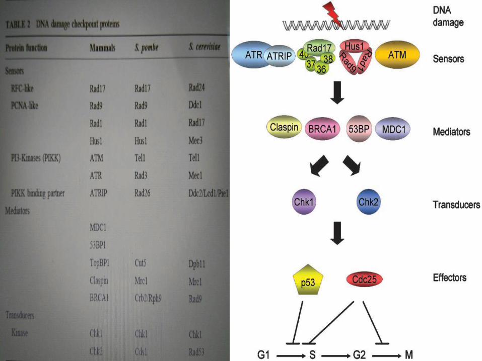
Components of the DNA damage checkpoints in human cellsThe damage is detected by sensors that, with the aid of mediators, transduce thesignal to transducers. The transducers, in turn, activate or inactivate other proteins(effectors) that directly participate in inhibiting the G1/S transition, S-phaseprogression, or the G2/M transition.

SENSORS
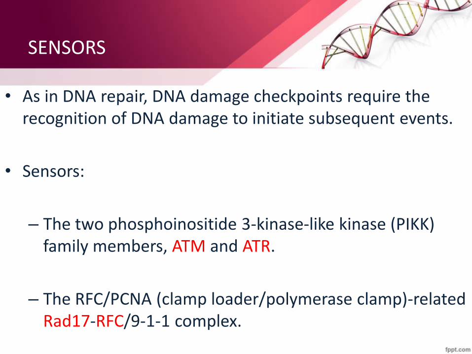
• As in DNA repair, DNA damage checkpoints require the recognition of DNA damage to initiate subsequent events.
• Sensors:
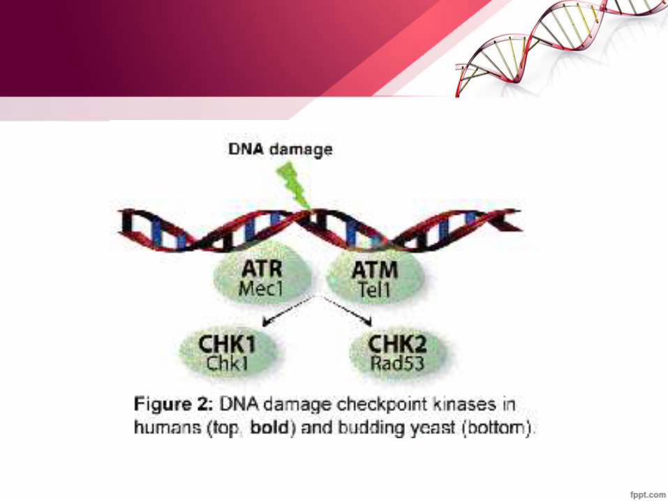
– The two phosphoinositide 3-kinase-like kinase (PIKK) family members, ATM and ATR.
– The RFC/PCNA (clamp loader/polymerase clamp)-related Rad17-RFC/9-1-1 complex.

ATM- Ataxia telangiectesia mutated protein
• Mutations in ATM cause ataxia-telangiectesia (A-T) in humans, a condition primarily characterized by:
• cerebellar degeneration
• immunodeficiency
• genome instability
• clinical radio sensitivity &
• cancer predisposition.
• The damage sensor ATM also functions as a signal transducer.
»
• It exhibits significant sequence homology to the phosphoinositide 3-kinases, but lacks lipid kinase activity.
• It does have protein kinase activity, and this activity is stimulated in vivo by agents that induce double-strand breaks.
• Upon exposure of cells to ionizing radiation, ATM phosphorylates many proteins, including Chk2, p53, NBS1 , BRCA1 and itself at serines and threonines in the sequence context of SQ or TQ.

ATR
• Discovered in the human genome database as a gene.
• Sequence homology to ATM and SpRad3, hence the name ATR (ATM and Rad3 related).
• Regions of homology to other PIKK family members.
• Knockout of ATR in mice results in embryonic lethality.
• Mutations causing partial loss of ATR activity in humans have been associated with the human autosomal recessive disorder Seckel syndrome.

• ATR, like ATM, is a protein kinase with specificity for S and T residues in SQ/TQ sequences.
• It phosphorylates essentially all of the proteins that are phosphorylated by ATM.
• ATR is activated in vivo by UV light rather than by ionizing radiation.
• It is the main PIKK family member that initiates signal transduction following UV irradiation. ATR serves an analogous role for base damages, at least from UV irradiation.
Rad-RFC Complex
• RAD17 is a protein that in humans is encoded by the RAD17gene.
• The protein encoded by this gene is highly similar to the gene product of Schizosaccharomyces pombe rad17, a cell cycle checkpoint gene required for cell cycle arrest and DNA damage repair in response to DNA damage.
• This protein shares strong similarity with DNA replication factor C (RFC), and can form a complex with RFCs.
• This protein binds to chromatin prior to DNA damage and is phosphorylated by ATR after the damage.

• This protein recruits the RAD1-RAD9-HUS1 checkpoint protein complex onto chromatin after DNA damage, which may be required for its phosphorylation.
• . The phosphorylation of this protein is required for the DNA-damage-induced cell cycle G2 arrest, and is thought to be a critical early event during checkpoint signalling in DNA-damaged cells.
• . The phosphorylation of this protein is required for the DNA-damage-induced cell cycle G2 arrest, and is thought to be a critical early event during checkpoint signalling in DNA-damaged cells.
9-1-1• The 9-1-1 (Rad9-Rad1-Hus1) complex is the checkpoint counterpart of
PCNA, a homotrimer with a ring-like structure.– Proliferating cell nuclear antigen (PCNA),or cyclin, is a non- histone
acidic nuclear protein that plays a key role in the control of eukaryotic DNA replication.
– It acts as a co-factor for DNA polymerase delta, which is responsible for leading strand DNA replication
• Although the Rad9, Rad1, and Hus1 proteins have little sequence homology to PCNA, or to one another, molecular modelling suggested that they may form a PCNA-like structure.
• When cells are exposed to either ionizing radiation or UV light, ATR and the 9-1-1 complex become associated with chromatin independently of one another.
• Rad17-RFC was found to be bound to chromatin at all times regardless of DNA damage.
MEDIATORS
• These proteins simultaneously associate with damage sensors and signal transducers at certain phases of the cell cycle and as a consequence help provide signal transduction specificity.
• The prototype mediator is the scRad9 protein, which functions along the signal transduction pathway from scMec1 (ATR) to scRad53 (Chk2).
• Another mediator, Mrc1 (mediator of replication checkpoint), found in both S. cerevisiae and S. pombe, is expressed only during S phase and is necessary for S-phase checkpoint signalling.

• p53 binding protein,53BP1 ; the topoisomerase binding protein,TopBP1 and the mediator of DNA damage checkpoint 1, MDC1, these proteins interact with damage sensors such as ATM, repair proteins such as BRCA1 and the M/R/N complex, signal transducers such as Chk2, and even effector molecules such as p53.
• In addition to these bona fide mediators, other proteins such as H2AX, BRCA1, the M/R/N complex, and SMC1 (structural maintenance of chromatin 1) play essential roles in the activation of checkpoint kinases.
– As these proteins also play direct roles in DNA repair, sister chromatid pairing, and segregation, they cannot simply be considered to be mediators.
• Human Claspin, which has marginal sequence homology to yeast Mrc1.
• Originally thought to be a mediator, appears to function more like a sensor.
SIGNAL TRANSDUCERS
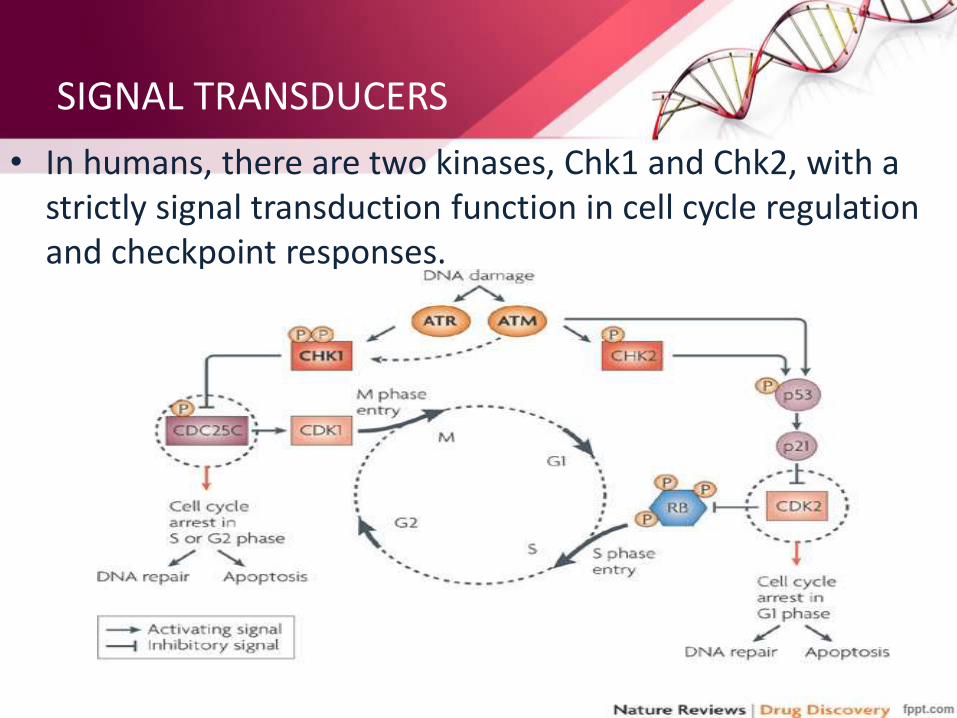
• In humans, there are two kinases, Chk1 and Chk2, with a strictly signal transduction function in cell cycle regulation and checkpoint responses.

EFFECTORS
• In humans, three phosphotyrosine phosphatases, Cdc25A, -B, and -C, dephosphorylate the cyclin-dependent kinases that act on proteins directly involved in cell-cycle transitions.
• Phosphorylation inactivates the Cdc25 proteins by excluding them from the nucleus, by causing proteolyticdegradation, or both.
• Unphosphorylated Cdc25 proteins promote the G1/S transition by dephosphorylating Cdk2 and promote the G2/M transition by dephosphorylating Cdc2 phosphotyrosine.
G1/S CHECKPOINT
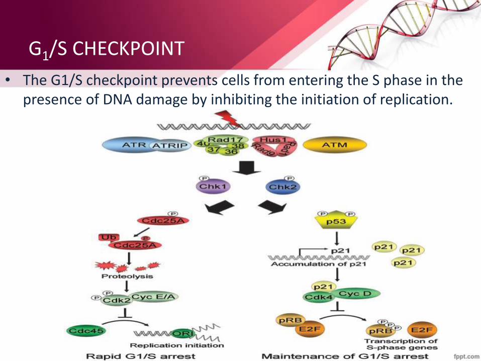
• The G1/S checkpoint prevents cells from entering the S phase in the presence of DNA damage by inhibiting the initiation of replication.
INTRA-S CHECKPOINT
• The intra-S-phase checkpoint is activated by damage encountered during the S phase or by unrepaired damage that escapes the G1/S checkpoint and leads to a block in replication.
• “a biochemical regulatory pathway which dictates the progression of cell cycle events (rather than phase transitions) in an orderly manner and that prevents the initiation of certain biochemical reactions before completion of the others within the cell”

The ATM-regulated intra-S-phase checkpoint.In response to double-strand breaks inducedby ionizing radiation, ATM triggers twocooperating parallel cascades to inhibitreplicative DNA synthesis. ATM, through theintermediacy of MDC1, H2AX, and 53BP1,phosphorylates Chk2 on Thr68 to induceubiquitin -mediated degradation of Cdc25Aphosphatase. The degradation locks the Sphase–promoting Cyclin E/Cdk2 in its inactive,phosphorylated form and prevents theloading of Cdc45 on the replication origin.ATM also initiates a second pathway byphosphorylating NBS1 of the M/R/N complex,as well as SMC1, BRCA1, and FANCD2.

G2/M CHECKPOINT
• The G2/M checkpoint prevents cells from undergoing mitosis in the presence of DNA damage.
• Depending on the type of DNA damage, the ATM-Chk2-Cdc25 signal transduction pathway and/or the ATR-Chk1-Cdc25 pathway is activated to arrest the cell cycle following DNA damage in G2.

• The G2/M checkpoint. The ionizing radiation- (ATM) and UV damage responsive sensor proteins (ATR-ATRIP, Rad17-RFC, and 9-1-1) are recruited to the damage site.
• The mediator proteins such as MDC1, BRCA1 and/or 53BP1 communicate the DNA damage signal to Chk1 and/or Chk2, thereby regulating the Cdc2/ CyclinB, Wee1, and Cdc25A proteins that are crucial for the G2/M transition by changing their expression, phosphorylation, and cellular localization.
REPLICATION CHECKPOINT (S/M CHECKPOINT)
• The replication checkpoint (also referred to as the S/M checkpoint) is the process by which mitosis is inhibited while DNA replication is ongoing or blocked.
• In both the G2/M and replication checkpoints, the ATR-Chk1-Cdc25 signal transduction pathway is utilized to inhibit mitosis, although the initiating signals for the two checkpoints are different.
• Ongoing replication or replication forks blocked by DNA damage or nucleotide starvation initiate the replication checkpoint.
CONCLUSION
• DNA damage activates several distinct biochemical pathways. First, DNA repair enzymes of varying complexities recognize and eliminate the damage.
• Second, DNA damage activates DNA damage checkpoints, which arrest cell cycle progression. Under most circumstances, checkpoints aid in cellular survival.
• Third, DNA damage activates transcription of certain genes (transcriptional response). The role of the transcriptional response in cell survival is unknown.
• Finally, apoptosis in metazoans is the programmed cell death that is activated by either cell death ligands or DNA damage, and serves to eliminate superfluous or deregulated and dangerous cells.