review open access presenilins and the g-secretase: · pdf filereview open access presenilins...
TRANSCRIPT

REVIEW Open Access
Presenilins and the g-secretase:still a complex problemDavid H Small1,2*, David W Klaver2,1, Lisa Foa1
Abstract
The presenilins form part of a complex of membrane proteins that are involved in the proteolytic cleavage of cell-surface molecules. This article reviews the history of the discovery of the presenilins, their role in the pathogenesisof Alzheimer’s disease and in the metabolism of the amyloid-b precursor protein. Unanswered questions abouttheir biochemical mechanism of action and their effects on Ca2+ homeostasis are examined.
Alzheimer’s disease (AD) is the most common cause ofdementia in the elderly. Typically 5-10% of the popula-tion over the age of 65 have dementia, and of thesecases, a large percentage have AD [1]. AD is charac-terised by the presence of proteinaceous deposits in thebrain [2]. The extracellular amyloid deposits, which arefound in the neuropil (amyloid plaques) and in associa-tion with small-medium size cerebral blood vessels (cer-ebral amyloid angiopathy), are composed of a 4 kDapolypeptide known as the amyloid-b protein (Ab) whichis derived by proteolytic cleavage from a much largeramyloid-b precursor protein (APP) [3]. Ab displays aspontaneous ability to aggregate into oligomers and lar-ger fibrillar structures, and it is generally thought thatthe accumulation of oligomeric Ab is chiefly responsiblefor the neurodegeneration that occurs in AD [4].For the generation of Ab, APP is first cleaved on the N-
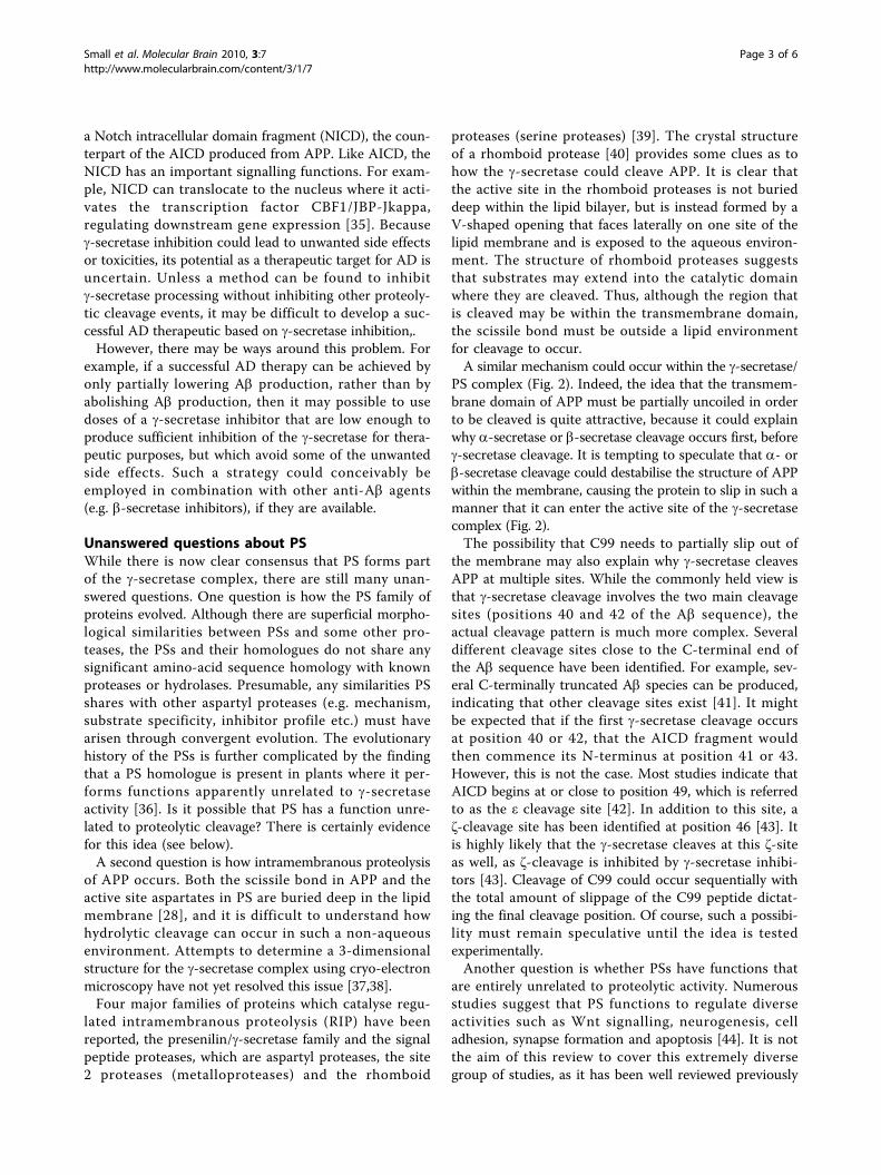
terminal side of the Ab sequence by the b-site APP cleav-ing enzyme-1 (BACE1), a transmembrane aspartyl pro-tease [3]. The resulting 99-amino acid residue C-terminalfragment (C99) is then cleaved by the g-secretase to yieldAb and a C-terminal APP intracellular domain (AICD)fragment (Fig. 1). The function of the AICD fragment isunclear, although it is thought to have a role in intracel-lular signalling. For example, AICD may be involved inthe regulation of gene transcription, synaptic plasticityand cytoskeletal dynamics [5].The major form of Ab possesses 40 amino-acid resi-
dues (Ab1-40). However, other minor species are alsoproduced which vary in the C-terminal sequence.
Production of a longer 42-residue species (Ab1-42) isthought to be intimately associated with AD pathogen-esis [6]. Ab1-42 aggregates more readily than Ab1-40, andincreased production of Ab1-42 may seed aggregation ofAb1-40 or other Ab species [4].
Genetic clues to the pathogenesis of ADApproximately 5% of all AD cases are autosomal domi-nant [7]. Soon after the complete APP sequence wascloned in 1987 [8], it became clear that at least onefamilial AD (FAD) locus was located on chromosome21 [9] and attention turned to the APP gene that hadpreviously been localised to a region within chromo-some 21. The first FAD mutation was identified withinthe APP gene [10], and soon after, a number of otherAPP mutations were also identified [11-13]. All of theFAD mutations in the APP gene cluster around theregion encoding the Ab sequence, suggesting that theyhave some effect on the aggregation or proteolytic pro-cessing of APP.APP mutations on chromosome 21 account for only a
small fraction of the total number of FAD cases. It wasclear that multiple FAD loci existed on other chromo-somes. The first evidence for an FAD locus on chromo-some 14 [14] came well before the identification of thelocus on chromosome 21. Then, in 1995, FAD mutationsin two presenilin (PS) genes located on chromosome 14(presenilin-1, PS1) and chromosome 1 (presenilin-2, PS2)were reported [15-17]. The PS genes encode proteinsthat are homologous to the C. elegans sel-12 gene, whichis known to be involved in Notch signalling [18]. Thisobservation provided that first clue that PS1 may beinvolved in cell-surface receptor signalling. To date, >100
* Correspondence: [email protected] Research Institute, University of Tasmania, Hobart, Tasmania 7001,Australia
Small et al. Molecular Brain 2010, 3:7http://www.molecularbrain.com/content/3/1/7
© 2010 Small et al; licensee BioMed Central Ltd. This is an Open Access article distributed under the terms of the Creative CommonsAttribution License (http://creativecommons.org/licenses/by/2.0), which permits unrestricted use, distribution, and reproduction inany medium, provided the original work is properly cited.

FAD mutations have been found in PS1 and 11 muta-tions in PS2 [19]. The PS genes encode proteins with 8or 9 transmembrane domains. The proteins are synthe-sized as ~50 kDa proteins that are subsequently cleavedby a presenilinase into a ~30 kDa N-terminal fragmentand a ~20 kDa C-terminal fragment, which remain asso-ciated with each other [20].PS mutations are linked to g-secretase activity [21].
Specifically, it has been observed that FAD mutations inPS1 increase the proportion of C99 that is cleaved by g-secretase at position 42 of the Ab sequence. The alteredcleavage pattern causes increased production of themore pathogenic Ab1-42. While the first impression maybe that these are “gain-of-function” mutations, such aconclusion is difficult to reconcile with the large numberof FAD mutations that have been identified, particularlyin PS1. Instead, mutations in PS are more likely to be“loss-of-function” mutations [22] in which a decrease inthe rate of g-secretase cleavage of APP leads to anincrease in the proportion of Ab1-42. PS1 knockout hasbeen shown to cause an 80% decrease in Ab production[23], while combined PS1 and PS2 knockout abolishesg-secretase activity and hence Ab production [24]. Inaddition, g-secretase activity co-purifies with a highmolecular weight complex that contains PS1 and severalother proteins (nicastrin, aph-1 and pen-2). It is nowknown that the g-secretase consists of a complex of pro-teins of which PS, nicastrin, aph-1 and pen-2 are the
principal components. Expression of all 4 proteins incells is necessary for g-secretase activity [25].Inhibitor studies demonstrate that the g-secretase is a
member of the aspartyl protease family [26]. All mem-bers of this family require two aspartyl residues forenzyme activity [27]. Some aspartyl proteases(e.g. BACE1) have two aspartyl residues within a singlesubunit, but other proteases have only one aspartyl resi-due, and therefore dimerization is needed to activate theenzyme. The amino-acid sequence of both PS1 and PS2contains two conserved aspartyl residues within twodomains predicted to be membrane spanning. Thesetwo aspartyl residues are thought to form part of thecatalytic domain [28]. In support of this idea, mutationof these two residues has been shown to cause loss ofg-secretase activity [29]. In addition, affinity labellingexperiments demonstrate that g-secretase inhibitors binddirectly to PS [30,31].While the exact number of g-secretase substrates is
unknown, a large number of transmembrane proteins arereportedly cleaved by the enzyme [32,33]. Some of theg-secretase substrates (other than APP) include APLP2,Notch, Delta, and tumour necrosis factor-a convertingenzyme (TACE). Of these proteins, Notch and Delta maybe the most important as some abnormalities or toxicitiesassociated with g-secretase inhibition or knockdowncould be due to failure of the Notch/Delta signallingpathway [34]. Cleavage of Notch by g-secretase produces
Figure 1 Amyloidogenic processing of the b-amyloid precursor protein (APP) by BACE1 and g-secretase. Initially, BACE1 cleaves APP onthe N-terminal end of the Ab sequence to yield a large secreted N-terminal fragment (sAPPb) and a smaller membrane-associated C-terminalstub (C99), which is then cleaved by the g-secretase complex to yield Ab and an APP intracellular domain (AICD). Secreted Ab aggregates in theextracellular environment to form neurotoxic oligomers.
Small et al. Molecular Brain 2010, 3:7http://www.molecularbrain.com/content/3/1/7
Page 2 of 6
a Notch intracellular domain fragment (NICD), the coun-terpart of the AICD produced from APP. Like AICD, theNICD has an important signalling functions. For exam-ple, NICD can translocate to the nucleus where it acti-vates the transcription factor CBF1/JBP-Jkappa,regulating downstream gene expression [35]. Becauseg-secretase inhibition could lead to unwanted side effectsor toxicities, its potential as a therapeutic target for AD isuncertain. Unless a method can be found to inhibitg-secretase processing without inhibiting other proteoly-tic cleavage events, it may be difficult to develop a suc-cessful AD therapeutic based on g-secretase inhibition,.However, there may be ways around this problem. For
example, if a successful AD therapy can be achieved byonly partially lowering Ab production, rather than byabolishing Ab production, then it may possible to usedoses of a g-secretase inhibitor that are low enough toproduce sufficient inhibition of the g-secretase for thera-peutic purposes, but which avoid some of the unwantedside effects. Such a strategy could conceivably beemployed in combination with other anti-Ab agents(e.g. b-secretase inhibitors), if they are available.
Unanswered questions about PSWhile there is now clear consensus that PS forms partof the g-secretase complex, there are still many unan-swered questions. One question is how the PS family ofproteins evolved. Although there are superficial morpho-logical similarities between PSs and some other pro-teases, the PSs and their homologues do not share anysignificant amino-acid sequence homology with knownproteases or hydrolases. Presumable, any similarities PSshares with other aspartyl proteases (e.g. mechanism,substrate specificity, inhibitor profile etc.) must havearisen through convergent evolution. The evolutionaryhistory of the PSs is further complicated by the findingthat a PS homologue is present in plants where it per-forms functions apparently unrelated to g-secretaseactivity [36]. Is it possible that PS has a function unre-lated to proteolytic cleavage? There is certainly evidencefor this idea (see below).A second question is how intramembranous proteolysis
of APP occurs. Both the scissile bond in APP and theactive site aspartates in PS are buried deep in the lipidmembrane [28], and it is difficult to understand howhydrolytic cleavage can occur in such a non-aqueousenvironment. Attempts to determine a 3-dimensionalstructure for the g-secretase complex using cryo-electronmicroscopy have not yet resolved this issue [37,38].Four major families of proteins which catalyse regu-
lated intramembranous proteolysis (RIP) have beenreported, the presenilin/g-secretase family and the signalpeptide proteases, which are aspartyl proteases, the site2 proteases (metalloproteases) and the rhomboid
proteases (serine proteases) [39]. The crystal structureof a rhomboid protease [40] provides some clues as tohow the g-secretase could cleave APP. It is clear thatthe active site in the rhomboid proteases is not burieddeep within the lipid bilayer, but is instead formed by aV-shaped opening that faces laterally on one site of thelipid membrane and is exposed to the aqueous environ-ment. The structure of rhomboid proteases suggeststhat substrates may extend into the catalytic domainwhere they are cleaved. Thus, although the region thatis cleaved may be within the transmembrane domain,the scissile bond must be outside a lipid environmentfor cleavage to occur.A similar mechanism could occur within the g-secretase/
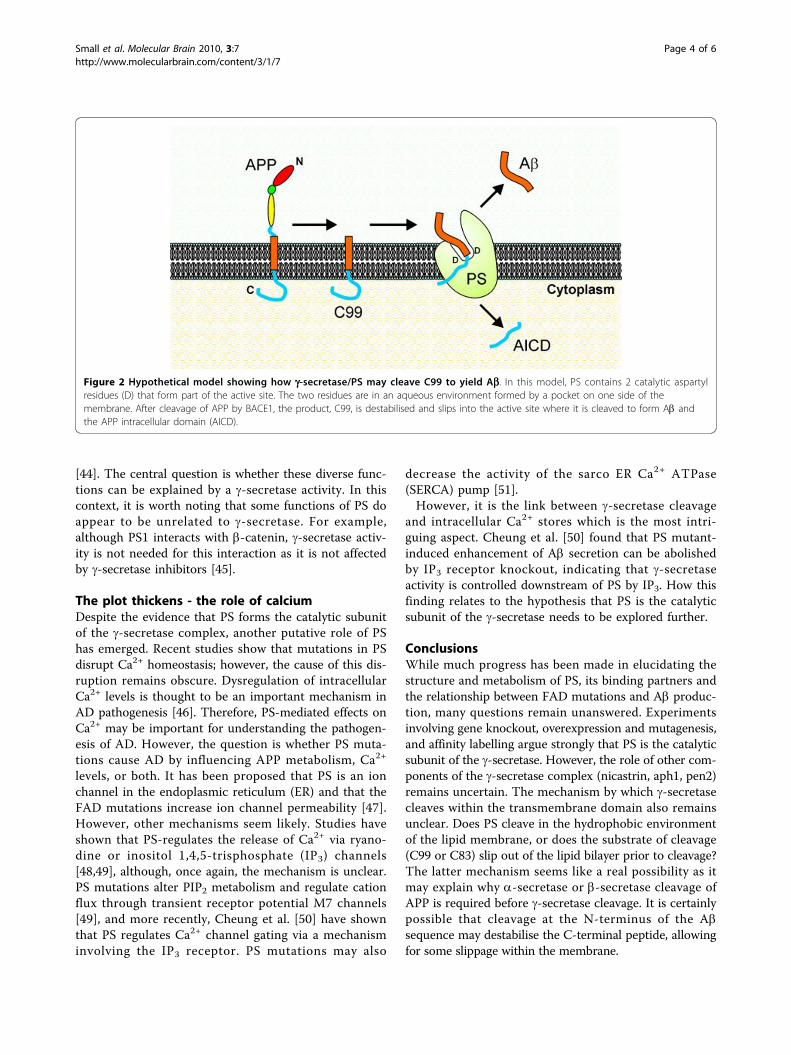
PS complex (Fig. 2). Indeed, the idea that the transmem-brane domain of APP must be partially uncoiled in orderto be cleaved is quite attractive, because it could explainwhy a-secretase or b-secretase cleavage occurs first, beforeg-secretase cleavage. It is tempting to speculate that a- orb-secretase cleavage could destabilise the structure of APPwithin the membrane, causing the protein to slip in such amanner that it can enter the active site of the g-secretasecomplex (Fig. 2).The possibility that C99 needs to partially slip out of
the membrane may also explain why g-secretase cleavesAPP at multiple sites. While the commonly held view isthat g-secretase cleavage involves the two main cleavagesites (positions 40 and 42 of the Ab sequence), theactual cleavage pattern is much more complex. Severaldifferent cleavage sites close to the C-terminal end ofthe Ab sequence have been identified. For example, sev-eral C-terminally truncated Ab species can be produced,indicating that other cleavage sites exist [41]. It mightbe expected that if the first g-secretase cleavage occursat position 40 or 42, that the AICD fragment wouldthen commence its N-terminus at position 41 or 43.However, this is not the case. Most studies indicate thatAICD begins at or close to position 49, which is referredto as the ε cleavage site [42]. In addition to this site, aζ-cleavage site has been identified at position 46 [43]. Itis highly likely that the g-secretase cleaves at this ζ-siteas well, as ζ-cleavage is inhibited by g-secretase inhibi-tors [43]. Cleavage of C99 could occur sequentially withthe total amount of slippage of the C99 peptide dictat-ing the final cleavage position. Of course, such a possibi-lity must remain speculative until the idea is testedexperimentally.Another question is whether PSs have functions that
are entirely unrelated to proteolytic activity. Numerousstudies suggest that PS functions to regulate diverseactivities such as Wnt signalling, neurogenesis, celladhesion, synapse formation and apoptosis [44]. It is notthe aim of this review to cover this extremely diversegroup of studies, as it has been well reviewed previously
Small et al. Molecular Brain 2010, 3:7http://www.molecularbrain.com/content/3/1/7
Page 3 of 6
[44]. The central question is whether these diverse func-tions can be explained by a g-secretase activity. In thiscontext, it is worth noting that some functions of PS doappear to be unrelated to g-secretase. For example,although PS1 interacts with b-catenin, g-secretase activ-ity is not needed for this interaction as it is not affectedby g-secretase inhibitors [45].
The plot thickens - the role of calciumDespite the evidence that PS forms the catalytic subunitof the g-secretase complex, another putative role of PShas emerged. Recent studies show that mutations in PSdisrupt Ca2+ homeostasis; however, the cause of this dis-ruption remains obscure. Dysregulation of intracellularCa2+ levels is thought to be an important mechanism inAD pathogenesis [46]. Therefore, PS-mediated effects onCa2+ may be important for understanding the pathogen-esis of AD. However, the question is whether PS muta-tions cause AD by influencing APP metabolism, Ca2+
levels, or both. It has been proposed that PS is an ionchannel in the endoplasmic reticulum (ER) and that theFAD mutations increase ion channel permeability [47].However, other mechanisms seem likely. Studies haveshown that PS-regulates the release of Ca2+ via ryano-dine or inositol 1,4,5-trisphosphate (IP3) channels[48,49], although, once again, the mechanism is unclear.PS mutations alter PIP2 metabolism and regulate cationflux through transient receptor potential M7 channels[49], and more recently, Cheung et al. [50] have shownthat PS regulates Ca2+ channel gating via a mechanisminvolving the IP3 receptor. PS mutations may also
decrease the activity of the sarco ER Ca2+ ATPase(SERCA) pump [51].However, it is the link between g-secretase cleavage
and intracellular Ca2+ stores which is the most intri-guing aspect. Cheung et al. [50] found that PS mutant-induced enhancement of Ab secretion can be abolishedby IP3 receptor knockout, indicating that g-secretaseactivity is controlled downstream of PS by IP3. How thisfinding relates to the hypothesis that PS is the catalyticsubunit of the g-secretase needs to be explored further.
ConclusionsWhile much progress has been made in elucidating thestructure and metabolism of PS, its binding partners andthe relationship between FAD mutations and Ab produc-tion, many questions remain unanswered. Experimentsinvolving gene knockout, overexpression and mutagenesis,and affinity labelling argue strongly that PS is the catalyticsubunit of the g-secretase. However, the role of other com-ponents of the g-secretase complex (nicastrin, aph1, pen2)remains uncertain. The mechanism by which g-secretasecleaves within the transmembrane domain also remainsunclear. Does PS cleave in the hydrophobic environmentof the lipid membrane, or does the substrate of cleavage(C99 or C83) slip out of the lipid bilayer prior to cleavage?The latter mechanism seems like a real possibility as itmay explain why a-secretase or b-secretase cleavage ofAPP is required before g-secretase cleavage. It is certainlypossible that cleavage at the N-terminus of the Absequence may destabilise the C-terminal peptide, allowingfor some slippage within the membrane.
Figure 2 Hypothetical model showing how g-secretase/PS may cleave C99 to yield Ab. In this model, PS contains 2 catalytic aspartylresidues (D) that form part of the active site. The two residues are in an aqueous environment formed by a pocket on one side of themembrane. After cleavage of APP by BACE1, the product, C99, is destabilised and slips into the active site where it is cleaved to form Ab andthe APP intracellular domain (AICD).
Small et al. Molecular Brain 2010, 3:7http://www.molecularbrain.com/content/3/1/7
Page 4 of 6
Finally, the role of PS in the release of intracellularcalcium stores needs to be understood. Of particularsignificance here are the findings that FAD mutationsinfluence inositol phosphate signalling and that inositolphosphate signalling can, in turn, regulate Ab produc-tion. While it is possible that FAD mutations have mul-tiple effects which converge on Ab metabolism, untilthe precise mechanism by which PS mutations influenceAb is understood, there will continue to be more ques-tions than answers.
AcknowledgementsDHS and LF are funded by project grants from the National Health andMedical Research Council of Australia.
Author details1Menzies Research Institute, University of Tasmania, Hobart, Tasmania 7001,Australia. 2Dept. Biochemistry and Molecular Biology, Monash University,Victoria 3800, Australia.
Authors’ contributionsDHS, DWK and LF wrote this article jointly. All authors read and approvedthe final manuscript.
Competing interestsThe authors declare that they have no competing interests.
Received: 22 December 2009 Accepted: 5 February 2010Published: 5 February 2010
References1. Storey E, Kinsella GJ, Slavin MJ: The neuropsychological diagnosis of
Alzheimer’s disease. J Alzheimers Dis 2001, 3:261-285.2. Masters CL, Simms G, Weinman NA, Multhaup G, McDonald BL,
Beyreuther K: Amyloid plaque core protein in Alzheimer disease andDown syndrome. Proc Natl Acad Sci USA 1985, 82:4245-4249.
3. Nunan J, Small DH: Regulation of APP cleavage by alpha-, beta- andgamma-secretases. FEBS Lett 2000, 483:6-10.
4. Jarrett JT, Lansbury PT Jr: Seeding “one-dimensional crystallization” ofamyloid: a pathogenic mechanism in Alzheimer’s disease and scrapie?Cell 1993, 73:1055-1058.
5. Muller T, Meyer HE, Egensperger R, Marcus K: The amyloid precursorprotein intracellular domain (AICD) as modulator of gene expression,apoptosis, and cytoskeletal dynamics - relevance for Alzheimer’s disease.Prog Neurobiol 2008, 85:393-406.
6. Walsh DM, Selkoe DJ: A beta oligomers - a decade of discovery.J Neurochem 2007, 101:1172-1184.
7. Bertram L: Alzheimer’s disease genetics current status and futureperspectives. Int Rev Neurobiol 2009, 84:167-184.
8. Kang J, Lemaire HG, Unterbeck A, Salbaum JM, Masters CL, Grzeschik KH,Multhaup G, Beyreuther K, Muller-Hill B: The precursor of Alzheimer’sdisease amyloid A4 protein resembles a cell-surface receptor. Nature1987, 325:733-736.
9. Patterson D, Gardiner K, Kao FT, Tanzi R, Watkins P, Gusella JF: Mapping ofthe gene encoding the beta-amyloid precursor protein and itsrelationship to the Down syndrome region of chromosome 21. Proc NatlAcad Sci USA 1988, 85:8266-8270.
10. Goate A, Chartier-Harlin MC, Mullan M, Brown J, Crawford F, Fidani L,Giuffra L, Haynes A, Irving N, James L, Mant R, Newton P, Rooke K, Roques P,Talbot C, Pericak-Vance M, Roses A, Williamson R, Rossor M, Owen M, Hardy J:Segregation of a missense mutation in the amyloid precursor proteingene with familial Alzheimer’s disease. Nature 1991, 349:704-706.
11. Murrell J, Farlow M, Ghetti B, Benson MD: A mutation in the amyloidprecursor protein associated with hereditary Alzheimer’s disease. Science1991, 254:97-99.
12. Chartier-Harlin MC, Crawford F, Houlden H, Warren A, Hughes D, Fidani L,Goate A, Rossor M, Roques P, Hardy J, et al: Early-onset Alzheimer’s
disease caused by mutations at codon 717 of the beta-amyloidprecursor protein gene. Nature 1991, 353:844-846.
13. Mullan M, Crawford F, Axelman K, Houlden H, Lilius L, Winblad B, Lannfelt L:A pathogenic mutation for probable Alzheimer’s disease in the APPgene at the N-terminus of beta-amyloid. Nat Genet 1992, 1:345-347.
14. Weitkamp LR, Nee L, Keats B, Polinsky RJ, Guttormsen S: Alzheimer disease:evidence for susceptibility loci on chromosomes 6 and 14. Am J HumGenet 1983, 35:443-453.
15. Sherrington R, Rogaev EI, Liang Y, Rogaeva EA, Levesque G, Ikeda M, Chi H,Lin C, Li G, Holman K, et al: Cloning of a gene bearing missensemutations in early-onset familial Alzheimer’s disease. Nature 1995,375:754-760.
16. Levy-Lahad E, Wasco W, Poorkaj P, Romano DM, Oshima J, Pettingell WH,Yu CE, Jondro PD, Schmidt SD, Wang K, et al: Candidate gene for thechromosome 1 familial Alzheimer’s disease locus. Science 1995, 269:973-977.
17. Rogaev EI, Sherrington R, Rogaeva EA, Levesque G, Ikeda M, Liang Y, Chi H,Lin C, Holman K, Tsuda T, et al: Familial Alzheimer’s disease in kindredswith missense mutations in a gene on chromosome 1 related to theAlzheimer’s disease type 3 gene. Nature 1995, 376:775-778.
18. Levitan D, Greenwald I: Facilitation of lin-12-mediated signalling by sel-12, a Caenorhabditis elegans S182 Alzheimer’s disease gene. Nature1995, 377:351-354.
19. Bertram L, Tanzi RE: Thirty years of Alzheimer’s disease genetics: theimplications of systematic meta-analyses. Nat Rev Neurosci 2008,9:768-778.
20. Ward RV, Davis JB, Gray CW, Barton AJ, Bresciani LG, Caivano M, Murphy VF,Duff K, Hutton M, Hardy J, et al: Presenilin-1 is processed into two majorcleavage products in neuronal cell lines. Neurodegeneration 1996,5:293-298.
21. Scheuner D, Eckman C, Jensen M, Song X, Citron M, Suzuki N, Bird TD,Hardy J, Hutton M, Kukull W, et al: Secreted amyloid beta-protein similarto that in the senile plaques of Alzheimer’s disease is increased in vivoby the presenilin 1 and 2 and APP mutations linked to familialAlzheimer’s disease. Nat Med 1996, 2:864-870.
22. Shen J, Kelleher RJ: The presenilin hypothesis of Alzheimer’s disease:evidence for a loss-of-function pathogenic mechanism. Proc Natl Acad SciUSA 2007, 104:403-409.
23. De Strooper B, Saftig P, Craessaerts K, Vanderstichele H, Guhde G,Annaert W, Von Figura K, Van Leuven F: Deficiency of presenilin-1 inhibitsthe normal cleavage of amyloid precursor protein. Nature 1998,391:387-390.
24. Herreman A, Serneels L, Annaert W, Collen D, Schoonjans L, De Strooper B:Total inactivation of gamma-secretase activity in presenilin-deficientembryonic stem cells. Nat Cell Biol 2000, 2:461-462.
25. Edbauer D, Winkler E, Regula JT, Pesold B, Steiner H, Haass C:Reconstitution of gamma-secretase activity. Nat Cell Biol 2003, 5:486-488.
26. Evin G, Cappai R, Li QX, Culvenor JG, Small DH, Beyreuther K, Masters CL:Candidate gamma-secretases in the generation of the carboxyl terminusof the Alzheimer’s disease beta A4 amyloid: possible involvement ofcathepsin D. Biochemistry 1995, 34:14185-14192.
27. Beher D, Graham SL: Protease inhibitors as potential disease-modifyingtherapeutics for Alzheimer’s disease. Expert Opin Investig Drugs 2005,14:1385-1409.
28. Wolfe MS: Therapeutic strategies for Alzheimer’s disease. Nat Rev DrugDiscov 2002, 1:859-866.
29. Wolfe MS, Xia W, Ostaszewski BL, Diehl TS, Kimberly WT, Selkoe DJ: Twotransmembrane aspartates in presenilin-1 required for presenilinendoproteolysis and gamma-secretase activity. Nature 1999, 398:513-517.
30. Esler WP, Kimberly WT, Ostaszewski BL, Diehl TS, Moore CL, Tsai JY,Rahmati T, Xia W, Selkoe DJ, Wolfe MS: Transition-state analogueinhibitors of gamma-secretase bind directly to presenilin-1. Nat Cell Biol2000, 2:428-434.
31. Li YM, Xu M, Lai MT, Huang Q, Castro JL, DiMuzio-Mower J, Harrison T,Lellis C, Nadin A, Neduvelil JG, et al: Photoactivated gamma-secretaseinhibitors directed to the active site covalently label presenilin 1. Nature2000, 405:689-694.
32. Hemming ML, Elias JE, Gygi SP, Selkoe DJ: Proteomic profiling of gamma-secretase substrates and mapping of substrate requirements. PLoS Biol2008, 6:e257.
33. Magold AI, Cacquevel M, Fraering PC: Gene expression profiling in cellswith enhanced gamma-secretase activity. PLoS One 2009, 4:e6952.
Small et al. Molecular Brain 2010, 3:7http://www.molecularbrain.com/content/3/1/7
Page 5 of 6

34. Imbimbo BP: Therapeutic potential of gamma-secretase inhibitors andmodulators. Curr Top Med Chem 2008, 8:54-61.
35. Lai EC: Notch signalling: control of cell communication and cell fate.Development 2004, 131:965-973.
36. Khandelwal A, Chandu D, Roe CM, Kopan R, Quatrano RS: Moonlightingactivity of presenilin in plants is independent of g-secretase andevolutionarily conserved. Proc Natl Acad Sci USA 2007, 104:13337-13342.
37. Osenkowski P, Li H, Ye W, Li D, Aeschbach L, Fraering PC, Wolfe MS,Selkoe DJ: Cryoelectron microscopy structure of purified gamma-secretase at 12 A resolution. J Mol Biol 2009, 385:642-652.
38. Lazarov VK, Fraering PC, Ye W, Wolfe MS, Selkoe DJ, Li H: Electronmicroscopic structure of purified, active gamma-secretase reveals anaqueous intramembrane chamber and two pores. Proc Natl Acad Sci USA2006, 103:6889-6894.
39. Ehrmann M, Clausen T: Proteolysis as a regulatory mechanism. Annu RevGenet 2004, 38:709-724.
40. Wang Y, Zhang Y, Ha Y: Crystal structure of a rhomboid familyintramembrane protease. Nature 2006, 444:179-180.
41. Wang R, Sweeney D, Gandy SE, Sisodia SS: The profile of soluble amyloidbeta protein in cultured cell media. Detection and quantification ofamyloid beta protein and variants by immunoprecipitation-massspectrometry. J Biol Chem 1996, 271:31894-31902.
42. Weidemann Eggert S, Reinhard FB, Vogel M, Paliga K, Baier G, Masters CL,Evin G: A novel epsilon-cleavage within the transmembrane domain ofthe amyloid precursor protein demonstrates homology with Notchprocessing. Biochemistry 2002, 41:2825-2835.
43. Xu X: g-Secretase catalyzes sequential cleavages of the Ab PPtransmembrane domain. J Alzheimer Dis 2009, 16:211-224.
44. Thinakaran G, Parent AT: Identification of the role of presenilins beyondAlzheimer’s disease. Pharmacol Res 2004, 50:411-418.
45. Meredith JE Jr, Wang Q, Mitchell TJ, Olson RE, Zaczek R, Stern AM,Seiffert D: Gamma-secretase activity is not involved in presenilin-mediated regulation of beta-catenin. Biochem Biophys Res Commun 2002,299:744-750.
46. Small DH, Gasperini R, Vincent AJ, Hung AC, Foa L: The role of Abeta-induced calcium dysregulation in the pathogenesis of Alzheimer’sdisease. J Alzheimers Dis 2009, 16:225-233.
47. Tu H, Nelson O, Bezprozvanny A, Wang Z, Lee SF, Hao YH, Serneels L, DeStrooper B, Yu G, Bezprozvanny I: Presenilins form ER Ca2+ leak channels,a function disrupted by familial Alzheimer’s disease-linked mutations.Cell 2006, 126:981-993.
48. Chan SL, Mayne M, Holden CP, Geiger JD, Mattson MP: Presenilin-1mutations increase levels of ryanodine receptors and calcium release inPC12 cells and cortical neurons. J Biol Chem 2000, 275:18195-18200.
49. Landman N, Jeong SY, Shin SY, Voronov SV, Serban G, Kang MS, Park MK,Di Paolo G, Chung S, Kim TW: Presenilin mutations linked to familialAlzheimer’s disease cause an imbalance in phosphatidylinositol 4,5-bisphosphate metabolism. Proc Natl Acad Sci USA 2006, 103:19524-19529.
50. Cheung KH, Shineman D, Muller M, Cardenas C, Mei L, Yang J, Tomita T,Iwatsubo T, Lee VM, Foskett JK: Mechanism of Ca2+ disruption inAlzheimer’s disease by presenilin regulation of InsP3 receptor channelgating. Neuron 2008, 58:871-883.
51. Green KN, Demuro A, Akbari Y, Hitt BD, Smith IF, Parker I, LaFerla FM:SERCA pump activity is physiologically regulated by presenilin andregulates amyloid beta production. J Cell Biol 2008, 181:1107-1116.
doi:10.1186/1756-6606-3-7Cite this article as: Small et al.: Presenilins and the g-secretase: still acomplex problem. Molecular Brain 2010 3:7. Submit your next manuscript to BioMed Central
and take full advantage of:
• Convenient online submission
• Thorough peer review
• No space constraints or color figure charges
• Immediate publication on acceptance
• Inclusion in PubMed, CAS, Scopus and Google Scholar
• Research which is freely available for redistribution
Submit your manuscript at www.biomedcentral.com/submit
Small et al. Molecular Brain 2010, 3:7http://www.molecularbrain.com/content/3/1/7
Page 6 of 6