tannic acid is a natural Я-secretase inhibitor that prevents
TRANSCRIPT

Tannic Acid Is a Natural �-Secretase Inhibitor That PreventsCognitive Impairment and Mitigates Alzheimer-likePathology in Transgenic Mice*
Received for publication, August 15, 2011, and in revised form, December 20, 2011 Published, JBC Papers in Press, January 4, 2012, DOI 10.1074/jbc.M111.294025
Takashi Mori,a,b1 Kavon Rezai-Zadeh,c Naoki Koyama,a Gary W. Arendash,d,e Haruyasu Yamaguchi,f
Nobuto Kakuda,g Yuko Horikoshi-Sakuraba,g Jun Tan,h,i and Terrence Townc,j,k2
From the Departments of aBiomedical Sciences and bPathology, Saitama Medical Center and University, Kawagoe, Saitama 350-8550, Japan, the cDepartment of Biomedical Sciences and Regenerative Medicine Institute Neural Program and the jDepartment ofNeurosurgery, Maxine Dunitz Neurosurgical Institute, Cedars-Sinai Medical Center, Los Angeles, California 90048, the dFloridaAlzheimer’s Disease Research Center and the eDepartment of Cell Biology, Microbiology, and Molecular Biology, University ofSouth Florida, Tampa, Florida 33620, the fGunma University School of Health Sciences, Maebashi, Gunma 371-8514, Japan, thegImmuno-Biological Laboratories Co., Ltd., Fujioka, Gunma 375-0005, Japan, the hRashid Laboratory for DevelopmentalNeurobiology, Silver Child Development Center and the iNeuroimmunology Laboratory, the Department of Psychiatry andNeurosciences, College of Medicine, University of South Florida, Tampa, Florida 33613, and the kDepartment of Medicine, DavidGeffen School of Medicine, University of California, Los Angeles, California 90048
Background: Recent focus has been given to anti-amyloidogenic naturally occurring polyphenols known as flavonoids.Results: The polyphenol tannic acid prevented behavioral impairment and mitigated Alzheimer disease-like pathology.Conclusion: Tannic acid may be prophylactic for Alzheimer disease by inhibiting �-secretase activity and mitigating brainpathology.Significance: This nutraceutical approach offers a new class of drug for inhibiting �-secretase with few if any side effects.
Amyloid precursor protein (APP) proteolysis is essential forproduction of amyloid-� (A�) peptides that form �-amyloidplaques in brains of Alzheimer disease (AD) patients. Recentfocus has been directed toward a group of naturally occurringanti-amyloidogenic polyphenols knownas flavonoids.Weorallyadministered the flavonoid tannic acid (TA) to the transgenicPSAPP mouse model of cerebral amyloidosis (bearing mutanthuman APP and presenilin-1 transgenes) and evaluated cogni-tive function and AD-like pathology. Consumption of TA for 6months prevented transgene-associated behavioral impairmentincluding hyperactivity, decreased object recognition, anddefective spatial reference memory, but did not alter nontrans-genicmouse behavior. Accordingly, brain parenchymal and cer-ebral vascular �-amyloid deposits and abundance of various A�species including oligomers were mitigated in TA-treatedPSAPPmice. These effects occurred with decreased cleavage ofthe �-carboxyl-terminal APP fragment, lowered soluble APP-�production, reduced �-site APP cleaving enzyme 1 protein sta-
bility and activity, and attenuated neuroinflammation. As invitro validation, we treated well characterized mutant humanAPP-overexpressing murine neuron-like cells with TA andfound significantly reduced A� production associated with lessamyloidogenic APP proteolysis. Taken together, these resultsraise the possibility that dietary supplementation with TA maybe prophylactic for AD by inhibiting �-secretase activity andneuroinflammation and thereby mitigating AD pathology.
Alzheimer disease (AD)3 is the most common dementia andis a growing worldwide public health concern (1). AD neuro-pathological hallmarks include extracellular deposits of amy-loid-� (A�) peptides, intracellular neurofibrillary tangles, neu-ronal and synaptic degeneration/loss, and neuroinflammation(2). Brain A� deposition likely results from increased peptideaccumulation/reduced clearance, endorsing toxic events thatdrive AD pathogenesis (3, 4). A� is produced from sequentialendoproteolytic cleavage of amyloid precursor protein (APP)by �- and �-secretases (5–9), and enters a dynamic equilibriumbetween soluble and deposited forms (10). In recent years,much attention has been directed toward soluble multimericforms of A� peptides as the toxic species. These so-called “A�oligomers” disrupt synaptic function and induce neurotoxicityin vivo (11–13).
* This work was supported, in whole or in part, by National Institutes of HealthGrants 5R00AG029726-04, 3R00AG029726-04S1, and 1R01NS076794-01from the NIA and the NINDS (to T. T.), Grant-in-aid for Scientific Research (C)22500320 (to T. M.) from the Japan Society for the Promotion of Scienceand Grant-in-aid for Scientific Research (B) 19300122 (to H. Y.) from theMinistry of Education, Culture, Sports, Science and Technology, Alzhei-mer’s Association Zenith Fellows Award ZEN-10-174633 (to T. T.), andAmerican Federation of Aging Research/Ellison Medical Foundation JulieMartin Mid-Career Award in Aging Research M11472 (to T. T.).
1 To whom correspondence may be addressed. Tel.: 81-49-228-3592; E-mail:[email protected].
2 Inaugural holder of the Ben Winters Endowed Chair in RegenerativeMedicine. To whom correspondence may be addressed: RegenerativeMedicine Institute, Cedars-Sinai Medical Center, 8700 Beverly Blvd., Ste-ven Spielberg Building, Rm. 361, Los Angeles, CA 90048. Tel.: 310-248-8581; E-mail: [email protected].
3 The abbreviations used are: AD, Alzheimer disease; CTF, carboxyl-terminalfragment; APP, amyloid precursor protein; A�, amyloid-�; BACE1, �-siteAPP cleaving enzyme 1; CAA, cerebral amyloid angiopathy; EGCG, (�)-epigallocatechin-3-gallate; TA, tannic acid; Iba1, ionized calcium-bindingadapter molecule 1; GFAP, glial fibrillary acidic protein; QPCR, quantitativereal-time PCR; ANOVA, analysis of variance; CC, cingulate cortex; H, hip-pocampus; EC, entorhinal cortex; sAPP, soluble APP.
THE JOURNAL OF BIOLOGICAL CHEMISTRY VOL. 287, NO. 9, pp. 6912–6927, February 24, 2012Published in the U.S.A.
6912 JOURNAL OF BIOLOGICAL CHEMISTRY VOLUME 287 • NUMBER 9 • FEBRUARY 24, 2012
by guest on March 22, 2018
http://ww
w.jbc.org/
Dow
nloaded from
Rooted in the “amyloid cascade hypothesis” of AD, whichpurports that cerebral A� accumulation sets a toxic down-stream cascade into motion (2–4), much focus has beendirected toward anti-amyloid therapies. Specific approachesinclude reducing cerebral A� production or enhancing A�clearance (14–19). Although synthetic drugs have been anti-amyloid agents of choice, these compounds can have significantundesirable side effects, especially when given long-term in adisease prevention paradigm. For example, the ADAPT trial totest nonsteroidal anti-inflammatory drugs for primary AD pre-vention was prematurely halted due to nonsteroidal anti-in-flammatory drug-associated cardiotoxicity (20, 21). Naturallyoccurring dietary compounds, or “nutraceuticals,” represent analternative class of molecules that typically have fewer sideeffects than designer drugs (22).Others and we have previously reported that nutraceuticals
including the green tea polyphenol (�)-epigallocatechin-3-gal-late (EGCG) (23, 24), the citrus bioflavonoid luteolin (25),grape-derived polyphenols (26, 27), and caffeine (28) have anti-amyloidogenic properties. Based on our findings that EGCGenhances �-secretase APP cleavage and mitigates cerebralamyloidosis in the Tg2576 mouse model of cerebral amyloido-sis (23, 24), we sought to investigate a structurally related com-pound, tannic acid (TA). TA is a plant-derived hydrolyzabletannin polyphenol (29) that is a gallic acid polymer glucoside(C76H52O46; Fig. 1). In addition to structural similarity betweenTA and EGCG (both contain gallate moieties), both com-pounds inhibit/destabilize A� fibrils in vitro (30–32). Toexplore whether TA impacted AD-like features, we orallyadministered the compound for 6 months to the doubly trans-genic (APP� PS1�E9) PSAPPmousemodel of cerebral amyloi-dosis and examined behavioral impairment, AD-like pathology,APP processing, and neuroinflammation. Additionally, we val-idated our results in vitro using mutant human APP-overex-pressing murine neuron-like cells.
EXPERIMENTAL PROCEDURES
Mice—Male double transgenic “Swedish” APPK670N/M671L(APPswe) plus Presenilin 1 exon 9 deleted (PS1�E9) B6C3-Tg85Dbo/J mice on a C57BL/6xC3H background (designatedPSAPP mice) were obtained from the Jackson Laboratory (BarHarbor, ME) and were bred with female C57BL/6 mice to yieldmutant PSAPP (APPswe � PS1�E9) and wild-type (WT) off-spring. PSAPP mice overproduce human A�1–40 and A�1–42peptides and develop progressive cerebral �-amyloid depositsand learning and memory impairment (33–36). All mice werecharacterized by PCR genotyping for mutant human APP andPS1 transgenes as described elsewhere (35). We strictly used
PSAPP and WT littermates obtained from this breeding strat-egy for all analyses. Thus, all mice used in this study are genet-ically comparable.TAwas obtained from Sigma, resuspended in distilled water,
and orally administered to 16 PSAPP mice (PSAPP-TAmice; 8males and 8 females). As a vehicle control, 16 additional PSAPPmice received distilled water (PSAPP-V mice; 8 males and 8females). In addition, 32WT littermates received TA (WT-TAmice; 8 males and 8 females) or distilled water (WT-V mice, 8males and 8 females). Beginning at 6 months of age, animalswere gavaged with TA (30 mg/kg) or vehicle once daily for 6months. In parallel, to examine if PSAPPmice were cognitivelyimpaired at the initiation of dosing and whether TA treatmentprevented versus delayed kinetics of disease progression, 12untreated PSAPPmice (PSAPP-6M, 6males and 6 females) and12 untreated WT mice (WT-6M, 6 males and 6 females) at 6months of age were included for analyses of behavior, �-amy-loid pathology, and neuroinflammation. Mice were housed in aspecific pathogen-free barrier facility under a 12/12-h light-dark cycle, with ad libitum access to food and water. All exper-imentswere performed in accordancewith the guidelines of theAnimal Use Ethics Committee of the Saitama Medical Univer-sity and of the NIH.Behavioral Analyses—Two weeks prior to sacrifice, a battery
of behavioral tests was conducted to assess exploratory activity,novel-object recognition and memory retention, and spatiallearning and memory in the six groups of mice detailed above.Exploratory activity was evaluated by individually placing miceinto a novel environment (the left corner of a white polyethyl-ene chamber; 54� 39� 20 cm). Their activity was recorded for20 min by an overhead video camera (BL-C131, Panasonic,Fukuoka, Japan) connected to a Windows PC, and horizontallocomotion and rearing scores were counted for each 2-mintime bin (37, 38). The next day, novel-object recognition andmemory retentionwere assessed as described (39). Briefly, eachmouse was habituated in a cage for 4 h, and then two differentshaped objects were concurrently provided to the mouse for 10min. The number of times that the mouse explored the object(defined as number of instances where a mouse directed itsnose 2 cm or less distance from the object) that was laterreplaced by a novel object was counted for the initial 5 min ofexposure (training phase). To test memory retention on thefollowing day, one of the original objects was replaced with adifferent shaped novel object, and then the number of explora-tions of the novel object was counted for 5 min (retention test).The recognition index, taken as an index ofmemory, is reportedas frequency (%) of explorations of the novel versus originalobjects.Subsequently, Morris water maze testing was performed
essentially as previously described (40, 41). The water mazeconsisted of a circular pool (80 cm diameter) filled with watermaintained at 23–26 °C. For the purpose of post hoc analyses,the pool was divided into quadrants (Q1 to Q4), and a 6-cmdiameter plexiglass platform was located 1 cm above the watersurface in the center of Q2. After a minimum of 20 min habit-uation to the room, mice naïve to the test were placed in thepool and allowed to search for the platform for 60 s. On the first2 days (four trials were conducted per day with a 20-min inter-
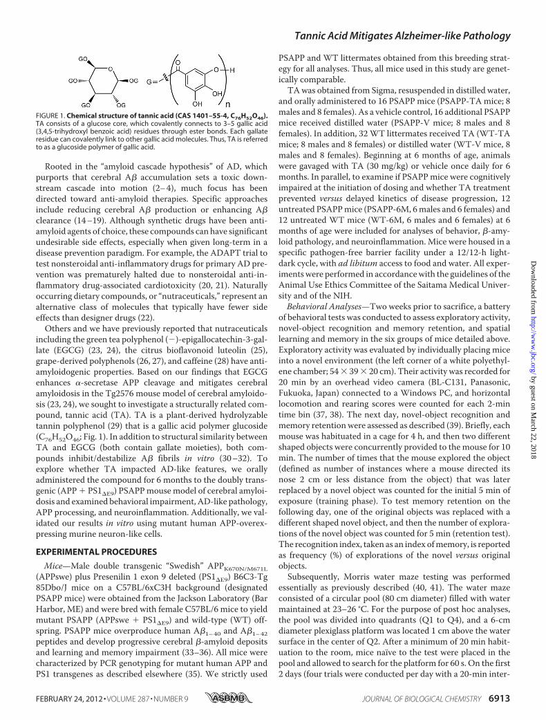
FIGURE 1. Chemical structure of tannic acid (CAS 1401–55-4, C76H52O46).TA consists of a glucose core, which covalently connects to 3–5 gallic acid(3,4,5-trihydroxyl benzoic acid) residues through ester bonds. Each gallateresidue can covalently link to other gallic acid molecules. Thus, TA is referredto as a glucoside polymer of gallic acid.
Tannic Acid Mitigates Alzheimer-like Pathology
FEBRUARY 24, 2012 • VOLUME 287 • NUMBER 9 JOURNAL OF BIOLOGICAL CHEMISTRY 6913
by guest on March 22, 2018
http://ww
w.jbc.org/
Dow
nloaded from

trial interval), a visible cue was placed on the platform and itslocation was randomly varied among four possible locations(counterbalanced across mice). The trial ended when a mouseclimbed the platform, or in the allocated 60 s, whichever camefirst. After finding and climbing on the platform, each mousewas allowed to remain there for 20 s, and was then returned toits cage. Animals that did not locate the platform within 60 swere guided to it and allowed to remain there for 20 s beforebeing returned to their cages. On the third day, submergedplatform testing was conducted for five consecutive days(learning phase; four trials per day with a 20-min inter-trialinterval). The location of the indiscernible platform remainedin Q2, 1 cm below the water surface, andmice were placed intothe pool in one of seven randomly selected locations (excludingthe position immediately adjacent to the platform). One dayafter the conclusion of the learning phase, memory retentionwas determined in a single 60-s probe trial. The submergedplatform was removed from the water maze, and mice wereplaced and released opposite the site where the platform hadbeen located and time spent in each quadrant was recorded forthe probe trial. All behavioral tests were performed in a room (6m � 4.5 m) with indirect lighting and multiple visible cues onthewalls. The examiner determined the time of swimming untilthe mouse reached the platform (latency) using a stopwatch. Inaddition, trials were recorded using an overhead video cameraand were analyzed using customized macro software inMicrosoft Excel. All trials were performed at the same time ofday (�1 h), during the animals’ light phase. So as not to inter-fere with behavioral testing, TA or vehicle treatment was car-ried out 1 h after behavioral testing.Tissue Preparation—Tissue was processed according to our
previously described methods (16, 18, 42, 43). At 12 months ofage, animals were anesthetized with sodium pentobarbital (50mg/kg) and euthanized by transcardial perfusion with ice-coldphysiological saline containing heparin (10 units/ml). Brainswere isolated and quartered (sagittally at the level of the longi-tudinal fissure of the cerebrum, and then coronally at the levelof the anterior commissure) using a mouse brain slicer (Muro-machi Kikai, Tokyo, Japan). Right anterior cerebral quarterswere weighed and snap-frozen at �80 °C for �- or �-secretaseactivity analyses. Right posterior cerebral quarters were furtherdivided into two pieces, and weighed and snap-frozen at�80 °C. One-half was sequentially extracted in Tris-bufferedsaline (TBS; 25 mM Tris-HCl, pH 7.4, 150 mM NaCl), 2% SDS-,and guanidine-soluble fractions for A� sandwich enzyme-linked immunosorbent assays (ELISAs). The other half wasused for holo-APP,�-site APP cleaving enzyme 1 (BACE1), and�-carboxyl-terminal fragment (�-CTF: phospho-C99 and C99)Western blots. Left anterior cerebral quarters were weighedand immersed in RNA stabilization solution (RNAlater�,Applied Biosystems, Foster City, CA) and then snap-frozen at�80 °C for proinflammatory cytokine and BACE1 quantitativereal-time PCR (QPCR) analyses. Left posterior cerebral quar-ters were immersion fixed in 4% paraformaldehyde in 0.1 M
phosphate buffer at 4 °C overnight, and routinely processed inparaffin for immunohistochemical analyses.Immunohistochemistry—For paraffin blocks, we sectioned
five coronal sections (per set) with a 100 �m interval and a
thickness of 5 �m for each brain region (for cingulate cortex(CC), bregma �0.10 to �0.82 mm; for hippocampus (H) andentorhinal cortex (EC), bregma �2.92 to �3.64 mm) (44). Weprepared three sets of five sections in each separate region foranalyses of A� deposits/�-amyloid plaques (for burden, plaquenumber, andmaximum diameter morphometry) as well as ion-ized calcium-binding adapter molecule 1 (Iba1, to mark reac-tive microglia) and glial fibrillary acidic protein (GFAP, anastrocytosis marker) burdens. Immunohistochemical stainingwas conducted according to the manufacturer’s protocol usinga Vectastain ABC Elite kit (Vector Laboratories, Burlingame,CA) coupled with the diaminobenzidine reaction, except thatthe biotinylated secondary antibody step was omitted for A�immunohistochemical staining. The following primary anti-bodies were used: a biotinylated human A� monoclonal anti-body (4G8; 1:200, CovanceResearch Products, Emeryville, CA),Iba1 polyclonal antibody (1:1,000, Wako, Osaka, Japan), andGFAP polyclonal antibody (1:500, Dako, Carpinteria, CA).Using additional sets of five sections, normal mouse or rabbitserum (isotype control) or phosphate-buffered saline (0.1 M
PBS, pH7.4) was used instead of primary or secondary antibodyor ABC reagent as negative controls.Image Analysis—Quantitative image analysis was done based
on previously validated methods (16, 18, 42, 43). Images wereacquired as digitized tagged-image format files to retain maxi-mum resolution using a BX60 microscope with an attachedCCD camera system (DP-70, Olympus, Tokyo, Japan), and dig-ital images were routed into a Windows PC for quantitativeanalyses using SimplePCI software (Hamamatsu Photonics,Hamamatsu, Shizuoka, Japan). We captured images of five5-�m sections through each anatomic region of interest (CC,EC, and H) based on anatomical criteria defined by Franklinand Paxinos (44), and obtained a threshold optical density thatdiscriminated staining frombackground. Each anatomic regionof interest was manually edited to eliminate artifacts. For A�,Iba1 (microgliosis), and GFAP (astrocytosis) burden analyses,data are reported as the percentage of labeled area captured(positive pixels) divided by the full area captured (total pixels).Selection bias was controlled for by analyzing each region ofinterest in its entirety.For �-amyloid plaque morphometric analyses, diameters
(based on maximum length) of �-amyloid plaques were meas-ured, and numbers of �-amyloid plaques falling into threemutually exclusive diameter categories (�25, 25–50, or �50�m) were tabulated. Results are presented as mean plaquenumber per mouse in each region examined. For cerebral amy-loid angiopathy (CAA) morphometric analysis, we countednumbers of A� antibody-stained cerebral vessels in each ana-tomic region of interest based on our previous methods (43);those data are shown asmean CAA deposit number permouse.Cell Culture—The N2a cell line that stably overexpresses
human “Swedish”-mutated APP-695 (SweAPP N2a cells) waskindly provided by Dr. Gopal Thinakaran (Department of Neu-robiology, University of Chicago). SweAPP N2a cells weregrown in Dulbecco’s modified Eagle’s medium supplementedwith 10% fetal calf serum, 2 mM glutamine, 100 units/ml ofpenicillin, 0.1 �g/ml of streptomycin, and 200 �g/ml of G418sulfate according to previously described methods (16, 23, 25).
Tannic Acid Mitigates Alzheimer-like Pathology
6914 JOURNAL OF BIOLOGICAL CHEMISTRY VOLUME 287 • NUMBER 9 • FEBRUARY 24, 2012
by guest on March 22, 2018
http://ww
w.jbc.org/
Dow
nloaded from

SweAPP N2a cells were seeded in 24-well tissue culture platesat 1 � 105 cells per well. Cultured cells were differentiated intoneuron-like cells by 2 h pre-treatment with neurobasal mediacontaining 300 �M dibutyryl cAMP and then treated with TA(1.563, 3.125, 6.25, 12.5, or 25 �M) or 0.1 M PBS (pH 7.4; asvehicle control) for 12 h in the same media prior to analyses.Lactate Dehydrogenase Release Assay—SweAPP N2a cells
were seeded in 24-well tissue culture plates at 1 � 105 cells perwell. Culture cells were differentiated into neuron-like cells by2 h pre-treatment with neurobasal media containing 300 �M
dibutyryl cAMP and then treated with TA (3.125, 6.25, 12.5, or25 �M) or 0.1 M PBS (pH 7.4; vehicle control) for 12 h in thesamemedia. Culture wells were then assayed for cell death by alactate dehydrogenase release assay (Promega) as described(45).Cell-free BACE1 Activity Assay—To directly test the effect of
TA on BACE1 activity, we used available kits based on secre-tase-specific peptides conjugated to DABCYL/EDANS fluoro-genic reporter molecules (Cayman Chemical, Ann Arbor, MI)in accordance with the manufacturer’s instructions. Briefly,BACE1 enzyme was incubated with various concentrations ofTA (1.563, 3.125, 6.25, 12.5, or 25 �M) or BACE1 inhibitor II(1.25 �m; as a positive control) in the presence of 1� reactionbuffer for 40 min prior to reading fluorescence values on aFLUOstar Omega (BMG LABTECH, San Diego, CA) fluores-cent microplate reader.ELISA—We separately quantified A�1–40 and A�1–42 in
brain homogenates and cultured SweAPP N2a cell superna-tants by sandwich ELISAs. Brain A�1–40 and A�1–42 specieswere detected by a three-step extraction protocol accordingto previously published methods (46, 47). Briefly, we homog-enized brains using TissueLyser LT (Qiagen, Valencia, CA;two times for 1 min at 50 Hz) in TBS solution containingprotease inhibitor mixture (Sigma), centrifuged homoge-nates at 18,800 � g for 60 min at 4 °C, and removed thesupernatants (TBS-soluble fraction). Resulting pellets weretreated with 2% SDS in H2O with the same protease inhibi-tors and homogenized using TissueLyser LT (one time for 1min at 50 Hz). We then centrifuged the homogenates at18,800 � g for 60 min at 4 °C and collected supernatants (2%SDS-soluble fraction). Finally, the remaining pellets weretreated with 5 M guanidine HCl and dissolved by occasionalmixing on ice for 30 min, then centrifuged at 18,800 � g for60 min at 4 °C, and supernatants were collected representingthe guanidine HCl-soluble fraction.A�1–40 and A�1–42 species were separately quantified in
individual samples in duplicate using ELISA kits (cataloguenumber 27718 for A�1–40 and number 27712 for A�1–42; IBL,Fujioka, Gunma, Japan) in accordance with the manufacturer’sinstructions (48). We also quantified A� oligomers in the 2%SDS-soluble fraction in duplicate individual samples by A� oli-gomer ELISA (catalogue number 27725; IBL) according to themanufacturer’s instructions (49). All samples fell within the lin-ear range of the standard curve. A�1–40 and A�1–42 ELISAvalues are reported as picograms of A�1-x/wet mg of brain, andthe A� oligomer concentration is reported as picomolar.Western Blot—Cultured SweAPPN2a cells were treated with
various doses of TA (1.563, 3.125, 6.25, 12.5, or 25 �M) or 0.1 M
PBS (pH 7.4; as a vehicle control) for 12 h. Cultured cells werethen lysed in ice-cold lysis buffer (containing 20 mM Tris-HCl,pH 7.5, 150mMNaCl, 1 mMNa2EDTA, 1mM EGTA, 1% TritonX-100, 2.5 mM sodium pyrophosphate, 1 mM �-glycerophos-phate, 1 mM Na3VO4, 1 �g/ml of leupeptin, and 1 mM PMSF).Mouse brain homogenates were lysed in TBS solution contain-ing protease inhibitor mixture (Sigma) followed by TNE buffer(10 mM Tris-HCl, 1% NP-40, 1 mM EDTA, and 150 mM NaCl),and aliquots corresponding to 10 �g of total protein were elec-trophoretically separated using 10 or 15% Tris glycine gelsbased on target protein molecular weights. Electrophoresedproteins were transferred to polyvinylidene difluoride mem-branes (Bio-Rad) that were subsequently blocked in blockingbuffer (1% (w/v) nonfat dry milk in TBS containing 0.1% (v/v)Tween 20) for 1 h at ambient temperature. After blocking,membranes were hybridized for 1 h at ambient temperaturewith primary antibodies: amino-terminal APP polyclonal anti-body (1:400, IBL), carboxyl-terminal soluble APP-� (sAPP-�)monoclonal antibody (2B3; 1:100, IBL) directed against aminoacids DAEFRHDSGYEVHHQK, carboxyl-terminal solubleAPP-� (sAPP-�)monoclonal antibody that recognizes Swedishmutant (ISEVNL) protein (6A1; 1:100, IBL), carboxyl-terminalBACE1 polyclonal antibody (1:400, IBL), amino-terminal A�monoclonal antibody (82E1; 1:150, IBL), carboxyl-terminalAPP polyclonal antibody (1:1,000, Merck Millipore, Billerica,MA), or actin polyclonal antibody as a loading control (1:500,Santa Cruz Biotechnology, Santa Cruz, CA). Membranes werethen rinsed three times for 30min each in TBS containing 0.1%(v/v) Tween 20 and incubated for 1 h at ambient temperaturewith appropriate horseradish peroxidase-conjugated second-ary antibodies. After additional rinsing as above, membraneswere incubated for 5 min at ambient temperature withenhanced chemiluminescence substrate (SuperSignal WestDura Extended Duration Substrate, Thermo Fisher Scientific,Waltham, MA), exposed to film, and developed.Secretase Activity Assays—For �- and �-secretase activity
analyses in brain homogenates, we used available kits based onsecretase-specific peptides conjugated to fluorogenic reportermolecules (DABCYL/EDANS; R & D Systems, Minneapolis,MN) according to our published methods (23, 43). Briefly,brains were lysed in ice-cold 1� cell extraction buffer for 10min and centrifuged at 18,800� g for 1min. Supernatants werecollected and kept on ice. Appropriate amounts of brain homo-genate, reaction buffer, and fluorogenic substratewere added induplicate to a 96-well plate and incubated in the dark at 37 °Cfor various periods of time. Following incubation, fluorescencewas monitored (335 nm excitation and 495 nm emission) at25 °C using a SH-9000 microplate fluorimeter with SF6 soft-ware (CORONA ELECTRIC, Hitachinaka, Ibaraki, Japan).Background was determined from negative controls (omissionof brain homogenate or fluorogenic substrate).QPCR—We quantified tumor necrosis factor-� (TNF-�),
interleukin-1� (IL-1�), BACE1, and �-actin mRNA levels byQPCR. Total RNA was extracted using the RNeasy Mini Kit(Qiagen), and first strand cDNA synthesis was carried out usingthe QuantiTect Reverse Transcription Kit (Qiagen) in accord-ance with the manufacturer’s instructions. We diluted cDNA1:1 in H2O and carried out QPCR for all genes of interest using
Tannic Acid Mitigates Alzheimer-like Pathology
FEBRUARY 24, 2012 • VOLUME 287 • NUMBER 9 JOURNAL OF BIOLOGICAL CHEMISTRY 6915
by guest on March 22, 2018
http://ww
w.jbc.org/
Dow
nloaded from

cDNA-specific TaqMan primer/probe sets (TaqMan GeneExpression Assays, Applied Biosystems) on an ABI 7500 FastReal-time PCR instrument (Applied Biosystems). Each 20-�lreaction mixture contained 2 �l of cDNA with 1 �l of TaqManGene Expression Assay reagent, 10 �l of TaqMan Fast Univer-sal PCR Master Mix (Applied Biosystems), and 7 �l of H2O.Thermocycler conditions consisted of: 95 °C for 15 s, followedby 40 cycles of 95 °C for 1 s and 60 °C for 20 s. TaqMan probe/primer sets were as follows: mouse TNF-� (catalogue numberMm00443258_m1), mouse IL-1� (number Mm00434228_m1),mouse BACE1 (number Mm00478664_m1), and mouse �-actin(numberMm00607939_s1; used as an internal reference control)(Applied Biosystems). Samples that were not subjected to reversetranscription were run in parallel as negative controls to rule outgenomic DNA contamination (data not shown). A “no templatecontrol” was also included for each primer set (data not shown).The cycle threshold number (CT) method (50) was used to deter-mine relative amounts of initial target cDNA in each sample.Results for BACE1 expression are expressed relative to vehicle-treatedWTmice,whereasTNF-� and IL-1� expressionvalues arenormalized toWT-6M littermates.Statistical Analysis—All experiments were performed by an
examiner blinded to sample identities, and codewas not brokenuntil the analyses were completed. Data are presented as themean � 1 S.E. A hierarchical analysis strategy was used fortime-dependent behavioral data in which the first step was arepeated-measures analysis of variance (ANOVA) to assess thesignificance of the main effects and interactive terms. If signif-icant, post hoc testingwas donewithTukey’sHSDorDunnett’sT3 methods, and appropriate p values are reported based onadjustment according to Levene’s test for equality of the vari-ance. For all other data, in instances of single mean compari-sons, Levene’s test followed by t test for independent sampleswas performed. In instances of multiple mean comparisons,one-way ANOVA was used, followed by post hoc comparisonof the means using Bonferroni’s or Dunnett’s T3 methods(where appropriateness was determined using Levene’s test). Ap value of less than 0.05 was considered to be significant. Allanalyses were performed using the Statistical Package for theSocial Sciences, release IBM SPSS 19.0 (IBM, Armonk, NY).
RESULTS
Oral Tannic Acid Treatment Mitigates Hyperactivity andCognitive Impairment in PSAPP Mice—We began by orallyadministering TA or vehicle to PSAPP orWTmice starting at 6months of age for a period of 6 months and subsequently con-ducted a behavioral testing battery. In addition, to examine cog-nitive status when dosing started, untreated PSAPP and WTmice at 6 months of age were included for analyses of behavior.When placed into a novel environment, PSAPP-V mice werehyperactive as measured by higher locomotion and rearingscores compared with the other 5 groups ofmice (Fig. 2A). Thisbehavioral phenotype has been observed in mouse models ofcerebral amyloidosis (e.g. Tg2576 or PSAPP) (18, 34, 51), andmay be associated with disinhibition resulting from corticaland/or hippocampal injury (38). Overall ANOVA showedmaineffects of time (p� 0.001), genotype (p� 0.001), and treatment(p � 0.001), and post hoc comparisons showed statistically sig-
nificant differences between PSAPP-V mice and the other 5mouse groups at each timepoint for locomotion scores (Fig. 2A,*, p � 0.05 for PSAPP-V versus PSAPP-TA, WT-V, WT-TA,PSAPP-6M, orWT-6Mmice) and for rearing scores (Fig. 2A, *,p � 0.05 for PSAPP-V versus PSAPP-TA, WT-V, WT-TA,PSAPP-6M, or WT-6M mice). Hyperactivity was fully pre-vented in PSAPP-TA mice, as they did not statistically differfromWT-VWT-TA, PSAPP-6M, or WT-6Mmice (p � 0.05).
We then tested learning and memory in the same cohort ofmice by a novel object recognition assay. If mice remember aninitial encounter with a novel object, they tend to preferentiallyexplore the new versus familiar object, typically operationalizedas “recognition index” (39). Although all groups performedsimilarly during the training phase of the test, in the retentionphase, one-way ANOVA followed by post hoc comparisonshowed statistically significant differences on recognition indexbetween PSAPP-V mice and the other 5 mouse groups as indi-cated (Fig. 2B, *, p � 0.05 for PSAPP-V versus PSAPP-TA,WT-V, WT-TA, PSAPP-6M, or WT-6M mice). Importantly,PSAPP-TAmice had significantly increased novel object explo-ration frequency versus PSAPP-V animals (Fig. 2B), but did notsignificantly differ from WT-V, WT-TA, PSAPP-6M, orWT-6M groups (p � 0.05), showing that TA also preventednovel object recognition impairment associated with PSAPPtransgene expression.We further tested animals in theMorris watermaze, a widely
accepted assay of spatial reference learning and memory inrodents (40, 41). For the learning phase of the test, overallANOVA showedmain effects of time (p� 0.001) and genotype(p � 0.001), and post hoc comparison revealed statistically sig-nificant differences between PSAPP-V mice and the other 5mouse groups as indicated (Fig. 2C, *, p � 0.05 for PSAPP-Vversus PSAPP-TA, WT-V, WT-TA, PSAPP-6M, or WT-6Mmice). PSAPP-Vmice had greater latency to reach the platformlocation after training than the other 5 mouse groups, whereasPSAPP-TA mice showed significant improvement, indicatingthat oral TA treatment inhibited PSAPP transgene-associatedimpaired spatial reference learning. For the probe trial (day 6 oftesting), the invisible platform was removed from the pool andplatform location memory was evaluated. When consideringQ2 (goal quadrant) data, one-wayANOVAand post hoc testingshowed statistically significant differences between PSAPP-Vmice and the other 5 mouse groups as indicated (Fig. 2D, *, p �0.05 for PSAPP-V versus PSAPP-TA, WT-V, WT-TA, PSAPP-6M, orWT-6Mmice). PSAPP-TAmice swam in the goal quad-rant significant longer than PSAPP-V mice, and their behaviordid not significantly differ fromWT-V,WT-TA, PSAPP-6M, orWT-6M mice, showing that TA treatment prevents PSAPPtransgene-associated spatial memory impairment.It is unlikely that behavioral differences in the Morris water
maze were due to motivational issues or to locomotor impair-ment, as there were no significant between group differences(p � 0.05) on swim speed during either the learning or probetrial phases of the test.Moreover, it is important to note that thedegree of thigmotaxis could indicate levels of anxiety andimpact interpretation of Morris water maze results. In thisregard, we did not observe evidence of thigmotaxis, operation-alized as prolonged movement of the mice along the pool cir-
Tannic Acid Mitigates Alzheimer-like Pathology
6916 JOURNAL OF BIOLOGICAL CHEMISTRY VOLUME 287 • NUMBER 9 • FEBRUARY 24, 2012
by guest on March 22, 2018
http://ww
w.jbc.org/
Dow
nloaded from

cumference, in anymice examined during either the learning orprobe trial phases of the test. Furthermore, untreated PSAPPmice at 6 months of age (PSAPP-6M) did not clearly show cog-nitive impairment as compared with WT mice at the same ageor toWT-V orWT-TAmice at 12 months of age by open field,object recognition, or either the learning or probe trial phasesof Morris water maze testing. Importantly, behavioral testingperformance in PSAPP-TAmice was not significantly differentfrom PSAPP-6M animals in each of the tests conducted. Thisresult can be interpreted as prevention of cognitive impairmentin PSAPPmice by a 6-month treatment regimen of TA. Finally,for all of the behavioral tests conducted, we used multipleANOVAmodels with gender as a categorical covariate, but didnot detect significant gender main effects or interactive terms(p � 0.05). We also stratified by gender and found a similarpattern of results as above in both males and females (data notshown).Tannic Acid Treatment Ameliorates A� Pathology in an
Accelerated Mouse Model of Cerebral Amyloidosis—We nextevaluatedA�/�-amyloid pathology by three strategies: A� anti-body immunoreactivity (conventional �-amyloid “burden”
analysis), �-amyloid morphometric analysis, and separateA�1–40 and A�1–42 sandwich ELISAs. PSAPP-V mice showedtypical �-amyloid deposition (33, 36), which was significantlyreduced by 51–58% in CC, EC, and H regions of PSAPP-TAmouse brains (Fig. 3, A and B, ***, p � 0.001). Of note,PSAPP-TA plaques were not completely attenuated versusPSAPP-6Mmice (Fig. 3,A and B), indicating that TAmitigatedas opposed to completely prevented cerebral amyloid deposi-tion. TA reduction in �-amyloid deposits was independent ofgender, being evident in both male and female PSAPP-TA ver-sus PSAPP-V mice (data not shown). To assess whetherreduced �-amyloid burden was specific to a particular plaquesize subset or occurredmore generally, we performedmorpho-metric analysis of �-amyloid plaques in PSAPP-V andPSAPP-TA mice. According to previously described methods(18, 16, 42, 43), plaques were assigned to one of three mutuallyexclusive categories according tomaximum diameter: small (�25 �m), medium (between 25 and 50 �m), or large (�50 �m).Data showed that all three subsets of plaques were significantlyreduced in PSAPP-TA versus PSAPP-V mice across all threebrain regions examined (Fig. 3,A andC, ***, p� 0.001, % reduc-
FIGURE 2. Tannic acid treatment prevents behavioral impairment in PSAPP mice. PSAPP mice received oral vehicle (PSAPP-V, n � 16) or TA (PSAPP-TA, n �16) treatment, and wild-type mice were given vehicle (WT-V, n � 16) or TA (WT-TA, n � 16) orally for 6 months beginning at 6 months of age, and subjected tobehavioral testing at 12 months of age. To examine cognitive status when dosing started, untreated 6-month-old PSAPP mice (PSAPP-6M, n � 12) and WT mice(WT-6M, n � 12) were included in the behavioral analyses. A, locomotion and rearing scores obtained from open field activity testing are shown. B, recognitionindex (%) in the object recognition test is shown (left, training phase; right, retention test phase). C, Morris water maze test data are shown from the submergedplatform (learning phase) and from D, a single 60-s probe trial test (conducted 1 day after termination of the learning phase). All statistical comparisons areversus PSAPP-V mice.
Tannic Acid Mitigates Alzheimer-like Pathology
FEBRUARY 24, 2012 • VOLUME 287 • NUMBER 9 JOURNAL OF BIOLOGICAL CHEMISTRY 6917
by guest on March 22, 2018
http://ww
w.jbc.org/
Dow
nloaded from
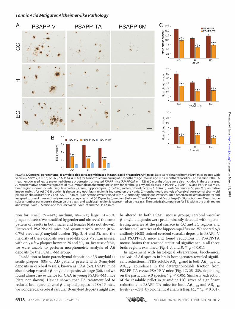
tion for: small, 39–44%; medium, 44–52%; large, 54–66%plaque subsets).We stratified by gender and observed the samepattern of results in both males and females (data not shown).Untreated PSAPP-6M mice had quantitatively minor (0.5–0.7%) cerebral �-amyloid burden (Fig. 3, A and B), and themajority of these deposits were seed-like dots �25 �m in size,with only a few plaques between 25 and 50 �m. Because of this,we were unable to perform morphometric analysis of A�deposits for the PSAPP-6M group.In addition to brain parenchymal deposition of �-amyloid as
senile plaques, 83% of AD patients present with �-amyloiddeposits in cerebral vessels, known as CAA (52). PSAPP micealso develop vascular �-amyloid deposits with age (36), and wefound almost no evidence for CAA in young PSAPP-6M mice(data not shown). Having shown that TA treatment led toreduced brain parenchymal �-amyloid plaques in PSAPPmice,wewondered if cerebral vascular�-amyloid depositsmight also
be altered. In both PSAPP mouse groups, cerebral vascular�-amyloid deposits were predominantly detected within pene-trating arteries at the pial surface in CC and EC regions andwithin small arteries at the hippocampal fissure.We scored A�antibody (4G8)-stained cerebral vascular deposits in PSAPP-Vand PSAPP-TA mice and found reductions in PSAPP-TAmouse brains that reached statistical significance in all threebrain regions examined (Fig. 4, A and B, **, p � 0.01).In agreement with histological observations, biochemical
analysis of A� species in brain homogenates revealed signifi-cant reductions in TBS-soluble A�1–40 and in both A�1–40 andA�1–42 abundance in the detergent-soluble fraction fromPSAPP-TA versus PSAPP-V mice (Fig. 4C, 25–33% dependingon the particular A� species; *, p � 0.05). Similarly, extractionof the insoluble pellet in guanidine HCl revealed significantreductions in PSAPP-TA mice for both A�1–40 and A�1–42levels (27–28%) by biochemical analysis (Fig. 4C, ***, p� 0.001).
FIGURE 3. Cerebral parenchymal �-amyloid deposits are mitigated in tannic acid-treated PSAPP mice. Data were obtained from PSAPP mice treated withvehicle (PSAPP-V, n � 16) or TA (PSAPP-TA, n � 16) for 6 months commencing at 6 months of age (mouse age � 12 months at sacrifice). To examine if the TAtreatment delayed versus prevented disease progression, untreated PSAPP mice (PSAPP-6M, n � 12) at 6 months of age were also included in these analyses.A, representative photomicrographs of 4G8 immunohistochemistry are shown for cerebral �-amyloid plaques in PSAPP-V, PSAPP-TA, and PSAPP-6M mice.Brain regions shown include: cingulate cortex (CC, top), hippocampus (H, middle), and entorhinal cortex (EC, bottom). Scale bar denotes 50 �m. B, quantitativeimage analysis for A� (4G8) burden is shown, and each brain region is indicated on the x axis. C, morphometric analysis of cerebral parencymal �-amyloidplaques is shown in PSAPP-V and PSAPP-TA mice. Brain sections were stained with 4G8 antibody, and plaques were counted based on maximum diameter andassigned to one of three mutually exclusive categories: small (�25 �m; top), medium (between 25 and 50 �m; middle), or large (�50 �m; bottom). Mean plaquesubset number per mouse is shown on the y axis, and each brain region is represented on the x axis. The statistical comparison for B is within the brain regionand versus PSAPP-TA mice, and for C, between PSAPP-V and PSAPP-TA mice.
Tannic Acid Mitigates Alzheimer-like Pathology
6918 JOURNAL OF BIOLOGICAL CHEMISTRY VOLUME 287 • NUMBER 9 • FEBRUARY 24, 2012
by guest on March 22, 2018
http://ww
w.jbc.org/
Dow
nloaded from

By contrast, A�1–42 levels in theTBS-soluble fractionwere sim-ilar between PSAPP-TA and PSAPP-V mice. We also analyzedPSAPP-6M mice by A� biochemistry, but found low abun-dance of A� species that was below the threshold of ELISAdetection in certain fractions (data not shown). Collectively,these data show that cerebral amyloidosis, including brainparenchymal and cerebral vascular �-amyloid deposits and A�peptide abundance, is delayed but not completely prevented byoral treatment with TA in PSAPP mice.Inhibition of Cerebral Amyloidogenic APP Metabolism in Tan-
nic Acid-treated PSAPPMice—Mitigated cerebral amyloidosis inPSAPP-TA mice could be due to 1) increased brain-to-blood A�efflux (10), 2) reduced expression of APP or PS1 transgenes, or 3)attenuated amyloidogenic APPmetabolism.We obtained periph-eral blood samples from PSAPP-V and PSAPP-TA mice at thetime of sacrifice and assayed plasma A�1–40 and A�1–42 species,but did not detect differences between groups (data not shown).Toexamine ifTA-attenuated cerebral amyloidosis couldbedue todecreased expressionof transgene-derivedAPPorPS1,weprobedbrain homogenates from PSAPP-V and PSAPP-TA mice usingamino-terminal APP polyclonal or carboxyl-terminal PS1 mono-
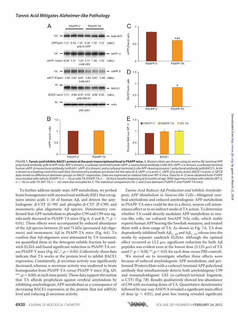
clonal antibodies and found no change inAPP or PS1 holoproteinlevels (Fig. 5A and data not shown).To determine whether APP metabolites including sAPP-�
and sAPP-�were affected byTA treatment, brain homogenateswere probed with monoclonal antibody 2B3 that detects thecarboxyl terminus of human sAPP-� and with monoclonalantibody 6A1 that recognizes the carboxyl terminus of humanSwedish mutant sAPP-�. Western blot and densitometry datashowed significantly reduced expression of sAPP-� inPSAPP-TA mouse brains, whereas sAPP-� abundance did notdiffer between groups (Fig. 5, A–C, ***, p � 0.001). To furtherdetermine whether amyloidogenic APP processing was inhib-ited by TA treatment, we probed brain homogenates with acarboxyl-terminal BACE1 polyclonal antibody. Densitometryshowed that BACE1 protein abundance was significantlydecreased in PSAPP-TA mice (Fig. 5, A and D, *, p � 0.05). Toassess whether this effect was due to TA attenuation of BACE1transcription, relative expression levels of BACE1 mRNA wereassayed in eachmouse group byQPCR.However, therewere nodetectable differences between groups (Fig. 5E), suggesting apost-transcriptional mode of TA action on BACE1.
FIGURE 4. Cerebral vascular �-amyloid deposits and brain A� levels are reduced in PSAPP mice given oral tannic acid treatment. A, representativephotomicrographs of 4G8 immunohistochemistry were taken from PSAPP-V and PSAPP-TA mouse hippocampi at 12 months of age, and cerebral vascular�-amyloid deposits are indicated (arrows). Scale bar denotes 200 �m. B, severity of cerebral amyloid angiopathy (mean CAA deposit number per mouse) isshown on the y axis with the brain region indicated on the x axis (CC, H, and EC). C, TBS-soluble, 2% SDS-soluble, and TBS-insoluble (but 5 M guanidineHCl-extractable) fractions from three-step extracted brain homogenates were examined by sandwich ELISA for human A�1– 40 and A�1– 42 levels. Data wereobtained from PSAPP mice treated with vehicle (PSAPP-V, n � 16) or with TA (PSAPP-TA, n � 16) for 6 months commencing at 6 months of age. All statisticalcomparisons are within the brain region and/or between PSAPP-V and PSAPP-TA mice.
Tannic Acid Mitigates Alzheimer-like Pathology
FEBRUARY 24, 2012 • VOLUME 287 • NUMBER 9 JOURNAL OF BIOLOGICAL CHEMISTRY 6919
by guest on March 22, 2018
http://ww
w.jbc.org/
Dow
nloaded from
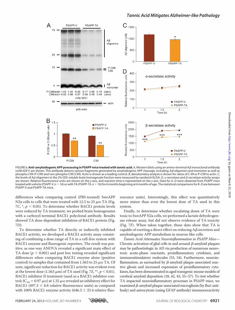
To further address steady-state APP metabolism, we probedbrain homogenates withmonoclonal antibody 82E1 that recog-nizes amino acids 1–16 of human A�, and detects the amy-loidogenic �-CTF (C-99) and phospho-�-CTF (P-C99) andmonomeric plus oligomeric A� species. Densitometry con-firmed that APPmetabolism to phospho-C99 and C99 was sig-nificantly deceased in PSAPP-TA mice (Fig. 6, A and B, **, p �0.01). These effects were accompanied by reduced abundanceof the A� species between 25 and 75 kDa (presumed A� oligo-mers) and monomeric A� in PSAPP-TA mice (Fig. 6A). Toconfirm that A� oligomers were attenuated by TA treatment,we quantified them in the detergent-soluble fraction by sand-wich ELISA and found significant reduction in PSAPP-TA ver-sus PSAPP-Vmice (Fig. 6C, *, p� 0.05). Collectively, these dataindicate that TA works at the protein level to inhibit BACE1expression. Consistently, �-secretase activity was significantlydecreased, whereas �-secretase activity was unaltered in brainhomogenates from PSAPP-TA versus PSAPP-V mice (Fig. 6D,***, p� 0.001 at each time point). These data support the notionthat TA affords prophylaxis against cerebral amyloidosis byinhibiting amyloidogenic APPmetabolism as a consequence ofdecreasing BACE1 expression at the protein (but not mRNA)level and reducing �-secretase activity.
Tannic Acid Reduces A� Production and Inhibits Amyloido-genic APP Metabolism in Neuron-like Cells—Mitigated cere-bral amyloidosis and reduced amyloidogenic APP metabolismin PSAPP-TAmice could be due to a direct, neuron cell auton-omous affect or to an indirectmode of TA action. To determinewhether TA could directly modulate APP metabolism in neu-ron-like cells, we cultured SweAPP N2a cells, which stablyexpress humanAPP bearing the Swedishmutation, and treatedthem with a dose-range of TA. As shown in Fig. 7A, TA dosedependently inhibited both A�1–40 and A�1–42 release into themedia by separate sandwich ELISAs. Although the optimaleffect occurred at 12.5 �M, significant reduction for both A�peptides was evident even at the lowest dose (3.125 �M) of TAused (*, p � 0.05; **, p � 0.01 for each dose versus PBS control).
We moved on to investigate whether these effects werebecause of reduced amyloidogenic APP metabolism, and per-formedWestern blots with a carboxyl-terminal APP polyclonalantibody that simultaneously detects both amyloidogenic C99and nonamyloidogenic C83 (�-carboxyl-terminal fragment,�-CTF) (Fig. 7B). Results qualitatively showed less abundanceof C99 with increasing doses of TA. Quantitative densitometryfollowed by one-way ANOVA revealed a significantmain effectof dose (p � 0.01), and post hoc testing revealed significant
FIGURE 5. Tannic acid inhibits BACE1 protein at the post-transcriptional level in PSAPP mice. A, Western blots are shown using an amino (N)-terminal APPpolyclonal antibody (pAb N-APP; holo-APP is shown), a carboxyl-terminal human sAPP-� monoclonal antibody (mAb 2B3; sAPP-� is shown), a carboxyl-terminalhuman sAPP-� monoclonal antibody (mAb 6A1; sAPP-� is shown), and a carboxyl-terminal �-site APP cleaving enzyme 1 polyclonal antibody (pAb BACE1). Actinis shown as a loading control for each blot. Densitometry analyses are shown for the ratio of: B, sAPP-� to actin; C, sAPP-� to actin; and D, BACE1 to actin. E, QPCRdata reveal no differences between groups on BACE1 expression. Data are expressed as relative fold over WT-V mice. Data for A–D were obtained from PSAPPmice treated with vehicle (PSAPP-V, n � 16) or with TA (PSAPP-TA, n � 16) for 6 months beginning at 6 months of age. Wild-type mice treated with vehicle (WT-V,n � 16) or with TA (WT-TA, n � 16) were also included for E. The statistical comparisons for C and D are between PSAPP-V and PSAPP-TA mice.
Tannic Acid Mitigates Alzheimer-like Pathology
6920 JOURNAL OF BIOLOGICAL CHEMISTRY VOLUME 287 • NUMBER 9 • FEBRUARY 24, 2012
by guest on March 22, 2018
http://ww
w.jbc.org/
Dow
nloaded from
differences when comparing control (PBS-treated) SweAPPN2a cells to cells that were treated with 12.5 to 25 �M TA (Fig.7C, *, p � 0.05). To determine whether BACE1 protein levelswere reduced by TA treatment, we probed brain homogenateswith a carboxyl-terminal BACE1 polyclonal antibody. Resultsshowed TA dose-dependent inhibition of BACE1 protein (Fig.7D).To determine whether TA directly or indirectly inhibited
BACE1 activity, we developed a BACE1 activity assay consist-ing of combining a dose range of TA in a cell-free system withBACE1 enzyme and fluorogenic reporters. The result was pos-itive, as one-way ANOVA revealed a significant main effect ofTA dose (p � 0.001) and post hoc testing revealed significantdifferences when comparing BACE1 enzyme alone (positivecontrol) to samples that contained from 1.563 to 25 �M TA. Ofnote, significant reduction for BACE1 activity was evident evenat the lowest dose (1.563 �m) of TA used (Fig. 7E, **, p � 0.01).BACE1 inhibitor II treatment (used as a BACE1 inhibitor con-trol; IC50 � 0.97�M) at 1.25�M revealed an inhibitory effect forBACE1 (497.3 � 8.8 relative fluorescence units) as comparedwith 100% BACE1 enzyme activity (646.3 � 25.4 relative fluo-
rescence units). Interestingly, this effect was quantitativelymore minor than even the lowest dose of TA used in thissystem.Finally, to determine whether escalating doses of TA were
toxic to SweAPPN2a cells, we performed a lactate dehydrogen-ase release assay, but did not observe evidence of TA toxicity(Fig. 7F). When taken together, these data show that TA iscapable of exerting a direct effect on reducing A� secretion andamyloidogenic APP metabolism in neuron-like cells.Tannic Acid Attenuates Neuroinflammation in PSAPP Mice—
Chronic activation of glial cells in and around �-amyloid plaquesmay be pathoetiologic in AD via production of numerous neuro-toxic acute-phase reactants, proinflammatory cytokines, andimmunostimulatory molecules (53, 54). Furthermore, neuroin-flammation, as earmarked by �-amyloid plaque-associated reac-tive gliosis and increased expression of proinflammatory cyto-kines, has been demonstrated in aged transgenicmousemodels ofcerebral amyloid deposition (18, 42, 43, 55–57). To test whetherTA impacted neuroinflammatory processes in PSAPP mice, weexamined�-amyloid plaque-associatedmicrogliosis (by Iba1 anti-body) and astrocytosis (using GFAP antibody) immunoreactivity
FIGURE 6. Anti-amyloidogenic APP processing in PSAPP mice treated with tannic acid. A, Western blots using an amino-terminal A� monoclonal antibody(mAb 82E1) are shown. This antibody detects various fragments generated by amyloidogenic APP cleavage, including: A� oligomers and monomer as well asphospho-C99 (P-C99) and non-phospho-C99 (C99). Actin is shown as a loading control. B, densitometry analysis is shown for ratios of C-99 or P-C99 to actin. C,the levels of A� oligomers in the 2% SDS-soluble brain homogenate fraction were measured by sandwich ELISA. D, �-secretase and �-secretase activity assaysare shown. Relative fluorescence units are shown on the y axis, and reaction time is represented on the x axis. Data for A–D were obtained from PSAPP micetreated with vehicle (PSAPP-V, n � 16) or with TA (PSAPP-TA, n � 16) for 6 months beginning at 6 months of age. The statistical comparisons for B–D are betweenPSAPP-V and PSAPP-TA mice.
Tannic Acid Mitigates Alzheimer-like Pathology
FEBRUARY 24, 2012 • VOLUME 287 • NUMBER 9 JOURNAL OF BIOLOGICAL CHEMISTRY 6921
by guest on March 22, 2018
http://ww
w.jbc.org/
Dow
nloaded from

(conventional microgliosis and astrocytosis burden analyses) andquantified brain expression of the proinflammatory cytokinesTNF-� and IL-1�. PSAPP-Vmice demonstrated elevated �-amy-loid plaque-associated reactive gliosis (microgliosis and astrocyto-sis), as evidencedby increasedexpressionof Iba1andGFAPinglialsomata and processes. Yet, microgliosis and astrocytosis were sig-nificantly reduced in PSAPP-TA mice compared with PSAPP-Vanimals (Figs. 8, A and B, and 9, A and B, ***, p � 0.001). Theseeffects were gender-independent, as a similar pattern of statisti-cally significant results was observed in both male and femalePSAPP-TA mice (data not shown). When considering brainmRNAabundance ofTNF-� and IL-1�, a similar pattern of statis-tically significant results was found (Fig. 9C, **, p � 0.01). Neuro-inflammation in PSAPP-TA mice was not reduced to levels ofuntreated PSAPP-6M mice, indicating mitigation but not com-plete prevention of this pathology (Figs. 8,A and B, and 9,A–C).Phagocytic microglia have been detected in the aged APP23
cerebral amyloidosismousemodel in small numbers (58) and inincreased abundance in aged Tg2576 mice bearing a CD11cpromoter-driven dominant-negative transforming growth fac-
tor-� receptor type II transgene (18) as well as in the ischemiccore after cerebral ischemia in the rat brain (59).However, reac-tive microglia in both groups of mice reported here had rami-fied thin and long cell processes and did not appear to be phag-ocytic based on morphological criteria (amoeboid structure,puffy cytoplasm, fewor no processes resembling brain-infiltrat-ing macrophages) (60).
DISCUSSION
Nutraceuticals are naturally occurring compounds that,because of their existence throughout evolution, tend to havefewer side effects than designer pharmaceuticals. The purposeof the present study was to evaluate the effects of TA, a plant-derived flavonoid nutraceutical, on behavioral impairment,AD-like pathology, and APP metabolism in vivo and in vitro.Collectively, our results demonstrate that oral TA preventsbehavioral impairment, mitigates cerebral amyloidosis, andpromotes nonamyloidogenic APP processing by inhibitingBACE1 expression and �-secretase activity without alteringBACE1 mRNA abundance in an accelerated mouse model of
FIGURE 7. Tannic acid reduces A� production and inhibits amyloidogenic APP metabolism in SweAPP N2a cells, and directly inhibits BACE1 in acell-free system. A, A�1– 40 and A�1– 42 species in cell supernatants from SweAPP N2a cells were separately measured by sandwich ELISA. B, promotion ofanti-amyloidogenic APP processing in SweAPP N2a cells treated with TA. Western blots using a carboxyl-terminal APP polyclonal antibody (pAb C-APP) showholo-APP and carboxyl-terminal fragments generated by amyloidogenic APP cleavage (C99, �-CTF and C83, �-CTF). Actin is included as an internal referencecontrol. C, densitometry analyses for the ratio of C99 to actin at various TA treatment doses are shown. D, BACE1 expression is inhibited in SweAPP N2a cellstreated with TA. A representative pAb BACE1 Western blot is shown. Actin is included as an internal reference control, and ratiometric densitometry data areshown below each lane. E, cell-free BACE1 activity assay results are shown. Relative fluorescence units are shown on the y axis. F, lactate dehydrogenase (LDH)release assay for SweAPP N2a cells treated from 0 to 25 �M of TA. All statistical comparisons are versus 0 �M of TA, and similar results were observed in 2–3independent experiments.
Tannic Acid Mitigates Alzheimer-like Pathology
6922 JOURNAL OF BIOLOGICAL CHEMISTRY VOLUME 287 • NUMBER 9 • FEBRUARY 24, 2012
by guest on March 22, 2018
http://ww
w.jbc.org/
Dow
nloaded from

cerebral amyloid deposition. In addition, TA dose-dependentlyinhibited A�1–40 and A�1–42 production and �-CTF cleavagein humanSweAPP-expressing neuron-like cells in vitro. Finally,TA attenuated neuroinflammation in PSAPP mice, including�-amyloid plaque-associated gliosis and expression of the pro-inflammatory cytokines TNF-� and IL-1�.TA is amember of the tannin category of plant-derived poly-
phenols, and is found in numerous herbaceous and woodyplants. Tannins are chemically divided into four groups basedon the structure of the monomer: hydrolysable tannins, con-densed tannins (proanthocyanidins), phlorotannins, and com-plex tannins. Among these, hydrolyzable tannins (includingTA) are derivatives of gallic acid (3,4,5-trihydroxyl benzoicacid), characterized by a variable number of gallate moietiesesterified to a core phenol (29). Similar to other polyphenols,tannins have antioxidant/free radical scavenging, antiviral/bac-terial, anticarcinogenic, antimutagenic, and anti-inflammatoryproperties (29, 61–63). Given these pleiotropic biological activ-ities, TA has yielded promising clinical results against cancer(61), myocardial infarction (64), and renal failure (65).In this study, we orally administeredTA tomice at 30mg/kg/
day via gavage, as this treatment strategy more precisely deliv-
ers the targeted amount of agent compared with ad libitumaccess in drinking water or chow. TA is well tolerated inrodents, with a lethal dose resulting in 50%mortality as high as2,260 mg/kg in the rat (66), and the dose that we administeredto mice is orders of magnitude lower. Similarly, the humantolerable daily intake of TA is 13.6 g/60 kg (67). Unwanted sideeffects are always a concern in the clinic, and it is important tonote that we did not observe evidence for adverse events asso-ciated with TA treatment in mice, including abnormal behav-ior, altered body weight or food intake, or mortality. Likewise,we did not detect pathological features in major organs such asthe brain, lung, heart, liver, digestive tract, and kidney uponpostmortem examination of TA-treated PSAPP or WT mice.These findings reinforce the notion that the dose of TA used inour study is safe; of course, our results are limited to mice.Because BACE1 likely has important physiologic functions, andTA may have long-term toxic effects due to inhibition ofBACE1, possible side effects of TA would need to be properlyinvestigated in humans.It is important to consider how TA exerts its biological
effects. The compound is a high molecular weight moleculethat does not easily penetrate the cell or freely cross the blood-
FIGURE 8. Attenuated reactive microgliosis in PSAPP mice treated with tannic acid. Data were obtained from PSAPP mice treated with vehicle (PSAPP-V,n � 16) or with TA (PSAPP-TA, n � 16) for 6 months commencing at 6 months of age. In addition, untreated PSAPP mice at 6 months of age (PSAPP-6M, n � 12)were included in the analysis. A, representative photomicrographs of Iba1 immunohistochemistry for �-amyloid plaque-associated microgliosis are shown inPSAPP-V and PSAPP-TA mice at 12 months of age as well as in PSAPP-6M mice. Brain regions shown include CC (top), H (middle), and EC (bottom). Scale bardenotes 50 �m. B, quantitative image analysis for Iba1 burden is shown for each brain region indicated on the x axis. All statistical comparisons are versusPSAPP-TA mice.
Tannic Acid Mitigates Alzheimer-like Pathology
FEBRUARY 24, 2012 • VOLUME 287 • NUMBER 9 JOURNAL OF BIOLOGICAL CHEMISTRY 6923
by guest on March 22, 2018
http://ww
w.jbc.org/
Dow
nloaded from

brain barrier. Nonetheless, TA is metabolized into much lowermolecular weight, absorbable tannins that are biologicallyactive in various organs including the brain (68). For example,hydrolysable tannins are degraded to gallic acid, pyrogallol,phloroglucinol, and finally to acetate and butyrate via sequen-tial enzymatic action (29). In particular, oral administration ofgallic acid led to intestinal absorption over a period of 60minin rodents (69), which equated to 76 min in humans (70). Fla-vonoids including gallate have also been shown to cross into thebrain in rodents (68, 71, 72). Thus, the gallate moiety is a likelycandidate formediating the bioactivity of TA in our system; yet,future study is warranted to characterize which chemical struc-ture in TA plays a pivotal role on mitigating A� pathology.
Our in vivo and in vitro results show that TA shifts APPmetabolism toward the nonamyloidogenic pathway. Specifi-cally, TA lowered BACE1 expression (without any change inBACE1mRNA expression) and�-secretase activity (but had noeffect on �-secretase activity), leading to reduced abundance ofthe amyloidogenic C99 and phospho-C99 APP CTFs, attenu-ated sAPP-� abundance, and lower levels of A� peptides. Inter-
estingly, TA exerts its effects on BACE1 both at the post-trans-lational level and also by directly attenuating enzymatic activity,as demonstrated in a cell-free BACE1 activity assay. AlthoughA� is generally regarded as the pathogenic species in bothmouse models of cerebral amyloidosis and in human AD, itremains possible that TA reduction of any combination of amy-loidogenic APP metabolites may be responsible for bringingabout reduced behavioral impairment in PSAPP mice.It is generally recognized that APP is a limiting reagent in the
cell, and it has been postulated that �- and �-secretases com-pete for APP proteolysis (73). Reduced �-secretase activitycould therefore be responsible for mitigated cerebral A�pathology in TA-treated PSAPPmice and attenuated A� secre-tion by SweAPP N2a cells. In this regard, we previously dem-onstrated that another polyphenol, EGCG (a bioactive fla-vonoid ingredient in green tea), was also able to promotenonamyloidogenic APP metabolism in the Tg2576 cerebralamyloidosis mouse model and in SweAPP N2a cells (23). Inter-estingly, the mechanism for the beneficial effects of EGCG onnonamyloidogenicAPPprocessing relied onpromoting activity
FIGURE 9. Tannic acid-treated PSAPP mice have reduced astrocytosis. Data for A and B were obtained from PSAPP mice treated with vehicle (PSAPP-V, n �16) or with TA (PSAPP-TA, n � 16) for 6 months beginning at 6 months of age. In addition, 6-month-old untreated PSAPP mice (PSAPP-6M, n � 12) and wild-type(WT-6M, n � 12) mice were included for C. A, representative photomicrographs of GFAP immunohistochemistry, taken from each brain region indicated on theleft (CC, top; H, middle; EC, bottom), for �-amyloid plaque-associated astrocytosis are shown for PSAPP-V and PSAPP-TA mice at 12 months of age and foruntreated PSAPP-6M mice. Scale bar denotes 50 �m. B, quantitative image analysis for GFAP burden is shown for each brain region indicated on the x axis. C,expression of brain proinflammatory TNF-� and IL-1� cytokine mRNAs is attenuated by TA treatment in PSAPP mice. Data are expressed as relative fold overWT-6M mice, and all statistical comparisons are versus PSAPP-TA mice.
Tannic Acid Mitigates Alzheimer-like Pathology
6924 JOURNAL OF BIOLOGICAL CHEMISTRY VOLUME 287 • NUMBER 9 • FEBRUARY 24, 2012
by guest on March 22, 2018
http://ww
w.jbc.org/
Dow
nloaded from

of the candidate �-secretase, a disintegrin and metalloprotease10 (23, 24). Given their complementary modes of action then, acombined TA/EGCG approach may make sense, at least inprinciple, as an A� lowering strategy.
It is generally accepted that newly produced A� exists indynamic equilibrium between soluble and deposited forms inthe brain, with continual transport of soluble A� out of thebrain and into the circulation (10). In this regard, it has beenreported that TA inhibits A� fibrillogenesis, and destabilizespreformed A� fibrils in vitro (30). We tested the impact of TAon various pools of A� peptides in vivo, and observed that TAtreatment nonselectively reduced A� in TBS-, SDS-, and gua-nidine HCl-soluble brain homogenate fractions from PSAPPmice. One interesting exception was TBS-soluble A�1–42,which remained unaltered by oral TA treatment. A�1–42 is typ-ically thought to have a higher propensity to form oligomersand higher molecular weight aggregates than A�1–40 (11, 74),and we went on to probe whether TA impacted A� oligomers.Data showed a reduced abundance of both monomeric andoligomeric A� by Western blot and reduced A� oligomers bysandwich ELISA in brain homogenates from TA-treatedPSAPPmice. These results may be because of a combination ofreduced APP metabolism to A� (concentration-dependenteffect on A� oligomer formation) and perhaps a more directeffect of TAon inhibiting assembly of structuredA� oligomers/aggregates as has been demonstrated for both TA and EGCG(30, 75). If this were the case, then it opens the possibility of anegative feedback loop where loss of A� oligomers mightdown-regulate BACE1 activity and/or expression.Although our in vivo and in vitro data suggest that TA has a
direct effect on reducing A� production by neuronal cells, itremains possible in vivo that the compound exerts its effects onnon-neuronal cell types as well. For example, TA has beenreported to have antioxidant and anti-inflammatory properties(76, 77), and we went on to determine whether the compoundimpacted neuroinflammation, an important AD pathoetiologichallmark (53, 54), in PSAPP mice. Interestingly, oral TA treat-ment reduced microgliosis, astrocytosis, and expression of theproinflammatory cytokines TNF-� and IL-1� in PSAPP mice.One interpretation of these results is that TA has an anti-in-flammatory effect independent of its anti-amyloidogenic prop-erty. Yet, it is important to note that neuroinflammation andcerebral amyloid generally correlate in mouse models and inhuman AD (18, 42, 43, 55–57, 78). Thus, it remains possiblethat reduced neuroinflammation is secondary to TA ameliora-tion of cerebral amyloidosis.It deserves mentioning that our present findings are given
added importance based on the intense �-secretase inhibitionfocus as an Alzheimer therapeutic approach. Unfortunately,designer drugs have yet to pan out in the clinic, and 16 com-pounds have been abandoned by pharmaceutical companies,mainly due to toxicity issues in pre-clinical rodentmodels of thedisease (alzforum.org). The present nutraceutical approachoffers a new class of drug for inhibiting �-secretase with few ifany side effects in PSAPP mice or in humans, and providesproof-of-concept that BACE1 is indeed still a “drug-abletarget.”
In conclusion, our data demonstrate that the plant-derivedpolyphenol, TA, opposes behavioral impairment and AD-likepathology in PSAPP mice. These beneficial effects occur withreduction in: cerebral A� pathology, cleavage of �-CTF,sAPP-�, BACE1 protein expression and activity, and neuroin-flammation. If A� pathology in these transgenic models is rep-resentative of the clinical syndrome, then our data raise thepossibility that dietary supplementationwithTAmay representa potentially safe and effective AD prophylaxis.
Acknowledgments—We thank Dr. Joshua J. Breunig for helpful dis-cussion of the manuscript, and Dr. Gopal Thinakaran for generouslygifting the SweAPP N2a cells.
REFERENCES1. Brookmeyer, R., and Gray, S. (2000) Methods for projecting the incidence
and prevalence of chronic diseases in aging populations: application toAlzheimer’s disease. Stat. Med. 19, 1481–1493
2. Selkoe, D. J. (2001) Alzheimer’s disease: genes, proteins, and therapy.Physiol. Rev. 81, 741–766
3. Rozemuller, J. M., Eikelenboom, P., Stam, F. C., Beyreuther, K., and Mas-ters, C. L. (1989)A4protein inAlzheimer’s disease: primary and secondarycellular events in extracellular amyloid deposition. J. Neuropathol. Exp.Neurol. 48, 674–691
4. Hardy, J., and Allsop, D. (1991) Amyloid deposition as the central event inthe aetiology of Alzheimer’s disease. Trends Pharmacol. Sci. 12, 383–388
5. De Strooper, B., Saftig, P., Craessaerts, K., Vanderstichele, H., Guhde, G.,Annaert, W., Von Figura, K., and Van Leuven, F. (1998) Deficiency ofpresenilin-1 inhibits the normal cleavage of amyloid precursor protein.Nature 391, 387–390
6. Sinha, S., and Lieberburg, I. (1999) Cellular mechanisms of �-amyloidproduction and secretion. Proc. Natl. Acad. Sci. U.S.A. 96, 11049–11053
7. Vassar, R., Bennett, B. D., Babu-Khan, S., Kahn, S., Mendiaz, E. A., Denis,P., Teplow, D.B., Ross, S., Amarante, P., Loeloff, R., Luo, Y., Fisher, S.,Fuller, J., Edenson, S., Lile, J., Jarosinski, M. A., Biere, A. L., Curran, E.,Burgess, T., Louis, J. C., Collins, F., Treanor, J., Rogers, G., and Citron, M.(1999) �-Secretase cleavage of Alzheimer’s amyloid precursor protein bythe transmembrane aspartic protease BACE. Science 286, 735–741
8. Vassar, R., Kovacs, D.M., Yan, R., andWong, P. C. (2009) The �-secretaseenzyme BACE in health and Alzheimer’s disease: regulation, cell biology,function, and therapeutic potential. J. Neurosci. 29, 12787–12794
9. Yan, R., Bienkowski, M. J., Shuck, M. E., Miao, H., Tory, M. C., Pauley,A. M., Brashier, J. R., Stratman, N. C., Mathews,W. R., Buhl, A. E., Carter,D. B., Tomasselli, A. G., Parodi, L. A., Heinrikson, R. L., and Gurney, M. E.(1999) Membrane-anchored aspartyl protease with Alzheimer’s disease�-secretase activity. Nature 402, 533–537
10. DeMattos, R. B., Bales, K. R., Cummins, D. J., Paul, S. M., and Holtzman,D.M. (2002) Brain to plasma amyloid-� efflux: ameasure of brain amyloidburden in a mouse model of Alzheimer’s disease. Science 295, 2264–2267
11. Walsh, D. M., Klyubin, I., Fadeeva, J. V., Cullen, W. K., Anwyl, R., Wolfe,M. S., Rowan,M. J., and Selkoe,D. J. (2002)Naturally secreted oligomers ofamyloid� protein potently inhibit hippocampal long-termpotentiation invivo. Nature 416, 535–539
12. Cleary, J. P., Walsh, D. M., Hofmeister, J. J., Shankar, G. M., Kuskowski,M. A., Selkoe, D. J., and Ashe, K. H. (2005) Natural oligomers of theamyloid-�protein specifically disrupt cognitive function.Nat.Neurosci.8,79–84
13. Shankar, G. M., Li, S., Mehta, T. H., Garcia-Munoz, A., Shepardson, N. E.,Smith, I., Brett, F. M., Farrell, M. A., Rowan, M. J., Lemere, C. A., Regan,C. M., Walsh, D. M., Sabatini, B. L., and Selkoe, D. J. (2008) Amyloid-�protein dimers isolated directly from Alzheimer’s brains impair synapticplasticity and memory. Nat. Med. 14, 837–842
14. Schenk, D., Barbour, R., Dunn,W., Gordon,G., Grajeda,H., Guido, T., Hu,K., Huang, J., Johnson-Wood, K., Khan, K., Kholodenko, D., Lee, M., Liao,Z., Lieberburg, I., Motter, R., Mutter, L., Soriano, F., Shopp, G., Vasquez,
Tannic Acid Mitigates Alzheimer-like Pathology
FEBRUARY 24, 2012 • VOLUME 287 • NUMBER 9 JOURNAL OF BIOLOGICAL CHEMISTRY 6925
by guest on March 22, 2018
http://ww
w.jbc.org/
Dow
nloaded from

N., Vandevert, C., Walker, S., Wogulis, M., Yednock, T., Games, D., andSeubert, P. (1999) Immunization with amyloid-� attenuates Alzheimer-disease-like pathology in the PDAPP mouse. Nature 400, 173–177
15. Weggen, S., Eriksen, J. L., Das, P., Sagi, S. A., Wang, R., Pietrzik, C. U.,Findlay, K. A., Smith, T. E., Murphy, M. P., Bulter, T., Kang, D. E., Mar-quez-Sterling, N., Golde, T. E., and Koo, E. H. (2001) A subset of NSAIDslower amyloidogenic A�42 independently of cyclooxygenase activity.Na-ture 414, 212–216
16. Tan, J., Town, T., Crawford, F., Mori, T., DelleDonne, A., Crescentini, R.,Obregon, D., Flavell, R. A., andMullan,M. J. (2002) Role of CD40 ligand inamyloidosis in transgenic Alzheimer’s mice.Nat. Neurosci. 5, 1288–1293
17. Kukar, T. L., Ladd, T. B., Bann, M. A., Fraering, P. C., Narlawar, R., Maha-rvi, G. M., Healy, B., Chapman, R., Welzel, A. T., Price, R. W., Moore, B.,Rangachari, V., Cusack, B., Eriksen, J., Jansen-West, K., Verbeeck, C.,Yager, D., Eckman, C., Ye, W., Sagi, S., Cottrell, B. A., Torpey, J., Rosen-berry, T. L., Fauq, A., Wolfe, M. S., Schmidt, B., Walsh, D. M., Koo, E. H.,and Golde, T. E. (2008) Substrate-targeting �-secretase modulators. Na-ture 453, 925–929
18. Town, T., Laouar, Y., Pittenger, C.,Mori, T., Szekely, C.A., Tan, J., Duman,R. S., and Flavell, R. A. (2008) Blocking TGF-�-Smad2/3 innate immunesignaling mitigates Alzheimer-like pathology. Nat. Med. 14, 681–687
19. Zhu, Y., Hou, H., Rezai-Zadeh, K., Giunta, B., Ruscin, A., Gemma, C., Jin,J., Dragicevic, N., Bradshaw, P., Rasool, S., Glabe, C. G., Ehrhart, J., Bick-ford, P., Mori, T., Obregon, D., Town, T., and Tan, J. (2011) CD45 defi-ciency drives amyloid-� peptide oligomers and neuronal loss in Alzhei-mer’s disease mice. J. Neurosci. 31, 1355–1365
20. Breitner, J., Evans, D., Lyketsos, C., Martin, B., and Meinert, C. (2007)ADAPT trial data. Am. J. Med. 120, e3
21. Montine, T. J., Sonnen, J. A., Milne, G., Baker, L. D., and Breitner, J. C.(2010) Elevated ratio of urinary metabolites of thromboxane and prosta-cyclin is associated with adverse cardiovascular events in ADAPT. PLoSOne 5, e9340
22. Georgiou, N. A., Garssen, J., andWitkamp, R. F. (2011) Pharma-nutritioninterface: the gap is narrowing. Eur. J. Pharmacol. 651, 1–8
23. Rezai-Zadeh, K., Shytle, D., Sun, N., Mori, T., Hou, H., Jeanniton, D.,Ehrhart, J., Townsend, K., Zeng, J., Morgan, D., Hardy, J., Town, T., andTan, J. (2005) Green tea epigallocatechin-3-gallate (EGCG) modulatesamyloid precursor protein cleavage and reduces cerebral amyloidosis inAlzheimer transgenic mice. J. Neurosci. 25, 8807–8814
24. Obregon, D. F., Rezai-Zadeh, K., Bai, Y., Sun,N., Hou,H., Ehrhart, J., Zeng,J., Mori, T., Arendash, G. W., Shytle, D., Town, T., and Tan, J. (2006)ADAM10 activation is required for green tea (�)-epigallocatechin-3-gal-late-induced �-secretase cleavage of amyloid precursor protein. J. Biol.Chem. 281, 16419–16427
25. Rezai-Zadeh, K., Douglas Shytle, R., Bai, Y., Tian, J., Hou, H., Mori, T.,Zeng, J., Obregon, D., Town, T., and Tan, J. (2009) Flavonoid-mediatedpresenilin-1 phosphorylation reduces Alzheimer’s disease�-amyloid pro-duction. J. Cell Mol. Med. 13, 574–588
26. Ono, K., Yoshiike, Y., Takashima, A., Hasegawa, K., Naiki, H., andYamada, M. (2003) Potent anti-amyloidogenic and fibril-destabilizing ef-fects of polyphenols in vitro: implications for the prevention and thera-peutics of Alzheimer’s disease. J. Neurochem. 87, 172–181
27. Marambaud, P., Zhao, H., and Davies, P. (2005) Resveratrol promotesclearance of Alzheimer’s disease amyloid-� peptides. J. Biol. Chem. 280,37377–37382
28. Arendash, G. W., Mori, T., Cao, C., Mamcarz, M., Runfeldt, M., Dickson,A., Rezai-Zadeh, K., Tan, J., Citron, B. A., Lin, X., Echeverria, V., andPotter, H. (2009) Caffeine reverses cognitive impairment and decreasesbrain amyloid-� levels in agedAlzheimer’s diseasemice. J. Alzheimers Dis.17, 661–680
29. Serrano, J., Puupponen-Pimiä, R., Dauer, A., Aura, A. M., and Saura-Ca-lixto, F. (2009) Tannins: current knowledge of food sources, intake, bio-availability and biological effects.Mol. Nutr. Food Res. 53, S310–S329
30. Ono, K., Hasegawa, K., Naiki, H., and Yamada, M. (2004) Anti-amyloido-genic activity of tannic acid and its activity to destabilize Alzheimer’s�-amyloid fibrils in vitro. Biochim. Biophys. Acta 1690, 193–202
31. Ehrnhoefer, D. E., Bieschke, J., Boeddrich, A., Herbst,M.,Masino, L., Lurz,R., Engemann, S., Pastore, A., and Wanker, E. E. (2008) EGCG redirects
amyloidogenic polypeptides into unstructured, off-pathway oligomers.Nat. Struct. Mol. Biol. 15, 558–566
32. Meng, F., Abedini, A., Plesner, A., Verchere, C. B., andRaleigh, D. P. (2010)The flavanol (�)-epigallocatechin 3-gallate inhibits amyloid formation byislet amyloid polypeptide, disaggregates amyloid fibrils, and protects cul-tured cells against IAPP-induced toxicity. Biochemistry 49, 8127–8133
33. Borchelt, D. R., Ratovitski, T., van Lare, J., Lee,M.K., Gonzales, V., Jenkins,N. A., Copeland, N. G., Price, D. L., and Sisodia, S. S. (1997) Acceleratedamyloid deposition in the brains of transgenic mice coexpressing mutantpresenilin 1 and amyloid precursor proteins. Neuron 19, 939–945
34. Arendash, G. W., King, D. L., Gordon, M. N., Morgan, D., Hatcher, J. M.,Hope, C. E., and Diamond, D. M. (2001) Progressive, age-related behav-ioral impairments in transgenic mice carrying both mutant amyloid pre-cursor protein and presenilin-1 transgenes. Brain Res. 891, 42–53
35. Jankowsky, J. L., Slunt, H. H., Gonzales, V., Jenkins, N. A., Copeland, N. G.,and Borchelt, D. R. (2004) APP processing and amyloid deposition inmicehaplo-insufficient for presenilin 1. Neurobiol. Aging 25, 885–892
36. Garcia-Alloza, M., Robbins, E. M., Zhang-Nunes, S. X., Purcell, S. M.,Betensky, R. A., Raju, S., Prada, C., Greenberg, S. M., Bacskai, B. J., andFrosch, M. P. (2006) Characterization of amyloid deposition in theAPPswe/PS1dE9 mouse model of Alzheimer disease. Neurobiol. Dis. 24,516–524
37. Laghmouch, A., Bertholet, J. Y., and Crusio, W. E. (1997) Hippocampalmorphology and open-field behavior in Mus musculus domesticus andMus spretus inbred mice. Behav. Genet. 27, 67–73
38. Kim, K. S., andHan, P. L. (2006) Optimization of chronic stress paradigmsusing anxiety- and depression-like behavioral parameters. J. Neurosci. Res.83, 497–507
39. De Rosa, R., Garcia, A. A., Braschi, C., Capsoni, S., Maffei, L., Berardi, N.,and Cattaneo, A. (2005) Intranasal administration of nerve growth factor(NGF) rescues recognitionmemory deficits in AD11 anti-NGF transgenicmice. Proc. Natl. Acad. Sci. U.S.A. 102, 3811–3816
40. Morris, R.G., Garrud, P., Rawlins, J. N., and O’Keefe, J. (1982) Place navi-gation impaired in rats with hippocampal lesions. Nature 297, 681–683
41. Good, M., and Honey, R. C. (1997) Dissociable effects of selective lesionsto hippocampal subsystems on exploratory behavior, contextual learning,and spatial learning. Behav. Neurosci. 111, 487–493
42. Mori, T., Town, T., Tan, J., Yada, N., Horikoshi, Y., Yamamoto, J., Shi-moda, T., Kamanaka, Y., Tateishi, N., and Asano, T. (2006) Arundic acidameliorates cerebral amyloidosis and gliosis in Alzheimer transgenicmice. J. Pharmacol. Exp. Ther. 318, 571–578
43. Mori, T., Koyama, N., Arendash, G. W., Horikoshi-Sakuraba, Y., Tan, J.,and Town, T. (2010) Overexpression of human S100B exacerbates cere-bral amyloidosis and gliosis in the Tg2576 mouse model of Alzheimer’sdisease. Glia 58, 300–314
44. Franklin, K. B. J., and Paxinos, G. (2001) The Mouse Brain in StereotaxicCoordinates, Academic Press, San Diego, CA
45. Tan, J., Town, T., Mori, T.,Wu, Y., Saxe,M., Crawford, F., andMullan,M.(2000) CD45 opposes �-amyloid peptide-induced microglial activationvia inhibition of p44/42 mitogen-activated protein kinase. J. Neurosci. 20,7587–7594
46. Kawarabayashi, T., Younkin, L. H., Saido, T. C., Shoji, M., Ashe, K. H., andYounkin, S. G. (2001) Age-dependent changes in brain, CSF, and plasmaamyloid � protein in the Tg2576 transgenic mouse model of Alzheimer’sdisease. J. Neurosci. 21, 372–381
47. Jankowsky, J. L., Slunt, H. H., Gonzales, V., Savonenko, A. V., Wen, J. C.,Jenkins, N. A., Copeland, N. G., Younkin, L. H., Lester, H. A., Younkin,S. G., and Borchelt, D. R. (2005) Persistent amyloidosis following suppres-sion of A� production in a transgenic model of Alzheimer disease. PLoSMed. 2, e355
48. Horikoshi, Y., Sakaguchi, G., Becker, A.G., Gray, A. J., Duff, K., Aisen, P. S.,Yamaguchi, H., Maeda, M., Kinoshita, N., andMatsuoka, Y. (2004) Devel-opment of A� terminal end-specific antibodies and sensitive ELISA forA� variant. Biochem. Biophys. Res. Commun. 319, 733–737
49. Xia, W., Yang, T., Shankar, G., Smith, I. M., Shen, Y., Walsh, D. M., andSelkoe, D. J. (2009) A specific enzyme-linked immunosorbent assay formeasuring�-amyloid protein oligomers in human plasma and brain tissueof patients with Alzheimer disease. Arch. Neurol. 66, 190–199
Tannic Acid Mitigates Alzheimer-like Pathology
6926 JOURNAL OF BIOLOGICAL CHEMISTRY VOLUME 287 • NUMBER 9 • FEBRUARY 24, 2012
by guest on March 22, 2018
http://ww
w.jbc.org/
Dow
nloaded from

50. Monney, L., Sabatos, C. A., Gaglia, J. L., Ryu, A., Waldner, H., Chernova,T., Manning, S., Greenfield, E. A., Coyle, A. J., Sobel, R. A., Freeman, G. J.,and Kuchroo, V. K. (2002) Th1-specific cell surface protein Tim-3 regu-lates macrophage activation and severity of an autoimmune disease. Na-ture 415, 536–541
51. King, D. L., Arendash, G. W., Crawford, F., Sterk, T., Menendez, J., andMullan, M. J. (1999) Progressive and gender-dependent cognitive impair-ment in the APPsw transgenic mouse model for Alzheimer’s disease. Be-hav. Brain Res. 103, 145–162
52. Ellis, R. J., Olichney, J. M., Thal, L. J., Mirra, S. S., Morris, J. C., Beekly, D.,and Heyman, A. (1996) Cerebral amyloid angiopathy in the brains of pa-tients with Alzheimer’s disease: the CERAD experience, Part XV.Neurol-ogy 46, 1592–1596
53. Griffin, W. S., Sheng, J. G., Royston, M. C., Gentleman, S. M., McKenzie,J. E., Graham, D. I., Roberts, G. W., and Mrak, R. E. (1998) Glial-neuronalinteractions in Alzheimer’s disease: the potential role of a ‘cytokine cycle’in disease progression. Brain Pathol. 8, 65–72
54. Akiyama, H., Barger, S., Barnum, S., Bradt, B., Bauer, J., Cole, G. M., Coo-per, N. R., Eikelenboom, P., Emmerling, M., Fiebich, B. L., Finch, C. E.,Frautschy, S., Griffin, W. S., Hampel, H., Hull, M., Landreth, G., Lue, L.,Mrak, R., Mackenzie, I. R., McGeer, P. L., O’Banion, M. K., Pachter, J.,Pasinetti, G., Plata-Salaman, C., Rogers, J., Rydel, R., Shen, Y., Streit, W.,Strohmeyer, R., Tooyoma, I., VanMuiswinkel, F. L., Veerhuis, R., Walker,D., Webster, S., Wegrzyniak, B., Wenk, G., and Wyss-Coray, T. (2000)Inflammation and Alzheimer’s disease. Neurobiol. Aging 21, 383–421
55. Benzing,W. C.,Wujek, J. R.,Ward, E. K., Shaffer, D., Ashe, K. H., Younkin,S. G., and Brunden, K. R. (1999) Evidence for glial-mediated inflammationin aged APPsw transgenic mice. Neurobiol. Aging 20, 581–589
56. Stalder, M., Phinney, A., Probst, A., Sommer, B., Staufenbiel, M., andJucker, M. (1999) Association of microglia with amyloid plaques in brainsof APP23 transgenic mice. Am. J. Pathol. 154, 1673–1684
57. Lim, G. P., Yang, F., Chu, T., Chen, P., Beech, W., Teter, B., Tran, T.,Ubeda, O., Ashe, K. H., Frautschy, S. A., and Cole, G. M. (2000) Ibuprofensuppresses plaque pathology and inflammation in a mouse model for Alz-heimer’s disease. J. Neurosci. 20, 5709–5714
58. Stalder, A. K., Ermini, F., Bondolfi, L., Krenger, W., Burbach, G. J., Deller,T., Coomaraswamy, J., Staufenbiel, M., Landmann, R., and Jucker, M.(2005) Invasion of hematopoietic cells into the brain of amyloid precursorprotein transgenic mice. J. Neurosci. 25, 11125–11132
59. Ito, D., Tanaka, K., Suzuki, S., Dembo, T., and Fukuuchi, Y. (2001) En-hanced expression of Iba1, ionized calcium-binding adapter molecule 1,after transient focal cerebral ischemia in rat brain. Stroke 32, 1208–1215
60. Streit, W. J. (2005) in Neuroglia (Kettermann, H., and Ransom, B. R., eds)pp. 60–71, Oxford University Press, New York
61. Nepka, C., Sivridis, E., Antonoglou, O., Kortsaris, A., Georgellis, A., Tait-zoglou, I., Hytiroglou, P., Papadimitriou, C., Zintzaras, I., and Kouretas, D.(1999) Chemopreventive activity of very low dose dietary tannic acid ad-ministration in hepatoma bearing C3H male mice. Cancer Lett. 141,57–62
62. Chen, S. C., and Chung, K. T. (2000) Mutagenicity and antimutagenicitystudies of tannic acid and its related compounds. Food Chem. Toxicol. 38,1–5
63. Andrade, R. G., Jr., Dalvi, L. T., Silva, J.M., Jr., Lopes, G. K., Alonso, A., andHermes-Lima, M. (2005) The antioxidant effect of tannic acid on the invitro copper-mediated formation of free radicals. Arch. Biochem. Biophys.
437, 1–964. Zhang, H., Zhu, S. J., Wang, D., Wei, Y. J., and Hu, S. S. (2009) Intramyo-
cardial injection of tannic acid attenuates postinfarction remodeling: anovel approach to stabilize the breaking extracellular matrix. J. Thorac.Cardiovasc. Surg. 137, 216–222, 222e1–2
65. Yokozawa, T., Fujioka, K., Oura, H., Nonaka, G., and Nishioka, I. (1991)Effects of rhubarb tannins on uremic toxins. Nephron 58, 155–160
66. Boyd, E. M., Bereczky, K., and Godi, I. (1965) The acute toxicity of tannicacid administered intragastrically. Can. Med. Assoc. J. 92, 1292–1297
67. Bian, Y., Masuda, A., Matsuura, T., Ito, M., Okushin, K., Engel, A. G., andOhno, K. (2009) Tannic acid facilitates expression of the polypyrimidinetract binding protein and alleviates deleterious inclusion of CHRNA1exon P3A due to an hnRNP H-disrupting mutation in congenital myas-thenic syndrome. Hum. Mol. Genet. 18, 1229–1237
68. Medic-Saric, M., Rastija, V., Bojic, M., and Males, Z. (2009) From func-tional food to medicinal product: systematic approach in analysis of poly-phenolics from propolis and wine. Nutr. J. 8, 33
69. Konishi, Y., Hitomi, Y., and Yoshioka, E. (2004) Intestinal absorption ofp-coumaric and gallic acids in rats after oral administration. J. Agric. FoodChem. 52, 2527–2532
70. Shahrzad, S., Aoyagi, K., Winter, A., Koyama, A., and Bitsch, I. (2001)Pharmacokinetics of gallic acid and its relative bioavailability from tea inhealthy humans. J. Nutr. 131, 1207–1210
71. Mandel, S., Amit, T., Reznichenko, L., Weinreb, O., and Youdim, M. B.(2006) Green tea catechins as brain-permeable, natural iron chelators-antioxidants for the treatment of neurodegenerative disorders.Mol. Nutr.Food Res. 50, 229–234
72. Ferruzzi, M. G., Lobo, J. K., Janle, E.M., Cooper, B., Simon, J. E.,Wu, Q. L.,Welch, C., Ho, L.,Weaver, C., and Pasinetti, G.M. (2009) Bioavailability ofgallic acid and catechins from grape seed polyphenol extract is improvedby repeated dosing in rats: implications for treatment in Alzheimer’s dis-ease. J. Alzheimers’s Dis. 18, 113–124
73. Gandhi, S., Refolo, L. M., and Sambamurti, K. (2004) Amyloid precursorprotein compartmentalization restricts �-amyloid production: therapeu-tic targets based on BACE compartmentalization. J. Mol. Neurosci. 24,137–143
74. Lesné, S., Koh, M. T., Kotilinek, L., Kayed, R., Glabe, C. G., Yang, A.,Gallagher, M., and Ashe, K.H. (2006) A specific amyloid-� protein assem-bly in the brain impairs memory. Nature 440, 352–357
75. Bieschke, J., Russ, J., Friedrich, R. P., Ehrnhoefer, D.E., Wobst, H., Neuge-bauer, K., and Wanker, E. E. (2010) EGCG remodels mature �-synucleinand amyloid-� fibrils and reduces cellular toxicity. Proc. Natl. Acad. Sci.U.S.A. 107, 7710–7715
76. Gali, H.U., Perchellet, E.M., Klish,D. S., Johnson, J.M., and Perchellet, J. P.(1992) Hydrolyzable tannins: potent inhibitors of hydroperoxide produc-tion and tumor promotion in mouse skin treated with 12-O-tetradecano-ylphorbol-13-acetate in vivo. Int. J. Cancer 51, 425–432
77. Sehrawat, A., Sharma, S., and Sultana, S. (2006) Preventive effect of tannicacid on 2-acetylaminofluorene induced antioxidant level, tumor promo-tion and hepatotoxicity: a chemopreventive study. Redox Rep. 11, 85–95
78. McGeer, P. L., Itagaki, S., Tago, H., and McGeer, E. G. (1987) Reactivemicroglia in patients with senile dementia of the Alzheimer type are pos-itive for the histocompatibility glycoprotein HLA-DR. Neurosci. Lett. 79,195–200
Tannic Acid Mitigates Alzheimer-like Pathology
FEBRUARY 24, 2012 • VOLUME 287 • NUMBER 9 JOURNAL OF BIOLOGICAL CHEMISTRY 6927
by guest on March 22, 2018
http://ww
w.jbc.org/
Dow
nloaded from
Yamaguchi, Nobuto Kakuda, Yuko Horikoshi-Sakuraba, Jun Tan and Terrence TownTakashi Mori, Kavon Rezai-Zadeh, Naoki Koyama, Gary W. Arendash, Haruyasu
Impairment and Mitigates Alzheimer-like Pathology in Transgenic Mice-Secretase Inhibitor That Prevents CognitiveβTannic Acid Is a Natural
doi: 10.1074/jbc.M111.294025 originally published online January 4, 20122012, 287:6912-6927.J. Biol. Chem.
10.1074/jbc.M111.294025Access the most updated version of this article at doi:
Alerts:
When a correction for this article is posted•
When this article is cited•
to choose from all of JBC's e-mail alertsClick here
http://www.jbc.org/content/287/9/6912.full.html#ref-list-1
This article cites 76 references, 18 of which can be accessed free at
by guest on March 22, 2018
http://ww
w.jbc.org/
Dow
nloaded from