review of the hydrothermal vent shrimp genus mirocaris
TRANSCRIPT

Cah. Biol. Mar. (2003) 44 : 199-215
Review of the hydrothermal vent shrimp genus Mirocaris,redescription of M. fortunata
and reassessment of the taxonomic status of the familyAlvinocarididae (Crustacea: Decapoda: Caridea)
Tomoyuki KOMAI1* and Michel SEGONZAC2
(1*) Corresponding author: Natural History Museum and Institute, Chiba,955-2 Aoba-cho, Chuo-ku, Chiba 260-8682, Japan
Fax: (81) 43 266 2481 - E-mail: [email protected](2) IFREMER, Centre du Brest, DRO/EP-Centob, F-29280 Plouzané, France
E-mail: [email protected]
Abstract: The hydrothermal vent shrimp genus Mirocaris is reviewed. Morphological comparison between the two nominalspecies in the genus, M. fortunata and M. keldyshi, was made based on the re-examination of the holotype and paratypes ofMirocaris fortunata and the paratypes of M. keldyshi. Samples newly collected from various sites on the Mid-Atlantic Ridgewere also examined. The validity of the genus Mirocaris has been confirmed. However, our study has revealed that thesupposed morphological characters distinguishing M. fortunata and M. keldyshi do not provide significant taxonomicdifferences and further comparison failed to detect morphological differences between the two taxa. Thus M. keldyshi is synonymized with M. fortunata, and so there is only one single species in the genus Mirocaris. This supportsthe suggestion by Shank et al. (1999), based on a molecular study, that the two taxa might be conspecific. A redescription of M. fortunata is provided to better establish the morphology of the species. The clarification led us toimprove the morphological descriptions of the caridean species associated with vent or seep environments and to reassessthe relationships among Mirocaris and the other shrimp taxa in the superfamily Bresilioidea. The generic diagnosis ofMirocaris is emended. Because a number of presumably apomorphic characters are shared by Mirocaris and otheralvinocaridid genera, the genus Mirocaris is now assigned to the family Alvinocarididae. The family Mirocarididae is syn-onymized with the Alvinocarididae and the diagnosis of this family is emended.
Résumé: Révision du genre Mirocaris, crevette des sources hydrothermales océaniques, redescription de M. fortunata etréexamen du statut de la famille des Alvinocarididae. Le genre Mirocaris, créé pour des crevettes hydrothermales, est révisé.Une étude morphologique comparative des deux espèces Mirocaris fortunata et M. keldyshi, basée sur le réexamen des holo-types et paratypes et sur l’étude de nombreux échantillons provenant de divers sites hydrothermaux de la dorsale médio-atlantique, a été effectuée. La validité du genre Mirocaris est confirmée. Il apparaît que les caractères qui distinguent M. keldyshi et M. fortunata ne sont pas des critères taxonomiques suffisants et une étude morphologique plus poussée n’apas révélé de différences morphologiques entre les deux taxa. Ceci est en accord avec l’interprétation de Shank et al. (1999)qui, à la suite d’une étude moléculaire, suggèrent la synonymie des deux espèces.Une redescription détaillée de M. fortunata est donnée et les relations entre Mirocaris et les autres genres de la superfamilledes Bresilioidea sont discutées. La diagnose générique de Mirocaris est modifiée. Comme plusieurs caractères apomorphes
Reçu le 20 mars 2002 ; accepté après révision le 28 mars 2003.Received 20 March 2002; accepted in revised form 28 March 2003.

Introduction
The genus Mirocaris was established by Vereshchaka(1997) to accommodate M. keldyshi Vereshchaka, 1997(type species of the genus), described as a new species fromhydrothermal vent site in TAG (Trans-Atlantic Geotraverse)on the Mid-Atlantic Ridge, and Chorocaris fortunataMartin & Christiansen, 1995, described from specimenscollected at several vent sites along the Mid-Atlantic Ridgenear the Azores. Vereshchaka (1997) also established a newmonotypic family Mirocarididae to accommodateMirocaris, recognizing four families within the superfamilyBresilioidea Calman, 1896, i.e. Bresiliidae Calman, 1896,Disciadidae Rathbun, 1902, Alvinocarididae Christoffersen,1986 and Mirocarididae. Later, Shank et al. (1999), usingthe mitochondrial cytochrome c oxydase subunit I (COI),analyzed the molecular phylogenetic relationships amongthe shrimp species associated with hydrothermal vents andcold brine or hydrocarbon seeps, including: - four species ofAlvinocaris (A. lusca Williams & Chace, 1982, A. markensisWilliams, 1988, A. stactophila Williams, 1988 and A. sp.from the Edison Sea Mount in the western Pacific), - twospecies of Chorocaris [C. vandoverae Martin & Hessler,1990 and C. chacei (Williams & Rona, 1986)] - twonominal species of Mirocaris, [M. fortunata (Martin &Christiansen, 1995), and M. keldyshi], - Opaepele loihiWilliams & Dobbs, 1995, - Rimicaris exoculata Williams &Rona, 1986 and - one unnamed species. This analysisindicated that (1) those species form a monophyleticassemblage; (2) a group including the two nominal speciesof Mirocaris and the unidentified species is sisterly relatedto a group containing the other taxa; and (3) M. fortunataand M. keldyshi might be conspecific.
In an attempt to reassess the specific status of Mirocariskeldyshi, we have re-examined the holotype and paratypesof Mirocaris fortunata and the paratypes of M. keldyshi. Supplemental samples from various sites of theMid-Atlantic Ridge have also been examined. During thisexamination we found that several important morphologicalcharacters of M. fortunata were insufficiently reported inthe original description of Martin & Christiansen (1995).The supposed differences used by Vereshchaka (1997) todistinguish M. keldyshi from M. fortunata have beencritically examined and further comparison failed to detectany significant differences between the type materials of thetwo taxa. Based on our morphological data and on the
molecular study of Shank et al. (1999), we thus concludethat M. fortunata and M. keldyshi are conspecific, theformer name taking priority over the latter. The validity ofthe genus Mirocaris has been confirmed, as certaincharacters clearly distinguish Mirocaris fortunata not onlyfrom Chorocaris, but also from other related genera, such asAlvinocaris, Opaepele and Rimicaris. Furthermore, it wasfound that the previous descriptions done by Martin &Christiansen (1995) and Vereshchaka (1997) omittedseveral important details possibly providing taxonomic orphylogenetic characters. Thus, we decided to provide a fullredescription and illustration of M. fortunata, and to emendthe generic diagnosis of Mirocaris. A comparison of ourmorphological information on Mirocaris with previousdescriptions of other shrimp taxa associated with vent andseep environments, assigned to the Alvinocarididae byVereshchaka (1997), has shown that the homology ofparticular structures of the mouthparts of those taxa had tobe clarified. Lastly, the morphological redescription enablesus to reassess the relationship between Mirocaris and therelated genera more precisely. We recognize theAlvinocarididae as a distinct family. The genus Mirocaris isassigned to the Alvinocarididae, as Mirocaris shares anumber of presumably apomorphic characters with the otheralvinocaridid genera. Thus the family Mirocarididae issynonymized with the family Alvinocarididae.
Material and methods
This study was made with the holotype and 46 paratypes ofMirocaris fortunata deposited in the Los Angeles CountyMuseum of Natural History (LACM), and two paratypes ofMirocaris keldyshi in the collection of the Muséum nationald’Histoire naturelle, Paris (MNHN). The type material of C. fortunata was collected during a series of dives on theAmerican Lucky Strike Cruise (see Martin & Christiansen,1995). The type material of M. keldyshi was collectedduring the British-Russian Program BRAVEX-94 (seeVereshchaka, 1997). Supplemental specimens of M. fortunata accumulated from MAR by the junior authorare deposited in MNHN and the Natural History Museumand Institute, Chiba (CBM). The newly obtained specimenswere all collected by using slurp gun.
For comparative purpose, the following species wereexamined:
200 TAXONOMY OF MIROCARIS
sont partagés par Mirocaris et les autres genres de la famille des Alvinocarididae, le genre Mirocaris est maintenant affec-té à cette famille. La famille des Mirocarididae est mise en synonymie avec celle des Alvinocarididae dont la diagnose estmodifiée.
Keywords: Mirocaris fortunata, M. keldyshi, taxonomy, synonym, redescription, Mid-Atlantic Ridge, hydrothermalism.
Alvinocaris markensis Williams, 1988: MICROSMOKE(DS Nautile), dive 8, 21.11.1995, Mid-Atlantic Ridge,Snake Pit hydrothermal vent field, Les Ruches site(23°22.90’N; 44°57.13’W), 3480 m, baited trap, 1 femaleCL 16.3 mm (MNHN-Na).
Chorocaris chacei (Williams & Rona, 1986): NOAAVENTS Program, RV Researcher, Mid-Atlantic Ridge,TAG Hydrothermal Field (26°08.3’N; 44°49.6’W), 3620-3650 m, 03.08.1985, dredge, 1 female CL 17.3 mm(holotype: National Museum of Natural History,Smithsonian Institution, USNM 228452).
Chorocaris vandoverae Martin & Hessler, 1990: DSAlvin, dive 1843, Alice springs vent field (18°12.599’N;144°42.431’E), Mariana Back-Arc Basin, 3640 m, netsmanipulated by mechanical arm of submersible,04.05.1987, 1 female CL 13.2 mm (holotype USNM243946).
Opaepele loihi Williams & Dobbs, 1995: DSRV PiscesV, dive #213, Hawaii, Loihi Seamount (18°55’N;155°16’W), 980 m, 28.08.1992, baited trap, 2 males 6.8, 8.9 mm, 2 females 9.2, 9.4 mm (paratypes USNM 251449).
Rimicaris exoculata Williams & Rona, 1986: PICO (DSNautile), dive PL 1264, Rainbow (36°13.40’N;33°54.07’W), Mid-Atlantic Ridge, 2285 m, 30.06.1998,slurp gun, 1 male CL 18.6 mm, 1 female 18.6 mm (CBM-ZC 6446); MICROSMOKE, dive PL 01, Snake Pit, siteElan, (23°22.20’N, 44°57.08’W), 3500 m, 14.11.1995, 6 juv. 7.3-8.8 mm (MNHN-Na).
Bresilia atlantica Calman, 1896: data unknown, 1 femaleCL 3.3 mm (MNHN-Na 3474). This specimen is in poorcondition. Morphological information on this species wassupplemented by literature examination (Kemp, 1910).
Bresilia corsicana Forest & Cals, 1977: RV Calypso,station SME 17561, Corsica Channel, Mediterranean, 450 m, 26.06.1961, dredge, 1 female (?) CL 3.2 mm(holotype; MNHN-Na 2777). The condition of the holotypeis very poor; the original description given by Forest & Cals(1977) was also examined.
Discias cf. exul Kemp, 1920: Yonara Strait, YaeyamaGroup, Ryukyu Islands, 15 m, 23.14.1998, SCUBA, coll. K. Nomura, 1 female CL 1.6 mm (CBM-ZC 5016).Morphological information on this species, as well as theother disciadid genera, was supplemented by literatureexamination (Kensley, 1983).
The abbreviation ovig. indicates ovigerous female(s).One measurement, postorbital carapace length (CL, distancefrom the level of posterior margin of the orbit to midpoint ofthe posterodorsal margin of the carapace, provides anindication of specimen size). The drawings were made withthe aid of a drawing tube mounted on a Leica MZ8stereomicroscope.
Description
Mirocaris fortunata (Martin & Christiansen, 1995)(Figs 1-5)
Chorocaris fortunata Martin & Christiansen, 1995: 221,figs 1-3.Mirocaris keldyshi Vereshchaka, 1997: 431, figs 1-5; Shanket al., 1999: 246 (table 1), 247 (table 2), 252, fig. 2.Mirocaris fortunata - Vereshchaka, 1997: 430; Shank et al.,1999: 246 (table 1), 247 (table 2), 252, fig. 2; Segonzac,1997: 196.
Type material examinedHolotype of Chorocaris fortunata: American Lucky StrikeCruise, dive 2607, Mid-Atlantic Ridge, Vent Site 3 (SintraSite), 37°17.30’N; 32°16.28’W, 1624 m, 02.06.1993, ovig.female CL 8.7 mm (LACM Cr 1993-045.1). Paratypes of C. fortunata: same data as holotype, 12 males CL3.8-5.2 mm, 34 females CL 3.3-8.1 mm including 8 ovig.CL 5.7-8.1 mm (LACM Cr 1993-045.3). Paratypes ofMirocaris keldyshi: BRAVEX-94, Station 3369,22.09.1994, TAG, 26°08’N; 44°49’W, 3650 m, baited trapinstalled 17.09.1994, retrieved 22.09.1994, 1 male CL7.2 mm, 1 female CL 8.3 mm (MNHN-Na).
Other material of Mirocaris fortunataDIVA 2: dive PL 13/924, Menez Gwen, 37°50’N; 31°31’W,850 m, 15.06.1994, 3 males CL 4.8-5.2 mm, 23 females CL5.2-8.7 mm including 2 ovig. CL 6.7, 8.7 mm (MNHN-Na14139). ATOS: dive PL 107-05, Rainbow, 36°13.44’N;33°54.20’W, 2285 m, 01.07.2001, slurp gun 1, 6 males CL4.2-5.3 mm, 5 females CL 4.3-7.6 mm, 2 juv. CL 3.0, 3.5 mm (MNHN-Na 14140); dive PL 107-05, Rainbow, id.,01.07.2001, slurp gun 2, 5 females CL 5.5-5.8 mm (MNHN-Na 14141); dive PL 119-17, Lucky Strike, Eiffel Tower site,37°17,20’N; 32°16.20’’W, 1689 m, 16.07.2001, slurp gun 1,33 females CL 6.0-10.7 mm including 7 ovig. 6.0-6.9 mm(MNHN-Na 14142). MICROSMOKE, dive PL 20,Logatchev, Irina 2 site, 14°45.19’N; 44°58.76’W, 3008 m, 2 males CL 3.9-6.6 mm, 5 females CL 3.6-7.3 mm (MNHN-Na 14143). DIVERSExpedition, DS Alvin, dive PL 3668,Logatchev, Irina 2 site, id., 07.07.2001, 5 males CL 4.4-6.2 mm, 19 females CL 4.9-7.8 mm including 2 ovig. 7.8 mm (MNHN-Na 14144); 1 male CL 4.5 mm, 2 femalesCL 6.6-7.0 mm (CBM-ZC 6445).
RedescriptionIntegument of body thin, but not membranous; surfaceshining, but inconspicuously pitted with shallowpunctuations.
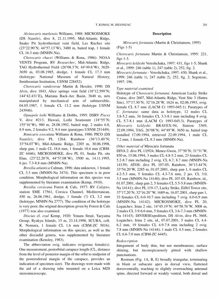
Rostrum (Fig. 1A, B, E) broadly triangular, terminatingin blunt or subacute apex in dorsal view, flatteneddorsoventrally, reaching to slightly overreaching antennalspine, directed forward or weakly ventral, both dorsal and
T. KOMAI, M. SEGONZAC 201
202 TAXONOMY OF MIROCARIS
Figure 1. Mirocaris fortunata (Martin & Christiansen, 1995). Holotype, ovigerous female (CL 8.7 mm; LACM 1993-045.1). A. cara-pace and cephalic appendages, lateral; B. carapace, dorsal (setae omitted); C. detail of surface structure of submedian region of carapace,dorsal; D. abdomen, lateral; E. anterior part of carapace and cephalic appendages, dorsal (setae partially omitted; right antenna removed).
Figure 1. Mirocaris fortunata (Martin & Christiansen, 1995). Holotype, femelle ovigère (CL 8.7 mm ; LACM 1993-045.1). A. vuelatérale de la carapace et des appendices céphaliques ; B. carapace, vue dorsale (sans les soies) ; C. détail de la structure superficielle dela région sub-médiane de la carapace, vue dorsale ; D. abdomen, vue de profil ; E. partie antérieure de la carapace et premiers appendicescéphaliques, vue dorsale (soies partiellement représentées; antenne droite non représentée).

ventral surfaces not dentate; dorsal surface weakly convex,without sharp carina.
Carapace (Figs 1A, B, 5A, B) somewhat compressedlaterally, with short transverse (vertical) rows of short setaeon lateral parts, and scattered short setae particularlyanteriorly (including rostrum) along midline; dorsal surfacerounded in males and non-ovigerous females, broadlycarinate in ovigerous females, general outline in lateral viewfaintly sinuous to weakly convex; in ovigerous females,submedian areas very shallowly depressed and ornamentedwith numerous longitudinal striae (Fig. 1B, C); orbitalmargin evenly rounded; antennal spine slightly directedmesially; pterygostomian angle not exceeding antennalspine; anterolateral margin between antennal spine andpterygostomian angle weakly concave; posteriorsubmarginal groove shallow, rather inconspicuous.
Thoracic sternite with pair of slender submedian spineson seventh somite (reduced in ovigerous females); medianspur on eighth thoracic somite (Fig. 4A) terminating inacute spine in males and non-spawning females, subacute orblunt spine in spawning females.
Abdomen (Fig. 1D) rounded dorsally in all somites.Pleura of anterior four somites all broadly rounded; on fifthsomite, acute or subacute posteroventral tooth. Sixth somite1.74-1.83 times longer than fifth somite, 1.40-1.43 timeslonger than proximal depth; posterolateral process short,terminating in small acute tooth; posteroventral cornerproduced, terminating in subacute point. First abdominalsternite with pair of rudimentary, slender submedian spines,similar spines better developed and more strongly curvedmesially on second and third sternites, again less developedspines on fourth sternite (those submedian spines greatlyreduced in spawning females); fifth sternite with distinctmedian keel terminating posteriorly in acute spine; sixthsternite flattened, thin, transparent, with small preanal spine.
Telson (Figs 1D, 2C) 1.25-1.36 times longer than sixthabdominal somite, slightly tapering posteriorly, widthbetween posterolateral corners 0.75-0.80 of greatest anteriorwidth; dorsal surface with very slight trace of medianlongitudinal concavity in posterior 0.75-0.80, bearing rowof 7-9 spines (excluding spines at posterolateral corner) oneither side along posterior 0.80 length; posterior margin(Fig. 2D) broadly convex, occasionally with shallowmedian emargination, bearing 12-19 spines in total; 1-3spines at posterolateral corner shorter than mesial spines,simple, while remaining mesial spines elongate, bearingminute marginal setules.
Eye-stalks (Fig. 1E) rather large but degenerated, broadlyfused mesially without trace of median separation (inholotype left eye abnormally smaller than right eye); corneaunfaceted, poorly organized retinal pigment discernibleinside, through cuticle; no distinct spine or tubercle onanterior surface of eye.
Antennular peduncles (Fig. 1A, E) stout, slightlyflattened dorsoventrally. Basal segment with distal widthnearly half of its length; dorsal surface fairly inflated indistal part , but remaining proximal part depressed below,continuous with deep groove separating basal segment andstylocerite; distal margin slightly oblique in dorsal view;distolateral tooth well developed, acute, overlapped bystylocerite, exceeding midlength of penultimate segment,distomesial tooth much shorter than distolateral tooth,usually blunt; stylocerite strong, tapering to slender pointreaching or overreaching level of midlength of penultimatepeduncular segment. Penultimate segment with scatteredshort setae on dorsal surface; distomesial tooth as large ascorresponding tooth on basal segment, terminating acutely.Ultimate segment slightly longer than wide. Flagella ratherstout, unequal, inserted side by side on oblique terminalmargin of distal segment; lateral flagellum shorter thanmesial, aesthetasc-bearing portion occupying 0.80-0.85 oftotal length of flagellum, article each with tufts ofaesthetascs on mesial face; mesial flagellum with annulimuch denser than those on lateral flagellum.
Antenna (Figs 1A, E, 2B) with basicerite stout, bearingblunt distolateral dorsal projection and acute distolateralventral tooth exceeding former projection. Carpocerite (fifthsegment of antennal peduncle) very stout, cylindrical,exceeding midlength of scaphocerite. Scaphocerite broadlyoval with greatest width across level of midlength; lateralmargin very slightly convex to sinuous, terminating in short,stout tooth separated by narrow incision and considerablyexceeded by rounded blade; mesial margin noticeablyconvex; dorsal surface with distinct median ridgeaccompanied by deep groove. Flagellum stouter thanantennular flagella, slightly longer than body, annuli dense.
Mandible (Fig. 2E, F) with incisor process broad,somewhat tapering distally, bearing 6-8 unequal, acute orsubacute teeth on mesial margin (distalmost tooth distinctlyseparated from remaining teeth); molar process slender,unarmed, extending as far as incisor process; basal article ofpalp with deep notch on mesial surface proximal tomidlength, distal article stout, shorter than basal article,bearing scattered plumose setae with variable length.
Maxillule (Fig. 2G) with coxal endite slightly taperingdistomesially, with dense setae on mesial margin; basialendite broad, mesial margin with 2 rows of small spines(spines more numerous and denser in internal row than inexternal row); external surface of basial endite withsubmarginal row of setae and few small spines adjacent tomesial margin; palp (Figs 2G, 4B) somewhat curved,slightly bilobed distally, bearing 2 setae; outer setae short,simple, arising subterminally from ventral surface slightlyproximal to base of somewhat produced outer lobule (inholotype, outer lobule broken off); inner lobule small,bearing a long plumose seta.
T. KOMAI, M. SEGONZAC 203

204 TAXONOMY OF MIROCARIS

Maxilla (Fig. 4C) with coxal endite composed of singlelobe separated from basial endite by deep incision andfollowing suture; basial endite consisting of 2 lobes,proximal lobe with roundly truncate mesial margin, distallobe subtriangular, with submarginal row of setae onexternal surface; palp slender, sinuously curved, slightlyexceeding distal lobe in length. Scaphognathite greatlyexpanded, anterior lobe subovate, with densely setosemargin bearing longest setae along distomesial sector,posterior lobe (broken off in holotype) elongatesubtriangular, fringed on mesial margin with very long setaebecoming further longer posteriorly.
First maxilliped (Fig. 2H) with coxal endite somewhatthickened, with short setae on external surface and longersetae on mesial face; basial endite moderately broad,strongly convex and densely setose on external surface,mesial margin convex to concave, densely fringed withsetae; palp (not visible in ventral view) slender, weaklycurved mesially, bearing short apical bristles; exopodgreatly expanded, 1.40-2.10 times as long as broad, broadlyrounded and fringed with double row of long plumose setae,lacking flagellum, concavity on external surface sometimesdeep; epipod large, foliaceus, weakly bilobed.
Second maxilliped (Fig. 2I) somewhat pediform, 6-segmented; coxa somewhat expanded mesially, withnumerous setae on mesial face; basis and ischiumcompletely fused, this fused segment longest and broadest,with row of setae on mesial and lateral margins; merusabout half length of basis-ischium fused segment, with longsetae on lateral face; carpus short, with long plumose setaeon distal surface, proximomesial margin weakly tosomewhat produced on external surface, partially coveringbasal part of propodus; propodus obliquely articulated todactylus, with row of setae on mesial margin; dactyluslonger than propodus, slightly curved, tapering to roundedapex, bearing numerous short setae on mesial to distalmargins; exopod absent; epipod subtriangular, with slenderrudiment of podobranch reaching midlength to distal marginof basis-ischium fused segment and occasionally bearing 1or 2 small papillae possibly representing rudimentaryfilaments.
Third maxilliped (Fig. 4D, E) 4-segmented (broken inholotype), slightly overreaching anterior margin of
scaphocerite. Coxa stout; lateral surface (Fig. 4E) withprominent slender process directed laterally; epipod (Fig.4F) with 3-5 curved bristles distally. Antepenultimatesegment (basis-ischium-merus fused segment) somewhatflattened dorsoventrally, strongly sinuously curved in dorsalview, setose, with slender spine at distolateral ventralcorner. Penultimate segment (= carpus) weakly curvedventrally, with dense setae on mesial face. Ultimate segmentslightly curved, gradually tapering distally and terminatingin small corneous spine, with scattered long setae on lateralsurface and obliquely transverse tracts of short stiff setae; 2-4 spinules adjacent to base of terminal spine.
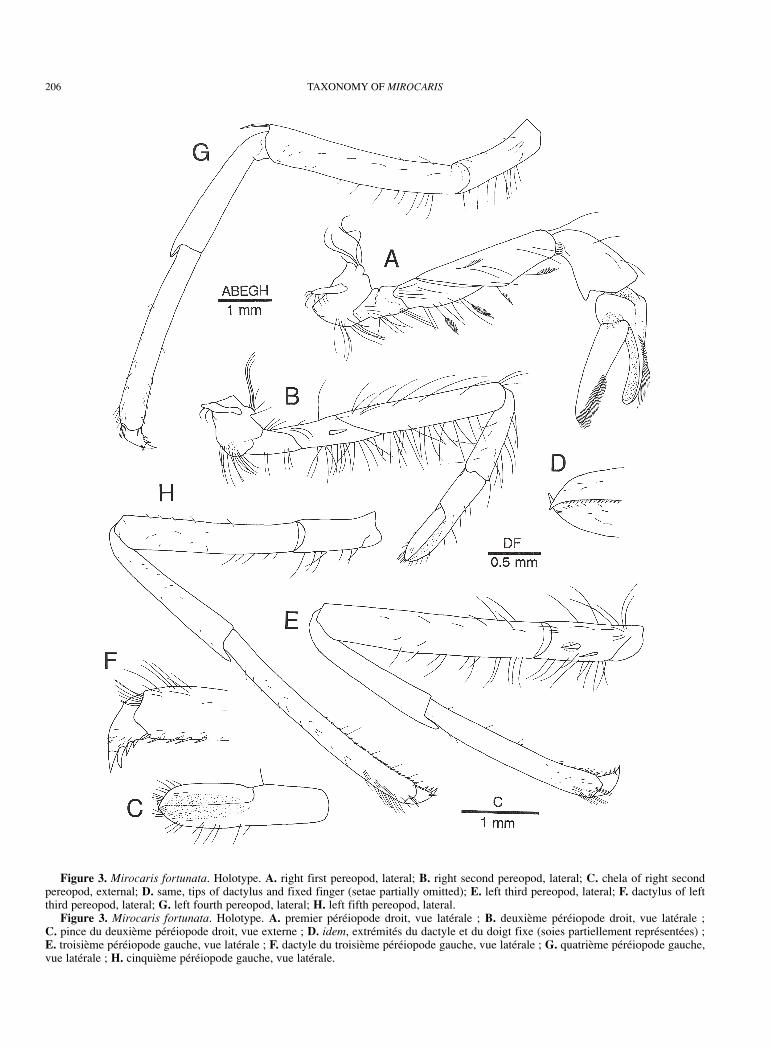
First pereopod (Figs 3A, 4G, H) short, stout, slightlyoverreaching (when extended) distal margin of scaphoceriteat most, with chela and carpus oriented toward midline.Articulation between ischium and merus strongly oblique.Ischium and merus with scattered plumose setae on lateraland ventral surfaces. Merus somewhat compressed laterallyand slightly tapering distally, ventral surface slightlyconcave for reception of flexed carpus. Carpus (Figs 3A, 4I)shorter than merus, somewhat inflated, irregularly funnel-shaped, dorsal surface bent at right angle near taperedproximal end articulating with merus; distolateral marginslightly produced medially; distomesial margin morestrongly produced, forming broadly triangular lobe; mesialface as in generic diagnosis. Palm short, strongly inflated,with patch of minute setae on mesial surface ventrally.Fingers curved and closing without hiatus; internal surfacesdeeply concave; external surface of each finger convex;cutting edges uniformly offset, each armed with row ofuniform, minute, erect, closely set tooth; cutting edge offixed finger bordered with narrow, thin corneous plateincluding tip; internal surface with submarginal row ofsparse short setae along cutting edge; external surface offixed finger with some submarginal rows of longer setae.Dactylus 1.20-2.80 times longer than palm, uniformlynarrowed distally, considerably flattened in distal 0.50-0.75;internal surface with submarginal row of short, sparse setaealong cutting edge; external surface with some submarginalrows of longer setae along cutting edge in distal half.
Second pereopod (Fig. 3B) slightly slender than otherpereopods, reaching distal margin of scaphocerite at most.Articulation between ischium and merus oblique. Ischium
T. KOMAI, M. SEGONZAC 205
Figure 2. Mirocaris fortunata. Holotype. A. anterior part of carapace and eye, right side (setae omitted); B. part of right antenna, dor-sal (setae omitted); C. telson and left uropod, dorsal (setae omitted); D. posterior margin of telson, dorsal; E. right mandible, internal; F.same, external; G. right maxillule, external (outer distal lobule of palp broken off); H. right first maxilliped, external; inset, palp, internal;I. right second maxilliped, external; upper inset, dactylus and propodus, mesial; lower inset, epipod and podobranch, internal.
Figure 2. Mirocaris fortunata. Holotype. A. partie antérieure droite de la carapace et œil (soies non représentées). B. une partie de l’an-tenne droite, vue dorsale (soies non représentées) ; C. telson et uropode gauche, vue dorsale (soies non représentées) ; D. bord postérieurdu telson, vue dorsale ; E. mandibule droite, face interne ; F. idem, face externe ; G. maxillule droite, face externe (lobe distal du palpeabsent) ; H. premier maxillipède droit, face externe ; en haut à droite, palpe, face interne; I. deuxième maxillipède droit, face externe ; enhaut à droite, dactyle et propodus , vue mésiale ; en bas à gauche, épipodite et podobranchie, vue interne.
206 TAXONOMY OF MIROCARIS
Figure 3. Mirocaris fortunata. Holotype. A. right first pereopod, lateral; B. right second pereopod, lateral; C. chela of right secondpereopod, external; D. same, tips of dactylus and fixed finger (setae partially omitted); E. left third pereopod, lateral; F. dactylus of leftthird pereopod, lateral; G. left fourth pereopod, lateral; H. left fifth pereopod, lateral.
Figure 3. Mirocaris fortunata. Holotype. A. premier péréiopode droit, vue latérale ; B. deuxième péréiopode droit, vue latérale ; C. pince du deuxième péréiopode droit, vue externe ; D. idem, extrémités du dactyle et du doigt fixe (soies partiellement représentées) ; E. troisième péréiopode gauche, vue latérale ; F. dactyle du troisième péréiopode gauche, vue latérale ; G. quatrième péréiopode gauche,vue latérale ; H. cinquième péréiopode gauche, vue latérale.

usually with one movable spine strongly pressed on lateralsurface. Merus 5.10-5.30 times as long as maximal height,with sparse long setae on dorsal surface and row of shortersetae, present also on ischium on ventral surface. Carpuswith sparse setae on dorsal and lateral surfaces. Chela (Fig.3C) 1.10-1.20 times longer than carpus, slightly broadeneddistally, 3.30-3.70 times longer than greatest width; fingerslonger than palm, each terminating in small corneous spine(Fig. 3D), crossing at tip; external surfaces slightlydepressed toward cutting edges, with scattered minute setaeand longer setae on distal part of fingers; cutting edges eachwith row of minute corneous spinules at least in distal half.
Third to fifth pereopods similar in structure, butincreasing in length from anterior pair to posterior pair.Third pereopod (Figs 3E, 5C) at most overreaching distalmargin of scaphocerite by length of dactylus and full lengthof propodus, somewhat compressed laterally; ischium with1 or 2 spines on lateral surface ventrally; merus 4.50-5.70times longer than greatest height, with sparse setae; carpus-propodus combined slightly shorter than merus-ischiumcombined; carpus 0.75-0.80 times as long as propodus;propodus (Fig. 4J) increasing slightly in depth toward distalend, with 2 rows of spinules on ventral surface (spinules ofmesial row fewer than those of lateral row); dactylus (Fig.3F) stout, 0.22-0.37 times as long as propodus, unguis ratherclearly demarcated, sometimes elongate, ventral marginwith 3-4 accessory spinules becoming larger distally.Fourth pereopod (Fig. 3G) at most overreaching distalmargin of scaphocerite by length of dactylus and half ofpropodus; ischium with 0-1 spine; carpus-propoduscombined subequal in length to merus-ischium combined.Fifth pereopod (Fig. 3H) at most overreaching distal marginof scaphocerite by length of dactylus and half of propodus;ischium unarmed; carpus-propodus combined longer thanmerus-ischium combined; ventral surface of propodus (Fig.4K) with double or triple row of setulose spinules on lateralside and single row of simple spinules on mesial side.
Branchial formula summarized in Table 1. Pleurobranchson fourth to eighth thoracic somites asymmetrically Y-branched, noticeably increasing in length posteriorly, apicesdirected forward. Arthrobranchs on third to seventh thoracicsomites moderately developed, nearly symmetrically U-branched, but last one on seventh somite distinctly smallerthan preceding ones. Epipods on first to fourth pereopodsstrap-like, similar to that on third maxilliped in shape.Setobranchs on first to fifth pereopods corresponding toepipods on third maxilliped to fourth pereopod respectively.
Endopod of first pleopod in males (Fig. 5E) with row ofsparse plumose setae on both margins, terminatingdistomesially in subtriangular lobe bearing 1 apical and 2-3subdistal bristles, all bristles essentially directed to midlineof body; in female, endopod (Fig. 5D) uniformly taperingwith margins fringed sparsely with plumose setae.
In males, second to fourth pleopods bearing greatlyreduced, rudimentary appendix interna (cf. Fig. 5F, G) andfifth pleopods bearing normally developed appendix internabearing terminal cluster of cincinnuli; in females, appendixinterna absent on second to fourth pleopods; fifth pleopodwith normally developed appendix interna. Appendixmasculina on second pleopod (Fig. 5F, G) arising fromproximal 0.30 of mesial margin of endopod, exceedingmidlength of endopod, bearing 8-10 long bristles distally.
Uropod (Fig. 2C) with both rami elongate oval,exceeding posterior margin of telson; endopod shorter andnarrower than exopod; exopod with straight lateral marginterminating in tiny acuminate tooth; long movable spinearising just mesial to distolateral tooth; suture distinct,sinuous.
VariationIn the ovigerous females, the submedian regions of thecarapace are very shallowly depressed, and the surface ofthe integument of this area is ornamented with irregularpattern of thin longitudinal striae; the midline of thecarapace forms a broad, rounded carina (Figs 1B, 5B). In themales and non-ovigerous females such a modification is notfound (Fig. 5A). The reason of this peculiar modificationremains unknown.
There seem to be two forms of ambulatory legs in thespecimens examined, but careful observation of abundantsamples has revealed the presence of intermediate formsbetween the two extremes as shown in Figs 3E and 5C.Moreover, we have been unable to associate the differencewith any other morphological characters, and the occurrenceof various forms of ambulatory legs, even in the samesamples, dissuaded us from considering this feature.
The holotype of Chorocaris fortunata is an aberrantspecimen. The carapace is somewhat deformed, and thus thesubmedian depressed areas on the carapace areasymmetrically formed, and the posterodorsal margin of thecarapace is also asymmetrical (Fig. 1B), as illustrated byMartin & Christiansen (1995). The eyes are dissimilar withthe left distinctly smaller than the right. This asymmetry ispresumably due to injury and regeneration of the left eye.
DistributionKnown from hydrothermal vent sites along the Mid-AtlanticRidge between 38°N and 14°N: Menez Gwen, 37°50’N-31°31’W, 850 m; Lucky Strike, 37°17’N-32°16’W, 1700 m(Martin & Christiansen, 1995; Shank et al., 1999; presentstudy); Rainbow, 36°13’N-33°54’W, 2289 m, (presentstudy); Broken Spur, 29°10’N- 43°10’W, 3000 m, (Martin& Christiansen, 1995; Shank et al., 1999); TAG, 26°08’N-44°49’W, 3650 m (Vereshchaka, 1997; Shank et al., 1999);Snake Pit, 23°22’N-44°57’W, 3480 m (unpublished data:the junior author observed once a juvenile of M. fortunata);Logatchev, 14°45’N-44°58’W, 3008 m (Shank et al., 1999;present study).
T. KOMAI, M. SEGONZAC 207

208 TAXONOMY OF MIROCARIS

Discussion
Specific status of Mirocaris keldyshiVereshchaka (1997) cited the following seven characters indistinguishing M. keldyshi from M. fortunata, although hedid not examine the type specimens of the latter taxon: (1)structure of two distal setae on palp of maxillule (setaesimple in M. fortunata, plumose in M. keldyshi); (2)proportion of exopod of first maxilliped (3 times as long asbroad in M. fortunata, 2 times in M. keldyshi); (3) on secondmaxilliped, relative length of podobranch (attributed to anexopod by Vereshchaka, see below) and epipod(podobranch 1.0 times as long as epipod in M. fortunata,1.5-2.0 times in M. keldyshi); (4) carpus and merus ofsecond maxilliped fused in M. fortunata, but separated in M. keldyshi; (5) patch of setae on ventral surface of palm offirst pereopod (absent in M. fortunata, present in M. keldyshi); (6) armature of ischium of second pereopod (amovable spine absent in M. fortunata, present in M. keldyshi); (7) number of setulose spines on posteriormargin of telson (10 spines in M. fortunata vs. 12-18 in M. keldyshi).
We have examined these differences critically, and foundthat none provides any taxonomic significance as discussedbelow. The morphological variation of the mouthparts waschecked using the seven specimens from Logatchevcollected during MICROSMOKE Cruise (see “Materialexamined”).
First character. As described above, the apical seta on theinner lobe of the maxillule palp is actually plumose in theholotype of M. fortunata, as well as in the holotype of M. keldyshi (see Vereshchaka, 1997, fig. 2B) and in otherspecimens we examined. However, the subterminal seta onthe outer lobe of the maxillule palp is simple in ourspecimens. Vereshchaka’s description of M. keldyshi is notconsistent with the illustration which is exact (Vereshchaka,1997, fig. 2B), because the outer seta illustrated as simple isconsidered as plumose in the text.
Second character. According to the illustration of the firstmaxilliped by Vereshchaka (1997, fig. 3A), the length andwidth of the exopod represent the distance between anterior
margin and base of the endopod, and the greatest width,respectively. However, according to the figure by Martin &Christiansen (1995, fig. 2g), the ratio should be 1.5 for M. fortunata, a value conformable to the holotype of M. keldyshi. Further examination of other specimens(including the paratypes of M. keldyshi) has shown that theproportional ratio of the exopod is quite variable, rangingfrom 1.40 to 2.10, and that the shape of the entire exopod iseasily affected by the preservation conditions.
Third character. According to the illustrations byVereshchaka (1997, fig. 3B) and Martin & Christiansen(1995, fig. 2i, j), the length of the podobranch (exopod,according to Vereshchaka) is similar in the holotypes of M. keldyshi and M. fortunata, although the epipod appearssmaller in the holotype of M. keldyshi than in the holotypeof M. fortunata. The difference in the ratio given byVereshchaka (1997) does not reflect the length of thepodobranch, but actually the size of the epipod. The epipodis soft and fragile, and thus easily affected by preservationin ethanol. In fact, we have found that the size of the epipodin the examined specimens varies individually and thus thischaracter does not provide any taxonomic significance.
Fourth character. Interpretation on the segmentation ofthe second maxilliped by Vereshchaka (1997) is confusing:the second maxilliped is described as five-segmented, in thefamilial description, but the illustration (Vereshchaka, 1997,fig. 3B) shows a second maxilliped composed of at least sixsegments. In all the specimens examined, we observed thatthe second maxilliped is six-segmented with carpus andmerus clearly separated. The pattern of segmentation is notconsistent with Vereshchaka’s figure where the coxa is notillustrated and the carpus appears subdivided, althoughVereshchaka confirmed it is not (personal communication).In the specimens we examined (Fig. 2I), the carpus is notsubdivided and the six-segmented condition of thisappendage is due to a complete fusion of the ischium andbasis.
Fifth character. It has been found that there is actually anoval patch of short setae on the ventromesial face of thepalm of the first pereopod in the type specimens of M. fortunata and other examined specimens. This patch of
T. KOMAI, M. SEGONZAC 209
Figure 4. Mirocaris fortunata. A-F, female from Lucky Strike (ATOS, PL 119-17) (CL 10.4 mm; MNHN-Na 14142); G-J, Holotype.A. eighth thoracic sternite, ventral; B. palp of left maxillule, external; C. left maxilla, external; D. left third maxilliped, lateral; E. coxaand antepenultimate segment of third maxilliped, dorsal (setae partially omitted); F. epipod of third maxilliped, ventral; G. chela of rightfirst pereopod, external; H. same, internal; I. carpus of right first pereopod, mesial; J. ventral surface of propodus of left third pereopod(setae partially omitted); K. ventral surface of propodus of left fifth pereopod (setae partially omitted).
Figure 4. Mirocaris fortunata. A-F, femelle de Lucky Strike (ATOS, PL 119-17) (CL 10.4 mm; MNHN-Na 14142); G-J, Holotype. A. huitième sternite thoracique, vue ventrale ; B. palpe de la maxillule gauche, vue externe ; C. maxille gauche, vue externe ; D. troisièmemaxillipède gauche, vue latérale ; E. coxa et antépénultième segment du troisième maxillipède, vue dorsale (soies partiellement représen-tées) ; F. épipodite du troisième maxillipède, vue ventrale ; G. pince du premier péréiopode droit, vue externe ; H. idem, vue interne ; I. carpe du premier péréiopode droit, vue mésiale ; J. surface ventrale du propodus du troisième péréiopode gauche (soies partiellementreprésentées) ; K. surface ventrale du propodus du cinquième péréiopode gauche (soies partiellement représentées).

210 TAXONOMY OF MIROCARIS
Figure 5. Mirocaris fortunata. A. female from Lucky Strike (as in Fig. 4) ; B. paratype, ovigerous female (CL 8.1 mm; LACM1993.045.3); C. paratype, ovigerous female (CL 5.7 mm; LACM 1993.045.3); E-G, male from Logatchev (DIVERSExpedition, PL 3668)(CL 5.4 mm; MNHN-Na 14144). A, B. carapace, dorsal (setae omitted in both); limits of submedian regions indicated in B, but striaeomitted ; C. left third pereopod, lateral; D. E. endopod of left first pleopod, ventral; F. endopod of left second pleopod, ventral; G. appendix masculina on left second pleopod, mesial.
Figure 5. Mirocaris fortunata. A,-D. femelle de Lucky Strike (comme Fig. 4) ; B. paratype, femelle ovigère (CL 8.1 mm ; LACM1993.045.3) ; C. paratype, femelle ovigère (CL 5.7 mm ; LACM 1993.045.3) ; E-G. mâle de Logatchev (DIVERSExpedition, PL 3668)(CL 5.4 mm; MNHN-Na). A, B. carapace, vue dorsale (soies non représentées) ; limites des zones sub-médianes indiquées en B, maisstries non représentées ; C. troisième péréiopode gauche, vue latérale ; D. E. vue ventrale des endopodites du premier pléopode droit, vueventrale ; F. endopodite du deuxième pléopode gauche, vue ventrale ; G. appendix masculina du deuxième pléopode gauche, vue mésiale.

setae probably represents a grooming apparatus togetherwith the setal assemblage on the carpus. This structure maybe easily overlooked without a careful observation.
Sixth character. Our examination has shown that theischium of the second pereopod is armed with a movablespine ventrolaterally in the type specimens of M. fortunatawhich was not mentioned or illustrated in the originaldescription by Martin & Christiansen (1995). This spinemay be easily overlooked, as it is usually pressed into ashallow cavity on the ischium.
Seventh character. Martin & Christiansen (1995)illustrated 10 spines on the posterior margin of telson of M. fortunata holotype, although the authors did not specifythe number of spines in the descriptive text. Ourexamination has shown that there are 13 spines in theholotype of M. fortunata, and 12-19 spines in the paratypes.Martin & Christiansen failed to illustrate the shorter spinesat the posterolateral corners of the telson (one on the left andtwo on the right). Thus there is no difference in thischaracter between M. fortunata and M. keldyshi.
Our morphological examination of the two taxa revealsthat the differences cited by Vereshchaka (1997) separatingM. fortunata and M. keldyshi do not provide any taxonomicsignificance. We could not find any other significantdifferences during our examination of the type and othermaterials. Our morphological analysis strongly indicatesthat M. fortunata and M. keldyshi are conspecific.Therefore, M. keldyshi is considered to be a junior synonymof M. fortunata. Our conclusion supports the results of thephylogenetic analysis using the mitochondrial COI gene(Shank et al., 1999).
Shank et al. (1999) used three of the seven characterscited by Vereshchaka (1997) for making preliminarydistinction between M. fortunata and M. keldyshi: numberof telson spines, presence or absence of movable spines onischium of second pereopod and presence or absence of an
oval patch on palm of first pereopod. As discussed above,however, there are no real differences in these characters inthe specimens examined by us. The specimens used byShank et al. (1999) have not been available for study. It isnecessary to reexamine those specimens in order to makeclear whether the differences are true.
Gebruk et al. (2000) also suggested that M. fortunata and M. keldyshi were distinguishable by morphology and colorin life, but they did not comment any further.
Homology of particular structures of mouthparts inalvinocaridid shrimpsThe homology of the following morphological structures ofshrimp species from vent and seep environments, assignedto the Bresiliidae or Alvinocarididae, is here clarified.
The presence of an exopod on the second maxilliped inthe species of Alvinocaris, Opaepele, Mirocaris andRimicaris, for example, was reported by several authors(Williams & Chace, 1982; Williams, 1988; Kikuchi & Ohta,1995; Williams & Dobbs, 1995; Vereshchaka, 1996, 1997;Kikuchi & Hashimoto, 2000). However, Segonzac et al.(1993) pointed out that this “exopod” is not a true exopod,but a rudimentary podobranch, since it arises in fact fromthe basal part of the epipod, not from the basis.
The coxal (or proximal) endite of the maxilla wasdescribed as divided into two lobes in different species byseveral authors (Williams & Chace, 1982; Williams &Rona, 1986; Williams, 1988; Martin & Hessler, 1990;Williams & Dobbs, 1995; Kikuchi & Ohta, 1995;Vereshchaka, 1996, 1997; Kikuchi & Hashimoto, 2000).However, our study demonstrated that the proximal lobeand the two distal lobes are primarily separated from eachother by a deep notch followed by a suture in Alvinocarismarkensis, Chorocaris chacei, C. vandoverae, Mirocarisfortunata and Rimicaris exoculata. Therefore, we considerthe proximal lobe as a one-lobed coxal endite, and the twodistal lobes as basial endite, a usual structure in carideanspecies (Komai, 1994).
The third maxilliped has been reported as bearing anexopod in the species of Alvinocaris, Chorocaris, Mirocarisand Rimicaris (Williams & Chace, 1982; Williams & Rona,1986; Williams, 1988; Martin & Hessler, 1990; Kikuchi &Ohta, 1995; Vereshchaka, 1996, 1997; Kikuchi &Hashimoto, 2000). Williams & Dobbs (1995) did notmention an exopod on the third maxilliped in Opaepeleloihi. Our examination of Alvinocaris markensis,Chorocaris chacei, C. vandoverae, Mirocaris fortunata,Rimicaris exoculata (and even Opaepele loihi)demonstrated that the short projection arising from thelateral surface of the coxa, interpreted as an exopod byprevious authors, is not a true exopod, but represents astructure probably originating from the epipod. Thisprojection may be homologous to the coxal lateral processreported in other caridean taxa (Komai, 1994). In taxa
T. KOMAI, M. SEGONZAC 211
Table 1. Mirocaris fortunata (Martin & Christiansen, 1995).Branchial formula; epipods and corresponding setobranchs, as wellas exopods, are also indicated (r: rudimentary).
Tableau 1. Mirocaris fortunata (Martin & Christiansen, 1995).Formule branchiale ; les épipodites et sétobranchies correspon-dantes, ainsi que les exopodites, sont aussi indiqués (r: réduite).
Thoracic somites 1 2 3 4 5 6 7 8Maxillipeds Pereopods
1 2 3 1 2 3 4 5
Pleurobranchs - - - + + + + +Arthrobranchs - - 1 1 1 1 1 -Podobranchs - r - - - - - -Epipods + + + + + + + -Setobranchs - - - + + + + +Exopods + - - - - - - -

associated to vent and seep environments, the projection isslender and laterally or ventrally directed (Fig. 4E), while inother carideans, the coxal lateral process, if present, is oftensemioval in shape and flattened dorsoventrally (Komai,1994).
Invalidity of the family Mirocarididae and status of thegenus MirocarisThe recognition of the Bresiliidae and Disciadidae asseparate families have been long accepted (e.g. Holthuis,1955; Forest, 1977). Following the discovery of the bizarrepolychelate shrimp Pseudocheles Chace & Brown, 1978,the family Disciadidae was synonymized with theBresiliidae by Chace & Brown (1978) rather thanestablishing a new monotypic family for the genusPseudocheles, characterized by the chelate third to fifthpereopods, a character of uncertain significance at familylevel. Subsequently, the Bresiliidae sensu Chace & Brown(1978) was accepted by many carcinologists (e.g. Williams& Chace, 1982; Williams & Rona, 1986; Williams, 1988;Burukovsky, 1988; Wicksten, 1989; Martin & Hessler,1990; Williams & Dobbs, 1995). On the other hand,according to a morphological phylogenetic analysis, theBresiliidae sensu lato were divided into three families,Bresiliidae s. s., Disciadidae and Alvinocarididae (Chris-toffersen, 1986, 1990). Subsequently, Vereshchaka (1997)established a new monotypic family Mirocarididae toaccommodate the genus Mirocaris. He distinguished fourfamilies on the basis of the development of pereopodalexopods and epipods: Alvinocarididae and Mirocarididaewere distinguished from Bresiliidae and Disciadidae by theabsence of pereopodal exopods; further the Mirocarididaewas separated from the Alvinocarididae [including thegenera Alvinocaris, Chorocaris, Iorania (= Rimicaris; seeShank et al., 1998), Opaepele and Rimicaris] by thepresence of pereopodal epipods and the absence ofappendices internae on the second to fourth pleopods in thefemales. Gebruk et al. (2000) cited opinion of sometaxonomic experts (A. B. Williams, J. W. Martin, F. A.Chace, Jr. and A. L. Vereshchaka) who agreed in placingvent or seep shrimps in the families Alvinocarididae andMirocarididae, separating them from non-vent or seepgenera which remain in the family Bresiliidae.
A phylogenetic analysis is necessary to establishapomorphies, and to identify homoplasy and reversals.However, a comprehensive treatment of the phylogeneticrelationships among the bresilioid taxa is beyond the scopeof this paper. We follow Christoffersen (1986, 1990),Segonzac et al. (1993) and Vereshchaka (1997) inrecognizing Bresiliidae, Disciadidae and Alvinocarididae asseparate families, because several distinctive characters ofAlvinocarididae sensu Vereshchaka (1997) have been foundduring our study.
The characters of Alvinocarididae include:1. Telson relatively broad, bearing numerous setae or
spines on broadly rounded posterior margin (vs telsonrelatively slender, bearing 2 or 3 pairs of spines on pointedposterior margin in Bresiliidae and Disciadidae);
2. Eyes greatly reduced, lacking faceted structure oncorneal surface in adults vs. eyes normally developed inadults in Bresiliidae and Disciadidae (except for the cavedwelling shrimp Agostocaris williamsi Hart & Manning,1986).
3. Basal segment of antennular peduncle with distolateralspine and rounded projection on dorsal surface proximal tobase of stylocerite (vs structures secondarily reduced inadults of Rimicaris exoculata), and with a deep grooveseparating stylocerite and main part of basal segment (vsnone of these structures present in Bresiliidae andDisciadidae);
4. Penultimate segment of antennular peduncle armedwith small but distinct distomesial spine (vs spine absent inBresiliidae and Disciadidae);
5. Exopod of first maxilliped greatly expanded, subovatein outline, and fringed with single or double row of longplumose setae (vs exopod narrow, fringed with single row ofsparse setae in Bresiliidae and Disciadidae);
6. Basis and ischium of second maxilliped completelyfused, with fine row of numerous setae on mesial margin ofcoxa and basis-ischium segment (vs second maxilliped 7-segmented in Bresiliidae, and 6-segmented with merus-ischium fused in the Disciadidae; no fringe of setae onmesial margin of basal segments in both families); exopodabsent (vs exopod present in Bresiliidae and Disciadidae);podobranch rudimentary, simple or sparsely papillate (vspodobranch absent in Bresiliidae and Disciadidae);
7. Coxa of third maxilliped with cluster of fine long setaeon mesial face and a prominent slender projection directedlaterally or ventrally on lateral face (vs no cluster of finelong setae on coxa of third maxilliped in Bresiliidae andDisciadidae, but normal coxal lateral projection on lateralface); distal two segments of third maxilliped arched (vs notarched in Bresiliidae and Disciadidae);
8. First pereopod with chela highly specialized, “bird-head with bent beak” shaped (flamingo-like), at least inyoung stages, ventral face of closed fingers forming deepexcavation (vs morphology variable, but quite different inBresiliidae and Disciadidae); carpus with shallow concavityfilled with cluster of fine stiff setae in ventral part and oneto three small movable spines arising at posterior border ofconcavity (vs no concavity or spines but sparse setae onmesial face of carpus in Bresiliidae and Disciadidae).
It is remarkable that these characters are all present inMirocaris. Particularly, the similarity in the structure of thefirst and second pereopods is striking, as it has beeneffectively used in diagnosing caridean families (see
212 TAXONOMY OF MIROCARIS

Holthuis, 1993). Vereshchaka (1997) considered thepossession of pereopodal epipods and the greatly reducedappendices internae on the second to fourth pleopods assignificant characters defining the family Mirocarididae.Indeed, the presence of pereopodal epipods represents aclear-cut difference separating Mirocaris from otheralvinocaridid, bresiliid and disciadid genera (cf. Chace,1992). However, in other caridean taxa, it is known that thedevelopment and number of pereopodal epipods arevariable even in the same genus, for example the hippolytidEualus, Heptacarpus and Lebbeus (cf. Butler, 1980), andthe pandalid Plesionika (cf. Chace, 1985). Therefore, thesignificance of this character at family level is questionable. On the other hand, as Vereshchaka (1997) himself noted, atendency toward reduction of appendices internae, althoughin lesser degree, is also found in Alvinocaris, Chorocaris,Opaepele and Rimicaris. Appendices internae on the secondto fourth pleopods are slender and simple, lackingcincinnuri in Alvinocaris, Chorocaris and Opaepele ; theyare slender and simple in second and third pleopods inRimicaris. In Mirocaris, the development of the appendicesinternae on the second to fourth pleopods is differentbetween male and female: in males the second to fourthpleopods bear rudimentary appendices internae, and infemales the pleopods are devoid of appendices internae.Thus a trend towards the reduction of appendices internaecan be recognized in a group including alvinocaridids andMirocaris. Considering the morphological similaritybetween Mirocaris and other alvinocaridid genera and theuncertain significance of the characters cited byVereshchaka (1997) at family level, we propose to assignMirocaris to the Alvinocarididae, and to synonymize themonotypic family Mirocarididae with the Alvinocarididae.
The validity of the genus Mirocaris has been confirmed.Besides the presence of pereopodal epipods andcorresponding setobranchs and the greatly reducedappendices internae on the second to fourth pleopods,Mirocaris differs from all other alvinocaridid genera inhaving, in ovigerous females, the peculiar longitudinaldepression on either side of the midline of the carapace,ornamented with microscopic longitudinal striae, andhaving a row of submarginal setae along cutting edge on theexternal surface of the dactylus and fixed finger of the firstchela. Mirocaris is distinguishable from Alvinocaris andOpaepele by the completely toothless rostrum and the bluntpterygostomian angle of the carapace. From Chorocaris,Opaepele and Rimicaris, it differs in the presence of only asingle row, rather than two to four rows, of accessoryspinules on the dactyli of the third to fifth pereopods.
As a result of the above comparisons, emended diagnosesthe family Alvinocarididae and the genus Mirocaris aregiven below.
Family Alvinocarididae Christoffersen, 1986
Bresiliidae - Williams & Chace, 1982: 145-146 (part);Williams & Rona, 1986: 460-461 (part); Martin & Hessler,1990: 2, 9 (part); Chace, 1992: 70; Holthuis, 1993: 69 (part).Alvinocarididae Christoffersen, 1986: 273, 277; Segonzacet al., 1993: 535; Vereshchaka, 1997: 428; (type genus:Alvinocaris Williams & Chace, 1982).Mirocarididae Vereshchaka, 1997: 426 (type genus:Mirocaris Vereshchaka, 1997).
Emended diagnosisCarapace unarmed on lateral surface. Telson not stronglynarrowed posteriorly, posterior margin usually rounded,with numerous spines or plumose setae. Eyes greatlyreduced, lacking faceted structure on corneal region inadults. Basal segment of antennular peduncle usually withdistolateral spine and rounded projection on dorsal surfaceproximal to base of stylocerite, and with deep grooveseparating stylocerite and main part of basal segment;penultimate segment of antennular peduncle with small butdistinct distomesial spine. Mandible distinctly divided inincisor and molar processes; incisor process broad, dentatemesially; molar process slender, nearly conical; palp 2-articulated. Maxilla with coxal endite composed of singlelobe. First maxilliped with greatly expanded, subovateexopod. Second maxilliped 6-segmented, somewhatpediform; basis and ischium completely fused; fine row ofnumerous setae on mesial margin of coxa and basis-ischiumfused segment; exopod absent; podobranch consisting ofrudimentary, simple or sparsely papillate bud. Thirdmaxilliped 4-segmented; coxa bearing cluster of fine longsetae on mesial face and prominent slender projectiondirected laterally or ventrally on lateral face; distal twosegments arched; ultimate segment trigonal in cross section;exopod absent. No exopods on pereopods. Chela of firstpereopod specialized, its outline usually shaped like “bird-head with bent beak” (flamingo-like) at least in youngstages, and ventral face of closed fingers forming deepexcavation; cutting edges of fingers microscopicallypectinate; carpus bearing shallow concavity which is filledby a cluster of fine stiff setae in the ventral part andprovided with 1-3 small movable spines arising at posteriorborder of concavity. Second pereopod chelate, slender thanfirst pereopod. Arthrobranch on third to seventh thoracicsomites. Appendices internae showing tendency towardreduction, at least those on second and third pleopodsslender, lacking distal cluster of cincinnuri.
CompositionAlvinocaris Williams & Chace, 1982; Rimicaris Williams &Rona, 1986; Chorocaris Martin & Hessler, 1990; OpaepeleWilliams & Dobbs, 1995; Mirocaris Vereshchaka, 1997.
T. KOMAI, M. SEGONZAC 213

Genus Mirocaris Vereshchaka, 1997
Mirocaris Vereshchaka, 1997: 429 [type species: Mirocariskeldyshi Vereshchaka, 1997 (= Mirocaris fortunata (Martin& Christiansen, 1995)].
Emended diagnosisRostrum flattened dorsoventrally, triangular in dorsal view,not distinctly carinate or dentate dorsally, not reaching distalmargin of basal segment of antennular peduncle; ventralsurface without tooth. Carapace somewhat compressedlaterally, with shallow hepatic groove; no distinct mediancarina, but in ovigerous females, shallow longitudinaldepression ornamented with minute longitudinal striae oneither side of midline; antennal spine acuminate;pterygostomian angle weakly produced anteriorly, rounded.Telson with dorsolateral spines forming sinuous row. Eyesrather large but degenerate, broadly fused mesially.Antennal scaphocerite broadly oval, with distinctdorsolateral tooth. Third maxilliped to fourth pereopodswith hooked epipods and first to fifth pereopods withcorresponding setobranchs. Dactyli of third to fifthpereopods compressed laterally, each with single row ofaccessory spinules on ventral margin. Second to fourthpleopods lacking appendices internae in females; in malesrudimentary appendix interna present on second pleopod,but no appendices internae on third and fourth pleopods;appendix interna of fifth pleopod normally developed,bearing distal cluster of cincinnuri.
CompositionMirocaris fortunata (Martin & Christiansen, 1995).
Acknowledgements
We sincerely thank G. Davis and J. W. Martin, LACM,M. de Saint Laurent and R. Cleva, Laboratoire de Zoologie(Arthropodes), MNHN, Paris, for making available the typematerials of M. fortunata and M. keldyshi for study. Specialthanks are also due to the chief scientists of various cruiseswho collected the material examined in this study: D. Desbruyères, IFREMER, Brest (Cruise DIVA 2), Pierre-Marie Sarradin, IFREMER (Cruise ATOS, program of theEU-funded VENTOX project EVK3CT 1999-00003), D. Prieur Roscoff (Cruise MICROSMOKE), C. Van Dover,College of William and Mary, Williamsburg, USA(DIVERSExpedition). We also thank A. Crosnier, BIMM,MNHN, for cordial reception in his laboratory and his warmhospitality, and for comments on the manuscript. Thanks arealso extended to three reviewers and the executive editor fortheir valuable comments for improvements.
References
Burukovsky R.N. 1988. New representatives of the familyBresiliidae (Crustacea, Decapoda) from the north-east Atlantic.
Zoologischeskii Zhurnal, 67: 456-460.Butler T.H. 1980. Shrimps of the Pacific coast of Canada.
Canadian Bulletin of Fisheries and Aquatic Sciences, 202: 1-280, pls 1-8.
Calman W.T. 1896. On deep-sea Crustacea from the south west ofIreland. Transactions of the Royal Irish Academy, 31: 1-20.
Chace F.A. Jr. 1985. The caridean shrimps (Crustacea: Decapoda)of the Albatross Philippine Expedition, 1907-1910, Part 3:Families Thalassocarididae and Pandalidae. SmithsonianContributions to Zoology, 411: 1-143.
Chace F.A. Jr. 1992. On the classification of the Caridea(Decapoda). Crustaceana, 63: 70-80.
Chace F.A. Jr. & Brown D.E. 1978. A new polychelate shrimpfrom the Great Barrier Reef of Australia and its bearing on thefamily Bresiliidae (Crustacea, Decapoda, Caridea). Procee-dings of the Biological Society of Washington, 91: 756-766.
Christoffersen M.L. 1986. Phylogenetic relationships betweenOplophoridae, Atyidae, Pasiphaeidae, Alvinocarididae fam. n.,Bresiliidae, Psalidopodidae and Disciadidae (Crustacea,Caridea, Atyoidea). Boletim de Zoologia, Universidade de SaoPaulo, 10: 273-281.
Christoffersen M.L. 1990. A new superfamily classification of theCaridea (Crustacea: Pleocyemata) based on phylogeneticpattern. Zeitschrift für Zoologische Systematik undEvolutionsforschung, 28: 94-106.
Forest J. 1977. Un groupement injustifié: la superfamille desBresilioidea. Remarques critiques sur le statut des famillesréunies sous le nom (Crustacea Decapoda Caridea). Bulletin duMuséum national d’Histoire naturelle, 3e série, Zoologie, 332:869-888.
Forest J. & Cals P. 1977. Une deuxième espèce du genre BresiliaCalman, B. corsicana sp. nov. comparison avec B. atlanticaCalman (Crustacea Decapoda Bresiliidae). Bulletin du Muséumnational d’Histoire naturelle, 3e série, Zoologie, 316: 549-656.
Gebruk A.V., Southward E.C., Kennedy H. & Southward A.J.2000. Food sources, behavior, and distribution of hydrothermalvent shrimps at the Mid-Atlantic Ridge. Journal of the MarineBiological Association of the United Kingdom, 80: 485-499.
Hart C.W. & Manning R.B. 1986. Two new shrimps(Procarididae and Agostocarididae, new family) from marinecaves of the western North Atlantic. Journal of CrustaceanBiology, 6: 408-416.
Holthuis L.B. 1955. The recent genera of the caridean andstenopodidean shrimps (Class Crustacea, Order Decapoda,Supersection Natantia) with keys for their determination.Zoologische Verhandelingen, 26: 1-157.
Holthuis L.B. 1993. The recent genera of the caridean andstenopodidean shrimps (Crustacea, Decapoda) with anappendix on the order Amphionidacea. NationaalNatuurhistorisch Museum: Leiden. 328 pp.
Kemp S. 1910. The Decapoda Natantia of the coasts of Ireland.Fisheries Ireland Scientific Investigations, 1908 [1910]: 1-190,pls 1-23.
Kemp S. 1920. Notes on Crustacea Decapoda in the IndianMuseum, XIV: On the occurrence of the caridean genus Disciasin Indian waters. Records of the Indian Museum, 19: 137-143.
Kensley B. 1983. New records of bresiliid shrimp from Australia,South Africa, Caribbean, and Gulf of Mexico (Decapoda:
214 TAXONOMY OF MIROCARIS

Natantia: Caridea). Smithsonian Contributions to Zoology, 394:1-31.
Kikuchi T. & Hashimoto J. 2000. Two new caridean shrimps ofthe family Alvinocarididae (Crustacea, Decapoda) from ahydrothermal field at the Minami-Ensei Knoll in the Mid-Okinawa Trough, Japan. Species Diversity, 5: 135-148.
Kikuchi T. & Ohta S. 1995. Two caridean shrimps of the familiesBresiliidae and Hippolytidae from a hydrothermal field on theIheya Ridge, off the Ryukyu Islands, Japan. Journal ofCrustacean Biology, 15: 771-785.
Komai T. 1994. Phylogeny and cladistic classification of thesuperfamily Pandaloidea (Crustacea: Decapoda: Caridea).Doctoral thesis. Hokkaido University: Hakodate. 289 pp.
Martin J.W. & Christiansen J.C. 1995. A new species of theshrimp genus Chorocaris Martin & Hessler, 1990 (Crustacea:Decapoda: Bresiliidae) from hydrothermal vent fields alongMid-Atlantic Ridge. Proceedings of the Biological Society ofWashington, 108: 220-227.
Martin J. W. & Hessler R. R. 1990. Chorocaris vandoverae, anew genus and species of hydrothermal vent shrimp (Crustacea:Decapoda: Bresiliidae) from the western Pacific., 417: 1-11.
Martin J.W., Signorovitch J. & Patel H. 1997. A new species ofRimicaris (Crustacea: Decapoda: Bresiliidae) from the SnakePit Hydrothermal vent field on the Mid-Atlantic Ridge.Proceedings of the Biological Society of Washington, 110: 399-411.
Rathbun M.J. 1902. Papers from the Hopkins-StanfordGalapagos Expedition 1898-1899. VIII. Brachyura andMacrura. Proceedings of the Washington Academy of Sciences,4: 274-292.
Segonzac M. 1997. Mirocaris fortunata (Martin & Christiansen,1995). In: Handbook of Deep-sea Hydrothermal Vent Fauna(D. Desbruyères & M. Segonzac eds), p. 196. ÉditionsIFREMER, Brest.
Segonzac M., de Saint Laurent M. & Casanova B. 1993.L’énigme du comportement trophique des crevettes
Alvinocarididae des sites hydrothermaux de la dorsale médio-atlantique. Cahiers de Biologie Marine, 34: 535-571.
Shank T.M., Lutz R.A. & Vrijenhoek R.C. 1998. Molecularsystematics of shrimp (Decapoda; Bresiliidae) from deep-seahydrothermal vents: I. Enigmatic “small orange” shrimp fromthe Mid-Atlantic Ridge are juvenile Rimicaris exoculata.Molecular Marine Biology and Biotechnology, 7: 88-96.
Shank T.M., Black, M.B., Halanych K.M., Lutz R.A. &Vrijenhoek R.C. 1999. Miocene radiation of deep-seahydrothermal vent shrimp (Caridea: Bresiliidae): Evidencefrom mitochondrial cytochrome oxydase subunit I. MolecularPhylogenetics and Evolution, 13: 244-254.
Vereshchaka A.L. 1996. A new genus and species of carideanshrimp (Crustacea: Decapoda: Alvinocarididae) from northAtlantic hydrothermal vents. Journal of the Marine BiologicalAssociation of the United Kingdom,76: 951-961.
Vereshchaka A.L. 1997. A new family for a deep-sea carideanshrimp from North Atlantic hydrothermal vents. Journal of theMarine Biological Association of the United Kingdom, 77: 425-438.
Wicksten M.K. 1989. Encantada spinoculata, a new genus andspecies of shrimp from the Galapagos Islands (Caridea:Bresiliidae). Journal of Crustacean Biology, 9: 136-147.
Williams A. B. 1988. New marine decapod crustaceans fromwaters influenced by hydrothermal discharge, brine, andhydrocarbon seepage. Fishery Bulletin, 86: 263-287.
Williams A. B. & Chace F.A. Jr. 1982. A new caridean shrimp ofthe family Bresiliidae from thermal vents of the Galapagos Rift.Journal of Crustacean Biology, 2: 136-147.
Williams A.B. & Dobbs F.C. 1995. A new genus and species ofcaridean shrimp (Crustacea: Decapoda: Bresiliidae) fromhydrothermal vents on Loihi Seamount, Hawaii. Proceedings ofthe Biological Society of Washington, 108: 228-237.
Williams A.B. & Rona P. 1986. Two new caridean shrimps(Bresiliidae) from a hydrothermal vent on the Mid-AtlanticRidge. Journal of Crustacean Biology, 6: 446-462.
T. KOMAI, M. SEGONZAC 215