review article drug delivery innovations for enhancing the
TRANSCRIPT
Review ArticleDrug Delivery Innovations for Enhancing the AnticancerPotential of Vitamin E Isoforms and Their Derivatives
Christiana M. Neophytou and Andreas I. Constantinou
Department of Biological Sciences, Faculty of Pure and Applied Sciences, University of Cyprus, 1678 Nicosia, Cyprus
Correspondence should be addressed to Andreas I. Constantinou; [email protected]
Received 20 February 2015; Revised 12 April 2015; Accepted 15 April 2015
Academic Editor: Rohit S. Mulik
Copyright © 2015 C. M. Neophytou and A. I. Constantinou.This is an open access article distributed under the Creative CommonsAttribution License, which permits unrestricted use, distribution, and reproduction in any medium, provided the original work isproperly cited.
Vitamin E isoforms have been extensively studied for their anticancer properties. Novel drug delivery systems (DDS) thatinclude liposomes, nanoparticles, and micelles are actively being developed to improve Vitamin E delivery. Furthermore, severaldrug delivery systems that incorporate Vitamin E isoforms have been synthesized in order to increase the bioavailability ofchemotherapeutic agents or to provide a synergistic effect. D-alpha-tocopheryl polyethylene glycol succinate (Vitamin E TPGS orTPGS) is a synthetic derivative of natural alpha-tocopherol which is gaining increasing interest in the development of drug deliverysystems and has also shown promising anticancer effect as a single agent. This review provides a summary of the properties andanticancer effects of the most potent Vitamin E isoforms and an overview of the various formulations developed to improve theirefficacy, with an emphasis on the use of TPGS in drug delivery approaches.
1. Introduction
Vitamin E exists in nature in 8 natural isoforms that haveexhibited diverse therapeutic properties in numerous studiesover the last decades. In vitro, in vivo, and clinical studies ofthe Vitamin E isoforms have highlighted their antioxidant,anti-inflammatory, neuroprotective, and antithrombotic abil-ities among others [1–3]. Importantly, Vitamin E natural iso-forms have been shown to act in a preventive and therapeuticmanner against several types of cancer and are still beingwidely investigated for their potential efficacy in this disease[4].
Even though in vitro studies have highlighted the greatanticancer potential of Vitamin E isoforms, their anticancerefficacy in animal models is hindered due to low compoundbioavailability and solubility. The in vivo absorption of Vita-min E isoforms is limited due to their lipophilic nature andinsolubility in aqueous solvents [5, 6]. Their formulations inethanol, DMSO, or vegetable oil emulsions limit their appli-cations in the clinical setting. Furthermore, when Vitamin Eisoforms are present in high concentration in the intestine,their absorption is reduced because their transport becomessaturated [7, 8]. As described in detail below, Vitamin E
isoforms also display nonspecific distribution in tissues andwhen administered at high concentrations they are rapidlymetabolized and excreted in urine.
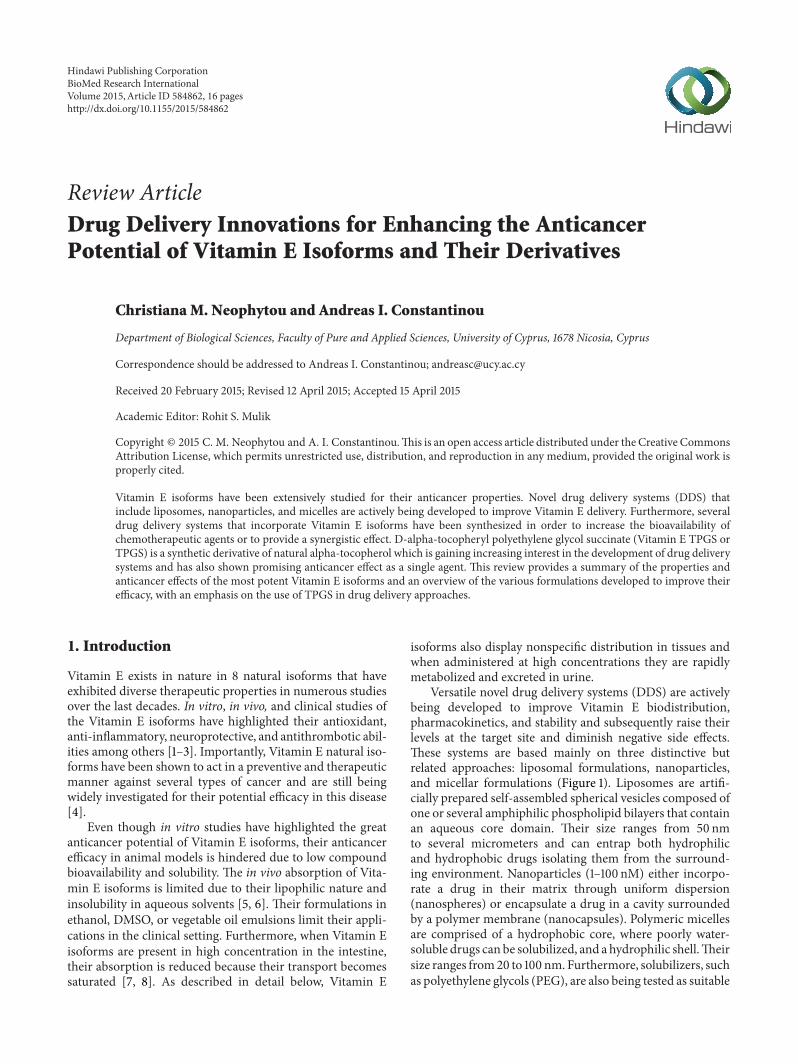
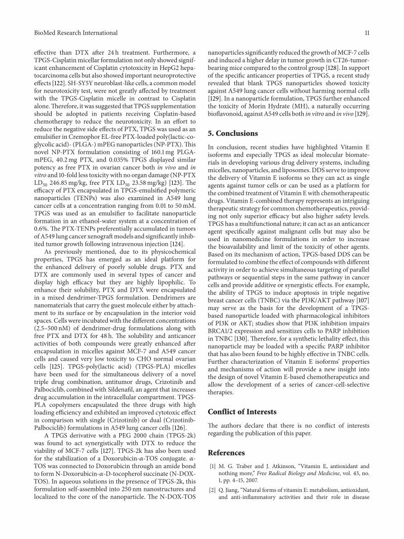
Versatile novel drug delivery systems (DDS) are activelybeing developed to improve Vitamin E biodistribution,pharmacokinetics, and stability and subsequently raise theirlevels at the target site and diminish negative side effects.These systems are based mainly on three distinctive butrelated approaches: liposomal formulations, nanoparticles,and micellar formulations (Figure 1). Liposomes are artifi-cially prepared self-assembled spherical vesicles composed ofone or several amphiphilic phospholipid bilayers that containan aqueous core domain. Their size ranges from 50 nmto several micrometers and can entrap both hydrophilicand hydrophobic drugs isolating them from the surround-ing environment. Nanoparticles (1–100 nM) either incorpo-rate a drug in their matrix through uniform dispersion(nanospheres) or encapsulate a drug in a cavity surroundedby a polymer membrane (nanocapsules). Polymeric micellesare comprised of a hydrophobic core, where poorly water-soluble drugs can be solubilized, and a hydrophilic shell.Theirsize ranges from20 to 100 nm. Furthermore, solubilizers, suchas polyethylene glycols (PEG), are also being tested as suitable
Hindawi Publishing CorporationBioMed Research InternationalVolume 2015, Article ID 584862, 16 pageshttp://dx.doi.org/10.1155/2015/584862
2 BioMed Research International
Drug
Liposome Micelle Nanocapsule Nanosphere
Figure 1: Schematic structure of liposomes, micelles, nanocapsules,and nanospheres. Liposomes are composed of one or more lipidbilayer structures surrounding an aqueous core where the drug isencapsulated. Micelles contain a hydrophilic shell and a hydropho-bic core for carrying lipophilic drugs. Nanocapsules encapsulate adrug in an inner space surrounded by a polymer membrane whilenanospheres are solid polymers that incorporate a drug in theirmatrix through uniform dispersion. Modified from [131].
formulations for human application. Besides increasing drugbioavailability, novel drug delivery systems aim to provide asynergistic effect by formulating natural Vitamin E isoformsor synthetic derivatives with other chemotherapeutic agents.This review compares and contrasts the properties andanticancer effects of the most potent Vitamin E isoforms andderivatives with emphasis on the most promising deliveryapproaches being developed to improve their efficacy.
2. Vitamin E
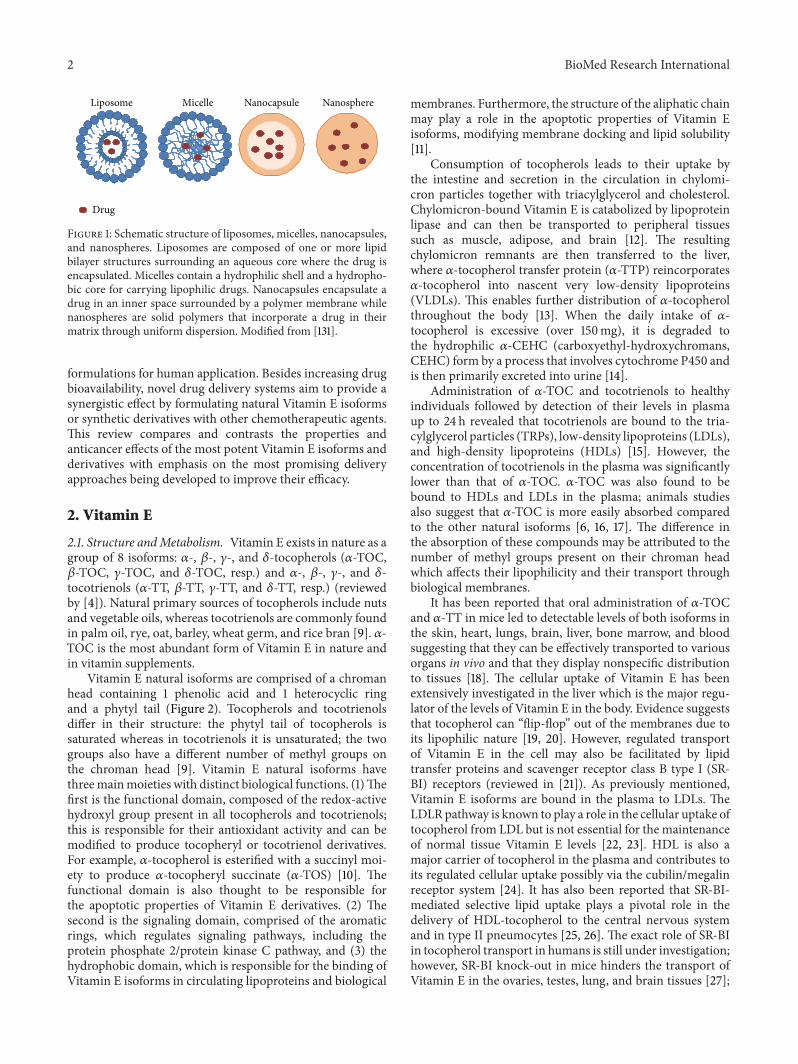
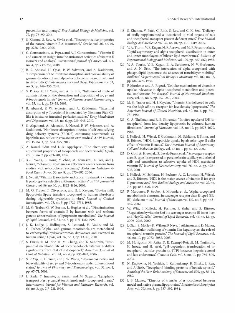
2.1. Structure andMetabolism. Vitamin E exists in nature as agroup of 8 isoforms: 𝛼-, 𝛽-, 𝛾-, and 𝛿-tocopherols (𝛼-TOC,𝛽-TOC, 𝛾-TOC, and 𝛿-TOC, resp.) and 𝛼-, 𝛽-, 𝛾-, and 𝛿-tocotrienols (𝛼-TT, 𝛽-TT, 𝛾-TT, and 𝛿-TT, resp.) (reviewedby [4]). Natural primary sources of tocopherols include nutsand vegetable oils, whereas tocotrienols are commonly foundin palm oil, rye, oat, barley, wheat germ, and rice bran [9]. 𝛼-TOC is the most abundant form of Vitamin E in nature andin vitamin supplements.
Vitamin E natural isoforms are comprised of a chromanhead containing 1 phenolic acid and 1 heterocyclic ringand a phytyl tail (Figure 2). Tocopherols and tocotrienolsdiffer in their structure: the phytyl tail of tocopherols issaturated whereas in tocotrienols it is unsaturated; the twogroups also have a different number of methyl groups onthe chroman head [9]. Vitamin E natural isoforms havethreemainmoieties with distinct biological functions. (1)Thefirst is the functional domain, composed of the redox-activehydroxyl group present in all tocopherols and tocotrienols;this is responsible for their antioxidant activity and can bemodified to produce tocopheryl or tocotrienol derivatives.For example, 𝛼-tocopherol is esterified with a succinyl moi-ety to produce 𝛼-tocopheryl succinate (𝛼-TOS) [10]. Thefunctional domain is also thought to be responsible forthe apoptotic properties of Vitamin E derivatives. (2) Thesecond is the signaling domain, comprised of the aromaticrings, which regulates signaling pathways, including theprotein phosphate 2/protein kinase C pathway, and (3) thehydrophobic domain, which is responsible for the binding ofVitamin E isoforms in circulating lipoproteins and biological
membranes. Furthermore, the structure of the aliphatic chainmay play a role in the apoptotic properties of Vitamin Eisoforms, modifying membrane docking and lipid solubility[11].
Consumption of tocopherols leads to their uptake bythe intestine and secretion in the circulation in chylomi-cron particles together with triacylglycerol and cholesterol.Chylomicron-bound Vitamin E is catabolized by lipoproteinlipase and can then be transported to peripheral tissuessuch as muscle, adipose, and brain [12]. The resultingchylomicron remnants are then transferred to the liver,where 𝛼-tocopherol transfer protein (𝛼-TTP) reincorporates𝛼-tocopherol into nascent very low-density lipoproteins(VLDLs). This enables further distribution of 𝛼-tocopherolthroughout the body [13]. When the daily intake of 𝛼-tocopherol is excessive (over 150mg), it is degraded tothe hydrophilic 𝛼-CEHC (carboxyethyl-hydroxychromans,CEHC) form by a process that involves cytochrome P450 andis then primarily excreted into urine [14].
Administration of 𝛼-TOC and tocotrienols to healthyindividuals followed by detection of their levels in plasmaup to 24 h revealed that tocotrienols are bound to the tria-cylglycerol particles (TRPs), low-density lipoproteins (LDLs),and high-density lipoproteins (HDLs) [15]. However, theconcentration of tocotrienols in the plasma was significantlylower than that of 𝛼-TOC. 𝛼-TOC was also found to bebound to HDLs and LDLs in the plasma; animals studiesalso suggest that 𝛼-TOC is more easily absorbed comparedto the other natural isoforms [6, 16, 17]. The difference inthe absorption of these compounds may be attributed to thenumber of methyl groups present on their chroman headwhich affects their lipophilicity and their transport throughbiological membranes.
It has been reported that oral administration of 𝛼-TOCand 𝛼-ΤΤ in mice led to detectable levels of both isoforms inthe skin, heart, lungs, brain, liver, bone marrow, and bloodsuggesting that they can be effectively transported to variousorgans in vivo and that they display nonspecific distributionto tissues [18]. The cellular uptake of Vitamin E has beenextensively investigated in the liver which is the major regu-lator of the levels of Vitamin E in the body. Evidence suggeststhat tocopherol can “flip-flop” out of the membranes due toits lipophilic nature [19, 20]. However, regulated transportof Vitamin E in the cell may also be facilitated by lipidtransfer proteins and scavenger receptor class B type I (SR-BI) receptors (reviewed in [21]). As previously mentioned,Vitamin E isoforms are bound in the plasma to LDLs. TheLDLRpathway is known to play a role in the cellular uptake oftocopherol from LDL but is not essential for the maintenanceof normal tissue Vitamin E levels [22, 23]. HDL is also amajor carrier of tocopherol in the plasma and contributes toits regulated cellular uptake possibly via the cubilin/megalinreceptor system [24]. It has also been reported that SR-BI-mediated selective lipid uptake plays a pivotal role in thedelivery of HDL-tocopherol to the central nervous systemand in type II pneumocytes [25, 26]. The exact role of SR-BIin tocopherol transport in humans is still under investigation;however, SR-BI knock-out in mice hinders the transport ofVitamin E in the ovaries, testes, lung, and brain tissues [27];
BioMed Research International 3
O
HO
O
HO
Tocopherols
TocotrienolsR1
R1
R2
R2
CH3
CH3
CH3
CH3
CH3 CH3 CH3
CH3
CH3 CH3 CH3
CH3
𝛼: R1 = CH3, R2 = CH3𝛽: R1 = CH3, R2 = H𝛾: R1 = H, R2 = CH3𝛿: R1 = H, R2 = H
Figure 2: Structure of the natural isoforms of Vitamin E.
interestingly, the expression of SR-BI is affected by Vitamin Estatus in rat liver and HepG2 cells [28].
The intracellular transport of Vitamin E has also beeninvestigated in the liver. Following internalization in hepa-tocytes, Vitamin E reaches the lysosomal vesicles throughthe endocytic pathway. TTP facilitates intracellular transportof Vitamin E between membrane vesicles in vitro [29]. Ithas been suggested that TPP, which contains a putativelysosomal targeting motif, transiently associates with themembrane vesicle by utilizing accessory proteins to anchorto the membrane and obtains Vitamin E from the endocyticvesicles [30–33]. Functional lysosomal proteins NPC1 andNPC2 are also required for the release of tocopherol fromthe endosomes/lysosomes [34, 35]. At the subcellular level,Vitamin E localizes at the golgi apparatus, endoplasmicreticulum, mitochondria, and lysosomes [36].
2.2. Biological Properties and Anticancer Activity. VitaminE natural isoforms display great structural homology andhave similar functions such as antioxidant activity. However,studies suggest that Vitamin E isoforms also have distinctbiological activities that are not common between them. Forexample, 𝛼-TOC (1) can inhibit the function of PKC, 5-lipo-oxygenase, and phospholipase in a posttranslational leveland can activate phosphatase 2A and (2) can inhibit cellularproliferation, platelet aggregation, and the monocyte attach-ment [37]. The above functions of Vitamin E suggest that,in addition to their antioxidant properties, these isoformscan interact with enzymes, structural proteins, lipids, andtranscription factors. 𝛾-ΤOC has also been found to possessanti-inflammatory and antineoplastic activities [38].
Vitamin E natural isoforms and synthetic derivatives(VitE-ISDs) have been intensively investigated for their anti-cancer properties against several types of cancer includingbreast, prostate, lung, colon, gastric, and ovarian cancers andhave been found to affect survival and proliferation pathwaysboth in vitro and in vivo (reviewed by [4]).The initial evidenceregarding the anticancer potential of Vitamin E was derivedfrom studies showing that people with a diet rich in VitaminE natural isoforms have a lower risk of colon cancer [39, 40].Further support for the potential use of Vitamin E in cancerchemoprevention or therapy came from studies showing thatdietary treatment with Vitamin E led to a reduction in theincidence of prostate cancer or a delay in its progression
[41–43]. Based on the literature, 𝛾-ΤOC and 𝛿-ΤOC aremore potent inducers of apoptosis compared to 𝛽- and 𝛼-tocopherols. Even though 𝛼-TOC is a potent antioxidant,accumulating evidence in the literature suggests that 𝛼-TOCcannot induce apoptosis [44]. Furthermore, tocotrienols aremore effective proapoptotic agents than tocopherols; 𝛾-ΤΤand 𝛿-ΤΤ are more potent than 𝛼- and 𝛽-forms.
Themajor apoptotic pathways affected by themost potentnatural and synthetic Vitamin E isoforms have been exten-sively described in other reviews [2, 4, 45]. 𝛾-ΤΤ is a well-studied natural VitaminE isoformknown to induce apoptosisvia the extrinsic and intrinsic pathway, by inhibiting the NF-𝜅B transcription factor and the AKT pathway thereby lower-ing the levels of antiapoptotic proteins such as c-FLIP, Bcl-2,Bcl-xL, and IAPs and allowing the activation of caspases 9 and3 [46, 47]. 𝛾-ΤΤ also acts via the intrinsic pathway by inducingthe translocation of Bax to the mitochondria and MOMP[48]. Vitamin E compounds have been found to induce bothcaspase-dependent and -independent pathways of apoptosis;however, the precise mechanism of CI-PCD induced by thesecompounds is still under investigation.
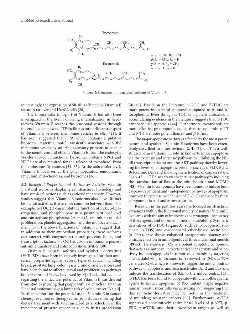
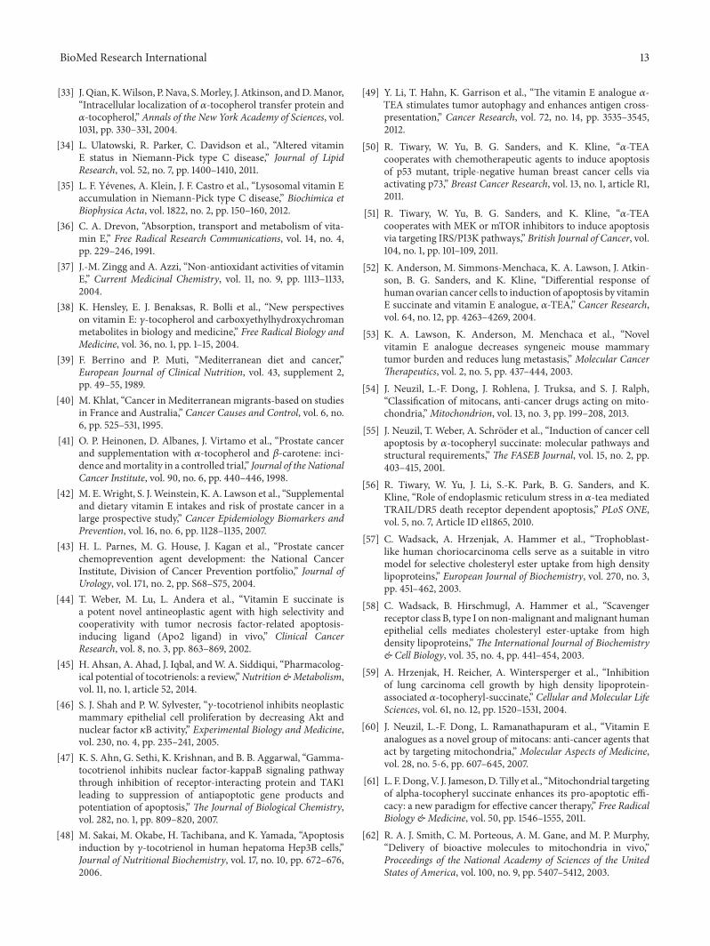
Research in the past few years has focused on structuralvariations within the functional moiety of natural Vitamin Eisoforms with the aim of improving the proapoptotic potencyof these agents and improving their bioavailability. Syntheticderivatives of 𝛼-TOC (Figure 3), such as 𝛼-tocopherol suc-cinate (𝛼-TOS) and 𝛼-tocopheryl ether-linked acetic acid(𝛼-TEA), have shown enhanced proapoptotic potency andanticancer action in tumorigenic cell lines and animalmodels[49–53]. Derivative 𝛼-TOS is a potent apoptotic compoundthat acts as a mitocan; it exerts anticancer activity and selec-tively induces apoptosis in tumor cells mainly by targetingand destabilizing mitochondria (reviewed in [54]). 𝛼-TOSgenerates ROS, which is known to trigger the mitochondrialpathway of apoptosis, and also inactivates Bcl-2 and Bax andinduces the translocation of Bax to the mitochondria [55].𝛼-TEA has been found to cooperate with chemotherapeuticagents to induce apoptosis of P53 mutant, triple negativehuman breast cancer cells via activating P73 suggesting thatthis synthetic derivative may be useful in the treatmentof multidrug resistant cancers [50]. Furthermore, 𝛼-TEAsuppressed constitutively active basal levels of p-AKT, p-ERK, p-mTOR, and their downstream targets as well as
4 BioMed Research International
O
O
OOH
O
O
OHO
O
H3C
CH3
CH3
H3C
CH3 CH3 CH3
CH3
CH3
CH3
CH3
CH3CH3 CH3 CH3
CH3
𝛼-Tocopherol succinate (𝛼-TOS)
𝛼-tocopheryl ether-linked acetic acid (𝛼-TEA)
Figure 3: Structure of Vitamin E synthetic derivatives.
induced apoptosis in breast cancer cells [51]. 𝛼-TEA was alsofound to stimulate tumor autophagy and enhance antigencross-presentation in murine mammary and lung cancercells [49] and induce apoptosis via ER stress by enhancingDR5/caspase-8 proapoptotic signaling in breast cancer cells[56]. Therefore, there is great interest for further evaluationof different alpha-tocopherol synthetic derivatives in in vitroand in vivo systems either as single agents or in combinationwith other drugs.
3. Improving the Efficacy of Vitamin ECompounds with Novel Delivery Systems
The potential of Vitamin E isoforms in cancer preventionand therapy and the problems regarding their solubility andabsorption have prompted the development of novel deliverysystems to intensify their effects. In addition to enhancingtheir own anticancer effects, several drug delivery systemsthat incorporate Vitamin E isoforms are being developedin order to improve the efficacy of other agents, either byincreasing their bioavailability or by acting in a synergisticmanner. We will review below these systems that includenovel formulations for both tocopherols and tocotrienols.Wewill present the most important preclinical studies and theirapplications in cancer therapeutics.
3.1. Tocopherol Delivery Systems. As previously described, 𝛼-tocopherol is esterified with a succinyl moiety to produce 𝛼-tocopheryl succinate or 𝛼-TOS. The proapoptotic propertiesof 𝛼-TOS are well known and there are numerous studiesshowing its enhanced anticancer potency in many cancertypes includingmelanoma, breast, prostate, gastric, mesothe-lioma, and colorectal (reviewed in [4]). In addition, severalformulations of 𝛼-TOS have been tested in order to improveits distribution and efficacy at the target site. Like other Vita-min E isoforms, 𝛼-TOS is subjected to intestinal hydrolysis;in order to counter this effect and to increase its uptakeby rapidly divided tumor cells, high-density lipoprotein-(HDL)- associated 𝛼-TOS was synthesized. Tumor cells have
an excessive need for cholesterol and selectively uptake HDL-associated compounds [57, 58]. 𝛼-TOS in association withHDL was successfully delivered to A549 lung cancer cells invitro and LL2 mouse lung carcinoma cells in vivo via SR-BI, the prime receptor mediating selective lipid uptake fromHDL, and inhibited tumor cell growth [59].
It has also been reported that analogues of VitaminE act as mitocans; they induce apoptosis in tumor cellsby affecting mitochondria stability (reviewed in [60]). Toenhance this effect, a mitochondrially targeted 𝛼-TOS wassynthesized by tagging the hydrophobic chain of the parentalcompound with a triphenylphosphonium group (TPP+) [61].MitoVES was 10- to 30-fold more effective than its untaggedcounterpart. TPP+ has been shown to promote the selectivemitochondrial uptake of antioxidant compounds driven bythe mitochondrial membrane potential [62]. Conjugationof 𝛼-TOS with TPP+ (MitoVES) potentiated its apoptoticeffect in a mitochondria-depended mechanism involving thegeneration of ROS and upregulation of the Noxa protein invarious malignant cell lines; its effect was also potentiated intwo animal cancer models (Table 1) [61]. In a different study,MitoVES induced apoptosis in mesothelioma cells moreefficiently than 𝛼-TOS by destabilizing the mitochondrialmembrane potential and generating ROS. The mitochondri-ally targeted 𝛼-TOS also suppressed mesothelioma growthin nude mice with high efficacy [63]. MitoVES selectivelysuppressed the proliferation of cancer cells at subapoptoticdoses by affecting mitochondrial DNA (mtDNA) transcripts.Specifically, MitoVES strongly suppressed the level of thedisplacement loop transcript and of mtDNA genes coding forsubunits of mitochondrial complexes causing disruption ofmitochondrial function. In addition, MitoVES decreased theexpression of mitochondrial transcription factor A (TFAM)and diminished mitochondrial biogenesis. Importantly, theinhibition of mitochondrial transcription was replicated invivo; MitoVES lowered the level of mtDNA transcripts inHER2 overexpressing breast cancer cells but not in normaltissue [64].
BioMed Research International 5
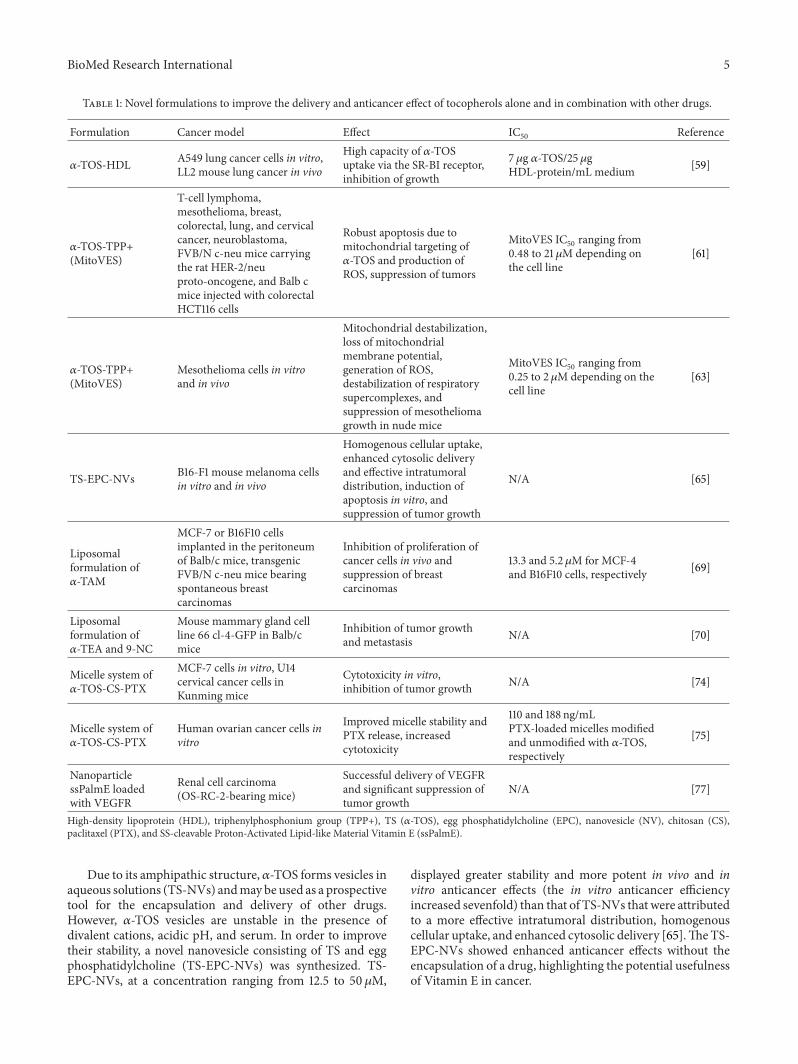
Table 1: Novel formulations to improve the delivery and anticancer effect of tocopherols alone and in combination with other drugs.
Formulation Cancer model Effect IC50 Reference
𝛼-TOS-HDL A549 lung cancer cells in vitro,LL2 mouse lung cancer in vivo
High capacity of 𝛼-TOSuptake via the SR-BI receptor,inhibition of growth
7𝜇g 𝛼-TOS/25𝜇gHDL-protein/mL medium [59]
𝛼-TOS-TPP+(MitoVES)
T-cell lymphoma,mesothelioma, breast,colorectal, lung, and cervicalcancer, neuroblastoma,FVB/N c-neu mice carryingthe rat HER-2/neuproto-oncogene, and Balb cmice injected with colorectalHCT116 cells
Robust apoptosis due tomitochondrial targeting of𝛼-TOS and production ofROS, suppression of tumors
MitoVES IC50 ranging from0.48 to 21 𝜇M depending onthe cell line
[61]
𝛼-TOS-TPP+(MitoVES)
Mesothelioma cells in vitroand in vivo
Mitochondrial destabilization,loss of mitochondrialmembrane potential,generation of ROS,destabilization of respiratorysupercomplexes, andsuppression of mesotheliomagrowth in nude mice
MitoVES IC50 ranging from0.25 to 2 𝜇M depending on thecell line
[63]
TS-EPC-NVs B16-F1 mouse melanoma cellsin vitro and in vivo
Homogenous cellular uptake,enhanced cytosolic deliveryand effective intratumoraldistribution, induction ofapoptosis in vitro, andsuppression of tumor growth
N/Α [65]
Liposomalformulation of𝛼-TAM
MCF-7 or B16F10 cellsimplanted in the peritoneumof Balb/c mice, transgenicFVB/N c-neu mice bearingspontaneous breastcarcinomas
Inhibition of proliferation ofcancer cells in vivo andsuppression of breastcarcinomas
13.3 and 5.2 𝜇M for MCF-4and B16F10 cells, respectively [69]
Liposomalformulation of𝛼-TEA and 9-NC
Mouse mammary gland cellline 66 cl-4-GFP in Balb/cmice
Inhibition of tumor growthand metastasis N/Α [70]
Micelle system of𝛼-TOS-CS-PTX
MCF-7 cells in vitro, U14cervical cancer cells inKunming mice
Cytotoxicity in vitro,inhibition of tumor growth N/Α [74]
Micelle system of𝛼-TOS-CS-PTX
Human ovarian cancer cells invitro
Improved micelle stability andPTX release, increasedcytotoxicity
110 and 188 ng/mLPTX-loaded micelles modifiedand unmodified with 𝛼-TOS,respectively
[75]
NanoparticlessPalmE loadedwith VEGFR
Renal cell carcinoma(OS-RC-2-bearing mice)
Successful delivery of VEGFRand significant suppression oftumor growth
N/A [77]
High-density lipoprotein (HDL), triphenylphosphonium group (TPP+), TS (𝛼-TOS), egg phosphatidylcholine (EPC), nanovesicle (NV), chitosan (CS),paclitaxel (PTX), and SS-cleavable Proton-Activated Lipid-like Material Vitamin E (ssPalmE).
Due to its amphipathic structure, 𝛼-TOS forms vesicles inaqueous solutions (TS-NVs) andmay be used as a prospectivetool for the encapsulation and delivery of other drugs.However, 𝛼-TOS vesicles are unstable in the presence ofdivalent cations, acidic pH, and serum. In order to improvetheir stability, a novel nanovesicle consisting of TS and eggphosphatidylcholine (TS-EPC-NVs) was synthesized. TS-EPC-NVs, at a concentration ranging from 12.5 to 50𝜇M,
displayed greater stability and more potent in vivo and invitro anticancer effects (the in vitro anticancer efficiencyincreased sevenfold) than that of TS-NVs that were attributedto a more effective intratumoral distribution, homogenouscellular uptake, and enhanced cytosolic delivery [65].TheTS-EPC-NVs showed enhanced anticancer effects without theencapsulation of a drug, highlighting the potential usefulnessof Vitamin E in cancer.
6 BioMed Research International
Vitamin E analogues can be easily incorporated into lipidbilayers to produce liposomes of various surface modifica-tions and size distributions. Liposomes can encapsulate asingle drug for monotherapy or several drugs for multi-targeted effects. The drugs incorporated into liposomes arepassively delivered to tumor tissue by enhanced permeationand retention (EPR) effect. The EPR effect is the property bywhich certain molecules accumulate in tumors rather than innormal tissues, due to the presence of more blood vessels intumor tissue [66, 67]. In order for tumor cells to grow quickly,they stimulate the production of blood vessels for theirnutritional and oxygen supply. These newly formed tumorvessels are usually abnormal in form and architecture whichleads to abnormal molecular and fluid transport dynamics,especially for macromolecular drugs. Thus, the EPR effecthelps to carry micelles, nanoparticles, and liposomes andspread them inside the cancer tissue.
A derivative of 𝛼-TOC, a-tocopheryl maleamide (𝛼-TAM), contains a noncleavable amide bond and has exhibitedenhanced apoptotic properties in vitro [68]. In a liposo-mal formulation, 𝛼-TAM induced apoptosis in human andmurine cancer cells in vitro and suppressed tumors inmouse models. Importantly, 𝛼-TAM’s nonspecific toxicityand immunotoxicity were diminished when incorporatedinto liposomes [69]. Another derivative of Vitamin E withpotent anticancer activity, 𝛼-TEA, in combination with 9-nitrocamptothecin (9-NC), a derivative of camptothecin,was formulated into liposomes using dilauroylphosphatidyl-choline and administered by aerosol in tumor-bearing mice.Liposome-formulated𝛼-TEA and 9-NC significantly reducedthe growth, induced apoptosis, and inhibited metastasis ofmouse mammary gland cell line (66 cl-4-GFP). Treatment of66 cl-4-GFP cells in culture for 3 days with a combination of𝛼-TEA (10mg/mL, singly produces 38% apoptosis) and 9-NC(15.6, 31.3, 62.5, or 125 ng/mL; singly produces 2–7% apopto-sis) produced 47%, 58%, 64%, and 69% apoptosis. In controlanimals, the incidence of macroscopic lung metastasis was83% compared to 8% in 𝛼-TEA-, 9-NC-, or combination-treated mice [70]. It was calculated that approximately 36 g𝛼-TEA and 0.4 g 9-NC per mouse per day were deposited inthe lungs, respectively.
Micelles are commonly used as a platform for thedelivery of hydrophobic drugs. They display good solu-bilization efficiency and high stability upon dilution andtheir nanoscale dimensions permit the efficient accumulationin tumor tissues via the EPR effect [71–73]. Chitosan iswidely used in micellar drug delivery applications due toits excellent biocompatibility, nontoxicity, biodegradability,and low immunogenicity. 𝛼-TOS has been synthesized incombination with paclitaxel (PTX) in chitosan derivativepolymeric micelles for cancer cell delivery. This conjugateself-assembled in aqueous medium to form micelles andPTX was incorporated into the micellar core for intravenousdelivery. Commonly, PTX is diluted in a 50 : 50 mixture withCremophor EL which has severe toxic side effects. A poly-meric micelle system of 𝛼-tocopherol succinate-amphiphilicchitosan (CS-TOS) loaded with PTX displayed comparablecytotoxicity to PTX-Cremophor EL and free PTX againstMCF-7 cells in vitro and significantly inhibited the growth
of U14 tumors in vivo at doses of 10 and 20mg/kg with notoxic side effects [74]. This further highlights the usefulnessof 𝛼-tocopherol succinate in replacing harmful lipophilicsolvents. In an aim of improving the therapeutic efficiencyand reducing side effects of PTX, 𝛼-TOS-grafted chitosanoligosaccharide was synthesized and physically loaded byPTX and 𝛼-TOS.The incorporation of 𝛼-TS into the micellesled to an increase of the hydrophobic interaction betweenPTX and the micelles core, thereby improving micelle sta-bility, reducing micelle size, and facilitating PTX releasefrom the micelles. In addition, 𝛼-TS/PTX-loaded micellesdisplayed higher cytotoxicity against human ovarian cancercells in vitro than that of PTX-loadedmicelles and PTX alone[75].𝛼-TOS has also been used as a hydrophobic scaf-
fold for a novel molecular platform, the SS-cleavableProton-Activated Lipid-like Material (ssPalm). This pDNA-encapsulating nanoparticle was designed to degrade in thecytosolic environment. The lipid envelope coating the con-densed DNA/polycation complex was a stable bilayer com-posed of the ssPalm molecule which mounts dual sensingmotifs that respond to various intracellular environmentsand allow for the release of the encapsulated molecule inthe cytoplasm [76]. ssPalm contains a proton-sponge unit(tertiary amines) that functions in response to an acidicenvironment, such as in an endosome or lysosome, and disul-fide bonding that can be cleaved in a reducing environmentsuch as the cytosol. ssPalmE, where Vitamin E (𝛼-tocopherolsuccinate) was used as a hydrophobic scaffold, successfullydelivered a solute form of VEGFR in vivo and showedenhanced antitumor action in mice bearing tumors estab-lished from a renal cell carcinoma (OS-RC-2). The ssPalmEformulation was significantly more effective than ssPalmM,whereMyristic acid was the hydrophobic scaffold, suggestingthat 𝛼-TOS worked synergistically with the VEGFR agonistagainst renal cell carcinoma in vivo [77]. LNPssPalmM andLNPssPalmE were administered intravenously at a dose of37.5 𝜇g of pDNA/mouse 3 times at every 3 days.
3.2. Tocotrienol Delivery Systems. Novel DDS aim to targetcompounds to tumor sides thereby diminishing side effectson healthy tissues. To achieve specific distribution to tumors,tocotrienol extract from palm oil or tocotrienol-rich fraction(TRF) was encapsulated within vesicles bearing transfer-rin (Table 2). The transferrin receptor is a cell membrane-associated glycoprotein involved in iron homeostasis andthe regulation of cell growth; since transferrin receptors areoverexpressed up to 100-fold in several types of cancer cellscompared to normal cells, these vesicles could selectivelylocalize to tumors. This novel formulation of TRF led to a 3-fold higher uptake and improved its toxicity more than 100-fold in A431 (epidermoid carcinoma), T98G (glioblastoma),and A2780 (ovarian carcinoma) cell lines compared to TRFsolution. The vesicle formulations of TRF were all signifi-cantly more effective than the free drug by at least 80 times,with IC
50ranging from 0.05 ± 0.02 to 1.42 ± 0.30 𝜇g/mL
depending on the cell line. Intravenous administration oftransferrin-bearing vesicles loaded with TRF induced tumorregression and improvement of animal survival in a murine
BioMed Research International 7
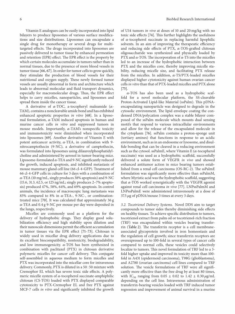
Table 2: Novel formulations to improve the delivery and anticancer effect of tocotrienols alone and in combination with other drugs.
Formulation Cancer model Effect IC50 Reference
UnilamellarTRF-vesiclesbearing transferrin
A431 (epidermoid carcinoma),T98G (glioblastoma), andA2780 (ovarian carcinoma)cells, A431 cells implanted inBALB/c mice
Threefold higher TRF uptakeand more than 100-foldimproved cytotoxicity in vitro,tumor regression, andimprovement of animalsurvival
Ranging from 0.05 ± 0.02 to 1.42± 0.30 𝜇g/mL depending on thecell line
[78]
MultilamellarTRF-vesiclesbearing transferrin
A431 human epidermoidcarcinoma, T98G humanglioblastoma, B16-F10 mousemelanoma cells, and A431 orB16-F10-luc-G5 tumors inBALB/c mice
Improved TRF uptake andcytotoxicity in vitro, slowergrowth of A431 and B16-F10tumors, and long-termsurvival of 100% of theanimals
Ranging from 0.89 ± 0.11 to 4.09± 0.65 𝜇g/mL depending on thecell line
[79]
Lipidnanoemulsionsloaded with TRFand Simvastatin
MCF-7 and MDA-MB-231breast cancer cells Decrease of TRF IC50
Decreased from 14 to 10 𝜇M inMCF-7 and from 7 to 4.8 𝜇M inMDA-MB-231 cells whenSimvastatin was added
[84]
Nanoemulsifiedformulation ofT3-rich palm oil(Tocomin-NE)
Human cutaneous carcinomain vitro Increased cytotoxicity
Tocomin-NE 42.6 ± 3.8mM and47.3 ± 3.2mM, Tocomin control217.4mM and 278.5mM in A431cells and SCC-4 cells, respectively
[85]
EPI-NPscoadministeredwith tocotrienols
Hep G2 (HCC) cells in vitro,HCC mouse model in Albinomice
Enhanced antiproliferativeeffect in vitro, enhancedapoptosis, and reduced VEGFlevel in vivo
Free EPI viability >90%EPI-NPs 0.9 𝜇g/mL, EPI-NPstocotrienols 2 𝜇g/mL
[86]
Tocotrienol-rich fraction (TRF), Epirubicin (EPI), nanoparticles (NPs), and Hepatocellular carcinoma (HCC).
xenograft model and was well tolerated by animals [78].However, tumor regression lasted only for the duration ofthe treatment. To improve the therapeutic efficacy, TRFwas encapsulated in multilamellar rather than unilamellartransferrin-bearing vesicles. Multilamellar vesicles containmore than one phospholipid bilayers compared to unilamel-lar; this may improve tocotrienol loading within the lipidicmembranes. This novel formulation not only significantlyimproved tocotrienol uptake by transferrin-expressing tumorcells but also improved the in vitro therapeutic efficacy offree tocotrienol from 17- to 72-fold depending on the cellline. Importantly, this novel TRF formulation led to completetumor suppression for 40% of B16-F10 murine melanomatumors and 20% of A431 human epidermoid carcinomatumors, with long-term survival of the animals [79].
In addition to their inherent anticancer properties,tocotrienols can also potentiate the anticancer activity ofother drugs thereby lowering their effective concentrationand limiting toxicity. Tocotrienols can enhance the effectof several compounds including cox-2 inhibitor celecoxib,tyrosine kinase inhibitor gefitinib, and statins such as Sim-vastatin [80–82]. Simvastatin is a potent inhibitor of 3-hydroxy-3-methylglutaryl-coenzyme A (HMGCoA) reduc-tase that displays anticancer activity but its clinical useis limited by high-dose toxicity. In an effort to producebioactive, injectable nanoparticles that carry both TRF andSimvastatin, parenteral lipid nanoemulsions loadedwith bothcompounds were synthesized; nanoemulsions are comprisedof nanoscale droplets in the range of 1–100 nm and have
many applications in pharmaceutics (reviewed in [83]).Lipid nanoemulsions containing Simvastatin at subthera-peutic doses decreased the IC
50of TRF in MCF-7 (from
14 to 10 𝜇M) and MDA-MB-231 (from 7 to 4.8 𝜇M) breastcancer cells [84]. Different nanoemulsions for optimizedincorporation of tocotrienol- (T3-) rich palm oil or Tocominobtained with different homogenization strategies have alsobeen tested for their anticancer activities. Adopted hybridnanoemulsification of Tocomin (Tocomin-NE) displayed2,2-diphenyl-1-picrylhydrazyl- (DPPH-) radical scavengingcapacity which effectively permeated cell membranes bydiffusion and demonstrated significantly stronger cytotoxicprofiles (at least 5-fold lower IC
50values, compared to those
estimated for the Tocomin-control). This hybrid nanoemul-sified formulation of T3-rich palm oil may be used in topicaldelivery against skin carcinomas [85].
Tocotrienols have also been formulated with Epirubicin(EPI) to reduce its toxicity. EPI is an anthracycline derivativeused commonly in the treatment ofHepatocellular carcinoma(HCC) but displays serious side effects including cardiomy-opathy and congestive heart failure. To specifically targethepatocytes, EPI was loaded in chitosan-PLGA nanoparticleslinked with asialofetuin (EPI-NPs); furthermore, to reducecardiotoxicity, targeted EPI-NPs were coadministered withtocotrienols. Combined therapy of tocotrienols with EPI-NPs enhanced apoptosis, reduced VEGF level in a dosedependent manner, and provided protection against oxida-tive stress and inflammation induced by EPI in the heart[86].

8 BioMed Research International
4. D-Alpha-Tocopheryl Polyethylene GlycolSuccinate (TPGS)
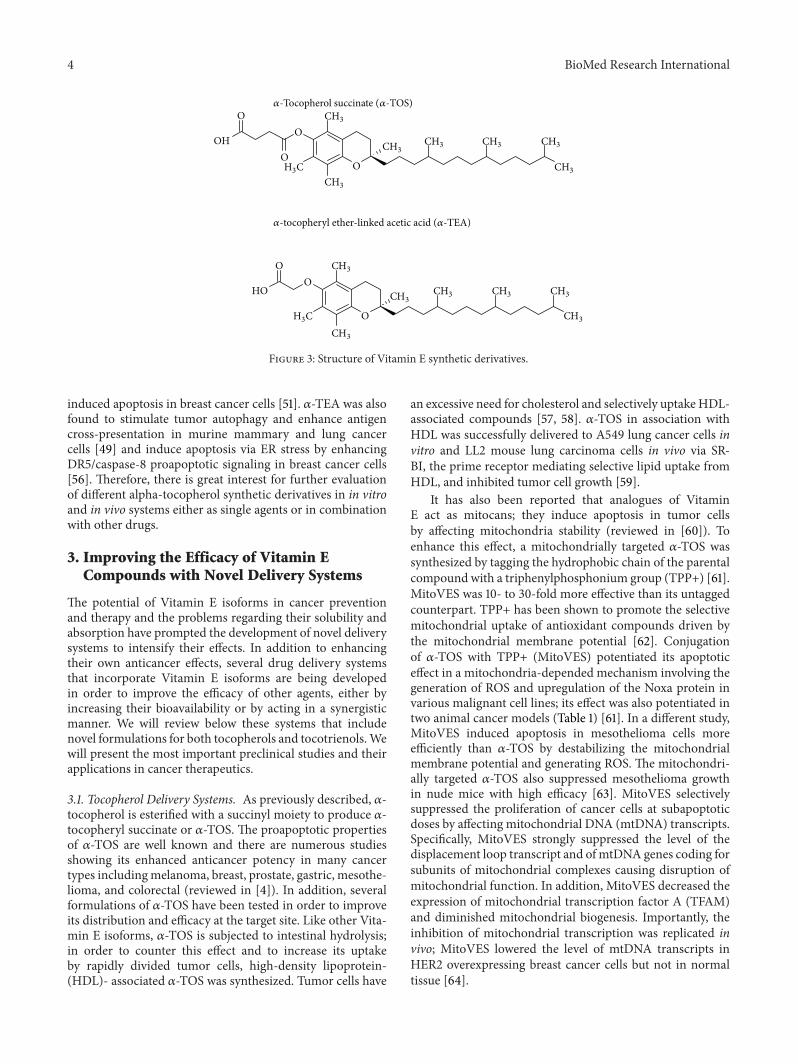
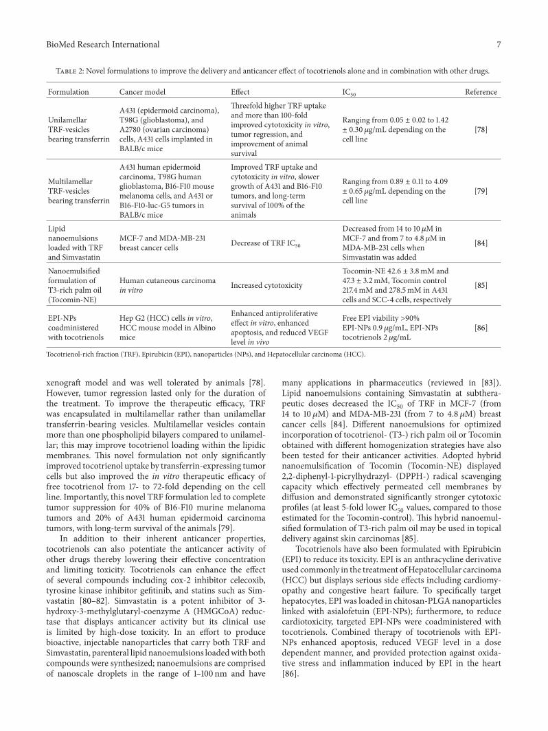
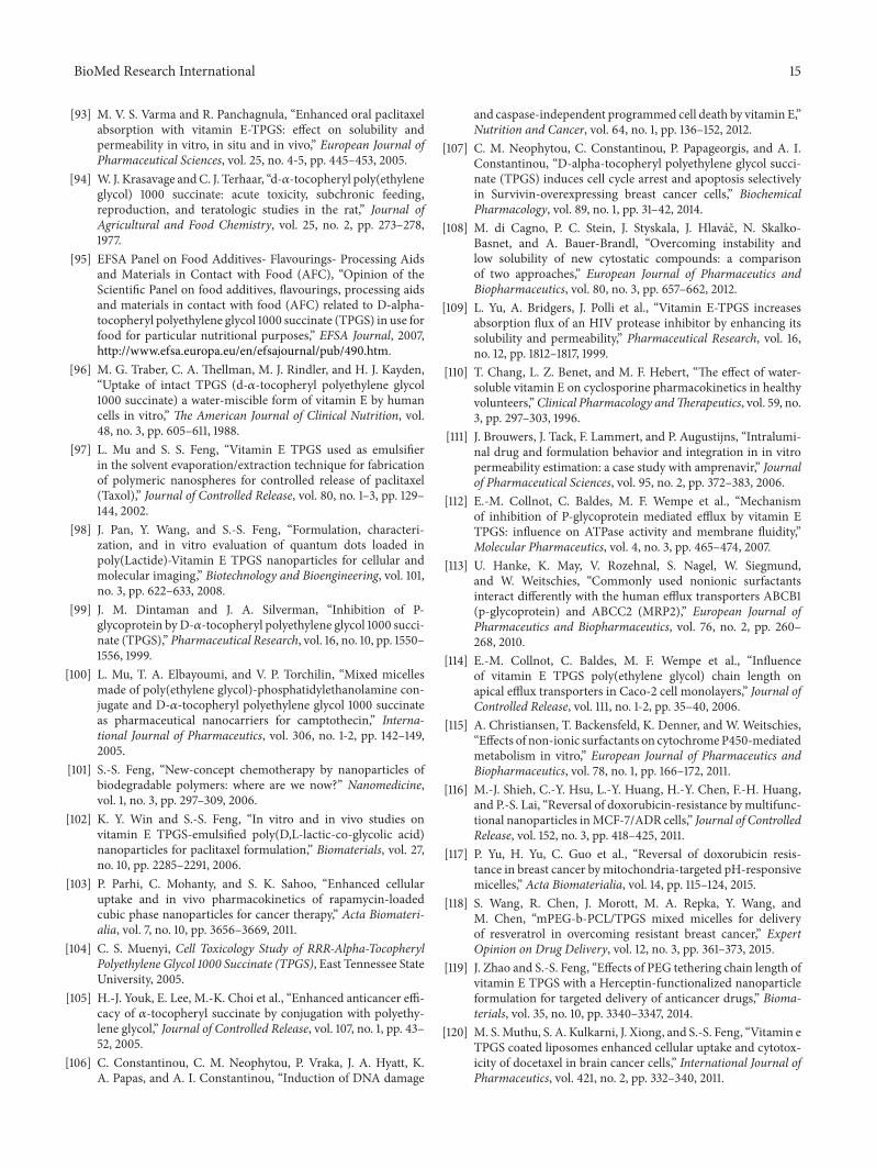
4.1. Physicochemical Properties. D-alpha-tocopheryl polyeth-ylene glycol succinate (Vitamin E TPGS or TPGS) is asynthetic derivative of natural alpha-tocopherol which isgaining increasing interest in the development of drug deliv-ery systems (reviewed by [87]). TPGS, prepared from theesterification of 𝛼-TOS and polyethylene glycol (PEG) 1000,possesses the advantages of PEG and Vitamin E in drugdelivery applications, including the ability to extend the half-life of the drug in the plasma.
The PEG 1000 has an average molecular weight of about1000 Dalton and is synthesized from fossil fuel while alpha-tocopherol succinate is produced by the alpha-tocopherolwhich is esterified to succinic acid. TPGS has an averagemolecular weight of 1513 and an amphiphilic structure oflipophilic alkyl tail and hydrophilic polar head; the PEG 1000portion of the molecule is water soluble, while the alpha-tocopherol portion is fat soluble [87]. Structurally, TPGSis very similar to the tocopherols; it has a phytyl tail anda chroman ring but the hydroxyl group on the chromanhead is esterified to the polyethylene 1000 succinate moiety(Figure 4). The molecular formula is C33 O5 H54 (CH
2
CH2O)n, where “𝑛” represents the number of polyethylene
oxide moieties attached to the acid group of alpha tocopherylsuccinate.
TPGS is a waxy solid that completely dissolves in waterand forms its own micelles; the hydrophilic regions ofthe molecule are in contact with the surrounding solvent,sequestering the hydrophobic tail regions in the micellecentre. It is also miscible with oils, other surfactants, andcosolvents. TPGS has a melting point around 37–41∘C andis heat stable at temperatures up to 199∘C [88]. This is veryimportant for applications in the pharmaceutical industrybecause it can be processed at thermally stable temperatureswithout degradation. TPGS is stable at pH 4.5–7.5; however,it degrades in alkaline or highly acidic environments wherethe ester linkages are hydrolyzed [88].
TPGS was developed in the 1950s by Eastman ChemicalCompany (Kingsport, TN) as awater-soluble formofVitaminE in order to be administered to individuals who can-not absorb naturally occurring lipophilic alpha-tocopherol.These include patients with cystic fibrosis, Crohn’s dis-ease, short bowel disease, pancreatic enzyme deficiency, orcholestatic liver disease [89–91]. Importantly, the UnitedStates’ Food and Drug Administration (FDA) as well asthe European Food Safety Authority (EFSA) has alreadyapproved TPGS as a safe pharmaceutical adjuvant used indrug formulation and has estimated the safety limits forTPGS use for research purposes [92, 93]. It has been reportedthat the acute oralmedian lethal dose (LD
50), which is defined
as the quantity of an agent that will kill 50 percent of the testsubjects within a designated period, is>7 g/kg for young adultrats of both sexes [94]. Following studies both in humansand in animals to address the bioavailability and safety ofTPGS, the EFSA concluded that the overall no-observed-adverse-effect level (NOAEL) of TPGS is 1000mg/kg bw/day.In addition, TPGS was found to be nongenotoxic [95].
The mechanism of TPGS to enter the enterocyte andbecome absorbed has also been investigated: TPGS maybe hydrolyzed to free alpha-tocopherol in the stomach bynonenzymatic hydrolysis, in the proximity of the brushborder epithelium by esterase hydrolysis, and on the surfaceof the enterocytes via a lipase or entire TPGSmicelles can passthrough cell membranes thereby enabling the absorption ofthe intact TPGS molecule [96].
In recent years, TPGS has been intensively applied indeveloping various drug delivery systems as an absorp-tion enhancer, emulsifier, solubilizer, additive, permeationenhancer, and stabilizer. The coadministration of TPGShas been shown to enhance drug solubility, to inhibit P-glycoprotein (P-gp) mediated multidrug resistance (MDR),and to increase the oral bioavailability of anticancer drugs[97–100]. As an effective emulsifier, TPGS has been shown togreatly enhance the performance of nanoparticles, resultingin much higher cellular uptake of the drug as well as moredesirable in vivo pharmacokinetics [101–103]. Importantly,emerging evidence in the literature suggests that TPGS notonly may be useful in cancer chemotherapy as a carrierdrug but also may act synergistically or enhance the effect ofanticancer drugs.
4.2. Anticancer Activity as a Single Agent. As a single agent,TPGS has been found to inhibit the growth of humanprostate and lung carcinoma cells both in vitro and in vivo[105, 106]. TPGS inhibited the growth of A549 human lungcarcinoma cells in vivo and, in an in vitro cell culture,more potently than 𝛼-TOS. Compared to 𝛼-TOS, TPGSwas more effective at inducing apoptosis in A549 cells asmeasured by DNA fragmentation; furthermore, at 40 𝜇Mconcentration, TPGS induced the production of ROS [105].Since PEG conjugation may affect the rate of cell uptake ofdrugs, the time-dependent accumulation of𝛼-TOS andTPGSin H460 cells was measured. However, the time-dependentuptake of TPGS into cells did not differ from that of 𝛼-TOS. Furthermore, since TPGS may be hydrolyzed afterentering the cells, the intracellular concentration of TPGSwasmeasured following treatment for up to 8 h and no detectablelevels of 𝛼-TOS were found [105]. This indicates that TPGSis not rapidly hydrolyzed to 𝛼-TOS and that the enhancedantitumor efficacy of TPGS was not due to its increaseduptake into cells.
The ability of TPGS to induce apoptosis was also investi-gated in androgen receptor negative (AR−) DU145, PC3, andandrogen receptor positive (AR+) LNCaP prostate cancercells [106]. TPGS induced caspase-3 activity solely in the AR+LNCaP cells; however, TPGS induced dominant caspase-independent programmed cell death (CI-PCD) in all celllines examined, suggesting that themolecular pathways beinginduced by the compound may depend on the cellularmicroenvironment. In a recent study conducted by our group,TPGS at 20 𝜇M induced G1/S cell cycle arrest and apoptosisin breast cancer cell lines MCF-7 and MDA-MB-231. Aninvestigation of the molecular mechanism of action of TPGSrevealed that induction of G1/S phase cell cycle arrest isassociated with upregulation of P21 and P27Kip1 proteins.Induction of apoptosis by TPGS involved the inhibition of
BioMed Research International 9
O
O
O
O
OH(OCH2CH2)n
(CH2)2
H3CCH3
CH3
CH3CH3 CH3 CH3
CH3
Figure 4: D-alpha-tocopheryl polyethylene glycol succinate structure. TPGS is synthesized from the esterification of 𝛼-TOS and PEG 1000.Modified from [104].
phospho-AKT and the downregulation of the antiapoptoticproteins Survivin and Bcl-2 [107]. Importantly, TPGS didnot reduce the viability or proliferation of “normal” (non-tumorigenic) immortalized cells (MCF-10A and MCF-12F)suggesting that its effects are cancer cell-specific.
4.3. Applications in Drug Delivery Systems. TPGS has beenused in various drug delivery systems as a solubilizer/absorp-tion enhancer, as a permeation enhancer, and as a potentP-gp inhibitor. Solubilizers/absorption enhancers are func-tional agents included in formulations to increase the sol-ubility or improve the absorption of a substance. TPGScan increase the solubility of drugs such as cyclosporines,taxanes, steroids, and antibiotics by incorporation into TPGSmicelles [108]. TPGS has been used as a solubilizer forPTX, celecoxib, corticosteroids, capuramycin analog SQ641,and Propofol (reviewed by [88]). Furthermore, TPGS hasbeen shown to increase the absorption flux of HIV proteaseinhibitor Amprenavir and to enhance the bioavailability ofcyclosporine in humans and of Colchicine in rats [109, 110].TPGS has also been used as a permeation enhancer; it isincorporated into formulations to promote their permeationthrough the skin or intestinal walls. TPGS was found toenhance the permeability of Amprenavir and Colchicine andhas potential as an enhancer of drug permeability in colonictissue [88, 111].
TPGS has also been used in combination with other com-pounds due to its ability to inhibit P-gp, an ATP-dependentdrug efflux pump, also known asmultidrug resistance protein1 (MDR1) or ATP-binding cassette subfamily B member 1(ABCB1). P-gp is responsible for the transport of a widevariety of substrates across extracellular and intracellularmembranes. It is widely expressed in hepatocytes, the renalproximal tubular cells, intestinal epithelium, adrenal gland,and capillary endothelial cells comprising the blood-brainand blood-testis barrier. P-gp is often overexpressed in cancercells and confers MDR by decreasing drug accumulationof chemotherapeutic drugs such as PTX, Etoposide, Dox-orubicin (DOX), and Vinblastine. TPGS can inhibit theP-gp ATPase (P-gp energy source of active transport) bybinding to the nontransport active binding site, resulting ininhibition of P-gp mediated drug transport and multidrugresistance [99, 112]. Specifically, combination of TPGS withDOX, Vinblastine, PTX, or Colchicine was found to enhancetheir cytotoxicity in NIH 3T3 cells transfected with thehuman MDR1 cDNA [99]. In a different study, TPGS andhigh concentrations of polysorbate 80 inhibited in vitro bothefflux transporters, ABCB1 (P-gp) and ABCC2 (MRP2), thatplay an essential role in the limitation of oral bioavailabilityof drugs [113]. So far, TPGS synthesized with PEG-1000 is
the most potent efflux pump inhibitor [114]. Furthermore,TPGS can inhibit cytochrome P450 3A (CYP3A) that hasbeen associated with deactivation of several anticancer drugs[115].
Recent studies suggest that the coadministration of TPGSwith chemotherapeutic agents may overcome drug resistanceby increasing their cellular entry and retention and by actingin a synergistic manner (Table 3). The addition of TPGS(0.03% (w/w)) in a nanocarrier loaded with DOX decreasedP-gp activity in MCF-7/ADR cells, increased the nuclearaccumulation of the drug, and increased the therapeuticefficacy against the resistant cell line [116]. Cells were treatedwith free Dox (from 0.13 𝜇M to 10 𝜇M) and TPGS-coatedporphyrin-polylactide nanoparticles (PPLA-NPs) or TPGS-coated Dox-PPLA-NPs (from 0.07 𝜇M to 4.3 𝜇M). Whenthe PPLA-NPs were coated with TPGS, cell viability wassignificantly reduced even at the lowest concentrations [116].In an alternative approach to overcome resistance in MCF-7/ADR cells, DOX was loaded in mitochondria-targeted pH-responsive (PDPA) micelles containing TPGS. In the acidicpH of early endosomes, the DOX payload was releasedwhile the TPGS component synergistically improved thecytotoxicity of the agent by reducing the mitochondrialtransmembrane potential. PDPA/TPGS micelles reduced theIC50
of DOX in MCF-7/ADR cells by a sixfold magnitude[117]. A TPGS-mixed micelle system loaded with Resveratrolhas also been formulated. Resveratrol has shown efficacy inovercoming chemoresistance in breast cancer but its effectsare hindered by poor absorption and rapid metabolism.Resveratrol-loaded mixed micelles composed of methoxypoly (ethylene glycol)-b-polycaprolactone (mPEG-PCL) andTPGS increased Resveratrol uptake by MCF-7/ADR cellsand induced higher rates of apoptosis. Blank mixed micellesdid not affect the proliferation of MCF-7/ADR cells (<10%inhibition), whereas Resveratrol alone (0–100𝜇M) inhibitedthe proliferation of MCF-7/ADR cells in a dose dependentmanner. Resveratrol-loaded mixed micelles enhanced thecytotoxicity of Resveratrol against MCF-7/ADR cells at all ofthe tested concentrations. Furthermore, Resveratrol-loadedmixedmicelles inhibited the activity of P-gp and significantlyenhanced DOX cellular accumulation and cytotoxicity inMCF-7/ADR cells [118].
TPGS has also been used in several formulations inorder to achieve targeted delivery of chemotherapeutic drugsand reduce negative side effects. Recently, TPGS-basedcopolymers were synthesized, conjugated with Herceptinfor targeted delivery of anticancer drugs such as Docetaxel(DTX) to cancer cells overexpressing the HER-2 receptor.Docetaxel was used at concentrations ranging from 0.025 to

10 BioMed Research International
Table 3: TPGS-based delivery systems of anticancer drugs.
Formulation Drug Cancer type Effect IC50 Reference
Nanoparticle DOX MCF-7/ADR cells in vitro
Decreased P-gp activity,increased drug nuclearaccumulation, andincreased therapeuticefficacy
N/A [116]
Mitochondria-targetedpH-responsivemicelles
DOX MCF-7/ADR cells in vitroand in vivo
Reduced mitochondrialtransmembrane potential,synergistic cytotoxicity
DOX 73.2 𝜇g/mL,PDPA-TPGS-DOX16.7𝜇g/mL
[117]
TPGS-mPEG-PCLmicelles Resveratrol MCF-7/ADR cells in vitro
Increased Resveratroluptake, enhancedapoptosis, and inhibition ofP-gp activity
N/A [118]
TPGS copolymersconjugated withHerceptin loadedwith DTX
DTX SK-BR-3 cancer cellsTargeted delivery, efficientcellular uptake, andimproved cytotoxicity
Nanoparticles withoutHerceptin 3.29 𝜇g/mL andwith Herceptin 0.341𝜇g/mL
[119]
TPGS coatedliposomes of DTX DTX C6 glioma cells Enhanced cellular uptake
and cytotoxicity
DTX alone 37.04 ± 1.05,TPGS coated liposomes5.93 ± 0.57 𝜇g/mL
[120]
Transferrin-conjugated TPGSmicelles
DTX MDA-MB-231 cells in vitroand in xenograft SCIDmice
Targeted delivery, highercellular uptake, highercytotoxicity, and reducedtumor size
DTX alone 13.63 ± 0.12,nontargeted micelles 0.89 ±0.10, and targeted micelles0.19 ± 0.04 𝜇g/mL
[121]
TPGS micelle Cisplatin HepG2 hepatocarcinomacells
Enhanced cytotoxicity,neuroprotective effects
Cisplatin alone 3.95 𝜇g/mL,TPGS micelle 1.36 𝜇g/mL [122]
TPGS-emulsifiedPLGA-mPEGnanoparticles
PTX IGROV1 ovarian cancercells in vitro and in vivo Decreased toxicity N/A [123]
TPGS-emulsifiedpolymericnanoparticles(TENPs)
PTX A549 lung cancer xenograftmodels Inhibition of tumor growth N/A [124]
Dendrimer-TPGSmicelles PTX, DTX MCF-7 and A549 cancer
cellsEnhanced solubility andincreased cytotoxicity N/A [125]
TPGS-PLAmicelles
Crizotinib,Palbociclib,and Sildenafil
A549 lung cancer cells Improved cytotoxic effect
IC50 (𝜇M): Crizotinib 26.07,Palbociclib, 17.65, doubletreatment 17.47, and tripletreatment 13.23
[126]
𝛼-TOS-TPGS-2knanoparticles DOX MCF-7 cells,
CT26-tumor-bearing miceReduced cell growth, delayin tumor growth
In vitro IC50 (𝜇M): TPGS 22± 2, TPGS + TOS10.1 ± 0.8,TPGS + TOS + DOX1.4 ± 0.4
[128]
Nanoparticle MH A549 in vitro and in vivo Enhanced toxicityIn vitro IC50 (𝜇M): free MH56.23, TPGS-MHnanoparticles 28.89
[129]
Methoxy poly (ethylene glycol)-b-polycaprolactone (mPEG-PCL), Doxorubicin (DOX), Docetaxel (DTX), paclitaxel (PTX), poly(lactic-co-glycolic acid)(PLGA), poly(lactic acid) (PLA), and Morin Hydrate (MH).
2.5 𝜇g/mL. Among the nanoparticles synthesized, those withchain length PEG 1000 resulted in the best therapeutic effects.Compared to the formulations with chain lengths 2000, 3350,and 5000, the IC
50values of the 1000-chain polymer were
68.1%, 90%, and 92.6% lower, respectively [119]. TPGS-coatedliposomes enhanced cellular uptake and cytotoxicity of DTX
in brain cancer cells [120], while targeted delivery of DTXby transferrin-conjugated TPGS micelles led to significantlyhigher cellular uptake and higher cytotoxicity against MDA-MB-231 cells in vitro and reduced tumor size in xenograftSCID mice [121]. Based on the IC
50values, the nontargeted
and targeted micelles could be 15.31- and 71.73-fold more
BioMed Research International 11
effective than DTX after 24 h treatment. Furthermore, aTPGS-Cisplatinmicellar formulation not only showed signif-icant enhancement of Cisplatin cytotoxicity in HepG2 hepa-tocarcinoma cells but also showed important neuroprotectiveeffects [122]. SH-SY5Yneuroblast-like cells, a commonmodelfor neurotoxicity test, were not greatly affected by treatmentwith the TPGS-Cisplatin micelle in contrast to Cisplatinalone.Therefore, it was suggested that TPGS supplementationshould be adopted in patients receiving Cisplatin-basedchemotherapy to reduce the neurotoxicity. In an effort toreduce the negative side effects of PTX, TPGS was used as anemulsifier in Cremophor EL-free PTX-loaded poly(lactic-co-glycolic acid)- (PLGA-)mPEGnanoparticles (NP-PTX).Thisnovel NP-PTX formulation consisting of 160.1mg PLGA-mPEG, 40.2mg PTX, and 0.035% TPGS displayed similarpotency as free PTX in ovarian cancer both in vivo and invitro and 10-fold less toxicity with no organ damage (NP-PTXLD50
246.85mg/kg, free PTX LD50
23.58mg/kg) [123]. Theefficacy of PTX encapsulated in TPGS-emulsified polymericnanoparticles (TENPs) was also examined in A549 lungcancer cells at a concentration ranging from 0.01 to 50mM.TPGS was used as an emulsifier to facilitate nanoparticleformation in an ethanol-water system at a concentration of0.6%. The PTX-TENPs preferentially accumulated in tumorsofA549 lung cancer xenograftmodels and significantly inhib-ited tumor growth following intravenous injection [124].
As previously mentioned, due to its physicochemicalproperties, TPGS has emerged as an ideal platform forthe enhanced delivery of poorly soluble drugs. PTX andDTX are commonly used in several types of cancer anddisplay high efficacy but they are highly lipophilic. Toenhance their solubility, PTX and DTX were encapsulatedin a mixed dendrimer-TPGS formulation. Dendrimers arenanomaterials that carry the guest molecule either by attach-ment to its surface or by encapsulation in the interior voidspaces. Cells were incubatedwith the different concentrations(2.5–500 nM) of dendrimer-drug formulations along withfree PTX and DTX for 48 h. The solubility and anticanceractivities of both compounds were greatly enhanced afterencapsulation in micelles against MCF-7 and A549 cancercells and caused very low toxicity to CHO normal ovariancells [125]. TPGS-poly(lactic acid) (TPGS-PLA) micelleshave been used for the simultaneous delivery of a noveltriple drug combination, antitumor drugs, Crizotinib andPalbociclib, combined with Sildenafil, an agent that increasesdrug accumulation in the intracellular compartment. TPGS-PLA copolymers encapsulated the three drugs with highloading efficiency and exhibited an improved cytotoxic effectin comparison with single (Crizotinib) or dual (Crizotinib-Palbociclib) formulations in A549 lung cancer cells [126].
A TPGS derivative with a PEG 2000 chain (TPGS-2k)was found to act synergistically with DTX to reduce theviability of MCF-7 cells [127]. TPGS-2k has also been usedfor the stabilization of a Doxorubicin-𝛼-TOS conjugate. 𝛼-TOS was connected to Doxorubicin through an amide bondto formN-Doxorubicin-𝛼-D-tocopherol succinate (N-DOX-TOS). In aqueous solutions in the presence of TPGS-2k, thisformulation self-assembled into 250 nm nanostructures andlocalized to the core of the nanoparticle. The N-DOX-TOS
nanoparticles significantly reduced the growth ofMCF-7 cellsand induced a higher delay in tumor growth in CT26-tumor-bearingmice compared to the control group [128]. In supportof the specific anticancer properties of TPGS, a recent studyrevealed that blank TPGS nanoparticles showed toxicityagainst A549 lung cancer cells without harming normal cells[129]. In a nanoparticle formulation, TPGS further enhancedthe toxicity of Morin Hydrate (MH), a naturally occurringbioflavonoid, against A549 cells both in vitro and in vivo [129].
5. Conclusions
In conclusion, recent studies have highlighted Vitamin Eisoforms and especially TPGS as ideal molecular biomate-rials in developing various drug delivery systems, includingmicelles, nanoparticles, and liposomes.DDS serve to improvethe delivery of Vitamin E isoforms so they can act as singleagents against tumor cells or can be used as a platform forthe combined treatment of Vitamin E with chemotherapeuticdrugs. Vitamin E-combined therapy represents an intriguingtherapeutic strategy for common chemotherapeutics, provid-ing not only superior efficacy but also higher safety levels.TPGS has amultifunctional nature; it can act as an anticanceragent specifically against malignant cells but may also beused in nanomedicine formulations in order to increasethe bioavailability and limit of the toxicity of other agents.Based on its mechanism of action, TPGS-based DDS can beformulated to combine the effect of compoundswith differentactivity in order to achieve simultaneous targeting of parallelpathways or sequential steps in the same pathway in cancercells and provide additive or synergistic effects. For example,the ability of TPGS to induce apoptosis in triple negativebreast cancer cells (TNBC) via the PI3K/AKT pathway [107]may serve as the basis for the development of a TPGS-based nanoparticle loaded with pharmacological inhibitorsof PI3K or AKT; studies show that PI3K inhibition impairsBRCA1/2 expression and sensitizes cells to PARP inhibitionin TNBC [130]. Therefore, for a synthetic lethality effect, thisnanoparticle may be loaded with a specific PARP inhibitorthat has also been found to be highly effective in TNBC cells.Further characterization of Vitamin E isoforms’ propertiesand mechanisms of action will provide a new insight intothe design of novel Vitamin E-based chemotherapeutics andallow the development of a series of cancer-cell-selectivetherapies.
Conflict of Interests
The authors declare that there is no conflict of interestsregarding the publication of this paper.
References
[1] M. G. Traber and J. Atkinson, “Vitamin E, antioxidant andnothing more,” Free Radical Biology and Medicine, vol. 43, no.1, pp. 4–15, 2007.
[2] Q. Jiang, “Natural forms of vitamin E: metabolism, antioxidant,and anti-inflammatory activities and their role in disease
12 BioMed Research International
prevention and therapy,” Free Radical Biology & Medicine, vol.72, pp. 76–90, 2014.
[3] S. Khanna, S. Roy, A. Slivka et al., “Neuroprotective propertiesof the natural vitamin E 𝛼-tocotrienol,” Stroke, vol. 36, no. 10,pp. 2258–2264, 2005.
[4] C. Constantinou, A. Papas, and A. I. Constantinou, “Vitamin Eand cancer: an insight into the anticancer activities of vitamin Eisomers and analogs,” International Journal of Cancer, vol. 123,no. 4, pp. 739–752, 2008.
[5] B. S. Abuasal, H. Qosa, P. W. Sylvester, and A. Kaddoumi,“Comparison of the intestinal absorption and bioavailability ofgamma-tocotrienol and alpha-tocopherol: in vitro, in situ andin vivo studies,” Biopharmaceutics and Drug Disposition, vol. 33,no. 5, pp. 246–256, 2012.
[6] S. P. Yap, K. H. Yuen, and A. B. Lim, “Influence of route ofadministration on the absorption and disposition of 𝛼-, 𝛾- and𝛿-tocotrienols in rats,” Journal of Pharmacy and Pharmacology,vol. 55, no. 1, pp. 53–58, 2003.
[7] B. Abuasal, P. W. Sylvester, and A. Kaddoumi, “Intestinalabsorption of 𝛾-Tocotrienol is mediated by Niemann-Pick C1-like 1: in situ rat intestinal perfusion studies,” Drug Metabolismand Disposition, vol. 38, no. 6, pp. 939–945, 2010.
[8] S. Alqahtani, A. Alayoubi, S. Nazzal, P. W. Sylvester, and A.Kaddoumi, “Nonlinear absorption kinetics of self-emulsifyingdrug delivery systems (SEDDS) containing tocotrienols aslipophilicmolecules: in vivo and in vitro studies,”AAPS Journal,vol. 15, no. 3, pp. 684–695, 2013.
[9] A. Kamal-Eldin and L.-A. Appelqvist, “The chemistry andantioxidant properties of tocopherols and tocotrienols,” Lipids,vol. 31, no. 7, pp. 671–701, 1996.
[10] X.-F. Wang, L. Dong, Y. Zhao, M. Tomasetti, K. Wu, and J.Neuzil, “Vitamin E analogues as anticancer agents: lessons fromstudies with 𝛼-tocopheryl succinate,” Molecular Nutrition &Food Research, vol. 50, no. 8, pp. 675–685, 2006.
[11] J. Neuzil, “Vitamin E succinate and cancer treatment: a vitaminE prototype for selective antitumour activity,” British Journal ofCancer, vol. 89, no. 10, pp. 1822–1826, 2003.
[12] M. G. Traber, T. Olivecrona, and H. J. Kayden, “Bovine milklipoprotein lipase transfers tocopherol to human fibroblastsduring triglyceride hydrolysis in vitro,” Journal of ClinicalInvestigation, vol. 75, no. 5, pp. 1729–1734, 1985.
[13] M. G. Traber, G. W. Burton, L. Hughes et al., “Discriminationbetween forms of vitamin E by humans with and withoutgenetic abnormalities of lipoprotein metabolism,” The Journalof Lipid Research, vol. 33, no. 8, pp. 1171–1182, 1992.
[14] J. K. Lodge, J. Ridlington, S. Leonard, H. Vaule, and M.G. Traber, “Alpha- and gamma-tocotrienols are metabolizedto carboxyethyl-hydroxychroman derivatives and excreted inhuman urine,” Lipids, vol. 36, no. 1, pp. 43–48, 2001.
[15] S. Fairus, R. M. Nor, H. M. Cheng, and K. Sundram, “Post-prandial metabolic fate of tocotrienol-rich vitamin E differssignificantly from that of 𝛼-tocopherol,” American Journal ofClinical Nutrition, vol. 84, no. 4, pp. 835–842, 2006.
[16] S. P. Yap, K. H. Yuen, and J. W. Wong, “Pharmacokinetics andbioavailability of 𝛼-, 𝛾- and 𝛿-tocotrienols under different foodstatus,” Journal of Pharmacy and Pharmacology, vol. 53, no. 1,pp. 67–71, 2001.
[17] I. Ikeda, Y. Imasato, E. Sasaki, and M. Sugano, “Lymphatictransport of 𝛼-, 𝛾-, and 𝛿-tocotrienols and 𝛼-tocopherol in rats,”International Journal for Vitamin and Nutrition Research, vol.66, no. 3, pp. 217–221, 1996.
[18] S. Khanna, V. Patel, C. Rink, S. Roy, and C. K. Sen, “Deliveryof orally supplemented 𝛼-tocotrienol to vital organs of ratsand tocopherol-transport protein deficient mice,” Free RadicalBiology and Medicine, vol. 39, no. 10, pp. 1310–1319, 2005.
[19] V. A. Tiurin, V. E. Kagan, N. F. Avrova, andM. P. Prozorovskaia,“Lipid asymmetry and alpha-tocopherol distribution in outerand inner monolayers of bilayer lipid membranes,” Bulletin ofExperimental Biology and Medicine, vol. 105, pp. 667–669, 1988.
[20] V. A. Tyurin, V. E. Kagan, E. A. Serbinova, N. V. Gorbunov,and A. N. Erin, “The interaction of alpha-tocopherol withphospholipid liposomes: the absence of transbilayer mobility,”Biulleten’ Eksperimental’noı Biologii i Meditsiny, vol. 102, no. 12,pp. 689–692, 1986.
[21] P. Mardones and A. Rigotti, “Cellular mechanisms of vitamin euptake: relevance in alpha-tocopherol metabolism and poten-tial implications for disease,” Journal of Nutritional Biochem-istry, vol. 15, no. 5, pp. 252–260, 2004.
[22] M. G. Traber and H. J. Kayden, “Vitamin E is delivered to cellsvia the high affinity receptor for low-density lipoprotein,” TheAmerican Journal of Clinical Nutrition, vol. 40, no. 4, pp. 747–751, 1984.
[23] C. A. Thellman and R. B. Shireman, “In vitro uptake of [3H]𝛼-tocopherol from low density lipoprotein by cultured humanfibroblasts,” Journal of Nutrition, vol. 115, no. 12, pp. 1673–1679,1985.
[24] I. Kolleck, H. Wissel, F. Guthmann, M. Schlame, P. Sinha, andB. Rustow, “HDL-holoparticle uptake by alveolar type II cells:effect of vitamin E status,” The American Journal of RespiratoryCell and Molecular Biology, vol. 27, no. 1, pp. 57–63, 2002.
[25] D. Goti, A. Hrzenjak, S. Levak-Frank et al., “Scavenger receptorclass B, type I is expressed in porcine brain capillary endothelialcells and contributes to selective uptake of HDL-associatedvitamin E,” Journal of Neurochemistry, vol. 76, no. 2, pp. 498–508, 2001.
[26] I. Kolleck, M. Schlame, H. Fechner, A. C. Looman, H. Wissel,and B. Rustow, “HDL is the major source of vitamin E for typeII pneumocytes,” Free Radical Biology and Medicine, vol. 27, no.7-8, pp. 882–890, 1999.
[27] P. Mardones, P. Strobel, S. Miranda et al., “Alpha-tocopherolmetabolism is abnormal in scavenger receptor class B type I (SR-BI)-deficient mice,” Journal of Nutrition, vol. 132, no. 3, pp. 443–449, 2002.
[28] W. Witt, I. Kolleck, H. Fechner, P. Sinha, and B. Rustow,“Regulation by vitamin E of the scavenger receptor BI in rat liverand HepG2 cells,” Journal of Lipid Research, vol. 41, no. 12, pp.2009–2016, 2000.
[29] J.Qian, S.Morley, K.Wilson, P.Nava, J. Atkinson, andD.Manor,“Intracellular trafficking of vitamin E in hepatocytes: the role oftocopherol transfer protein,”The Journal of Lipid Research, vol.46, no. 10, pp. 2072–2082, 2005.
[30] M. Horiguchi, M. Arita, D. E. Kaempf-Rotzoll, M. Tsujimoto,K. Inoue, and H. Arai, “pH-dependent translocation of 𝛼-tocopherol transfer protein (𝛼-TTP) between hepatic cytosoland late endosomes,” Genes to Cells, vol. 8, no. 10, pp. 789–800,2003.
[31] N. Kaplowitz, H. Yoshida, J. Kuhlenkamp, B. Slitsky, I. Ren,and A. Stolz, “Tocopherol-binding proteins of hepatic cytosol,”Annals of the New York Academy of Sciences, vol. 570, pp. 85–94,1989.
[32] J. B. Massey, “Kinetics of transfer of 𝛼-tocopherol betweenmodel and native plasma lipoproteins,” Biochimica et BiophysicaActa, vol. 793, no. 3, pp. 387–392, 1984.
BioMed Research International 13
[33] J.Qian, K.Wilson, P.Nava, S.Morley, J. Atkinson, andD.Manor,“Intracellular localization of 𝛼-tocopherol transfer protein and𝛼-tocopherol,” Annals of the New York Academy of Sciences, vol.1031, pp. 330–331, 2004.
[34] L. Ulatowski, R. Parker, C. Davidson et al., “Altered vitaminE status in Niemann-Pick type C disease,” Journal of LipidResearch, vol. 52, no. 7, pp. 1400–1410, 2011.
[35] L. F. Yevenes, A. Klein, J. F. Castro et al., “Lysosomal vitamin Eaccumulation in Niemann-Pick type C disease,” Biochimica etBiophysica Acta, vol. 1822, no. 2, pp. 150–160, 2012.
[36] C. A. Drevon, “Absorption, transport and metabolism of vita-min E,” Free Radical Research Communications, vol. 14, no. 4,pp. 229–246, 1991.
[37] J.-M. Zingg and A. Azzi, “Non-antioxidant activities of vitaminE,” Current Medicinal Chemistry, vol. 11, no. 9, pp. 1113–1133,2004.
[38] K. Hensley, E. J. Benaksas, R. Bolli et al., “New perspectiveson vitamin E: 𝛾-tocopherol and carboxyethylhydroxychromanmetabolites in biology and medicine,” Free Radical Biology andMedicine, vol. 36, no. 1, pp. 1–15, 2004.
[39] F. Berrino and P. Muti, “Mediterranean diet and cancer,”European Journal of Clinical Nutrition, vol. 43, supplement 2,pp. 49–55, 1989.
[40] M. Khlat, “Cancer in Mediterranean migrants-based on studiesin France and Australia,” Cancer Causes and Control, vol. 6, no.6, pp. 525–531, 1995.
[41] O. P. Heinonen, D. Albanes, J. Virtamo et al., “Prostate cancerand supplementation with 𝛼-tocopherol and 𝛽-carotene: inci-dence andmortality in a controlled trial,” Journal of the NationalCancer Institute, vol. 90, no. 6, pp. 440–446, 1998.
[42] M. E.Wright, S. J.Weinstein, K. A. Lawson et al., “Supplementaland dietary vitamin E intakes and risk of prostate cancer in alarge prospective study,” Cancer Epidemiology Biomarkers andPrevention, vol. 16, no. 6, pp. 1128–1135, 2007.
[43] H. L. Parnes, M. G. House, J. Kagan et al., “Prostate cancerchemoprevention agent development: the National CancerInstitute, Division of Cancer Prevention portfolio,” Journal ofUrology, vol. 171, no. 2, pp. S68–S75, 2004.
[44] T. Weber, M. Lu, L. Andera et al., “Vitamin E succinate isa potent novel antineoplastic agent with high selectivity andcooperativity with tumor necrosis factor-related apoptosis-inducing ligand (Apo2 ligand) in vivo,” Clinical CancerResearch, vol. 8, no. 3, pp. 863–869, 2002.
[45] H. Ahsan, A. Ahad, J. Iqbal, andW. A. Siddiqui, “Pharmacolog-ical potential of tocotrienols: a review,”Nutrition &Metabolism,vol. 11, no. 1, article 52, 2014.
[46] S. J. Shah and P. W. Sylvester, “𝛾-tocotrienol inhibits neoplasticmammary epithelial cell proliferation by decreasing Akt andnuclear factor 𝜅B activity,” Experimental Biology and Medicine,vol. 230, no. 4, pp. 235–241, 2005.
[47] K. S. Ahn, G. Sethi, K. Krishnan, and B. B. Aggarwal, “Gamma-tocotrienol inhibits nuclear factor-kappaB signaling pathwaythrough inhibition of receptor-interacting protein and TAK1leading to suppression of antiapoptotic gene products andpotentiation of apoptosis,” The Journal of Biological Chemistry,vol. 282, no. 1, pp. 809–820, 2007.
[48] M. Sakai, M. Okabe, H. Tachibana, and K. Yamada, “Apoptosisinduction by 𝛾-tocotrienol in human hepatoma Hep3B cells,”Journal of Nutritional Biochemistry, vol. 17, no. 10, pp. 672–676,2006.
[49] Y. Li, T. Hahn, K. Garrison et al., “The vitamin E analogue 𝛼-TEA stimulates tumor autophagy and enhances antigen cross-presentation,” Cancer Research, vol. 72, no. 14, pp. 3535–3545,2012.
[50] R. Tiwary, W. Yu, B. G. Sanders, and K. Kline, “𝛼-TEAcooperates with chemotherapeutic agents to induce apoptosisof p53 mutant, triple-negative human breast cancer cells viaactivating p73,” Breast Cancer Research, vol. 13, no. 1, article R1,2011.
[51] R. Tiwary, W. Yu, B. G. Sanders, and K. Kline, “𝛼-TEAcooperates with MEK or mTOR inhibitors to induce apoptosisvia targeting IRS/PI3K pathways,” British Journal of Cancer, vol.104, no. 1, pp. 101–109, 2011.
[52] K. Anderson, M. Simmons-Menchaca, K. A. Lawson, J. Atkin-son, B. G. Sanders, and K. Kline, “Differential response ofhuman ovarian cancer cells to induction of apoptosis by vitaminE succinate and vitamin E analogue, 𝛼-TEA,” Cancer Research,vol. 64, no. 12, pp. 4263–4269, 2004.
[53] K. A. Lawson, K. Anderson, M. Menchaca et al., “Novelvitamin E analogue decreases syngeneic mouse mammarytumor burden and reduces lung metastasis,” Molecular CancerTherapeutics, vol. 2, no. 5, pp. 437–444, 2003.
[54] J. Neuzil, L.-F. Dong, J. Rohlena, J. Truksa, and S. J. Ralph,“Classification of mitocans, anti-cancer drugs acting on mito-chondria,”Mitochondrion, vol. 13, no. 3, pp. 199–208, 2013.
[55] J. Neuzil, T. Weber, A. Schroder et al., “Induction of cancer cellapoptosis by 𝛼-tocopheryl succinate: molecular pathways andstructural requirements,” The FASEB Journal, vol. 15, no. 2, pp.403–415, 2001.
[56] R. Tiwary, W. Yu, J. Li, S.-K. Park, B. G. Sanders, and K.Kline, “Role of endoplasmic reticulum stress in 𝛼-tea mediatedTRAIL/DR5 death receptor dependent apoptosis,” PLoS ONE,vol. 5, no. 7, Article ID e11865, 2010.
[57] C. Wadsack, A. Hrzenjak, A. Hammer et al., “Trophoblast-like human choriocarcinoma cells serve as a suitable in vitromodel for selective cholesteryl ester uptake from high densitylipoproteins,” European Journal of Biochemistry, vol. 270, no. 3,pp. 451–462, 2003.
[58] C. Wadsack, B. Hirschmugl, A. Hammer et al., “Scavengerreceptor class B, type I on non-malignant andmalignant humanepithelial cells mediates cholesteryl ester-uptake from highdensity lipoproteins,” The International Journal of Biochemistry& Cell Biology, vol. 35, no. 4, pp. 441–454, 2003.
[59] A. Hrzenjak, H. Reicher, A. Wintersperger et al., “Inhibitionof lung carcinoma cell growth by high density lipoprotein-associated 𝛼-tocopheryl-succinate,” Cellular and Molecular LifeSciences, vol. 61, no. 12, pp. 1520–1531, 2004.
[60] J. Neuzil, L.-F. Dong, L. Ramanathapuram et al., “Vitamin Eanalogues as a novel group of mitocans: anti-cancer agents thatact by targeting mitochondria,” Molecular Aspects of Medicine,vol. 28, no. 5-6, pp. 607–645, 2007.
[61] L. F.Dong,V. J. Jameson,D. Tilly et al., “Mitochondrial targetingof alpha-tocopheryl succinate enhances its pro-apoptotic effi-cacy: a new paradigm for effective cancer therapy,” Free RadicalBiology & Medicine, vol. 50, pp. 1546–1555, 2011.
[62] R. A. J. Smith, C. M. Porteous, A. M. Gane, and M. P. Murphy,“Delivery of bioactive molecules to mitochondria in vivo,”Proceedings of the National Academy of Sciences of the UnitedStates of America, vol. 100, no. 9, pp. 5407–5412, 2003.

14 BioMed Research International
[63] J. Kovarova, M. Bajzikova, M. Vondrusova et al., “Mitochon-drial targeting of 𝛼-tocopheryl succinate enhances its anti-mesothelioma efficacy,” Redox Report, vol. 19, no. 1, pp. 16–25,2014.
[64] J. Truksa, L. F. Dong, J. Rohlena et al., “Mitochondrially targetedvitamin e succinate modulates expression of mitochondrialDNA transcripts andmitochondrial biogenesis,”Antioxidants&Redox Signaling, vol. 22, no. 11, pp. 883–900, 2015.
[65] S. Hama, S. Utsumi, Y. Fukuda et al., “Development of anovel drug delivery system consisting of an antitumor agenttocopheryl succinate,” Journal of Controlled Release, vol. 161, no.3, pp. 843–851, 2012.
[66] Y. Matsumura and H. Maeda, “A new concept for macro-molecular therapeutics in cancer chemotherapy: mechanism oftumoritropic accumulation of proteins and the antitumor agentsmancs,” Cancer Research, vol. 46, no. 12 I, pp. 6387–6392, 1986.
[67] K. Greish, “Enhanced permeability and retention (EPR) effectfor anticancer nanomedicine drug targeting,”Methods inMolec-ular Biology, vol. 624, pp. 25–37, 2010.
[68] A. Tomic-Vatic, J. Eytina, J. Chapman, E. Mahdavian, J. Neuzil,and B. A. Salvatore, “Vitamin E amides, a new class of vitaminE analogues with enhanced proapoptotic activity,” InternationalJournal of Cancer, vol. 117, no. 2, pp. 188–193, 2005.
[69] J. Turanek, X.-F. Wang, P. Knotigova et al., “Liposomal formu-lation of 𝛼-tocopheryl maleamide: in vitro and in vivo toxico-logical profile and anticancer effect against spontaneous breastcarcinomas inmice,” Toxicology and Applied Pharmacology, vol.237, no. 3, pp. 249–257, 2009.
[70] K. A. Lawson, K. Anderson, R. M. Snyder et al., “Novel vitaminE analogue and 9-nitro-camptothecin administered as liposomeaerosols decrease syngeneic mouse mammary tumor burdenand inhibit metastasis,” Cancer Chemotherapy and Pharmacol-ogy, vol. 54, no. 5, pp. 421–431, 2004.
[71] H. Montazeri Aliabadi, D. R. Brocks, and A. Lavasanifar,“Polymeric micelles for the solubilization and delivery ofcyclosporine A: pharmacokinetics and biodistribution,” Bioma-terials, vol. 26, no. 35, pp. 7251–7259, 2005.
[72] V. P. Torchilin, “Micellar nanocarriers: pharmaceutical perspec-tives,” Pharmaceutical Research, vol. 24, no. 1, pp. 1–16, 2007.
[73] H. Maeda, J. Wu, T. Sawa, Y. Matsumura, and K. Hori, “Tumorvascular permeability and the EPR effect in macromoleculartherapeutics: a review,” Journal of Controlled Release, vol. 65, no.1-2, pp. 271–284, 2000.
[74] N. Liang, S. Sun, X. Li, H. Piao, F. Cui, and L. Fang, “Alpha-tocopherol succinate-modified chitosan as a micellar deliverysystem for paclitaxel: preparation, characterization and invitro/in vivo evaluations,” International Journal of Pharmaceu-tics, vol. 423, no. 2, pp. 480–488, 2012.
[75] J. Emami, M. Rezazadeh, M. Rostami et al., “Co-delivery ofpaclitaxel and 𝛼-tocopherol succinate by novel chitosan-basedpolymeric micelles for improving micellar stability and effica-cious combination therapy,” Drug Development and IndustrialPharmacy, pp. 1–11, 2014.
[76] H. Akita, R. Ishiba, H. Hatakeyama et al., “A neutral envelope-type nanoparticle containing pH-responsive and SS-cleavablelipid-like material as a carrier for plasmid DNA,” AdvancedHealthcare Materials, vol. 2, no. 8, pp. 1120–1125, 2013.
[77] H. Akita, R. Ishiba, R. Togashi et al., “A neutral lipid envelope-type nanoparticle composed of a pH-activated and vitaminE-scaffold lipid-like material as a platform for a gene carriertargeting renal cell carcinoma,” Journal of Controlled Release,vol. 200, pp. 97–105, 2015.
[78] J. Y. Fu, D. R. Blatchford, L. Tetley, andC. Dufes, “Tumor regres-sion after systemic administration of tocotrienol entrapped intumor-targeted vesicles,” Journal of Controlled Release, vol. 140,no. 2, pp. 95–99, 2009.
[79] J. Y. Fu, W. Zhang, D. R. Blatchford, L. Tetley, G. McConnell,and C. Dufes, “Novel tocotrienol-entrapping vesicles can erad-icate solid tumors after intravenous administration,” Journal ofControlled Release, vol. 154, no. 1, pp. 20–26, 2011.
[80] A. B. Shirode and P. W. Sylvester, “Synergistic anticancer effectsof combined 𝛾-tocotrienol and celecoxib treatment are associ-ated with suppression in Akt and NF𝜅B signaling,” Biomedicineand Pharmacotherapy, vol. 64, no. 5, pp. 327–332, 2010.
[81] S. V. Bachawal, V. B. Wali, and P. W. Sylvester, “Combined𝛾-tocotrienol and erlotinib/gefitinib treatment suppresses Statand Akt signaling inmurine mammary tumor cells,”AnticancerResearch, vol. 30, no. 2, pp. 429–437, 2010.
[82] V. B. Wali and P. W. Sylvester, “Synergistic antiproliferativeeffects of 𝛾-tocotrienol and statin treatment on mammarytumor cells,” Lipids, vol. 42, no. 12, pp. 1113–1123, 2007.
[83] M. M. Fryd and T. G. Mason, “Advanced nanoemulsions,”Annual Review of Physical Chemistry, vol. 63, pp. 493–518, 2012.
[84] A. Y. Alayoubi, J. F. Anderson, S. D. Satyanarayanajois, P. W.Sylvester, and S. Nazzal, “Concurrent delivery of tocotrienolsand simvastatin by lipid nanoemulsions potentiates their antitu-mor activity against human mammary adenocarcenoma cells,”European Journal of Pharmaceutical Sciences, vol. 48, no. 3, pp.385–392, 2013.
[85] J. Pham, A. Nayel, C. Hoang, and T. Elbayoumi, “Enhancedeffectiveness of tocotrienol-based nano-emulsified system fortopical delivery against skin carcinomas,” Drug Delivery, pp. 1–11, 2014.
[86] M. Nasr, N. Nafee, H. Saad, and A. Kazem, “Improved anti-tumor activity and reduced cardiotoxicity of epirubicin usinghepatocyte-targeted nanoparticles combined with tocotrienolsagainst hepatocellular carcinoma in mice,” European Journal ofPharmaceutics and Biopharmaceutics, vol. 88, no. 1, pp. 216–225,2014.
[87] Z. Zhang, S. Tan, and S. S. Feng, “Vitamin E TPGS as amolecular biomaterial for drug delivery,” Biomaterials, vol. 33,pp. 4889–4906, 2012.
[88] Y. Guo, J. Luo, S. Tan, B. O. Otieno, and Z. Zhang, “Theapplications of Vitamin e TPGS in drug delivery,” EuropeanJournal of Pharmaceutical Sciences, vol. 49, no. 2, pp. 175–186,2013.
[89] M. G. Traber, T. D. Schiano, A. C. Steephen, H. J. Kayden, andM. Shike, “Efficacy of water-soluble vitamin E in the treatmentof vitamin E malabsorption in short-bowel syndrome,” TheAmerican Journal of Clinical Nutrition, vol. 59, no. 6, pp. 1270–1274, 1994.
[90] R. J. Sokol, N. Butler-Simon, C. Conner et al., “Multicentertrial of d-𝛼-tocopheryl polyethylene glycol 1000 succinate fortreatment of vitamin E deficiency in children with chroniccholestasis,” Gastroenterology, vol. 104, no. 6, pp. 1727–1735,1993.
[91] R. J. Sokol, N. A. Butler-Simon, D. Bettis, D. J. Smith, andA. Silverman, “Tocopheryl polyethylene glycol 1000 succinatetherapy for vitamin E deficiency during chronic childhoodcholestasis: neurologic outcome,”The Journal of Pediatrics, vol.111, no. 6, pp. 830–836, 1987.
[92] P. P. Constantinides, J. Han, and S. S. Davis, “Advances in the useof tocols as drug delivery vehicles,” Pharmaceutical Research,vol. 23, no. 2, pp. 243–255, 2006.
BioMed Research International 15
[93] M. V. S. Varma and R. Panchagnula, “Enhanced oral paclitaxelabsorption with vitamin E-TPGS: effect on solubility andpermeability in vitro, in situ and in vivo,” European Journal ofPharmaceutical Sciences, vol. 25, no. 4-5, pp. 445–453, 2005.
[94] W. J. Krasavage andC. J. Terhaar, “d-𝛼-tocopheryl poly(ethyleneglycol) 1000 succinate: acute toxicity, subchronic feeding,reproduction, and teratologic studies in the rat,” Journal ofAgricultural and Food Chemistry, vol. 25, no. 2, pp. 273–278,1977.
[95] EFSA Panel on Food Additives- Flavourings- Processing Aidsand Materials in Contact with Food (AFC), “Opinion of theScientific Panel on food additives, flavourings, processing aidsand materials in contact with food (AFC) related to D-alpha-tocopheryl polyethylene glycol 1000 succinate (TPGS) in use forfood for particular nutritional purposes,” EFSA Journal, 2007,http://www.efsa.europa.eu/en/efsajournal/pub/490.htm.
[96] M. G. Traber, C. A. Thellman, M. J. Rindler, and H. J. Kayden,“Uptake of intact TPGS (d-𝛼-tocopheryl polyethylene glycol1000 succinate) a water-miscible form of vitamin E by humancells in vitro,” The American Journal of Clinical Nutrition, vol.48, no. 3, pp. 605–611, 1988.
[97] L. Mu and S. S. Feng, “Vitamin E TPGS used as emulsifierin the solvent evaporation/extraction technique for fabricationof polymeric nanospheres for controlled release of paclitaxel(Taxol),” Journal of Controlled Release, vol. 80, no. 1–3, pp. 129–144, 2002.
[98] J. Pan, Y. Wang, and S.-S. Feng, “Formulation, characteri-zation, and in vitro evaluation of quantum dots loaded inpoly(Lactide)-Vitamin E TPGS nanoparticles for cellular andmolecular imaging,” Biotechnology and Bioengineering, vol. 101,no. 3, pp. 622–633, 2008.
[99] J. M. Dintaman and J. A. Silverman, “Inhibition of P-glycoprotein byD-𝛼-tocopheryl polyethylene glycol 1000 succi-nate (TPGS),” Pharmaceutical Research, vol. 16, no. 10, pp. 1550–1556, 1999.
[100] L. Mu, T. A. Elbayoumi, and V. P. Torchilin, “Mixed micellesmade of poly(ethylene glycol)-phosphatidylethanolamine con-jugate and D-𝛼-tocopheryl polyethylene glycol 1000 succinateas pharmaceutical nanocarriers for camptothecin,” Interna-tional Journal of Pharmaceutics, vol. 306, no. 1-2, pp. 142–149,2005.
[101] S.-S. Feng, “New-concept chemotherapy by nanoparticles ofbiodegradable polymers: where are we now?” Nanomedicine,vol. 1, no. 3, pp. 297–309, 2006.
[102] K. Y. Win and S.-S. Feng, “In vitro and in vivo studies onvitamin E TPGS-emulsified poly(D,L-lactic-co-glycolic acid)nanoparticles for paclitaxel formulation,” Biomaterials, vol. 27,no. 10, pp. 2285–2291, 2006.
[103] P. Parhi, C. Mohanty, and S. K. Sahoo, “Enhanced cellularuptake and in vivo pharmacokinetics of rapamycin-loadedcubic phase nanoparticles for cancer therapy,” Acta Biomateri-alia, vol. 7, no. 10, pp. 3656–3669, 2011.
[104] C. S. Muenyi, Cell Toxicology Study of RRR-Alpha-TocopherylPolyethylene Glycol 1000 Succinate (TPGS), East Tennessee StateUniversity, 2005.
[105] H.-J. Youk, E. Lee, M.-K. Choi et al., “Enhanced anticancer effi-cacy of 𝛼-tocopheryl succinate by conjugation with polyethy-lene glycol,” Journal of Controlled Release, vol. 107, no. 1, pp. 43–52, 2005.
[106] C. Constantinou, C. M. Neophytou, P. Vraka, J. A. Hyatt, K.A. Papas, and A. I. Constantinou, “Induction of DNA damage
and caspase-independent programmed cell death by vitamin E,”Nutrition and Cancer, vol. 64, no. 1, pp. 136–152, 2012.
[107] C. M. Neophytou, C. Constantinou, P. Papageorgis, and A. I.Constantinou, “D-alpha-tocopheryl polyethylene glycol succi-nate (TPGS) induces cell cycle arrest and apoptosis selectivelyin Survivin-overexpressing breast cancer cells,” BiochemicalPharmacology, vol. 89, no. 1, pp. 31–42, 2014.
[108] M. di Cagno, P. C. Stein, J. Styskala, J. Hlavac, N. Skalko-Basnet, and A. Bauer-Brandl, “Overcoming instability andlow solubility of new cytostatic compounds: a comparisonof two approaches,” European Journal of Pharmaceutics andBiopharmaceutics, vol. 80, no. 3, pp. 657–662, 2012.
[109] L. Yu, A. Bridgers, J. Polli et al., “Vitamin E-TPGS increasesabsorption flux of an HIV protease inhibitor by enhancing itssolubility and permeability,” Pharmaceutical Research, vol. 16,no. 12, pp. 1812–1817, 1999.
[110] T. Chang, L. Z. Benet, and M. F. Hebert, “The effect of water-soluble vitamin E on cyclosporine pharmacokinetics in healthyvolunteers,”Clinical Pharmacology andTherapeutics, vol. 59, no.3, pp. 297–303, 1996.
[111] J. Brouwers, J. Tack, F. Lammert, and P. Augustijns, “Intralumi-nal drug and formulation behavior and integration in in vitropermeability estimation: a case study with amprenavir,” Journalof Pharmaceutical Sciences, vol. 95, no. 2, pp. 372–383, 2006.
[112] E.-M. Collnot, C. Baldes, M. F. Wempe et al., “Mechanismof inhibition of P-glycoprotein mediated efflux by vitamin ETPGS: influence on ATPase activity and membrane fluidity,”Molecular Pharmaceutics, vol. 4, no. 3, pp. 465–474, 2007.
[113] U. Hanke, K. May, V. Rozehnal, S. Nagel, W. Siegmund,and W. Weitschies, “Commonly used nonionic surfactantsinteract differently with the human efflux transporters ABCB1(p-glycoprotein) and ABCC2 (MRP2),” European Journal ofPharmaceutics and Biopharmaceutics, vol. 76, no. 2, pp. 260–268, 2010.
[114] E.-M. Collnot, C. Baldes, M. F. Wempe et al., “Influenceof vitamin E TPGS poly(ethylene glycol) chain length onapical efflux transporters in Caco-2 cell monolayers,” Journal ofControlled Release, vol. 111, no. 1-2, pp. 35–40, 2006.
[115] A. Christiansen, T. Backensfeld, K. Denner, and W. Weitschies,“Effects of non-ionic surfactants on cytochromeP450-mediatedmetabolism in vitro,” European Journal of Pharmaceutics andBiopharmaceutics, vol. 78, no. 1, pp. 166–172, 2011.
[116] M.-J. Shieh, C.-Y. Hsu, L.-Y. Huang, H.-Y. Chen, F.-H. Huang,and P.-S. Lai, “Reversal of doxorubicin-resistance bymultifunc-tional nanoparticles inMCF-7/ADR cells,” Journal of ControlledRelease, vol. 152, no. 3, pp. 418–425, 2011.
[117] P. Yu, H. Yu, C. Guo et al., “Reversal of doxorubicin resis-tance in breast cancer bymitochondria-targeted pH-responsivemicelles,” Acta Biomaterialia, vol. 14, pp. 115–124, 2015.
[118] S. Wang, R. Chen, J. Morott, M. A. Repka, Y. Wang, andM. Chen, “mPEG-b-PCL/TPGS mixed micelles for deliveryof resveratrol in overcoming resistant breast cancer,” ExpertOpinion on Drug Delivery, vol. 12, no. 3, pp. 361–373, 2015.
[119] J. Zhao and S.-S. Feng, “Effects of PEG tethering chain length ofvitamin E TPGS with a Herceptin-functionalized nanoparticleformulation for targeted delivery of anticancer drugs,” Bioma-terials, vol. 35, no. 10, pp. 3340–3347, 2014.
[120] M. S.Muthu, S. A. Kulkarni, J. Xiong, and S.-S. Feng, “Vitamin eTPGS coated liposomes enhanced cellular uptake and cytotox-icity of docetaxel in brain cancer cells,” International Journal ofPharmaceutics, vol. 421, no. 2, pp. 332–340, 2011.
16 BioMed Research International
[121] M. S. Muthu, R. V. Kutty, Z. Luo, J. Xie, and S. S. Feng,“Theranostic vitamin E TPGS micelles of transferrin conjuga-tion for targeted co-delivery of docetaxel and ultra bright goldnanoclusters,” Biomaterials, vol. 39, pp. 234–248, 2015.
[122] Y. Mi, J. Zhao, and S.-S. Feng, “Vitamin e TPGS prodrugmicelles for hydrophilic drug delivery with neuroprotectiveeffects,” International Journal of Pharmaceutics, vol. 438, no. 1-2,pp. 98–106, 2012.
[123] W. Lv, L. Cheng, and B. Li, “Development and evaluationof a novel TPGS-mediated paclitaxel-loaded PLGA-mPEGnanoparticle for the treatment of ovarian cancer,” Chemical andPharmaceutical Bulletin, vol. 63, no. 2, pp. 68–74, 2015.
[124] Y. Sun, B. Yu, G. Wang et al., “Enhanced antitumor efficacy ofvitamin E TPGS-emulsified PLGA nanoparticles for delivery ofpaclitaxel,” Colloids and Surfaces B: Biointerfaces, vol. 123, pp.716–723, 2014.
[125] D. Pooja, H. Kulhari, M. K. Singh, S. Mukherjee, S. S.Rachamalla, and R. Sistla, “Dendrimer-TPGS mixed micellesfor enhanced solubility and cellular toxicity of taxanes,”Colloidsand Surfaces B: Biointerfaces, vol. 121, pp. 461–468, 2014.
[126] D. de Melo-Diogo, V. M. Gaspar, E. C. Costa et al., “Combina-torial delivery of Crizotinib-Palbociclib-Sildenafil using TPGS-PLAmicelles for improved cancer treatment,” European Journalof Pharmaceutics and Biopharmaceutics, vol. 88, no. 3, pp. 718–729, 2014.
[127] Y. Mi, Y. Liu, and S.-S. Feng, “Formulation of Docetaxel by folicacid-conjugated D-𝛼-tocopheryl polyethylene glycol succinate2000 (Vitamin E TPGS
2𝑘) micelles for targeted and synergistic
chemotherapy,” Biomaterials, vol. 32, no. 16, pp. 4058–4066,2011.
[128] N. Duhem, F. Danhier, V. Pourcelle et al., “Self-assemblingdoxorubicin-tocopherol succinate prodrug as a new drug deliv-ery system: synthesis, characterization, and in vitro and in vivoanticancer activity,” Bioconjugate Chemistry, vol. 25, no. 1, pp.72–81, 2014.
[129] S. Abbad, C.Wang, A. Y.Waddad, H. Lv, and J. Zhou, “Prepara-tion, in vitro and in vivo evaluation of polymeric nanoparticlesbased on hyaluronic acid-poly(butyl cyanoacrylate) and D-alpha-tocopheryl polyethylene glycol 1000 succinate for tumor-targeted delivery of morin hydrate,” International Journal ofNanomedicine, vol. 10, pp. 305–320, 2015.
[130] Y. H. Ibrahim, C. Garcıa-Garcıa, V. Serra et al., “PI3K inhibitionimpairs BRCA1/2 expression and sensitizes BRCA-proficienttriple-negative breast cancer to PARP inhibition,” Cancer Dis-covery, vol. 2, no. 11, pp. 1036–1047, 2012.
[131] G. Orive, E. Anitua, J. L. Pedraz, and D. F. Emerich, “Biomate-rials for promoting brain protection, repair and regeneration,”Nature Reviews Neuroscience, vol. 10, no. 9, pp. 682–692, 2009.

Submit your manuscripts athttp://www.hindawi.com
PainResearch and TreatmentHindawi Publishing Corporationhttp://www.hindawi.com Volume 2014
The Scientific World JournalHindawi Publishing Corporation http://www.hindawi.com Volume 2014
Hindawi Publishing Corporationhttp://www.hindawi.com
Volume 2014
ToxinsJournal of
VaccinesJournal of
Hindawi Publishing Corporation http://www.hindawi.com Volume 2014
Hindawi Publishing Corporationhttp://www.hindawi.com Volume 2014
AntibioticsInternational Journal of
ToxicologyJournal of
Hindawi Publishing Corporationhttp://www.hindawi.com Volume 2014
StrokeResearch and TreatmentHindawi Publishing Corporationhttp://www.hindawi.com Volume 2014
Drug DeliveryJournal of
Hindawi Publishing Corporationhttp://www.hindawi.com Volume 2014
Hindawi Publishing Corporationhttp://www.hindawi.com Volume 2014
Advances in Pharmacological Sciences
Tropical MedicineJournal of
Hindawi Publishing Corporationhttp://www.hindawi.com Volume 2014
Medicinal ChemistryInternational Journal of
Hindawi Publishing Corporationhttp://www.hindawi.com Volume 2014
AddictionJournal of
Hindawi Publishing Corporationhttp://www.hindawi.com Volume 2014
Hindawi Publishing Corporationhttp://www.hindawi.com Volume 2014
BioMed Research International
Emergency Medicine InternationalHindawi Publishing Corporationhttp://www.hindawi.com Volume 2014
Hindawi Publishing Corporationhttp://www.hindawi.com Volume 2014
Autoimmune Diseases
Hindawi Publishing Corporationhttp://www.hindawi.com Volume 2014
Anesthesiology Research and Practice
ScientificaHindawi Publishing Corporationhttp://www.hindawi.com Volume 2014
Journal of
Hindawi Publishing Corporationhttp://www.hindawi.com Volume 2014
Pharmaceutics
Hindawi Publishing Corporationhttp://www.hindawi.com Volume 2014
MEDIATORSINFLAMMATION
of