reversal of triple-negative breast cancer emt by mir-200c ...greg d. degala1, jimin shin3, aik-choon...
TRANSCRIPT

Chromatin, Epigenetics, and RNA Regulation
Reversal of Triple-Negative Breast Cancer EMTby miR-200c Decreases Tryptophan Catabolismand a Program of ImmunosuppressionThomas J. Rogers1, Jessica L. Christenson1, Lisa I. Greene1, Kathleen I. O'Neill1,Michelle M.Williams1, Michael A. Gordon1, Travis Nemkov2, Angelo D'Alessandro2,Greg D. Degala1, Jimin Shin3, Aik-Choon Tan3, Diana M. Cittelly1, James R. Lambert1,and Jennifer K. Richer1
Abstract
Tryptophan-2,3-dioxygenase (TDO2), a rate-limitingenzyme in the tryptophan catabolism pathway, is induced intriple-negative breast cancer (TNBC) by inflammatory signalsand anchorage-independent conditions. TNBCs expressextremely low levels of the miR-200 family compared withestrogen receptor–positive (ERþ) breast cancer. In normalepithelial cells and ERþ breast cancers and cell lines, highlevels of the family member miR-200c serve to target andrepress genes involved in epithelial-to-mesenchymal transi-tion (EMT). To identify mechanism(s) that permit TNBC toexpress TDO2 and other proteins not expressed in the morewell-differentiated ERþ breast cancers, miRNA-200c wasrestored in TNBC cell lines. The data demonstrate that miR-200c targeted TDO2 directly resulting in reduced production
of the immunosuppressive metabolite kynurenine. Further-more, in addition to reversing a classic EMT signature, miR-200c repressed many genes encoding immunosuppressivefactors including CD274/CD273, HMOX-1, and GDF15.Restoration of miR-200c revealed a mechanism, wherebyTNBC hijacks a gene expression program reminiscent ofthat used by trophoblasts to suppress the maternal immunesystem to ensure fetal tolerance during pregnancy.
Implications: Knowledge of the regulation of tumor-derivedimmunosuppressive factors will facilitate development ofnovel therapeutic strategies that complement currentimmunotherapy to reduce mortality for patients withTNBC.
IntroductionTriple-negative breast cancer (TNBC), which lacks estrogen
and progesterone receptors (ER and PR) and amplification ofthe growth factor receptor HER2 (human EGFR 2), recurs moreoften and more rapidly than other subtypes, with a peak risk ofrecurrence at 1 to 3 years postdiagnosis (1). If metastatic at thetime of initial diagnosis, TNBC has a median survival of 13months (2, 3). This high metastatic potential can be facilitatedby an oncogenic epithelial-to-mesenchymal transition (EMT)that enhances anchorage independence and invasiveness. Par-tial EMT or mixed populations potentiate metastasis in breastcancer preclinical models (4) and circulating tumor cells iso-lated from patients with breast cancer were enriched for mes-
enchymal markers upon chemoresistance (5). Estrogen recep-tor–positive (ERþ) breast cancer cell lines maintain epithelialmarkers and undergo significant programmed cell death termed"anoikis" under anchorage-independent conditions (6). Incontrast, TNBC lines aberrantly express mesenchymal andneuronal proteins that provide survival signals (7, 8) andmetabolic changes (9, 10) including increased tryptophancatabolism (11).
Increasing evidence supports a role for tryptophan catabo-lism in tumor progression, whereby the tryptophan catabolitekynurenine (Kyn) facilitates anchorage-independent survivalby binding to and activating the aryl hydrocarbon receptor(AhR; refs. 11–13). Although the enzymes indoleamine 2,3-dioxygenase 1 (IDO1) and IDO2 can mediate the rate-limitingstep of tryptophan catabolism, we reported that in TNBC theincreased intracellular and secreted levels of Kyn critical foranchorage-independent survival are mediated by tryptophan-2,3-dioxygenase (TDO2) activity (11). Kyn can also exert itseffects in a paracrine fashion, by binding to AhR in CD8þ T cellsto suppress antitumor activity (12, 14, 15), and by expandingthe regulatory T cell population to exhaust the antitumorresponse (14).
Members of the miR-200 family are high in normal epithelialcells and the more well-differentiated ERþ breast cancers, butextremely low in TNBC, and miR-200c is the most differentiallyexpressed between human ERþ versus TNBC cell lines (16–18).Considered the "guardians of the epithelial phenotype," thismiRNA family suppresses EMT by direct targeting of Zinc Finger
1Department of Pathology, University of Colorado Anschutz Medical Campus,Aurora, Colorado. 2Department of Biochemistry and Molecular Genetics,University of Colorado Anschutz Medical Campus, Aurora, Colorado.3Department of Medical Oncology, University of Colorado Anschutz MedicalCampus, Aurora, Colorado.
Note: Supplementary data for this article are available at Molecular CancerResearch Online (http://mcr.aacrjournals.org/).
Corresponding Author: Jennifer K. Richer, University of Colorado, AnschutzMedical Campus, 12800 E 19th Ave, Aurora, CO 80045. Phone: 303-724-3735;Fax: 303-724-3712; E-mail: [email protected]
doi: 10.1158/1541-7786.MCR-18-0246
�2018 American Association for Cancer Research.
MolecularCancerResearch
Mol Cancer Res; 17(1) January 201930
on August 10, 2020. © 2019 American Association for Cancer Research. mcr.aacrjournals.org Downloaded from
Published OnlineFirst September 13, 2018; DOI: 10.1158/1541-7786.MCR-18-0246

E-Box Binding Homeobox 1 and 2 (ZEB1/2) and other tran-scriptional repressors of E-cadherin (19, 20), as well as numerousmesenchymal, neuronal, and embryonic stem cell genes (21, 22).Loss or silencing of this miRNA family in TNBC permits aberrantproduction of nonepithelial proteins that facilitate multiplesteps in the metastatic cascade.
Here we report that restoration of miR-200c to TNBC celllines not only potently reversed a "pan-EMT gene signature"derived from multiple types of carcinomas, but also significant-ly decreased TDO2 and consequent production of Kyn. Fur-thermore, miR-200c repressed multiple genes encoding immu-nosuppressive factors known to be produced by early embryocells called trophoblasts. Trophoblasts undergo a normal EMTthat facilitates invasion into the uterus and suppresses thematernal immune system to ensure fetal tolerance. The com-monalities between tumor cells and trophoblasts are hypoth-esized to be due to adaptations to similar selective pressures,including limited nutrients, oxygen, and the need to avoidimmune recognition (23). Although the idea that "trophoblastmimicry" might facilitate tumor progression has been previ-ously recognized (24, 25), a global mechanism permittingtumor cells to coopt this immunosuppressive program, hadnot been identified.
Materials and MethodsCell lines
All cell lines were authenticated by Short Tandem RepeatDNA Profiling (Promega) at the University of Colorado CancerCenter (UCCC) Tissue Culture Core, and tested for Mycoplasmaevery 3 months. Only cells under five passages were used inthis study. Hs578T and SUM159PT cells were purchased fromthe UCCC Tissue Culture Core and the rest directly fromATCC. SUM159PT (SUM159) cells were grown in Ham F-12with 5% FBS, penicillin/streptomycin, hydrocortisone, insulin,HEPES, and L-glutamine supplementation. BT549 cellswere cultured in RPMI 1640 medium with 10% FBS, penicil-lin/streptomycin, and insulin. MDA-MB-453 (MDA-453) weregrown in DMEM with 10% FBS and penicillin/streptomycin.MDA-MB-231 (MDA-231) were grown in MEM with 5% FBS,HEPES, L-glutamine, nonessential amino acids, and insulin.MCF7 cells were obtained from Dr. Kate Horwitz at theUniversity of Colorado Anschutz Medical Campus (Aurora,CO) and were grown in DMEM with 10% FBS and L-glutamine.
ReagentsIL1b and TNFa were purchased from Affymetrix eBioscience
and used at a final concentration of 10 ng/mL. Negative, scram-bled control (SCR) or miR-200c mimics were purchased fromAmbion and used at a final concentration of 50 nmol/L. miRNAmimic and luciferase reporter transfections were performedusing either RNAi Max or Lipofectamine 3000 purchased fromThermo Fisher Scientific and experiments were conducted fol-lowing the manufacturer's protocols. TDO2 cDNA plasmid(TDO2-OE) and corresponding empty plasmid control vector(EV) control were obtained from Sino Biological (catalog nos.HG13215-UT and CV011).
Inducible system for miR-200c expressionBT549 cells were transduced with the doxycycline-inducible
lentiviral vector pTripZ (Dharmacon) encoding the precursor
sequence for miR-200c (pTripZ-200c). Control cells weretransduced with pTripZ empty vector (pTripZ-EV, pooledpopulation). Stable expression was selected using 1 mg/mLpuromycin, and multiple pTripZ-200c clones were tested toidentify which had low/absent background expression ofmiR-200c and at least 500-fold expression of miR-200c in thepresence of 1 mg/mL doxycycline inducer.
Sample preparation/UHPLC-MS analysis of cell linesCells were counted, pelleted at 1,500� g at 4�C, and stored at
�80�C until analysis. Prior to LC-MS analysis, samples wereplaced on ice and resuspended with methanol:acetonitrile:water (5:3:2, vol/vol) at a concentration of 2 � 106 cellsper mL. Media samples were extracted with the same solutionat a dilution of 1:25 (vol/vol). Suspensions were vortexedcontinuously for 30 minutes at 4�C. Insoluble material wasremoved by centrifugation at 10,000 � g for 10 minutes at 4�Cand supernatants were isolated for metabolomics analysis byUHPLC-MS. Briefly, the analytic platform employs a VanquishUHPLC system coupled online to a Q Exactive Mass Spectrom-eter (Thermo Fisher Scientific). Samples were resolved over aKinetex C18 column, 2.1 � 150 mm, 1.7 mm particle size(Phenomenex) equipped with a guard column (SecurityGuardUltracartridge – UHPLC C18 for 2.1 mm ID Columns – AJO-8782 – Phenomenex) using an aqueous phase (A) of water and0.1% formic acid and a mobile phase (B) of acetonitrile and0.1% formic acid. Samples were eluted from the column usingeither an isocratic elution of 5% B flowed at 250 mL per minuteand 25�C or a gradient from 5% to 95% B over 1 minute,followed by an isocratic hold at 95% B for 2 minutes, flowed at400 mL per minute and 30�C. The Q Exactive mass spectrometer(Thermo Fisher Scientific) was operated independently in pos-itive or negative ionmode, scanning in Full MSmode (2 mscans)from 60 to 900 m/z at 70,000 resolution, with 4 kV sprayvoltage, 15 shealth gas, and 5 auxiliary gas. Calibration wasperformed prior to analysis using the Pierce Positive andNegative Ion Calibration Solutions (Thermo Fisher Scientific).Acquired data were then converted from .raw to .mzXML fileformat using Mass Matrix. Metabolite assignments, isotopo-logue distributions, and correction for expected natural abun-dances of deuterium, 13C, and 15N isotopes were performedusing MAVEN. Graphs, heatmaps, and statistical analyses(either t test or ANOVA), metabolic pathway analysis, PLS-DA,and hierarchical clustering were performed using the MetaboA-nalyst 3.0 package (www.metaboanalyst.com). Hierarchicalclustering analysis was also performed through the softwareGENE-E (Broad Institute, Cambridge, MA). XY graphs wereplotted through GraphPad Prism 5.0 (GraphPad Software Inc.).
qRT-PCRTotal RNA was isolated using TRIzol RNA Isolation (Qiagen)
according to the manufacturer's instructions. cDNA was syn-thesized with the qScript cDNA SuperMix (QuantaBio).qRT-PCR was performed in an ABI 7600 FAST Thermal Cycler.SYBR Green quantitative gene expression analysis was per-formed using ABsolute Blue qRT-PCR SYBR Green Low ROXMix (Thermo Fisher Scientific) and the following primers:TDO2 forward, 50-CGGTGGTTCCTCAGGCTATC-30; reverse,50-CTTCGGTATCCAGTGTCGGG-30; IL6 forward, 50-AAGCCA-GAGCTGTGCAGATGA-30; reverse, 50-AACAACAATCTGAGGT-GCCCA; HMOX-1 forward, 50-CAGGCAGAGAATGCTGAGTTC-
miR-200c Targets TDO2 in TNBC
www.aacrjournals.org Mol Cancer Res; 17(1) January 2019 31
on August 10, 2020. © 2019 American Association for Cancer Research. mcr.aacrjournals.org Downloaded from
Published OnlineFirst September 13, 2018; DOI: 10.1158/1541-7786.MCR-18-0246

30; reverse, GDF-15 forward, 50-CTCAGGACGCTGAATGGC-TCT-30; reverse 50-GGGTCTTGCAAGGCTGAGCTG-30; PD-L1forward, 50-TATGGTGGTGCCGACTACAA-30; reverse, 50-TGGC-TCCCAGAATTACCAAG-30; ZEB1 forward, 50-TCCATGCTTAA-GAGCGCTAGCT-30; reverse, 50-ACCGTAGTTGAGTAGGTGTA-TGCCA-30; GAPDH, forward 50-GTCAGTGGTGGACCTGACCT-30; reverse 50-AGGGGTCTACATGGCAACTG-30; b-actin: for-ward, CTGTCCACCTTCCAGCAGATG; reverse, CGCAACTAA-GTCATAGTCCGC. Taqman quantitative gene expression anal-ysis for miR-200c and U6 was performed with primer-specificreverse transcriptase using the following TaqMan MicroRNAAssays (Applied Biosystems/Thermo Fisher Scientific):miR-200c assay ID #002300; U6 assay ID #001973. All experi-ments were performed in biological triplicate.
ImmunoblottingWhole-cell protein extracts (50 mg) were denatured, separated
on SDS-PAGE gels, and transferred to polyvinylidene fluoridemembranes. After blocking in 5% BSA in Tris-buffered saline–Tween, membranes were probed overnight at 4�C. The followingantibodies were used in this study: TDO2 (Sigma, catalog no.HPA039611, 1:1,000), HMOX-1 (Cell Signaling Technology,catalog no. 5061, 1:1,000), ZEB1 (Sigma, catalog no.HPA027524, 1:2,000), a-tubulin (Sigma, catalog no. T5168,1:10,000), and b-actin (Cell Signaling Technology, catalog no.3700, 1:4,000). Polyclonal antisera directed against GDF-15 weregenerated using the C-terminus peptide KTDTGVSLQTYDDLLA(Zymed). Affinity-purified GDF-15 antibody was prepared usingthe SulfoLink coupling resin according to the manufacturer'sinstructions (Thermo Fisher Scientific). Following secondary anti-body incubation, results were detected using Western LightingChemiluminescence Reagent Plus (PerkinElmer). Densitometryquantifications were performed using ImageJ (NIH). First, allbands were normalized to their respective a-Tubulin or b-actinloading control. The values shown in figures are reported as a foldchange compared with either untreated cells or cells transientlytransfected with scrambled mimic (SCR) or doxycycline.
Flow cytometrySingle-cell suspensions of BT549 cells were prepared and
stained using the anti PD-L1 (Clone MIH1, eBioscience) follow-ing the manufacturer's instructions, then fixed in 1% paraformal-dehyde and stored at 4�C until analysis. Flow cytometry wasconducted using the BD LSRFortessa Cell Analyzer (BD Bios-ciences) anddatawere analyzed using FlowJo software (Tree Star).
Gene expression analysisBT549-pTripZ-200c cells, plated in triplicate, were cultured in
doxycycline for 48 hours. RNAwas isolated using the RNeasy PlusKit (Qiagen) and quality was measured on an Agilent 2100Analyzer (Agilent Technologies) and RIN (RNA integrity number)ranged from 9.6 to 10.0. Gene expression wasmeasured using theAffymetrix Human Transcriptome 2.0 array hybridization per-formed by the University of Colorado Cancer Center Genomicsand Microarray core facility and was deposited in the GeneExpression Omnibus (GEO) database as GSE108271. Data wereanalyzed by one-way ANOVA performed at the gene and probe(exon) levels using Partek Genomics Suite software (Partek).Microarray data were normalized by Robust Multiarray Averagemethod using Affymetrix Power Tools. Multiple probe sets for thesame gene were collapsed using average expression. Genes with
FDR < 10% and fold change > 1.2 were selected as differentiallyexpressed between the two groups. Pathway analysis was per-formed using Gene Set Enrichment Analysis (GSEA) software andKEGG gene sets. Gene sets with P < 0.05 (after 1000 gene setpermutations) were deemed to be enriched in each group.
Pan-carcinoma EMT signature analysisTo determine the EMT states of the untreated and doxycycline-
treated BT549 cells, our gene array results were compared with thepublished pan-carcinoma EMT signatures from Mak and collea-gues (26). The "pan-carcinoma EMT signature" genes wereextracted from thenormalized gene expressiondata of theuntreat-ed and doxycycline-treated BT549 cells. Unsupervised clusteringwas performed using CLUSTER 3.0 with Spearman correlationwith average linkage. Cluster was visualized using Java TreeView(SourceForge).
Cloning of TDO2 30UTR and site-directed mutagenesisThe 30UTR of TDO2 was amplified from the BAC plasmid
RP11-401G24. Primers were designed to amplify the 30UTR ofTDO2 and to additionally add restriction enzyme cut sites forSac1 (50 end) and Xho1 (30 end): XhoI-TDO2: AGACCGCTCGA-GAATCGTCTGCAAAATCTATG; SalI-TDO2: GAACGCGTCGAC-TACAGGGAGAAAGATTAATAC. Once amplified, the insert wascloned into the pmiR-GLO vector (Promega) using the corre-sponding restriction enzyme cut sites. The pmiR-GLO vectorcontaining the 30UTR of TDO2 was confirmed by gel electropho-resis and its sequence confirmed by the DNA Sequencing Core atthe University of Colorado (Aurora, CO).
Luciferase reporter activityReporter activation was determined using the Dual-Luciferase
Reporter Assay System (Promega) according to themanufacturer'sprotocol. Briefly, cells were lysed for 15 minutes at room tem-perature using1�passive lysis buffer and centrifuged to eliminatecell debris. The supernatant was used immediately or diluted with1� passive lysis buffer for determination of luciferase activity. Foranalysis of the activity of miR-200c mimic on the wild-type ormutated TDO2 30UTR, bothwere cloned into the pmiR-Glo vectorand luciferase activity was normalized to the Renilla signal tocontrol for differences in transfection efficiency.
Measurement of anchorage-independent growthSoft-agar assays were performed in 0.5% bottom and 0.25%
top-layer agar (Difco Agar Noble; BD Biosciences). Cells wereembedded in the top-layer agar andmedia refreshed every 4 days.
Statistical analysisStatistical analysis was performed using GraphPad Prism 5.
Student t test, ANOVA with Tukey post hoc test, and two-wayANOVA with Bonferroni multiple comparison test were usedas noted. P values are denoted as follows: �, P < 0.05; ��, P <0.01; ���, P < 0.001; ����, P < 0.0001; ns, not significant.
ResultsRestoration of miR-200c to TNBC cell lines leads to widespreadreversal of EMT and an immunosuppressive signature thatincludes TDO2
BT549 breast cancer cells engineered to express miR-200c ina doxycycline-inducible manner were treated with or withoutdoxycycline for 48 hours. RNA was collected, gene expression
Rogers et al.
Mol Cancer Res; 17(1) January 2019 Molecular Cancer Research32
on August 10, 2020. © 2019 American Association for Cancer Research. mcr.aacrjournals.org Downloaded from
Published OnlineFirst September 13, 2018; DOI: 10.1158/1541-7786.MCR-18-0246

analysis performed, and unsupervised hierarchical clusteringof the top differentially expressed genes (FDR < 10% and foldchange > 1.2) is shown in heatmap form (Fig. 1A). Analysis ofmiR-200c and known target ZEB1 from BT549-TripZ-EV andBT549-TripZ-200c cells treated with or without doxycyclineshowed significantly increased miR-200c and a reduction inZEB1 in doxycycline-treated BT549-TripZ-200c cells exclusively(Fig. 1B). Western blot analysis confirmed decreased ZEB-1 andincreased E-cadherin proteins in doxycycline-treated BT549-TripZ-200c, but not BT549-TripZ-EV cells (Fig. 1C). GSEArevealed alterations in a number of pathways involved inimmune modulation upon induction of miR-200c, includingtryptophan metabolism, allograft rejection, complement andcoagulation pathways, and cytokine/cytokine receptorsignaling (Fig. 1D; Supplementary Fig. S2; SupplementaryTable S1). The list of altered genes involved in theseimmune-related pathways is available in Supplementary TableS2. For select genes encoding proteins with immunosuppres-sive functions, qRT-PCR was performed from RNA isolatedfrom an independent set of BT549-Tripz-200c cells treatedwith or without doxycycline. This experiment demonstratedsignificantly decreased levels of TDO2, programmed death-ligand 1 (PD-L1/CD274), heme oxygenase 1 (HMOX-1), andgrowth differentiation factor 15 (GDF-15) in the presence ofmiR-200c (Fig. 1E). Although GDF-15 was decreased by1.7-fold, it does not appear in the heatmap because its P valuefell slightly below the cutoff of FDR < 10% at FDR ¼ 12%, butwe followed up on its regulation because of its known immu-nomodulatory functions. Comparison of genes altered byinduction of miR-200c in BT549-TripZ-200c cells to a "pan-carcinoma EMT signature" derived from multiple types ofcarcinomas (26) revealed that induction of miR-200c increased60% (15/25) of the epithelial signature and decreased 80%(41/51) of the mesenchymal signature (SupplementaryFig. S1A).
Restoration of miR-200c in an immunocompetent mousemodel of triple-negative mammary carcinoma led to decreasedmetastatic outgrowth in the lungs (GSE62230; ref. 27).Although the immune system was not examined in that study,genes altered by restoration of miR-200c to human TNBC cellscompared with those altered by miR-200c in the mouse mam-mary carcinoma found a significant number of overlappinggenes, including TDO2, HMOX-1, PD-L1/CD274, and inhibitorof nuclear factor kappa B kinase subunit beta (IKBKB) amongothers (Supplementary Fig. S1B).
miR-200c directly targets TDO2 and decreases TNBC Kynproduction
To further validate the decrease in TDO2 upon miR-200crestoration observed in the initial gene expression findings,four TNBC cell lines (BT549, Hs578T, MDA-453, and SUM159)were transiently transfected with either miR-200c mimic orscrambled control mimic (SCR). Decreased TDO2 expressionwas detected in all four cell lines following miR-200c trans-fection (Fig. 2A). Western blot analysis of whole-cell lysatesshowed decreased TDO2 and ZEB1 in three TNBC cell linestested (Fig. 2B). A predicted miR-200bc/429–binding site wasidentified in the TDO2 30UTR (nucleotide position 50-395-400-30) using TargetScan. To test whether miR-200c directly bindsto and targets TDO2, the entire 30UTR of TDO2 was inserteddownstream of luciferase in the pmiR-GLO reporter plasmid
(Fig. 2C). When this reporter was transfected into BT549 cellswith miR-200c mimic or SCR, miR-200c mimic significantlydecreased luciferase activity. This effect was reversed uponmutation of the predicted binding site for miR-200c in theTDO2 30UTR (Fig. 2D). To test the impact of miR-200cand subsequent reduction in TDO2 protein on tryptophancatabolism, Kyn levels were measured by UHPLC-MS followingmiR-200c induction in the BT549 TripZ doxycycline-induciblesystem. Intracellular and secreted Kyn levels were signi-ficantly reduced with doxycycline induction of miR-200c inthe BT549 cells and the same effect was observed followingaddition of miR-200c mimic to the Hs578T and MDA-453TNBC cell lines (Fig. 2E).
Exogenous TDO2 in ERþ breast cancer cells increasedanchorage-independent growth
TDO2 mRNA and protein are very low or undetectable inERþ breast cancer cells and these cells do not survive well underanchorage-independent conditions compared with TNBClines (11). To determine whether increased TDO2 promotesanchorage-independent survival, MCF7 cells were transfectedwith either TDO2 expression vector (TDO2 OE) lacking the30UTR or empty vector (EV) for 24 hours and plated in softagar. After 11 days, colonies were stained and quantifiedto assess anchorage-independent growth. MCF7-TDO2 OEdemonstrated significantly enhanced growth on soft agar com-pared with MCF7 EV cells (Fig. 2F). Western blot analysis ofwhole-cell lysate collected prior to growth on soft agar con-firmed TDO2 overexpression in the MCF7-TDO2 OE cellsas compared with MCF7-EV (Fig. 2F). Furthermore, to func-tionally confirm the activity of the exogenous TDO2, we con-firmed by UHPLC-MS that MCF7-TDO2 OE cells had increasedintracellular and secreted Kyn levels compared with MCF7-EVcells (Fig. 2G).
Restoration of miR-200c decreases TDO2, HO-1, and GDF-15even in the presence of inflammatory stimuli
TDO2 is significantly upregulated in TNBC cells in forced-suspension culture as a result of increased NFkB activity (11).Given these findings, we examined whether miR-200c wouldrepress NFkB-mediated stimulation of TDO2 expression.BT549-TripZ-200c–inducible cells were treated with or withoutdoxycycline in the presence or absence of the NFkB-activatingcytokines IL1b and TNFa. Both TDO2 mRNA and proteinwere significantly upregulated following treatment with theNFkB-activating cytokines, but suppressed in the presence ofdoxycycline, indicating that miR-200c blocks TDO2 expressioneven in the context of inflammatory stimuli (Fig. 3A). As apositive control, we examined IL6, a known NFkB target, whichsignificantly increased following treatment with IL1b and TNFa(Fig. 3B). IL6 was also decreased with restoration of miR-200cin the initial array results and this was confirmed in an inde-pendent experiment shown in Fig. 3B. Although miR-200clevels significantly increased following treatment with doxycy-cline, they were not significantly affected by IL1b and TNFatreatment (Fig. 3C).
We further investigated whether miR-200c decreased multi-ple other genes encoding immunomodulatory proteins (Fig.1A; Supplementary Fig. S2). HMOX-1, which encodes hemeoxygenase 1 (HO-1), was reduced by restoration of miR-200c inthe microarray analysis (fold change �2.13; P ¼ 9.40E-05).
miR-200c Targets TDO2 in TNBC
www.aacrjournals.org Mol Cancer Res; 17(1) January 2019 33
on August 10, 2020. © 2019 American Association for Cancer Research. mcr.aacrjournals.org Downloaded from
Published OnlineFirst September 13, 2018; DOI: 10.1158/1541-7786.MCR-18-0246
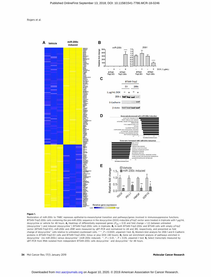
Figure 1.
Restoration of miR-200c to TNBC represses epithelial-to-mesenchymal transition and pathways/genes involved in immunosuppressive functions.BT549-TripZ-200c cells containing the pre-miR-200c sequence in the doxycycline (DOX)-inducible pTripZ vector were treated in triplicate with 1 mg/mLdoxycycline or vehicle for 48 hours. A, Heatmap of differentially expressed genes (Padj < 0.10 and fold change > 1.2) between untreated(doxycycline�) and induced (doxycyclineþ) BT549-TripZ-200c cells in triplicate. B, In both BT549-TripZ-200c and BT549 cells with empty pTripZvector (BT549-TripZ-EV), miR-200c and ZEB1 were measured by qRT-PCR and normalized to U6 and 18S, respectively, and presented as foldchange of doxycyclineþ cells relative to untreated counterpart cells. ���� , P < 0.0001, unpaired t test. C, Western blot analysis for ZEB-1 and E-Cadherinproteins in BT549-TripZ-EV cells and BT549-TripZ-200c minus or plus DOX (48 hours). D, Gene set enrichment analysis of pathways enriched indoxycycline� (no miR-200c) versus doxycyclineþ (miR-200c induced). �� , P < 0.01, � , P < 0.05, unpaired t test. E, Select transcripts measured byqRT-PCR from RNA isolated from independent BT549–200c cells doxycycline� and doxycyclineþ for 48 hours.
Rogers et al.
Mol Cancer Res; 17(1) January 2019 Molecular Cancer Research34
on August 10, 2020. © 2019 American Association for Cancer Research. mcr.aacrjournals.org Downloaded from
Published OnlineFirst September 13, 2018; DOI: 10.1158/1541-7786.MCR-18-0246

Figure 2.
Restoration of miR-200c to TNBC decreases TDO2 by direct targeting of TDO2 at a binding site in the 30UTR, resulting in reduction of intracellular andsecreted Kyn. A, Relative TDO2 mRNA levels determined by qRT-PCR in multiple TNBC cell lines following transfection with either negative SCRcontrol mimic or miR-200c for 48 hours. ��� , P < 0.001, �� , P < 0.01, unpaired t test. B, TDO2 and ZEB1 in multiple TNBC lines following transfectionwith negative control or miR-200c for 72 hours. C, Schematic of TDO2 30UTR depicting predicted binding site of miR-200c at position 395-400 cloned intopmiR-Glo-luciferase vector with site-directed mutations indicated by asterisks. D, Luciferase activity of the WT TDO2 30UTR or mutated TDO2 30UTRcontaining pmiR-Glo following transfection of BT549 cells with either negative control or miR-200c for 48 hours. ���� , P < 0.0001, one-way ANOVA analysis.E, Relative Kyn levels in BT549 cells as determined by UPLC-MS expressing a stable doxycycline-inducible lentiviral empty vector pTripZ vector ormiR-200c-pTripZ vector after 72 hours of doxycycline induction (left). Relative Kyn levels measured in Hs578T and MDA-453 following transfection with SCRcontrol or miR-200c mimic 72 hours prior (middle, right). F, MCF7 cells transfected with expression vector for TDO2 (OE) or empty vector (EV) andimmunoblot probed with TDO2-detecting antibody 24 hours after transfection (top). MCF7 cells with TDO2 OE or EV control were grown in soft agar andcolony number determined after 11 days (bottom). G, Intracellular and secreted Kyn measured by UPLC-MS in MCF7-EV or MCF7-TDO2-OE cells after 72 hours.Data are shown on a log10 scale.
miR-200c Targets TDO2 in TNBC
www.aacrjournals.org Mol Cancer Res; 17(1) January 2019 35
on August 10, 2020. © 2019 American Association for Cancer Research. mcr.aacrjournals.org Downloaded from
Published OnlineFirst September 13, 2018; DOI: 10.1158/1541-7786.MCR-18-0246

Figure 3.
Restoration of miR-200c abrogates expression of immunomodulatory gene and protein expression in TNBC cells. A, Relative TDO2 mRNA levels inBT549-TripZ-200c cells after 72 hours of no treatment, doxycycline (DOX), NFkB stimulation [TNFa, IL1b (10 ng/mL each)] for the last 48 hours, or thecombination of doxycycline and NFkB stimulation (top). Relative TDO2 levels were normalized to b-actin. ��� , P < 0.001, ��, P < 0.01 by unpaired t test.Immunoblot for TDO2 and ZEB1 with a-Tubulin as a loading control in BT549-TripZ-200c cells after 72 hours of no treatment, doxycycline, NFkBstimulation (TNFa and IL1b), or the combination of DOX and NFkB stimulation (bottom). Fold changes are indicated compared with vehicle treatedafter normalizing to loading control. B, IL6mRNA levels in BT549-TripZ-200c cells after 72 hours of no treatment, doxycycline, NFkB stimulation (TNFa, IL1b),or the combination of DOX and NFkB activation. Relative IL6 levels were normalized to b-actin. ��� , P < 0.001, �� , P < 0.01 by unpaired t test. C, RelativemiR-200c in BT549-TripZ-200c cells after 72 hours of no treatment, doxycycline alone, NFkB stimulation (TNFa, IL1b), or the combination ofdoxycycline plus NFkB activation. Relative miR-200c levels were normalized to U6 snRNA. D, HMOX-1 qRT-PCR in multiple TNBC cell lines followingtransient transfection with either negative control scrambled (SCR) or miR-200c mimic after 48 hours (left) and immunoblot for HO-1 (encoded by HMOX-1) inTNBC lines following transient transfection with SCR or miR-200c mimic for 72 hours (right). Fold changes were calculated over SCR after normalizing to thea-Tubulin loading control. E, Immunoblot for HO-1 protein in BT549-Tripz-EV cells as compared with BT549-Tripz-200c cells treated with or without DOX(þ/�; 72 hours) with or without TNFa for the last 48 hours. F, Immunoblot for GDF-15 in MDA-453 cells following transfection with SCR control or miR-200cmimic for 72 hours with or without NFkB stimulation for the last 48 hours (left) and in BT549-TripZ-200c cells after vehicle versus doxycycline for 72 hourswith or without NFkB stimulation for the last 48 hours (right). Fold change was calculated over vehicle-treated cells after normalization to a-Tubulin loadingcontrol.
Rogers et al.
Mol Cancer Res; 17(1) January 2019 Molecular Cancer Research36
on August 10, 2020. © 2019 American Association for Cancer Research. mcr.aacrjournals.org Downloaded from
Published OnlineFirst September 13, 2018; DOI: 10.1158/1541-7786.MCR-18-0246

Following transient transfection of BT549, Hs578T, MDA-453,and SUM159 TNBC lines with miR-200c mimic, HMOX-1was significantly reduced in all lines (Fig. 3D, left). HO-1protein (encoded by HMOX-1) was also decreased by miR-200c (Fig. 3D, right). Because TNFa increased HO-1 proteinlevels, but IL1b decreased them (Supplementary Fig. S3), weexamined HO-1 in empty vector containing BT549-TripZ-EVcells versus miR-200c containing BT549-TripZ-200c cells trea-ted with or without doxycycline, then plus or minus TNFa. Itwas clear that even in the face of TNFa stimulation, miR-200csuppressed production of HO-1 (Fig. 3E).
GDF-15, which encodes an anti-inflammatory cytokine alsoknown as placental TGF-beta (PTGFB) due to its high level ofexpression in placenta, decreased in BT549-TripZ-200c cellsfollowing induction of miR-200c in the array analysis (foldchange�1.7); however, the P value fell slightly below the cutoffof P < 0.1, with P ¼ 0.12. We examined whether NFkBstimulation affected GDF-15 levels and whether miR-200cwould limit this process. BT549-TripZ-200c–inducible cellswere treated with or without doxycycline in the presence orabsence of the NFkB-stimulating cytokines IL1b and TNFa for48 hours. GDF-15 protein was upregulated by 4.4-fold follow-ing cytokine treatment, but the effect was suppressed in thepresence of doxycycline induction of miR-200c (Fig. 3F, right).The same was true in MDA-453 cells, in which the NFkB-activating cytokines significantly upregulated GDF-15, butaddition of miR-200c mimic repressed both GDF-15 baselineand cytokine-induced levels (Fig. 3F, left).
Restoration of miR-200c decreases expression of PD-L1/2PD-L1 was recently identified as a direct target of miR-200c
in lung cancer and MCF7 breast cancer cells (28, 29). BecausePD-L1 was significantly decreased in BT549-200c cells follow-ing doxycycline induction in the array analysis (Fig. 1A), wetreated BT549-TripZ-200c cells with IFNg to induce PD-L1expression, and confirmed that miR-200c significantly reducedthe IFNg-mediated upregulation of PD-L1 (Fig. 4A). In addi-tion, IFNg-induced PD-L2 expression was significantly reducedin the presence of miR-200c (Fig. 4B). Reduction in PD-L1ligand following restoration of miR-200c was confirmed byflow cytometry (Fig. 4C). Figure 4D illustrates a model of howmiR-200c suppression of TDO2-dependent tryptophan cata-bolism and resulting Kyn production, along with suppressionof GDF-15 and HO-1, might affect T cells in a noncontact-dependent fashion in addition to reducing the contact-dependent immunosuppressors, PD-L1 and 2.
DiscussionHere we report a novel mechanism of regulation of tumor
tryptophan catabolism, involving post transcriptional regula-tion by miR-200c. The miR-200 family is well-described as acritical determinant of the epithelial phenotype that directlytargets and suppresses transcriptional repressors of E-cadherinsuch as ZEB1 and ZEB2, Twist, and Snail, to mediate normalEMT that facilitates rearrangement of epithelial cells duringdevelopment, as well as oncogenic EMT in carcinomas tofacilitate metastasis (19, 20). Many mesenchymal and neuronalgenes involved in motility, invasiveness, anoikis resistance,chemoresistance, and stem cell–like properties such as tropo-myosin-related kinase B (TrkB), neurotrophin 3 (NTF3),moesin,
tubulin beta 3 class III (TUBB3), and suppressor Of zeste 12protein homolog (SUZ12) are targeted by miR-200c (21, 30).In many dedifferentiated, aggressive carcinomas, includingTNBC, miR-200 family members are often lost or repressed bymicrodeletions or silenced via methylation (31, 32). Here,restoration of miR-200c to TNBC cells repressed ZEB1, restoredE-cadherin, profoundly reversed a patient-derived, pan-cancerEMT signature (26), and significantly repressed genes encodingimmunosuppressive factors. GSEA of genes reduced by induc-tion of miR-200c in TNBC revealed a change in a number ofpathways involved in immunomodulation including allograftrejection, complement and coagulation pathways, and trypto-phan metabolism.
Analysis of genes increased by induction of miR-200cdemonstrated the expected gain of epithelial characteristics,but also gave indications of increased glycolysis and oxidativephosphorylation. Although this alteration was unexpected, arecent study of the metabolic properties of an immortalizedbreast epithelial cell line and its EMT counterpart also foundglycolysis and oxidative phosphorylation to be associated withthe epithelial phenotype, while amino acid anaplerosis andfatty acid oxidation drove the mesenchymal phenotype (33).Interestingly, the tryptophan transporter LAT1 was deemed"essential for mesenchymal cell survival (33)."
Multiple clinical trials evaluating inhibitors of IDO activ-ity such as Indoximod (D-1-methyl-tryptophan, NewLinkGenetics) and INCB024360 (Incyte) either have already beenconducted or are underway. However, these compounds donot inhibit TDO2, which evolved separately to metabolizetryptophan (34, 35). Data presented here, in our previousreport, and by others, suggest that TDO2 may be relevant inTNBC and other types of cancer (11–13). Thus, new drugs thatinhibit both IDO and TDO2 may be more effective in somecancer types including TNBC. TDO2-mediated production ofKyn supports tumor cell anchorage-independent growth andinvasion in an autocrine manner by binding to and activatingAhR (11–13). Indeed AhR was recently linked to inflammationand tryptophan metabolism in breast cancer (36). Kyn can alsoact in a paracrine fashion by binding AhR in T cells to suppressT cell–mediated tumor rejection (12, 37) via expansion ofregulatory T cells (14) and direct effects on cytotoxic T cells(38). In addition to increased TDO2 upon NFkB activation inTNBC (11), we now report posttranscriptional control of TDO2by miR-200c, explaining why this enzyme is expressed in TNBC,but not ERþ lines, which have high levels of miR-200c (16).TDO2 is decreased as a consequence of direct targeting of theTDO2 30UTR by miR-200c and Kyn production is consequentlyreduced. Conversely, transfection of TDO2 lacking the 30UTRcontaining miR-200c–binding site into ERþ cells increasedexpression of Kyn and growth on soft agar.
GSEA of genes altered by miR-200c showed a reduction inpathways involved in immunosuppression and tolerance. Inter-estingly, normal EMT enables extravillar cytotrophoblasts toinvade into the maternal decidual stroma and produce immu-nosuppressive substances that ensure embryo maintenance/fetaltolerance during pregnancy (23).Wedemonstrate that restorationof miR-200c suppresses the ability of TNBC to make similarimmunosuppressive factors. The miR-200 family typically servesas the "brakes" by which EMT is kept at bay in normal epitheliumor ERþ breast cancer.We propose that loss or silencing of themiR-200 family permits TNBC to undergo EMT and coopt this
miR-200c Targets TDO2 in TNBC
www.aacrjournals.org Mol Cancer Res; 17(1) January 2019 37
on August 10, 2020. © 2019 American Association for Cancer Research. mcr.aacrjournals.org Downloaded from
Published OnlineFirst September 13, 2018; DOI: 10.1158/1541-7786.MCR-18-0246

invasive/immunosuppressive program. In support of this theory,low levels of the miR-200 family member, miR-141, permitsuccessful trophoblast uterine invasion and immunosuppressionto facilitate successful tolerance of the fetus, whereas miR-141 is
abnormally high in preeclamptic placentae (when these pro-cesses fail; ref. 39).
Mellor and Munn described the role of tryptophan catabolismin the prevention of T cell–driven complement activation and
Figure 4.
miR-200c regulates PD-L1 and 2 and other immunosuppressive factors in TNBC. BT549-TripZ-200c cells were treated with doxycycline (DOX) to induceexpression of miR-200c, or vehicle control. A, Relative PD-L1 mRNA levels were determined by qRT-PCR in 24 hours after doxycycline treatment began, cellswere treated with IFNg (10 ng/mL) or vehicle. Cells were harvested 72 hours after the commencement of doxycycline treatment. ���� , P < 0.0001, asdetermined by one-way ANOVA. B, Relative PD-L2 mRNA levels were determined by qRT-PCR in BT549 cells expressing a stably transduced, doxycycline-inducible miR-200c vector were treated with doxycycline to induce expression of miR-200c, or vehicle control. Twenty-four hours after doxycycline treatmentbegan, cells were treated with IFNg (10 ng/mL) or vehicle. Cells were harvested 72 hours after doxycycline treatment. ���� , P < 0.0001, as determined by one-way ANOVA. C, PD-L1 expression was measured by flow cytometry. BT549 cells expressing a stably transduced, doxycycline-inducible miR-200c vector weretreated with doxycycline to induce expression of miR-200c, or vehicle control. Twenty-four hours after doxycycline treatment began, cells were treated withIFNg (10 ng/mL) or vehicle. Cells were harvested 72 hours after the commencement of doxycycline treatment and stained for PD-L1. n¼ 3 samples, data shownare representative of two independent experiments. ��� , P < 0.001, ���� , P < 0.0001, as determined by unpaired t test. D, Model of factors repressed byrestoration of miR-200c to TNBC and their published effects on T cells.
Rogers et al.
Mol Cancer Res; 17(1) January 2019 Molecular Cancer Research38
on August 10, 2020. © 2019 American Association for Cancer Research. mcr.aacrjournals.org Downloaded from
Published OnlineFirst September 13, 2018; DOI: 10.1158/1541-7786.MCR-18-0246

inflammation during pregnancy (40). Inhibition of IDO orTDO2-mediated tryptophan catabolism in pregnant miceinduced fetal loss, supporting its role in maternal fetal tolerance(40). The immunosuppressive effects of both tryptophan deple-tion and increased tryptophan metabolites such as Kyn are wellestablished (12, 14, 41). TDO2 also plays a role in endometrialdecidualization where it suppresses the activation of decidual Tcells to maintain pregnancy (42).
HO-1 is an anti-inflammatory, immunosuppressive, proan-giogenic enzyme involved in suppression of CD8þ T cellsthrough expansion of T-regulatory cells necessary for fetaltolerance during pregnancy (43). Gestational stress in miceled to lower HO-1, increased cytotoxic CD8þ T cells, andintrauterine growth restriction (44). Here, we show that res-toration of miR-200c significantly decreased levels of HMOX-1(encoding HO-1) and HO-1 protein is overexpressed in breastcancer and predicts shorter overall survival (45). In clear cellrenal cell carcinoma cells, miR-200c directly targets HMOX-1(46). In a mouse mammary carcinomamodel, HO-1 inhibitionreduced tumor growth to a degree comparable with PD-1blockade (47). GDF-15 is highly expressed in placenta anddecidual stromal cells in the uterus and has been found topromote tolerogenic dendritic cells during pregnancy (48). Inmalignant gliomas, depletion of GDF-15 enhanced naturalkiller cell–mediated clearance of tumors (49) and is alsothought to help tumor cells evade immunosurveillance bymacrophages (50). In the normal prostate, GDF-15 is inverselycorrelated with inflammation (51); however, circulating GDF-15 levels are elevated in patients with prostate cancer comparedwith men without prostate cancer. GDF-15 was significantlydecreased by restoration of miR-200c to TNBC even in the faceof NFkB stimulation.
Our data support recent reports that PD-L1 (CD274) istargeted by miR-200c (28, 29), and we confirmed this findingat the protein level by flow cytometry. Restoration of miR-200calso reduced PD-L2 (CD273) induction by IFNg . Like PD-L1,PD-L2 is expressed on both antigen-presenting cells and tumorcells and interacts with PD-1 on T cells to inhibit antitumorfunction (52). High tumor PD-L2 indicated a better clinicalresponse in patients treated with pembrolizumab (Keytruda),suggesting that PD-1 blockade may function through inhibi-tion of both ligands (53).
Restoration of miR-200c decreased metastasis in an immu-nocompetent genetically engineered mouse model of claudin-low TNBC (27) and although changes in the immune responsewere not examined in that study, comparison of the data withthe human TNBC cells studied here revealed overlap in thegenes reduced upon induction of miR-200c, including TDO2,HMOX-1, IKBKB, PLCG1, and PD-L1 (Supplementary Fig. S1B).We observed that NFkB-stimulating cytokines upregulateTDO2, HO-1, and GDF-15 protein in TNBC, but this inductionby inflammatory stimuli is inhibited in the face of miR-200cas are baseline levels of these proteins. Although the ability ofmiR-200c to repress NFkB function may also be due tomiR-200c affecting upstream regulators of this pathway suchas IKKB, data presented here and by others indicate that TDO2,HMOX-1 (46), IL6 (54), and PD-L1 (28, 29) are directly target-ed by miR-200c. This insight into both the inflammation-mediated transcriptional and miRNA-mediated posttranscrip-tional regulation of specific immunosuppressive factorsproduced by TNBC cells may lead to new therapeutic strategies.
During pregnancy, infection can trigger preterm birth by acti-vating the immune system, so dampening the immune re-sponse to infection and non-self antigens is advantageous inthat setting. However, chronic inflammation may contribute tocancer by prompting epithelial cells to make immunosuppres-sive factors such as those described here, which would impedethe antitumor immune response. Furthermore, we previouslydocumented that NFkB activity increased in TNBC cells underanchorage-independent conditions (7, 11) and indeed TDO2and HMOX-1 increased in TNBC under these conditions aswell (8, 11). Thus, circulating tumor cells, which are able toresist the stress of anchorage-independent survival during dis-semination, may also be particularly able to generate immu-nosuppressive metabolites that contribute to immune evasion.Interestingly, HMOX-1 emerged in a recent synthetic lethalscreen for genes supporting metastasis in a mouse mammarycarcinoma model (55).
In a recent comparison of two isogenic mammary carcinomacell lines, the more mesenchymal line expressed higher PD-L1,lower MHC-1, and contained increased T-regulatory cells andM2macrophages in the tumor stroma (56); however, specific effectorsor regulatory mechanisms were not identified. This study revealsmultiple genes/proteinsmade by TNBC reminiscent of those usedby trophoblasts to achieve fetal tolerance including TDO2,PD-L1/2, HMOX-1, and GDF-15. Although future studies arenecessary to determine how to utilize this information to reversetumor-induced tolerance and reactivate the immune system,some of these enzymes (TDO2 and HO-1) and ligands (GDF-15)are targetable. The miR-200c family regulates the epigeneticmodifier PcG protein Enhancer of Zeste Homolog 2 (EZH2),which affects the adaptive response to immunotherapies in mel-anoma (57). Although other lines of evidence have linked EMTand immunosuppression in cancer (26, 56, 58), our study func-tionally links miR-200c to an early embryonic program of immu-nosuppressive factors that ensure fetal tolerance. Therapeuticstrategies targeting these tumor-derived noncontact-dependentimmunosuppressive factors may increase the efficacy of check-point inhibitors in TNBC.
Disclosure of Potential Conflicts of InterestNo potential conflicts of interest were disclosed.
Authors' ContributionsConception and design: T.J. Rogers, J.R. Lambert, J.K. RicherDevelopment ofmethodology: T.J. Rogers, L.I. Greene, K.I. O'Neill, T. Nemkov,A. D'Alessandro, G.D. Degala, A.-C Tan, J.K. RicherAcquisition of data (provided animals, acquired and managed pati-ents, provided facilities, etc.): T.J. Rogers, J.L. Christenson, K.I. O'Neill,M.M. Williams, T. Nemkov, A. D'Alessandro, A.-C Tan, D.M. Cittelly,J.R. Lambert, J.K. RicherAnalysis and interpretation of data (e.g., statistical analysis, biostatistics,computational analysis): T.J. Rogers, J.L. Christenson, L.I. Greene, K.I. O'Neill,M.M. Williams, M.A. Gordon, T. Nemkov, J. Shin, A.-C Tan, J.R. Lambert,J.K. RicherWriting, review, and/or revision of the manuscript: T.J. Rogers,J.L. Christenson, M.M. Williams, T. Nemkov, A.-C Tan, D.M. Cittelly,J.R. Lambert, J.K. RicherStudy supervision: J.K. Richer
AcknowledgmentsThe authors acknowledge the use of the University of Colorado
Cancer Center/NIH/NCI Cancer Core Support Grant P30 CA046934, specifi-cally the Tissue Culture Core and the DNA Sequencing Core at the University of
miR-200c Targets TDO2 in TNBC
www.aacrjournals.org Mol Cancer Res; 17(1) January 2019 39
on August 10, 2020. © 2019 American Association for Cancer Research. mcr.aacrjournals.org Downloaded from
Published OnlineFirst September 13, 2018; DOI: 10.1158/1541-7786.MCR-18-0246

Colorado-Anschutz Medical Campus (Aurora, CO). We thank Lei Zhao,University of Iowa (Iowa City, IA) for technical expertise and Nicole Spoelstraand Jill Slansky, PhD, for valuable editing. The study was supported by DODBCRP Breakthrough Level 2 Award W81XWH-15-1-0039 (to J.K. Richer); CUCancer Center's Women's Event/The Salah Foundation (to J.K. Richer); F99CA212230-01NCI Pre to Postdoctoral Fellow Transition Award (to T.J. Rogers);NRSA T32 CA190216-01A1 (to J.L. Christenson); NRSA F31 CA203486-01A1(to L.I. Greene).
The costs of publication of this article were defrayed in part by thepayment of page charges. This article must therefore be hereby markedadvertisement in accordance with 18 U.S.C. Section 1734 solely to indicatethis fact.
Received March 10, 2018; revised August 13, 2018; accepted September 6,2018; published first September 13, 2018.
References1. Liedtke C, Mazouni C, Hess KR, Andre F, Tordai A, Mejia JA, et al. Response
to neoadjuvant therapy and long-term survival in patients with triple-negative breast cancer. J Clin Oncol 2008;26:1275–81.
2. Dent R, Trudeau M, Pritchard KI, Hanna WM, Kahn HK, Sawka CA, et al.Triple-negative breast cancer: clinical features and patterns of recurrence.Clin Cancer Res 2007;13:4429–34.
3. Lin NU, Vanderplas A, Hughes ME, Theriault RL, Edge SB, Wong YN, et al.Clinicopathologic features, patterns of recurrence, and survival amongwomen with triple-negative breast cancer in the National ComprehensiveCancer Network. Cancer 2012;118:5463–72.
4. Neelakantan D, Zhou H, Oliphant MUJ, Zhang X, Simon LM, HenkeDM, et al. EMT cells increase breast cancer metastasis via paracrineGLI activation in neighbouring tumour cells. Nat Commun 2017;8:15773.
5. Yu M, Bardia A, Wittner BS, Stott SL, Smas ME, Ting DT, et al. Circulatingbreast tumor cells exhibit dynamic changes in epithelial andmesenchymalcomposition. Science 2013;339:580–4.
6. Frisch SM, Francis H. Disruption of epithelial cell-matrix interactionsinduces apoptosis. J Cell Biol 1994;124:619–26.
7. Howe EN, Cochrane DR, Cittelly DM, Richer JK. miR-200c targets a NF-kappaB up-regulated TrkB/NTF3 autocrine signaling loop to enhanceanoikis sensitivity in triple negative breast cancer. PLoS One 2012;7:e49987.
8. Barton VN, Christenson JL, GordonMA, Greene LI, Rogers TJ, Butterfield K,et al. Androgen receptor supports an anchorage-independent, cancer stemcell-like population in triple-negative breast cancer. Cancer Res 2017;77:3455–66.
9. Schafer ZT, Grassian AR, Song L, Jiang Z, Gerhart-Hines Z, Irie HY, et al.Antioxidant and oncogene rescue of metabolic defects caused by loss ofmatrix attachment. Nature 2009;461:109–13.
10. Farris JC, Pifer PM, Zheng L, Gottlieb E, Denvir J, Frisch SM. Grainy-head-like 2 reverses the metabolic changes induced by the oncogenicepithelial-mesenchymal transition: effects on anoikis. Mol Cancer Res2016;14:528–38.
11. D'Amato NC, Rogers TJ, Gordon MA, Greene LI, Cochrane DR,Spoelstra NS, et al. A TDO2-AhR signaling axis facilitates anoikisresistance and metastasis in triple-negative breast cancer. Cancer Res2015;75:4651–64.
12. Opitz CA, Litzenburger UM, Sahm F, Ott M, Tritschler I, Trump S, et al. Anendogenous tumour-promoting ligand of the human aryl hydrocarbonreceptor. Nature 2011;478:197–203.
13. Novikov O, Wang Z, Stanford EA, Parks AJ, Ramirez-Cardenas A,Landesman E, et al. An aryl hydrocarbon receptor-mediated amplificationloop that enforces cell migration in ER-/PR-/Her2- human breast cancercells. Mol Pharmacol 2016;90:674–88.
14. Mezrich JD, Fechner JH, Zhang X, Johnson BP, Burlingham WJ, BradfieldCA. An interaction between kynurenine and the aryl hydrocarbon receptorcan generate regulatory T cells. J Immunol 2010;185:3190–8.
15. Fallarino F, Grohmann U, Vacca C, Bianchi R, Orabona C, Spreca A,et al. T cell apoptosis by tryptophan catabolism. Cell Death Differ2002;9:1069–77.
16. CochraneDR,CittellyDM,Howe EN, SpoelstraNS,McKinsey EL, LaPara K,et al. MicroRNAs link estrogen receptor alpha status and Dicer levels inbreast cancer. Horm Cancer 2010;1:306–19.
17. Bockmeyer CL, Christgen M, Muller M, Fischer S, Ahrens P, Langer F, et al.MicroRNA profiles of healthy basal and luminal mammary epithelial cellsare distinct and reflected in different breast cancer subtypes. Breast CancerRes Treat 2011;130:735–45.
18. Castilla MA, Diaz-Martin J, Sarrio D, Romero-Perez L, Lopez-Garcia MA,Vieites B, et al. MicroRNA-200 family modulation in distinct breast cancerphenotypes. PLoS One 2012;7:e47709.
19. Gregory PA, Bert AG, Paterson EL, Barry SC, Tsykin A, Farshid G, et al. ThemiR-200 family and miR-205 regulate epithelial to mesenchymal transi-tion by targeting ZEB1 and SIP1. Nat Cell Biol 2008;10:593–601.
20. Park SM,Gaur AB, Lengyel E, PeterME. ThemiR-200 family determines theepithelial phenotype of cancer cells by targeting the E-cadherin repressorsZEB1 and ZEB2. Genes Dev 2008;22:894–907.
21. Howe EN, Cochrane DR, Richer JK. The miR-200 and miR-221/222microRNA families: opposing effects on epithelial identity. J MammaryGland Biol Neoplasia 2012;17:65–77.
22. Humphries B, Yang C. The microRNA-200 family: small molecules withnovel roles in cancer development, progression and therapy. Oncotarget2015;6:6472–98.
23. Piechowski J. Trophoblastic-like transdifferentiation: a key to oncogenesis.Crit Rev Oncol Hematol 2016;101:1–11.
24. Costanzo V, Bardelli A, Siena S, Abrignani S. Exploring the links betweencancer and placenta development. Open Biology 2018;8:180081.
25. Holtan SG, Creedon DJ, Haluska P, Markovic SN. Cancer and pregnancy:parallels in growth, invasion, and immune modulation and implicationsfor cancer therapeutic agents. Mayo Clin Proc 2009;84:985–1000.
26. Mak MP, Tong P, Diao L, Cardnell RJ, Gibbons DL, William WN, et al.A patient-derived, pan-cancer EMT signature identifies global molec-ular alterations and immune target enrichment following epithelial-to-mesenchymal transition. Clin Cancer Res 2016;22:609–20.
27. Knezevic J, Pfefferle AD, Petrovic I, Greene SB, Perou CM, Rosen JM.Expression of miR-200c in claudin-low breast cancer alters stem cellfunctionality, enhances chemosensitivity and reducesmetastatic potential.Oncogene 2015;34:5997–6006.
28. Chen L, Gibbons DL, Goswami S, Cortez MA, Ahn YH, Byers LA, et al.Metastasis is regulated viamicroRNA-200/ZEB1 axis control of tumour cellPD-L1 expression and intratumoral immunosuppression. Nat Commun2014;5:5241.
29. Noman MZ, Janji B, Abdou A, Hasmim M, Terry S, Tan TZ, et al. Theimmune checkpoint ligand PD-L1 is upregulated in EMT-activatedhuman breast cancer cells by a mechanism involving ZEB-1 andmiR-200. Oncoimmunology 2017;6:e1263412.
30. Iliopoulos D, Lindahl-Allen M, Polytarchou C, Hirsch HA, Tsichlis PN,Struhl K. Loss ofmiR-200 inhibitionof Suz12 leads to polycomb-mediatedrepression required for the formation and maintenance of cancer stemcells. Mol Cell 2010;39:761–72.
31. Vrba L, Jensen TJ, Garbe JC, Heimark RL, Cress AE, Dickinson S, et al. Rolefor DNA methylation in the regulation of miR-200c and miR-141 expres-sion in normal and cancer cells. PLoS One 2010;5:e8697.
32. HuX,MacdonaldDM,Huettner PC, Feng Z, ElNaqa IM, Schwarz JK, et al. AmiR-200 microRNA cluster as prognostic marker in advanced ovariancancer. Gynecol Oncol 2009;114:457–64.
33. Halldorsson S, Rohatgi N, Magnusdottir M, Choudhary KS, Gudjonsson T,Knutsen E, et al. Metabolic re-wiring of isogenic breast epithelial cell linesfollowing epithelial to mesenchymal transition. Cancer Lett 2017;396:117–29.
34. Suzuki S, Tone S, TakikawaO, Kubo T, Kohno I, Minatogawa Y. Expressionof indoleamine 2,3-dioxygenase and tryptophan 2,3-dioxygenase in earlyconcepti. Biochem J 2001;355:425–9.
35. Liu X, Shin N, Koblish HK, Yang G, Wang Q, Wang K, et al. Selectiveinhibition of IDO1 effectively regulatesmediators of antitumor immunity.Blood 2010;115:3520–30.
Rogers et al.
Mol Cancer Res; 17(1) January 2019 Molecular Cancer Research40
on August 10, 2020. © 2019 American Association for Cancer Research. mcr.aacrjournals.org Downloaded from
Published OnlineFirst September 13, 2018; DOI: 10.1158/1541-7786.MCR-18-0246

36. Vacher S, Castagnet P, Chemlali W, Lallemand F, Meseure D, PocardM, et al. High AHR expression in breast tumors correlates withexpression of genes from several signaling pathways namely inflam-mation and endogenous tryptophan metabolism. PLoS One 2018;13:e0190619.
37. Pilotte L, Larrieu P, Stroobant V, Colau D, Dolusic E, Frederick R, et al.Reversal of tumoral immune resistance by inhibition of tryptophan 2,3-dioxygenase. Proc Natl Acad Sci U S A 2012;109:2497–502.
38. Greene LI, Bruno T, Christenson JL, D'Alessandro A, Culp-Hill R, Torkko K,et al. A role for tryptophan-2,3-dioxygenase in CD8 T cell suppression andevidence of tryptophan catabolism in breast cancer patient plasma. MolCancer Res 2019;17:131–9.
39. Ospina-Prieto S, Chaiwangyen W, Herrmann J, Groten T, Schleussner E,Markert UR, et al. MicroRNA-141 is upregulated in preeclamptic placentaeand regulates trophoblast invasion and intercellular communication.Transl Res 2016;172:61–72.
40. Mellor AL, Sivakumar J, Chandler P, Smith K, Molina H, Mao D, et al.Prevention of T cell-driven complement activation and inflammation bytryptophan catabolism during pregnancy. Nat Immunol 2001;2:64–8.
41. Fallarino F, Grohmann U, You S, McGrath BC, Cavener DR, Vacca C, et al.The combined effects of tryptophan starvation and tryptophan catabolitesdown-regulate T cell receptor zeta-chain and induce a regulatory pheno-type in naive T cells. J Immunol 2006;176:6752–61.
42. Li DD, Gao YJ, Tian XC, Yang ZQ, Cao H, Zhang QL, et al. Differentialexpression and regulation of Tdo2 during mouse decidualization.J Endocrinol 2014;220:73–83.
43. Schumacher A, Wafula PO, Teles A, El-Mousleh T, Linzke N, ZenclussenML, et al. Blockage of heme oxygenase-1 abrogates the protective effectof regulatory T cells onmurine pregnancy and promotes the maturation ofdendritic cells. PLoS One 2012;7:e42301.
44. Solano ME, Kowal MK, O'Rourke GE, Horst AK, Modest K, Plosch T, et al.Progesterone and HMOX-1 promote fetal growth by CD8þ T cell mod-ulation. J Clin Invest 2015;125:1726–38.
45. Noh SJ, Bae JS, JamiyandorjU, ParkHS, KwonKS, Jung SH, et al. Expressionof nerve growth factor and heme oxygenase-1 predict poor survival ofbreast carcinoma patients. BMC Cancer 2013;13:516.
46. Gao C, Peng FH, Peng LK. MiR-200c sensitizes clear-cell renal cell carci-noma cells to sorafenib and imatinib by targeting heme oxygenase-1.Neoplasma 2014;61:680–9.
47. Muliaditan T, Opzoomer JW, Caron J, Okesola M, Kosti P, Lall S, et al.Repurposing tin mesoporphyrin as an immune checkpoint inhibitorshows therapeutic efficacy in preclinical models of cancer. Clin CancerRes 2018;24:1617–28.
48. Segerer SE, Rieger L, KappM, Dombrowski Y, Muller N, Dietl J, et al. MIC-1(a multifunctional modulator of dendritic cell phenotype and function) isproduced by decidual stromal cells and trophoblasts. Hum Reprod2012;27:200–9.
49. Roth P, Junker M, Tritschler I, Mittelbronn M, Dombrowski Y, Breit SN,et al. GDF-15 contributes to proliferation and immune escape ofmalignantgliomas. Clin Cancer Res 2010;16:3851–9.
50. Ratnam NM, Peterson JM, Talbert EE, Ladner KJ, Rajasekera PV, SchmidtCR, et al. NF-kappaB regulates GDF-15 to suppress macrophage surveil-lance during early tumor development. J Clin Invest 2017;127:3796–809.
51. Lambert JR, Whitson RJ, Iczkowski KA, La Rosa FG, Smith ML, Wilson RS,et al. Reduced expression of GDF-15 is associated with atrophic inflam-matory lesions of the prostate. Prostate 2015;75:255–65.
52. Latchman Y, Wood CR, Chernova T, Chaudhary D, Borde M, Chernova I,et al. PD-L2 is a second ligand for PD-1 and inhibits T cell activation.Nat Immunol 2001;2:261–8.
53. Yearley JH, Gibson C, Yu N, Moon C, Murphy E, Juco J, et al. PD-L2expression in human tumors: relevance to anti-PD-1 therapy in cancer.Clin Cancer Res 2017;23:3158–67.
54. Hong L, Sharp T, Khorsand B, Fischer C, Eliason S, Salem A, et al. Micro-RNA-200c Represses IL-6, IL-8, and CCL-5 expression and enhances oste-ogenic differentiation. PLoS One 2016;11:e0160915.
55. Knott SRV, Wagenblast E, Khan S, Kim SY, Soto M, Wagner M, et al.Asparagine bioavailability governs metastasis in a model of breast cancer.Nature 2018;554:378–81.
56. Dongre A, Rashidian M, Reinhardt F, Bagnato A, Keckesova Z, Ploegh HL,et al. Epithelial-to-Mesenchymal transition contributes to immunosup-pression in breast carcinomas. Cancer Res 2017;77:3982–9.
57. Zingg D, Arenas-Ramirez N, Sahin D, Rosalia RA, Antunes AT, Haeusel J,et al. The Histone Methyltransferase Ezh2 controls mechanisms ofadaptive resistance to tumor immunotherapy. Cell Rep 2017;20:854–67.
58. Akalay I, Janji B, Hasmim M, Noman MZ, Andre F, De Cremoux P, et al.Epithelial-to-mesenchymal transition and autophagy induction in breastcarcinoma promote escape from T-cell-mediated lysis. Cancer Res 2013;73:2418–27.
www.aacrjournals.org Mol Cancer Res; 17(1) January 2019 41
miR-200c Targets TDO2 in TNBC
on August 10, 2020. © 2019 American Association for Cancer Research. mcr.aacrjournals.org Downloaded from
Published OnlineFirst September 13, 2018; DOI: 10.1158/1541-7786.MCR-18-0246

2019;17:30-41. Published OnlineFirst September 13, 2018.Mol Cancer Res Thomas J. Rogers, Jessica L. Christenson, Lisa I. Greene, et al. ImmunosuppressionDecreases Tryptophan Catabolism and a Program of Reversal of Triple-Negative Breast Cancer EMT by miR-200c
Updated version
10.1158/1541-7786.MCR-18-0246doi:
Access the most recent version of this article at:
Cited articles
http://mcr.aacrjournals.org/content/17/1/30.full#ref-list-1
This article cites 58 articles, 22 of which you can access for free at:
E-mail alerts related to this article or journal.Sign up to receive free email-alerts
Subscriptions
Reprints and
To order reprints of this article or to subscribe to the journal, contact the AACR Publications Department at
Permissions
Rightslink site. Click on "Request Permissions" which will take you to the Copyright Clearance Center's (CCC)
.http://mcr.aacrjournals.org/content/17/1/30To request permission to re-use all or part of this article, use this link
on August 10, 2020. © 2019 American Association for Cancer Research. mcr.aacrjournals.org Downloaded from
Published OnlineFirst September 13, 2018; DOI: 10.1158/1541-7786.MCR-18-0246