retinoblastoma a study of two cases by electron...
TRANSCRIPT
Retinoblastoma
A study of two cases by electron microscopy
Raymond A. Allen, Harrison Latta, and Bradley R. Straatsma
The ultrastructure of two retinoblastomas includes cytologic features which are characteristicof neoplasia. Unusual degenerative changes were observed in both tumors. Rosettes xoerepresent in one case, and the cells which formed them showed some characteristics ofependyma of the primitive neural tube, and also of developing vision receptor cells. Structuresresembling pericentriolar bodies xoere associated with basal bodies of cilia in rosettes.
R'etinoblastoma, the most common intra-ocular tumor of childhood, has been wellcharacterized as regards its clinical be-havior and its varied structure by lightmicroscopy. This report is to record fea-tures of two such tumors observed byelectron microscopy. No previous suchstudy appears to have been published.
Materials, methods, andobservations by light microscopy
Case 1. A 3-year-old white boy was first foundto have an abnormal light reflex in the left eye1V2 months before he was seen at the Universityof California Medical Center, Los Angeles. Therewas no family history of retinoblastoma or signif-icant eye disease.
Physical examination disclosed no abnormalityexcept the ocular lesion. X-rays of the skull werenormal.
Examination of the eyes demonstrated normalvision in the right eye and no light perception in
From the Department of Pathology and theDivision of Ophthalmology, Department ofSurgery, University of California MedicalCenter, Los Angeles, Calif.
This project was supported by Grant-in-Aid No.C237 of the Bob Hope Fight-for-Sight Fundof the National Council to Combat Blindness,Inc., New York City.
the left eye. The right eye appeared normal inall respects. The left eye contained a large creamy-white and irregularly vascularized tumor masswhich filled the temporal aspect of the posteriorsegment, and obscured the optic nerve head. Thevitreous body contained numerous, rather small,discrete white particles of neoplasm.
Enucleation of the left eye was performedunder general anesthesia. Immediately after re-moval, the eye was opened, disclosing a softwhite tumor mass nearly filling the vitreous body.Bits of tumor tissue were immediately placedin 1 per cent osmium tetroxide fixative bufferedwith Veronal acetate and hydrochloric acid to pH7.36. Fixation at 0° C. was continued for 3Vfehours. Dehydration in graded alcohol series wasfollowed by propylene oxide for Epon embedding,according to the method of Luft.21 An estimatedperiod of 2 minutes elapsed between cutting theoptic nerve and placing the tumor fragments in-to fixative.
Sections about 1 M thick were cut from theblocks on a Porter-Blum microtome and stainedwith toluidine blue for light microscopy. Thinsections were cut and mounted on grids coatedwith celloidin. Most sections were stained withuranyl acetate,3'1 though some were stained withbasic lead oxide.17 The specimens were examinedwith an RCA EMU 3-B electron microscope.
Light microscopy disclosed a large tumor massextending inward from a broad attachment to theretina (Fig. 1) and occupying most of theposterior portion of the vitreous body. Theclosely packed tumor cells varied only slightly insize, shape, and staining. The cytoplasm was
728
Downloaded From: http://iovs.arvojournals.org/pdfaccess.ashx?url=/data/journals/iovs/932887/ on 06/07/2018

Number 6Retinoblastoma 729
sparse, faintly pink, and apparently shrunken. Thenuclei were generally round or oval, though somewere lobated and occasional ones appeared folded(Figs. 2 and 3). Most cells contained one ormore nucleoli which were seen best in theosmium-fixed toluidine blue-stained sections (Fig.3). Rosette formation was not observed. Occa-sional foci of calcium were present in necroticareas.
Within the retina the tumor extended to theoptic nerve head, but the optic nerve itself wasnot invaded. The tumor did not invade thechoroid.
Case 2. A 2V2-year-old white boy had beenwell throughout his life. One and one halfmonths before admission his parents noted a"strange red reflection" from his eyes as hewatched television. He was referred to the Uni-versity of California Medical Center for ophthal-mologic evaluation. There was no family historyof eye defects or neoplasms.
Physical examination disclosed a well-developedchild, apparently healthy except for the eye find-ings.
Examined under anesthesia, the left eye showeda large tumor mass occupying the temporal halfof the vitreous body and an associated retinal
detachment. Clumps of tumor could be seen float-ing in the vitreous body. In the right eye therewas a nodular area of retinoblastoma slightly in-ferior to the optic nerve head.
Enucleation of the left eye was advised, anda program of treatment for the right eye wasrecommended. This program, subsequently carriedout, included right internal carotid artery injectionof triethylenemelamine, x-irradiation, and photo-coagulation of the tumor nodule.
The left eye was enucleated under generalanesthesia and immediately after removal the eyewas opened. The lower temporal sector of theretina was replaced by a white tumor mass ap-proximately 1 cm. across. The growth lay prin-cipally on the inner surface of the retina andprotruded into the vitreous body.
The technical procedures followed were similarto those described for Case 1 with the followingexceptions: (1) a period of about 15 minuteselapsed between the time the optic nerve wasclamped and the bits of tissue were placed inosmium fixative; (2) fixation time in chilledosmium fixative was I to lVk hours.
Light microscopy disclosed a fairly large solidtumor mass extending into the vitreous bodyfrom- the posterior temporal portion of the retina
Fig. 1. Case 1. Posterior portion of eye, showing main tumor mass. The neoplasm has abroad retinal attachment and has grown over the optic nerve head, though the optic nerveitself was not invaded. Retinal detachment was present, but shrinkage during processingaccounts for most of the separation shown. (x7.5.)
Downloaded From: http://iovs.arvojournals.org/pdfaccess.ashx?url=/data/journals/iovs/932887/ on 06/07/2018

730 Allen, Latta, and Siraatsma Investigative OphthalmologyDecember 1962
I
I
I
Fig. 2. Case 1. Microscopic appearance of tumor,formalin-fixed paraffin embedded tissue. The cellsare fairly uniform in size and staining, butcytologic detail is lacking. Scattered pyknoticnuclei are evident. (Hematoxylin and eosin. x500;reduced %.)
(Fig. 4). The cells were similar to diose of Case1 but differed in that there was conspicuousrosette formation (Figs. 5 and 6). Areas ofnecrosis with calcification were present. Variable-sized clumps of tumor cells were free within thevitreous body. Large tumor cell aggregates werepresent in the intraretinal space, and at some sitesthe neoplasm had invaded the choroid. Thetumor extended to the margins of the optic nervehead, but the optic nerve was not invaded.
Observations by electron microscopyThe cellular characteristics of the two
tumors were essentially similar. The de-scriptions to follow, therefore, apply toboth tumors and differences will be citedseparately. The most significant differenceappeared to be rosette formations whichoccurred in Case 2.
Where the cells were best preserved, theneoplasm consisted of sheets of polyhedralcells with interposed degenerating cells orcell processes (Figs. 7 and 9). Much of thevariation in size and shape may be ex-
Fig. 3. The same tumor as shown in Fig. 2, butfixed in osmium and embedded in epon forelectron microscopy. The cells are much lessshrunken and much greater cell detail can beseen. Degenerating cells are even more evident.(Toluidine blue. x500; reduced %.)
plained by the thin sections which includedonly narrow portions of some cells.
The cytoplasm. The cytoplasm generallyconsisted of a narrow rim around thenucleus, but some cells had angular orrounded extensions from the main cell.This latter feature was more conspicuousin Case 2,. especially near rosette forma-tions (Fig. 23).
The mitochondria were the most promi-nent cytoplasmic component, and showedmoderate variation in number, location,and size in different cells. In some instancesthey appeared to occupy a sector of thecell, extending from the nuclear envelopealmost to the plasma membrane, and weredecreased in number or absent in otherportions of the cytoplasm. Where tumorcells were in relation to small bloodvessels, the mitochondria seemed morenumerous on the vascular side of the cell(Fig. 22).
Downloaded From: http://iovs.arvojournals.org/pdfaccess.ashx?url=/data/journals/iovs/932887/ on 06/07/2018
, Volume 1Number 6
In section the mitochondria containedprominent cristae and showed moderatevariability in size and shape. Rare bizarreforms were observed. In one mitochondriona cluster of distinct round osmiophilicbodies could be seen (Fig. 20). In earlystages of cell degeneration the mitochon-dria became swollen and empty (Fig. 20).
The Golgi complex could be identified insome cells, but it was generally small andinconspicuous (Fig. 20).
The endoplasmic reticulum was also arelatively inconspicuous cytoplasmic com-ponent. It consisted mainly of small vesiclesand occasional cisternae with denseribonucleoprotein (RNP) granules. Occa-sionally small vesicles were seen withoutRNP granules.
Centrioles could be identified in manycells, and were generally located near thenucleus. Cilia were present in the cellsforming rosettes in Case 2. These struc-tures will be further described in connec-tion with the rosettes.
Several varieties of cytoplasmic inclu-sions were observed. The most commonof these were spheroidal homogeneousosmiophilic bodies, possibly lipid. Therewere membranous cytoplasmic bodies con-taining more coarsely granular materialwhich may represent lysosomes. One cellcontained a pair of fibrillar or layeredbodies which were apparently containedwithin a membranous sac (Fig. 8). Vac-uoles of irregular size and shape werepresent in some cells, but were more con-spicuous in degenerating cells.
Cytoplasmic fibrils were present, espe-cially in cells forming rosettes, and wereless commonly found in other areas of thetumors.
The nucleus. In contrast to the compara-tively uniform appearance of the nuclei bylight microscopy of the sections stainedwith toluidine blue (Figs. 3 and 6), theelectron microscope disclosed considerablevariation in shape and structure (Figs. 7and 9). Many nuclei were round or oval,but deep cytoplasmic indentations werecommon. Often several indentations of
Retinoblastoma 731
varying depth were seen within a singlenucleus, in some cases resulting in a U- orY-shaped nuclear configuration. Occasionalcells appeared binucleated, apparently asthe result of sectioning both arms of theU (Figs. 7 and 9). Likewise, some ir-regular sections through cytoplasmic in-vaginations disclosed seemingly isolatedislands of cytoplasm within the nucleus.In many cells the nuclei were bi- or tri-lobated, the lobes connected only by a thinstrand of chromatin material, with its in-vesting nuclear envelope intact. Occa-sionally small bosses of nuclear materialwere present on the surface of the nucleus-In many cells small nuclear spurs orrounded projections were connected to themain nuclear mass by a thin stalk orpedicle. More complicated nuclear formswere common with complex divisions andsubdivisions of nuclear processes and cor-
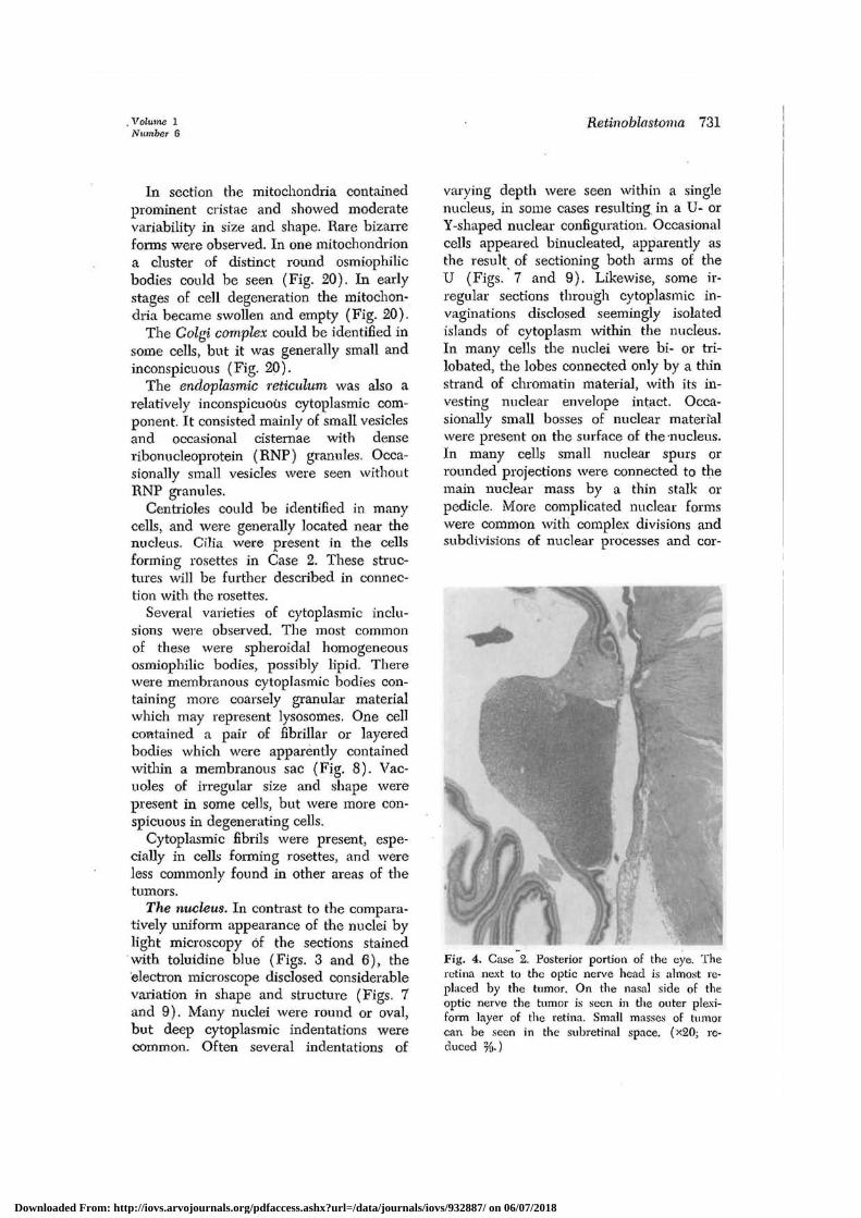
Fig. 4. Case 2. Posterior portion of the eye. Theretina next to the optic nerve head is almost re-placed by the tumor. On the nasal side of theoptic nerve the tumor is seen in the outer plexi-form layer of the retina. Small masses of tumorcan be seen in the subretinal space. (*20; re-duced %.)
Downloaded From: http://iovs.arvojournals.org/pdfaccess.ashx?url=/data/journals/iovs/932887/ on 06/07/2018

732 Allen, Latta, and Straatsma Investigative OphthalmologyDecember 1962
Fig. 5. Case 2. Rosette formation, as shown informalin-fixed paraffin-embedded section here,was" a conspicuous feature of tumor. Degeneratingcells and tissue necrosis are evident. (Hematoxylinand eosin, x500; reduced %.)
Fig. 6. The same tumor as shown in Fig. 5. Lessshrinkage of cells and better preservation of celldetail can be seen. Apparently healthy cells ap-pear adjacent to large areas of necrosis. (Osmiumfixation; tuluidine blue. x500; reduced %.)
responding cytoplasmic indentations (Figs.9 and 23).
The nuclear envelope consisted of twodistinct layers. Pores in the nuclear en-velope were observed in higher magnifica-tions. In some apparently well-preservedcells there was cisternal separation of thetwo layers which gave the appearance ofnuclear blebs (Fig. 10). This feature didnot seem accentuated in cells with changesof early degeneration.
Depending upon the stage of mitosis, thenuclear membrane was present to a vary-ing extent or appeared absent altogetherin some cells (Figs. 14 to 17). In manydegenerated cells both layers of the nuclearenvelope were clearly distinguishable afterthe nuclear chromatin had lost its organ-ized appearance and extensive degenera-tion had occurred in the cytoplasm (Figs.18 and 19).
Two fairly distinct patterns of nuclearchromatin distribution were observed: (1)
a fairly even distribution of small, denseparticles with one or more nucleoli (Fig.7); (2) aggregations into irregular coarseclumps with patchy condensations alongthe nucleai' envelope (Figs. 9 and 20).
The nucleoplasm contained fine fibrillaror granular material. Occasionally smallmembranous structures lay close to theinner layer of the nuclear envelope andmight represent imaginations of it (Figs.11, 12, and 16).
In cells with evenly distributed nuclearchromatin several nucleoli were often ap-parent. Usually this structure was situatedat the periphery of the nucleus, oftenadjacent to the nuclear envelope. It wasgenerally round or oval, but it occurredin a variety of less regular shapes.
Mitotic figures. These were fairly numer-ous in both tumors. Most of them were inmetaphase or telophase, but occasionally,earlier stages were noted (Figs. 14 and23)/
i t*
Downloaded From: http://iovs.arvojournals.org/pdfaccess.ashx?url=/data/journals/iovs/932887/ on 06/07/2018

Volume 1Number 6
Retinoblastoma 733
Cellular relationships. The relationshipsof the cells appeared comparatively simple.Surface differentiation was not observedexcept in the rosettes where terminal barswere present (Figs. 23 and 24). Mostlythe cells were fairly close together, theplasma membranes of adjacent cells form-ing more or less parallel lines. Portions ofother cells were sometimes interposedwhere the intercellular spaces widened.The tumor cells apparently did not formintercellular material.
Degenerative changes. Degeneration waswidespread in both tumors, and in prac-tically every field there were cells or por-tions of cells where such changes werepresent to some extent (Figs. 7, 10, and14).
One common form of degeneration(Figs. 18 and 19) consisted of fine andcoarse clumping of the nuclear chromatinwith disorganization. Both layers of thenuclear envelope were usually preserved,but convoluted and partly collapsed. Thecytoplasm showed loss of fine detail,though occasional, vesicles and clusters ofRNP remained. The plasma membrane re-mained, irregularly convoluted but ap-parently intact. At a later stage ofdegeneration the nucleus appeared as ir-regularly clumped dark granules within amembranous sac. Various-sized membra-nous sacs enclosed cytoplasmic constituents,some of which could be recognized asmitochondria. These latter structures ap-peared ballooned, and the cristae withinthem appeared as small cogs.
A slightly different degenerative patternoccurred when the nuclear chromatinclumped into variably sized dark homo-geneous masses (Figs. 7 and 14). In thesecells the membranous structures were lesswell preserved, and only a few smalltubular structures remained in the cyto-plasm, admixed with clumps of granularmaterial. That such changes were not dueto fixation was evident from the fact thatsuch degenerating cells lay immediatelyadjacent to or often completely surroundedby well-preserved cells. Degenerated neo-
plastic cells adjacent to blood vessels werenoted frequently.
Where degeneration was far more exten-sive and involved large areas, there wascellular fragmentation and loss of all finecell detail. Among the cellular debris therewere smudged, round, dark masses ofvariable size and poorly defined mem-branous and tubular remnants. Occasionallarger vacuolated bodies and myelinfigures with amorphous scattered granularmaterial were also present (Fig. 21).
Blood vessels. These were fairly numer-ous in the main tumor mass. They weremostly capillaries, lined by a single layerof endothelial cells resting upon a well-defined basement membrane which in someinstances was shared with pericytes. Bothof these cell types were contiguous withthe neoplastic cells.
The inner surface of the endotheliumwas irregular (Fig. 22) and spurs and flap-like processes projected into the lumen.In vessels which appeared collapsed, therewere more complicated endothelial infokl-ings. Several focal densities resemblingterminal bars were visible along the cleftforming the junctions of the endothelialcells. The capillary endothelial basementmembranes were distinct and appearedseparate from the basement membrane ofadjacent tumor cells (Fig. 22).
Rosettes. A striking feature of Case 2,not observed in Case 1, was the rosette,which consisted of a radial arrangement oftumor cells around a central lumen.Usually the circle of cells consisted of 9or 10 cell nuclei in a single plane, andcell processes from an additional 7 or 8cells could be identified as apparentlypaiticipating in the formation of the rosette(Fig. 23). Mitosis was often found in thecells of the rosette.
The nuclei were situated in the periph-eral broader portions of the cells near theouter cell margin. Cytoplasmic indentationsinto the nucleus were often striking, andoccurred principally on the luminal side ofthe nucleus, producing long fingerlike ex-tensions pointing to the lumen. Cyto-
Downloaded From: http://iovs.arvojournals.org/pdfaccess.ashx?url=/data/journals/iovs/932887/ on 06/07/2018

734 Allen, Latta, and Straatsma Investigative OphthalmologyDecember 1962
plasmic components also appeared orientedin the direction of the rosette lumen.
The mitochondria were mostly on theluminal side of the nucleus and had aradial arrangement. Admixed with themitochondria and also occupying theinner portion of the cytoplasm were manytubules and vesicles, including endoplasmicreticulum. Filamentous structures fre-quently extended radially within the cyto-plasm from the narrow luminal border ofsome cells (Fig. 24). Near the lumenterminal bars were prominent between thecells (Figs. 23 and 24).
The lumen of the rosette containedmicrovilli and broad clublike projectionsfrom some of its component cells. Only ascant amount of amorphous granular mate-rial was present within the nearly emptyspace.
Well-defined cilia extended into thelumen (Fig. 26). No more than one ciliumwas identified with any single cell. Thecilia and their basal bodies had distinctivestructural features (Figs. 24, 25, and 26).In one case a longitudinal section througha basal body showed structures resemblingpericentriolar bodies (Fig. 25). Near thelumen surface of a nearby cell there was astructure resembling a centriole withseveral pericentriolar bodies cut in crosssection (Fig. 24)
Discussion and comment
General features of neoplasia. As intumor diagnosis by light microscopy, thereis no known ultrastructural feature or com-bination of features specific for cancercells. The various abnormalities which oc-cur in tumor cells have been well sum-marized.-0 These may vary greatly in dif-
ferent neoplasms and their presence canbe considered only as general features ofcancer.JS
Retinoblastoma cells were characterizedby the following changes in the nuclei:enlargement, tabulation, and irregularityof shape, with pseudoinclusions of cyto-plasmic organelles. Additionally, the nu-cleoli were enlarged and were oftenmultiple.
Cytoplasmic changes were less striking.In general the normal cytoplasmic com-ponents (mitochondria, Golgi apparatus,endoplasmic reticulum) did not showsignificant alterations. One tumor cellshowed stratified inclusion bodies ap-parently enclosed within a membranousenvelope (Fig. 8) but the significance ofthese is not known. No inclusions sug-gestive of virus particles were seen.Mitotic figures were increased in number,but no abnormal forms were seen. Thesecytologic changes are in general agreementwith abnormalities observed in other neo-plasms by others.8's' 12>2G
Only the cells comprising the rosettesappeared to have specific orientation. Thetraditional view that rosette formation inretinoblastoma is a favorable prognosticfeature is probably sound, since accordingto Oberling and Bernhard29 "polarity isgradually lost in proportion to the degreeof anaplasia."
Degenerative changes. Necrosis and de-generation were conspicuous histologicfeatures of both tumors. By light micros-copy these areas were similar to suchchanges described by others.1'1
By electron microscopy degenerationand necrosis were even more widely evi-dent, large portions of both tumors show-
Fig. 7. Typical appearance of well-preserved retinoblastoma cells. The cells are of fairlyuniform shape and have a tile-like arrangement. The nuclei show more variation in shapeand have deep cytoplasmic imaginations. The appearance of a binucleated cell (upperright) is a result of sectioning across 2 lobes of a single nucleus. A portion of a degeneratingcell is present in upper right corner. Cytoplasmic bodies (box) are shown in inset (Fig. 8).Membranous structure (arrow) is shown in Fig. 13. (*8,000; reduced %.)
Fig. 8. Paired fibrillar cytoplasmic inclusions of unknown nature are apparently containedwithin a membranous envelope. (x54,000; reduced %.)
Downloaded From: http://iovs.arvojournals.org/pdfaccess.ashx?url=/data/journals/iovs/932887/ on 06/07/2018

Volume 1Number 6
Retinoblastoma 735
Figs. 7 and 8. For legends see opposite page.
Downloaded From: http://iovs.arvojournals.org/pdfaccess.ashx?url=/data/journals/iovs/932887/ on 06/07/2018
736 Allen, Ltitta, and Straatsmci Investigative OphthalmologyDecember 1962
•• ) f • *
Figs. 9-13, For legends see page 741.
Downloaded From: http://iovs.arvojournals.org/pdfaccess.ashx?url=/data/journals/iovs/932887/ on 06/07/2018

Volume 1Number 6
Retinoblastoma 737
Figs. 14-17. For legends see page 741.
Downloaded From: http://iovs.arvojournals.org/pdfaccess.ashx?url=/data/journals/iovs/932887/ on 06/07/2018

738 Allen, Latta, and Straatsma Investigative OphthalmologyDecember 1962
Figs. 18 and 19. For legends see page 741.
L.,
Downloaded From: http://iovs.arvojournals.org/pdfaccess.ashx?url=/data/journals/iovs/932887/ on 06/07/2018

Volume 1Number 6
Retinoblastoma 739
M
'¥•
Figs. 20-22. For legends see page 741,
1
Downloaded From: http://iovs.arvojournals.org/pdfaccess.ashx?url=/data/journals/iovs/932887/ on 06/07/2018

r740 Allen, Latta, and Straatsma Inoestigal-ioe Oyihtluilnwtogy
December 1962
•
p
Figs. 23-26. For legends see page 742,
Downloaded From: http://iovs.arvojournals.org/pdfaccess.ashx?url=/data/journals/iovs/932887/ on 06/07/2018

Volume i Retinoblastoma 741Number 6
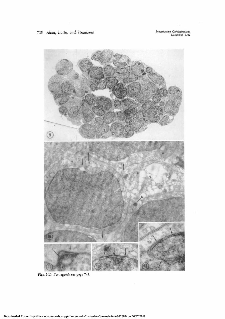
Fig. 9. Clump of tumor cells growing free in vitreous body demonstrates many nuclearirregularities which characterized both tumors studied. I, Bilobed nucleus with thin strand-like connection. 2, Small nuclear processes connected by thin strand. 3, Apparently binucleatedcells believed the result of sectioning across two lobes. Two fairly distinct patterns of nuclearchromatin are shown in adjacent cells, N and N'. (x2,600; reduced %.)
Fig. 10. Widened spaces (arrows) resembling blebs result from separation of the two layersof the nuclear envelope. Such changes seemed to occur in well-preserved cells and were notaccentuated in the process of degeneration. A portion of a cell at right shows numerousvacuoles (V) and swollen mitochondria. A round dark cytoplasmic inclusion body (b) ispresent in cell at lower right. (x7,600; reduced %.)
Fig. 11. Tubular structures included within nuclei are shown in this and the following figure.The space here (arrows) lies in an area of densely clumped chromatin. (x24,000; reduced %•)
Fig. 12. Parallel tubular structures (arrows) lie wholly within the nucleus and are separatedfrom the inner lamina of the nuclear envelope by a thin rim of nucleoplasm. Invaginationsinto the nucleus by the inner layer of the nuclear envelope may account for some of thesestructures. (x25,000; reduced %.)
Fig. 13. This membranous nuclear inclusion is shown in lower magnification in Fig. 7.Nucleoplasm is included within the double.membrane structure (arrows). (x55,000; reduced
Fig. 14. Early mitosis, late prophase (mi). The nuclear membrane can be seen on the right(arrows) but may have disappeared on the left side of the cell. The fine granules of nuclearchromatin are irregularly clumped. Degenerated cells (D) can be seen at upper right andlower left and border on cells which appear healthy. (x6,800; reduced %.)
Fig. 15. Late mitosis (telophase). The nuclear membranes shown in higher magnificationsin Figs. 16 and 17 are beginning to reform. The plasma membranes (arrows) appearwholly reconstituted. (xl0,000; reduced %.)
Figs. 16 and 17. Detail of reforming nuclear membranes of cells shown in Fig. 15. Nuclearchromatin is dense, and at several sites (arrows) the nuclear envelope appears to dip with-in nucleoplasm. (Fig. 16, x27,000; Fig. 17, x23,000; both reduced %.)
Fig. 18. Degeneration of progressive severity can be seen in cells from left to right. Cell inlower left corner (1) is apparently normal; adjacent cell (2) shows nuclear and cytoplasmicchanges of early degeneration, and the two cells (3 and 4) at right show changes compara-tively far advanced, (xl 1,000; reduced %.)
Fig. 19. Advanced degeneration of both nucleus and cytoplasm, with comparatively goodpreservation of nuclear and cytoplasmic membranes is shown. Most cells at right show bothlayers of nuclear envelope and well-defined plasma membranes. (xl4,000; reduced %.)
Fig. 20. The earliest changes of cellular degeneration are shown. The mitochondria (M) areswollen; a large cytoplasmic vacuole (V) is shown at left; the nuclear chromatin of all thecells is irregularly clumped. The Golgi apparatus (G) can be identified in cell at right. Smallround bodies (b) are seen in mitochondrion at left. (xl9,000; reduced %•)
Fig. 21. Junction between apparently healthy cells and areas of degeneration was often sharp.Even where degenerative changes are farthest advanced (right) membranous structurescan be recognized. Myelin figure (tnf) is shown above. The nature of the round gray bodies(arrows) is obscure. (x7,000; reduced %•)
Fig. 22. A capillary within the tumor is shown adjacent to a tumor cell, left. Endothelialspurs extend into the lumen (L). The layers of the basement membrane (bm) are distinct.The mitochondria (M) of the tumor cell are more numerous on the vascular side of the cell.Two centrioles (ce) can be seen. (x8,700; reduced %.)
Downloaded From: http://iovs.arvojournals.org/pdfaccess.ashx?url=/data/journals/iovs/932887/ on 06/07/2018

742 Allen, Latta, and Straatsma Investigative OphthalmologyDecember 1962
Fig. 23. Case 2. Typical appearance of rosette. The radially arranged cells surround acentral lumen (L), into which project blunt cellular processes, microvilli and cilia. A cell inmitosis (mi) is seen at right. The mitochondria (M) are principally on the luminal side of thenucleus. Arrow 2 points to detail shown in Fig. 24, arrow 2 to detail shown in Fig. 25.(x4,200; reduced %.)
Fig. 24. Detail of lumen (L) at arrow I shown in Fig. 23 is seen at higher magnification.Cellular processes and microvilli (mv) can be seen within lumen. Fibrillar cytoplasm (fi) canbe seen in two cells at left. Terminal bars (tb) are present between cells near lumen. In cellprocess at lower edge of lumen there is structure (arrow) resembling centriole with spoke-like arrangement of pericentriolar bodies (pb). Located near the surface of the cell, thesestructures appear associated with cilium. (xl5,000; reduced %.)
Fig. 25. Detail of edge of lumen at arrow 2 of rosette shown in Fig. 23. Portion of cilium(ci) in association with centriole (ce) can be seen. Pericentriolar bodies (pb) are seen in planenearly perpendicular to those shown in Fig. 24. (x40,000; reduced %.)
Fig. 26. Cilium (ci) projecting into rosette lumen (L) is shown with associated centrioles(ce, and cet). Pericentriolar bodies are not seen. (x31,000; reduced %.)
ing such changes. In considering the de-generative changes and the alterations indead and dying cells observed in bothtumors, we are unable to interpret thesequence of events. It remains uncertainat which stage a cell is "dead," if indeedcell death as such can be defined mor-phologically. Surprisingly little attentionhas been paid to the phenomenon ofcellular death and the changes of necrosiswhich ensue.27 Studies have been made ofthe fine structure of postmortem altera-tions but the details remain to be pub-lished.10
The earliest changes which we inter-preted as indicative of cellular degenera-tion are similar to those described byothers in acute injury.2-24 These consistedof slight clumping of the nuclear chromatin,swelling of the mitochondria, and forma-tion of large cytoplasmic vacuoles (Fig.20). The nuclear blebs in the tumorsseemed unrelated to degenerative phe-nomena, but they have been observed inacute cell injury.9'20'21> -
Later degenerative changes (Fig. 18,cells 3 and 4) consisted of clumping andcoarsening of the nuclear chromatin, widen-ing of the space between the 2 layers ofthe nuclear envelope, and marked swellingof the cytoplasm and of its components.These resemble later stages of cell in-
jury.24' 32 Still later degenerative changesappear to be those depicted in Fig. 19.These resemble changes which have beenobserved in early phases of tissue necrosis.7,9,32 j n m a n v areas degenerative changeswere those which seem to lead to pyknosisas described by light and electron micros-copy.21
A different pattern of degeneration isalso shown in Fig. 19. Some cells, thoughapparently in advanced degeneration, showremarkable preservation of nuclear andcytoplasmic membranes. The changesshown here do not seem to be related topyknosis, but there is as yet no directevidence to relate them to karyorrhexis orkaryolysis. The remarkable persistence ofnuclear and plasma membranes, and ofsome of the membranous cytoplasmic com-ponents, long after cellular organization islost, is one variety of necrotic cell reac-tion which appears not to have been previ-ously described.
The most advanced necrotic tumor celldegeneration is shown in Fig. 21. Border-ing on cells apparently healthy or with onlyminimal changes of degeneration, therewere wide expanses of cellular debris whichincluded membranous fragments of cells,myelin figures, vacuolated bodies, lipoidalmaterial, and granular material. The natureof the intermingled faintly granular gray
Downloaded From: http://iovs.arvojournals.org/pdfaccess.ashx?url=/data/journals/iovs/932887/ on 06/07/2018
Volume 1Number 6
Retinoblastoma 743
masses is not apparent. The appearance ofthese bodies by light microscopy is sug-gestive of the changes observed in latepyknosis.10
Possible cell origin
The structural features of the cells form-ing rosettes provide some clues as to thepossible origin of retinoblastoma. Thereare resemblances of these cells to thelining cells of the primitive neuraltube.4'10' 31 Some similarities, less striking,are evident when these cells are comparedwith ependyma.11' 2F>1 33
The cell process with its centrioles andcilium shown in Fig. 26 is similar to thatof developing visual cells in a 10-day-oldmouse as shown by Lasansky and DeRobertis18 in their Fig. 1. None of thematerial we viewed, however, showed thethe later stage of development wherethe cilium is expanded to contain saclikestructures, eventually to differentiate intothe outer segment of the visual cell (theirFig. 2).
The structures which resemble paricen-triolar bodies shown in Fig. 23, and inhigher magnifications in Figs. 24 and 25,are similar to those described by others.1'5*G
Somewhat similar, but not identical, struc-tures have been described in associationwith the primitive cilia from which therods and cones differentiate.18
That the cells forming rosettes resembleboth the ependymal cells and primitivevisual receptor cells may be explained bythe histogenesis of the retina. The de-veloping pars optica retinae undergoeshistologic changes similar to those whichoccur in other portions of the neural tube.The simple columnar neural epithelium dif-ferentiates into ependymal, mantle, andmarginal layers. The ependymal zone,which is in contact with the outer pig-mented layer, finally differentiates into therod and cone cells. The visual receptorcells may therefore be regarded as highlyspecialized ependymal elements.13
The controversy, therefore, as to whetherrosettes represent immature visual cells35
or lining cells of the primitive neuraltube30 is largely resolved to one of defini-tions.
Grateful acknowledgment is clue Miss AliceArvin for excellent technical assistance. We wishto thank Dr. W. A. McCormick of Arcadia, Cali-fornia, who made the surgical specimen ofCase 1 available to us for study.
REFERENCES1. Amano, S.: The structure of the centrioles
and spindle body as observed under theelectron and phase contrast microscopes; anew extension-fiber theory concerning mitoticmechanism in animal cells, Cytologia 22:193, 1957.
2. Ashford, T. P., and Porter, K. R.: Theresponse of hepatic cell fine structure toisolation perfusion (abstract), First annualmeeting, American Society for Cell Biology,Chicago, 111., Nov. 2-4, 1961, p. 10.
3. Ashworth, C. T., Luibel, F. J., and Sanders,E.: The fine structure of nuclei in certainhuman malignant neoplasms, Am. J. Clin.Path. 34: 9, 1960.
4. Bellairs, Ruth: The development of thenervous system in chick embryos, studied byelectron microscopy, J. Embryol. & Exper.Morphol. 7: 94, 1959.
5. Bernhard, W., and de Harven, E.: L'Ultra-structure du centriole et d'autres el6ments del'appareil achromatique, Fourth InternationalConference on Electron Microscopy, Berlin,1958. Cited by De Robertis, E. D. P.,Nowinski, U. W., and Salz, F. A.: GeneralCytology, ed. 3, Philadelphia, 1961, W. B.Saunders Company, p. 332.
6. Bessis, M., Briton-Gorius, J., and Thiery,J.-P.: Centriole, corps de Golgi, et aster desleukocytes, etude au microscope electronique,Revue d'hemat. 13: 363, 1958.
7. Bryant, R. E., Thomas, W. A., and O'Neal, R.M.: An electron microscopic study of myo-cardial ischemia of the heart, CirculationRes. 6: 699, 1958.
8. Dalton, A. J.: Organization in benign andmalignant cells, Lab. Invest. 8:. 510, 1959.
9. David, H., and Hecht, A.: SubmikroscopischeStrukturveriinderungen der Herzmuskelkapil-laren im Infarctgebiet, Zentralbl. allg. Path,u. path. Anat. 103: 68, 1961.
10. Duncan, D.: Electron microscope study ofthe embryonic neural tube and notocord,Texas Rep. Biol. & Med. 15: 307, 1957.
11. Fleischauer, K.: Untersuchungen am Epen-dym des Zwischen und Mittelhirns der Land-schildkrote (Testudo graeca), Ztschr. Zell-forsch. u. mikr. Anat. 46: 729, 1957.
Downloaded From: http://iovs.arvojournals.org/pdfaccess.ashx?url=/data/journals/iovs/932887/ on 06/07/2018

744 Allen, Latta, and Straatsma Investigative OphthalmologyDecember 1962
12. Friedmann, I.: Electron microscopy ofhuman biopsy material, Proc. Roy. Soc. Med.54: 1064, 1961.
13. Hamilton, W. J., Boyd, J. D., and Mossman,H. W.: Human embryology, Cambridge,1957, VV. Heffer & Sons, Ltd., p. 317.
14. Hogan, M. J., and Zimmerman, L. E.:Ophthalmic pathology, ed. 2, Philadelphia,1962, W. B. Saunders Company, pp. 516-525.
15. Hubner, C , and Bernhard, VV.: Das sub-mikroskopische Bild der Leberzelle nach tem-poriirer Durchblutungssperre, Beitr. path.Anat. u. allg. Path. 125: 1, 1961.
16. Ito, S., Revel, J. P., and Fawcett, D. W.:Persistence of cellular fine structure afterdeath (abstract), First annual meeting, Amer-ican Society for Cell Biology, Chicago, 111.,Nov. 2-4, 1961, p. 97.
17. Karnovsky, M. ].: Simple methods for "stain-ing with lead" at high pH for electron micros-copy, J. Biophys. & Biochem. Cytol. 11: 729,1961.
18. Lasansky, A., and De Robertis, E.: Sub-microscopic analysis of the genetic dystrophyof visual cells in C3H mice, J. Biophys. &Biochem. Cytol. 7: 679, 1960.
19. Leuchtenberger, Cecilie: A cytochemicalstudy of pycnotic nuclear degeneration,Chromosoma 3: 449, 1950.
20. Latta, H., and Kutsakis, Anne: Cytotoxiceffects of specific antiserum and 17-hydroxy-corticosterone on cells in tissue culture, Lab.Invest. 6: 12, 1957.
21. Latta, H.: Some cytologic effects of antibodies,in Mechanisms of hypersensitivity, Interna-tional Symposium sponsored by Henry FordHospital, Detroit, Mich., Boston, 1939, Little,Brown & Co., pp. 123-141.
22. Latta, H.: Cellular reaction to antibody in tis-sue culture studied with electron microscopy,J. Biophys. & Biochem. Cytol. 5: 405, 1959.
23. Luft, J., and Hechter, O.: An electron micro-scopic correlation of structure with functionin the isolated perfused cow adrenal. Pre-liminary observations, J. Biophys. & Biochem.Cytol. 3: 615, 1957.
24. Luft, J. H.: Improvements in epoxy resinembedding methods. J. Biophys. & Biochem.Cytol. 9: 409, 1961.
25. Luse, Sarah: Electron microscopic studies ofbrain tumors, Neurology 10: 881, 1960.
26. Luse, Sarah: Ultrastructural characteristicsof normal and neoplastic cells, in Allgoewer,M., editor: Progress in experimental tumorresearch, Vol. 2, New York, 1961, HafnerPublishing Co., Inc., pp. 1-35.
27. Majno, C , La Guttata, Monika, and Thomp-son, T. E.: Cellular death and necrosis:Chemical, physical, and morphologic changesin rat liver, Virchows Arch. path. Anat. 333:421, 1960.
28. Mercer, E. H.: The electron microscopy ofnormal and neoplastic cells, Proc. Roy. Soc.Med. 54: 1057, 1961.
29. Oberling, C, and Bernhard, W.: Themorphology of the cancer cells, in Brachet,J., and Mirsky, A. E., editors: The Cell,New York, 1961, Academic Press, Inc., vol.5, pp. 405-496.
30. Parkhill, E. M., and Benedict, W. L.:Gliomas of the retina. A histopathologicstudy, Am. J. Ophth. 24: 1354, 1941.
31. Sotelo, J. R., and Trujillo-Cenoz, O.: Electronmicroscope study on the development ofciliary components of the neural epitheliumof the chick embryo, Ztschr. Zellforsch. u.mikr. Anat. 49: 1, 1958.
32. Stone, R. S., Bencosme, S. A., Latta, H., andMadden, S. C : Renal tubular fine structurestudied during reaction to acute uraniuminjury, Arch. Path. 71: 160, 1961.
33. Tennyson, Virginia M., and Pappas, G. D.:An electron microscope study of ependymalcells of die fetal, early postnatal and adultrabbit, Ztschr. Zellforsch. u. mikr. Anat. 56:595, 1962.
34. Watson, M. L.: Staining of tissue sections forelectron microscopy with heavy metals, J.Biophys. & Biochem. Cytol. 4: 475, 1958.
35. Willis, R. A.: Pathology of tumours, ed. 2,St. Louis, 1959, The C. V. Mosby Company,p. 889.
Downloaded From: http://iovs.arvojournals.org/pdfaccess.ashx?url=/data/journals/iovs/932887/ on 06/07/2018