restorative smile case study · restorative smile case study neil gerrard presents his winning...
TRANSCRIPT

clinical Restorative Smile case study Neil Gerrard presents his winning entry for the Restorative Smile (single arch) category at the 2012 Smile Awards
History The patient, a 40-year-old male i n good
health, attended the practice w i t h the pr ior i ty
of improving the appearance of his smile. His
aim, to replace an existing anterior bridge
while creating a more symmetrical even smile
more visible w h e n smil ing, i.e. increase the
length of his upper anterior teeth, level the
occlusal plane and eliminate shadows i n the
buccal corridors.
Diagnosis and treatment plan A comprehensive dental health examination
was completed prior to commencement of
treatment. A n intra-oral assessment confirmed
a Class I occlusion w i t h compromised dental
health due to mult iple carious lesions and
failing restorations. Oral hygiene was of a fair
standard.
Examination of the TMJ's and muscles
mastication found no abnormality. A m i n o r
discrepancy between centric relation and
centric occlusion was noted (approximately
1mm), w i t h an anterior slide to the left
detectable fo l lowing bi-manual manipulat ion
of the mandible. W i t h a risk assessment and
clinical examination indicating no risk factors
associated w i t h the existing occlusal scheme
Figures 1-2: Full face views (before and after)
Figures 3-4: Smile views, anterior (before and after)
(the existing anterior bridge had been i n situ
for 20 years or more), a conformative approach
was taken uti l is ing the existing MIR
Analysis of his smile indicated an occlusal
plane w h i c h dropped posteriorly left and right
fo l lowing over eruption as a result of early loss
of the first molars i n the lower arch. A reverse
smile line was also noted 13 to 23, the canines
appearing longer than the central incisors
when smil ing. A ridge defect around the
existing bridge was also evident i n the 11/12
region, again from a result of long-term tooth
loss fo l lowing a traumatic in jury as a chi ld .
I n accordance w i t h the patients
requirements a number of treatment proposals
were presented, inc luding pre-restorative
alignment to level the occlusal plane and
gingival zeniths into the ideal posit ion w i t h
orthodontic treatment, thus negating the
need for crown lengthening i n the posterior
sextants. Implant placement to support the 11 ,
thus reducing the risks of abutment failure, and
conventional crown and bridge restorations to
restore those teeth compromised w i t h pr imary
and secondary carious lesions. Oral hygiene
and dietary advice w o u l d also be offered to
reduce future risk of caries i n conjunction w i t h
caries management programme as appropriate.
W i t h a complete understanding of the
benefits and compromises associated w i t h
each treatment, the patient opted to replace
the existing bridge w i t h another fixed bridge
(as this had functioned satisfactory for nearly
20 years), along w i t h crowns and veneers i n
the upper arch to correct those undesirable
features already noted.
Treatment w o u l d therefore consist of:
• Placement of indirect restorations to all teeth
treated w i t h i n the upper arch - a combination
of a zirconia-supported bridge w i t h layered
Dr Neil Gerrard is the principal of Clifton Dental Studio in Bristol. He is one of a select number of UK dentists to have achieved accreditation status with
JkV the British Academy of ^ » Cosmetic Dentistry (BACD).
Neil's interests are in advanced cosmetic dentistry, complex smile reconstruction with dental implants and smile rejuvenation. He completes over 100 hours of advanced training every year in the fields of cosmetic and implant dentistry, and is an examiner with the BACD, helping educate dentists, dental technicians and team members in the art and science of cosmetic dentistry. For further information please www. cliftonsmiles.com and www.bacd.com.
26 August 2012 adt

ceramic for 12 to 21 and E-max crowns/
veneers/onlays as required.
• Crown lengthening to reduce the left and
right posterior inclination of the gingival
tissues, thus a l lowing levelling of the occlusal
plane anterior-posteriorly wi thout shortening
of the visible c rown heights. This w o u l d also
offer the ability to widen the buccal corridors
as required while maintaining the correct
crown inclination of each tooth.
• Root treatments in the pre-molars and
molars as required if exposure were to occur
while creating adequate occlusal reduction to
lift the occlusal plane.
• Enamel plasty of the lower incisor teeth as
required to improve incisal edge relationship.
• Finally, a conformative occlusal scheme
w o u l d be fol lowed, as no evidence of
dysfunction could be deduced from the intra
oral examination or patient history.
The patient was also aware that failure to
restore 37 and replace 47 w o u l d increase the risk
of compensatory eruption in the right posterior
sextant and the need for further complex
restorative care w i t h 37. The patient requested
that treatment be phased, concentrating on the
upper arch at this point in t ime, followed by the
lower arch in the near future.
Treatment process Fol lowing hygiene care and dietary advice,
records were taken to produce a laboratory
fabricated diagnostic wax up. These included
upper and lower polyether impressions for
accuracy, a Kois facial analyser to aid occlusal
plane correction and pre-operative photos.
Additionally, the laboratory was instructed to
lengthen the incisors by 1.5mm as determined
w i t h a composite mock-up i n the m o u t h to
conf i rm ideal incisal edge position.
At the request of the patient treatment
was to be expedited i n as short a timeframe
as possible. Fol lowing confirmation that
the diagnostic wax-up fulf i l led all aesthetic
and functional requirements, treatment was
initiated. To save time, c rown lengthening
w o u l d be completed at the same time as the
preparation visit.
Uti l is ing a technique described by
Mangne 1 , bis-acrylic c rown material was
injected into a putty matrix taken from the
pre-operative diagnostic wax model . This was
positioned in to the m o u t h and allowed to
cure for three minutes. The matrix was then
removed, revealing a bis-acrylic template of
the final result. Depth gauge cuts were then
made through this template, uti l is ing round
diamond burs (Brassier) to aid a conservative
Figures 5-6: Smile views, right (before and after)
Figures 7-8: Smile views, left (before and after)
Figures 9-10: Retracted views, anterior (before and after)
Figures 11-12: Retracted views, right (before and after)
preparation design. The remaining template
material was then removed to allow finalisation
of the preparations.
It was felt that, due to the presence of
interproximal carious lesions and dentine/
cementum exposure fol lowing crown
lengthening, a cohesive cementation protocol
w o u l d offer greater long-term predictability,
rather than rely on dentine bonding. This
cementation protocol w o u l d therefore dictate
the need for ful l coverage restorations. The
depth gauge preparation technique is also
beneficial when used i n conjunction w i t h
ful l coverage restorations, enabling minimal
preparation (buccal, palatal and occlusal
reduction) of all tooth surfaces. In this instance
the use of this technique resulted in no pulpal
exposures (even the occlusal surface of 16)
w i t h all ful l coverage restorations. To maintain
the correct occlusal relationship, bite records
were taken in sections fo l lowing preparation
of a few teeth at a time.
For those teeth requiring surgical crown
lengthening, the gingival tissues were sculpted
to the ideal form using a diode laser, followed
by preparation of the restorative margins to the
correct level. Probing to bone w i t h a W i l l i a m s
probe confirmed that the restorative margin
was w i t h i n a distance of less than 1mm to
crestal bone. Leaving preparation margins in
such close prox imi ty w o u l d result in profound
biological w i d t h invasion. Surgical c rown
lengthening w o u l d therefore be required to
re-establish the correct crestal bone level i n
adt August 2012 27

relation to the restorative margin.
In accordance with papers published
by Kois 2 , restorative margins positioned
2.5mm from the crestal bone would enable
development of a healthy gingival complex
(biologic width).
At this point it was decided to complete
placement of a bis-acrylic trial smile and
leave the crown lengthening for a future
appointment. Short-term invasion of the
biological width would cause no negative
occurrences and the patient would be seen
again within seven days to complete this part
of the procedure.
Trial smile Prior to impression taking, a trial smile was
produced utilising the putty matrix and
bis-acrylic temporary crown material in an
appropriate shade. As the majority of the
teeth were of full coverage design the trial
smile would be cemented with conventional
eugonol Tempbond. The trial smile would be
cemented in sections, with the veneers on the
canines supported both mesially and distally
from the full coverage restorations.
Prior to cementation and impressions
the form of the pontic for 11 was modified
to an ovale form followed my creation of an
ovate pontic site in the gingival tissue with
a diode laser for haemostatic control. This
was designed in such a way as to push the
tissues aplically and buccally. Ideal care would
normally dictate a connective tissue graft with
such a defect, in this instance the patient
refused this procedure as the pontic site was
not visible when smiling. The technique of
directional pressure application can result in
a satisfactory tissue form, but is not always
predictable with inadequate pre-operative soft
tissues.
A polyether was used to complete the
final working impression, followed by stump
shade and additional photos as required. Once
poured this would be cross-mounted with
the new bite records against the previously
articulated lower model. Assessment of the
trial-smile confirmed the occlusal place to be
level, thus negating the need for intra-oral
adjustment and new face-bow.
At this point the trial-smile was seated
in situ with Tempbond and the patient was
allowed to leave.
One week later the patient returned for
surgical crown lengthening of 16, 15, 14 and
24, 25, 26.
Following LA, flaps were raised to allow
clear access to the crestal bone. Using
1 4 '
Figures 13-14: Retracted views, left (before and after)
Figures 15-16: Views, anterior (before and after)
Figures 17-18: Views, right (before and after)
Figures 19-20: Views, left (before and after)
appropriate burs and periodontal instruments
crestal bone was removed to a distance of
2.5mm of the restorative margins. Great care
was taken to achieve this critical distance and
remove adequate bone at the line angles to
prevent a chronic inflammatory reaction. This
was sutured with 5.0 Vicryl Rapid.
One-week post-op sutures were removed
and confirmation of uneventful healing was
noted. The patient confirmed that he was
delighted with the aesthetic result and reported
no post-operative complications apart from
some soreness of the gingival tissues to touch
(as to be expected).
Cementation of restorations Cementation of the final restorations was completed three weeks post-preparation appointment. At this time complete resolution of the soft tissues in the pre-molar/molar regions had not occurred following the crown lengthening procedure. However, this was of no concern as the literature confirms a 100% predictable outcome in relation to gingival margin position to crestal bone position when the restorative margin is placed 2-2.5mm from the crestal bone, Kois. 2
All teeth were isolated with retraction cord to prevent crevicular fluid contaminating the
28 August 2012 adt
cementation surface and to control haemostasis
fo l lowing particulate air abrasion. Air abrasion
w i t h 27um alumina oxide was used to prepare
the bond surface, removing all contaminates
w h i c h could compromise the tooth cement
interface, inc luding b lood, saliva and eugonol.
The veneers on 13 and 23 were cemented
first fo l lowing an adhesive protocol , total etch,
bond and light cure cementation. Fol lowing
clean-up, all remaining ful l coverage
restorations were seated using a self-etch dual -
cure composite cement. Care was taken w i t h
both techniques not to desiccate any exposed
dentine, thus reducing the incidence of post
operative sensitivity.
Fol lowing cementation the usual
refinements were completed, the occlusion was
checked and adjusted as required. Refinement
of restorative margins was performed w i t h fine
rubber points together w i t h confirmation that
excessive blanching of the ovate pontic site
had abated.
• I w o u l d like to thank my brother, Paul
Gerrard of Bremadent Dental Laboratory, w h o
provided the ceramic w o r k , w h i c h he used
as an accreditation case for the BACD. He is
one of only two technicians to be accredited
and together we are the only practice i n the
country to have both an accredited dentist and
technician w o r k i n g under the same roof.
Care to comment? @AesDenToday
Aesthetic Dentistry Awards2013
If you'd like to enter the 2013 Aesthetic Dentistry Awards (formerly The Smile Awards), please call Karen Sherwood on 01923 851743, email [email protected] or visit the website www.aestheticdentistrytoday.co.uk/awards.
References 1 Magne R Magne M (2006). Use of additive wax up
and direct intra-oral mock-up for enamel preservation
with porcelain laminate veneers The European Journal of
Esthetic Dentistry 1(1): 10-19.
2. Kois, John C (1994) Altering Gingival Levels: The
restorative connection Pari 1: BiologicalVariables
Journal of Esthetic Dentistry 6 ( 0 : 3 - 9
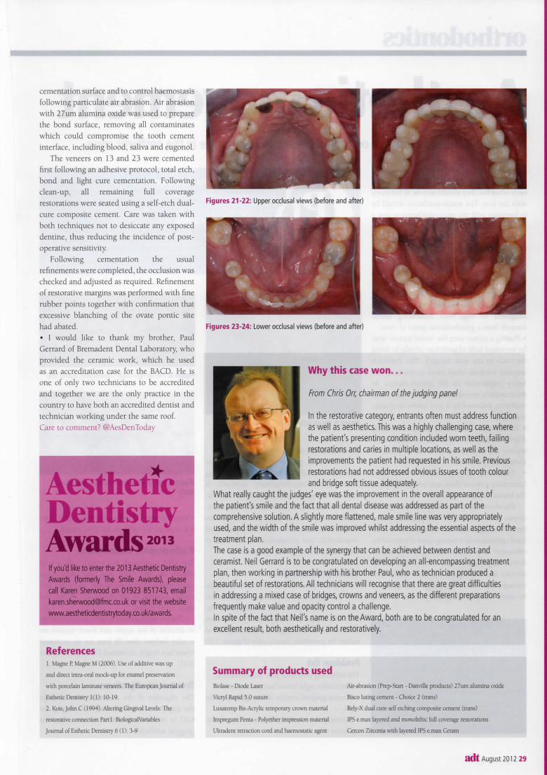
Figures 21-22: Upper occlusal views (before and after)
Figures 23-24: Lower occlusal views (before and after)
Why this case won.
From Chris Orr, chairman of the judging panel
In the restorative category, entrants often must address function as well as aesthetics. This was a highly challenging case, where the patient's presenting condition included worn teeth, failing restorations and caries in multiple locations, as well as the improvements the patient had requested in his smile. Previous restorations had not addressed obvious issues of tooth colour and bridge soft tissue adequately.
What really caught the judges' eye was the improvement in the overall appearance of the patient's smile and the fact that all dental disease was addressed as part of the comprehensive solution. A slightly more flattened, male smile line was very appropriately used, and the width of the smile was improved whilst addressing the essential aspects of the treatment plan. The case is a good example of the synergy that can be achieved between dentist and ceramist. Neil Gerrard is to be congratulated on developing an all-encompassing treatment plan, then working in partnership with his brother Paul, who as technician produced a beautiful set of restorations. All technicians will recognise that there are great difficulties in addressing a mixed case of bridges, crowns and veneers, as the different preparations frequently make value and opacity control a challenge. In spite of the fact that Neil's name is on the Award, both are to be congratulated for an excellent result, both aesthetically and restoratively.
Summary of products used Biolase - Diode Laser
Vicryl Rapid 5 0 suture
Luxatcmp Bis-Acrylic temporary crown material
lmpregum Pema - Polyether impression matenal
Ultradent retraction cord and haemostatic agent
Air-abrasion (Prep-Stan - Danville products) 27um alumina oxide
Bisco luting cement - Choice 2 (trans)
Rely-X dual cure self etching composite cement (trans)
IPS e.max layered and monolithic full coverage restorations
Cercon Zirconia with layered IPS e.max Ceram
adt August 2012 29