respiratory disorders 2 (or pulmonary potpourri) · respiratory disorders 2 (or pulmonary...
TRANSCRIPT
Respiratory Disorders 2(or Pulmonary Potpourri)
Thomas Lahiri MDProfessor of Pediatrics
Larner College of MedicineUniversity of Vermont

Disclosure I have no relevant financial relationships with the
manufacturers(s) of any commercial products(s) and/or provider of commercial services discussed in this CME activity.
I do not intend to discuss an unapproved/investigative use of a commercial product/device in my presentation.
Objectives Develop a differential for exertional
dyspnea Recognize signs of respiratory failure Know the evaluation and management of
sleep disordered breathing Understand key components of pulmonary
function testing
Lecture Content Exercise Induced
Dyspnea Obstructive Sleep
Apnea
Aspiration Pleural Fluid Pulmonary Function
Testing
Dyspnea with Exertion May be the earliest sign of respiratory conditions Asthma Vocal cord dysfunction (usually with stridor) Airway narrowing/collapse (e.g. severe malacia) Cardiac etiologies Pulmonary hypertension Neuromuscular weakness Deconditioning

Exertional Dyspnea Young children
Labored breathing Inability to keep up with peers
School aged/adolescent Chest tightness Throat closure Fatigue

Exertional Dyspnea Chest tightness, cough, wheeze: think of
a pulmonary etiology Most common: Exercise induced
bronchoconstriction (asthma) Airway issues , interstitial lung disease
Chest pain, syncope: r/o cardiac disease
Exertional Dyspnea Rapid onset & resolution with stridor: VCD Evaluation may include (after thorough
history): Spirometry Radiography EKG Exercise challenge
Differential Diagnosis Vocal cord dysfunction (often with stridor) Dynamic airway collapse (severe malacia) Bronchial stenosis/tracheal compression Pulmonary hypertension Neuromuscular disease Deconditioning
Bronchiolitis Viral etiology: RSV, influenza,
parainfluenza, metapneumovirus Presentation:
Apnea (especially under 4 months of age) Copious rhinorrhea Cough/wheeze Exam findings include +/- fever, nasal
secretions/flaring, wheeze, crackles, retractions, prolonged expiratory phase
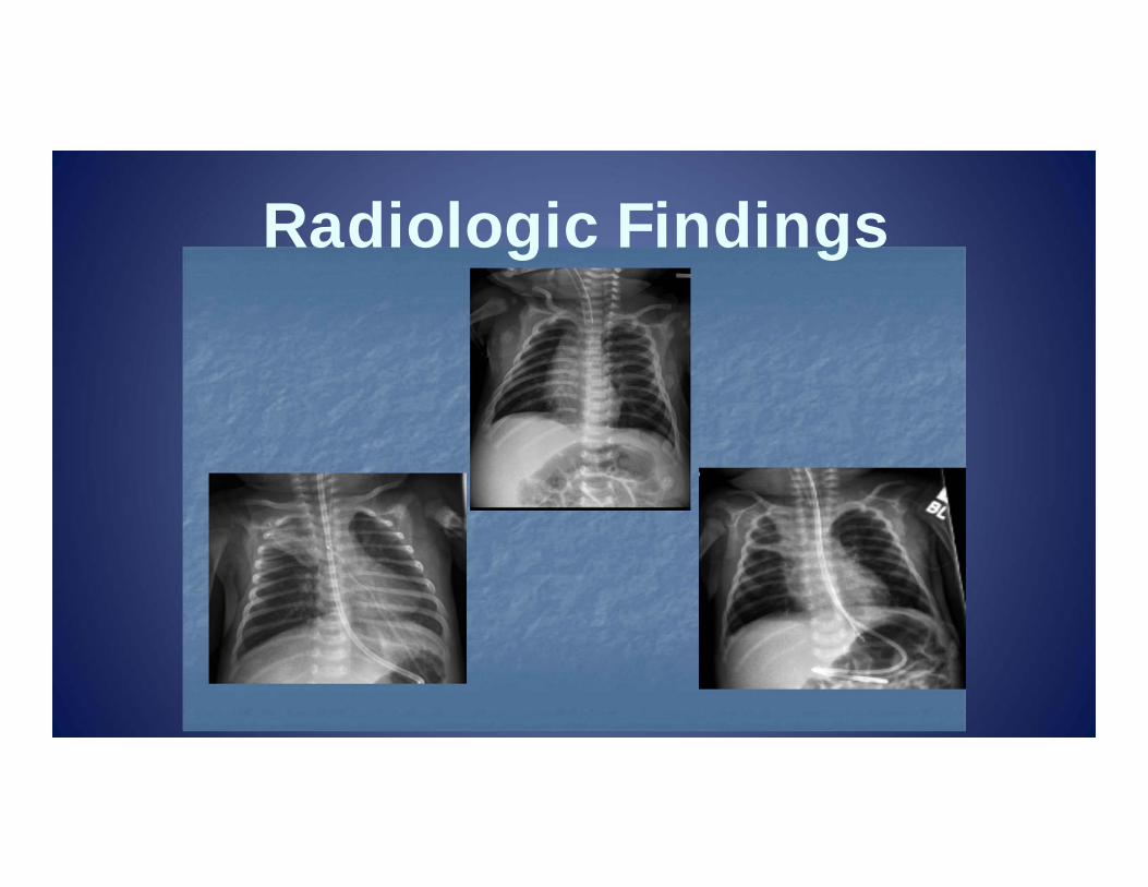
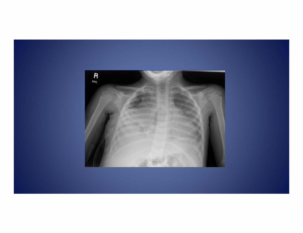
Radiologic Findings
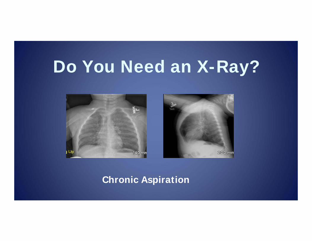
Do You Need an X-Ray?
Chronic Aspiration
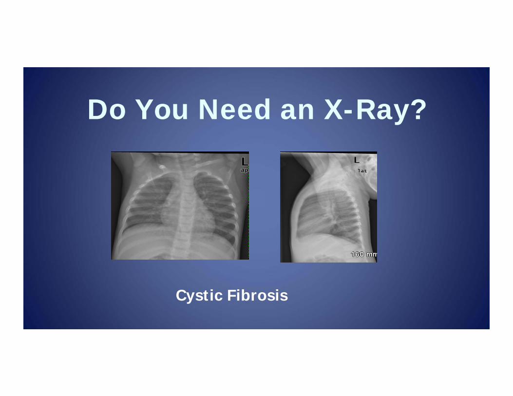
Do You Need an X-Ray?
Cystic Fibrosis
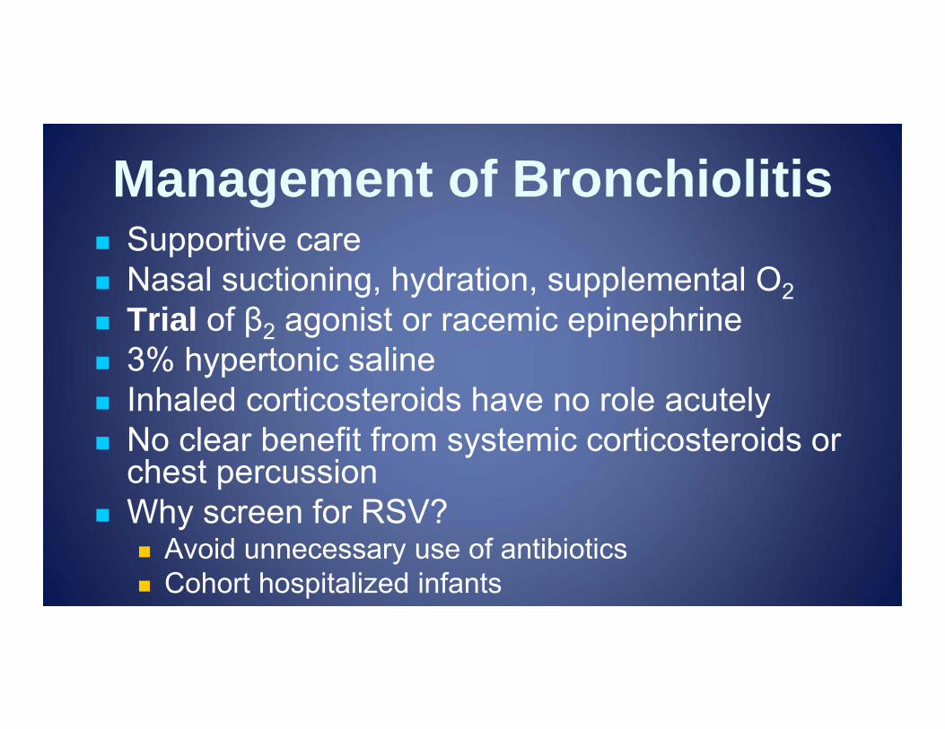
Management of Bronchiolitis Supportive care Nasal suctioning, hydration, supplemental O2 Trial of β2 agonist or racemic epinephrine 3% hypertonic saline Inhaled corticosteroids have no role acutely No clear benefit from systemic corticosteroids or
chest percussion Why screen for RSV?
Avoid unnecessary use of antibiotics Cohort hospitalized infants
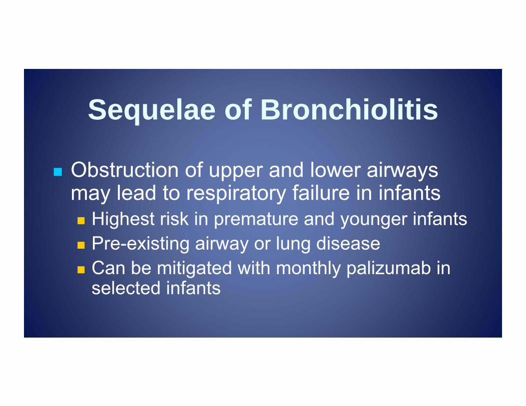
Sequelae of Bronchiolitis
Obstruction of upper and lower airways may lead to respiratory failure in infants Highest risk in premature and younger infants Pre-existing airway or lung disease Can be mitigated with monthly palizumab in
selected infants
Sequelae of Bronchiolitis Hospitalization may be required
If presenting with apnea Unable to maintain adequate oral intake Hypoxemia (SaO2 <90%) Concern for impending respiratory failure
Impending Respiratory Failure in Infants
Scenarios: Upper airway obstruction Lower respiratory involvement Sepsis Hypotonia
Impending Respiratory Failure in Infants
Increased accessory muscle use Inability to coordinate feeding Decreased arousability Hypoxemia/hypercarbia
normal PCO2 with marked tachypnea
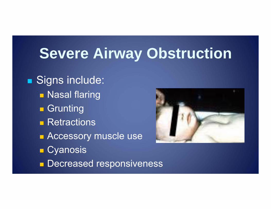
Severe Airway Obstruction Signs include:
Nasal flaring Grunting Retractions Accessory muscle use Cyanosis Decreased responsiveness
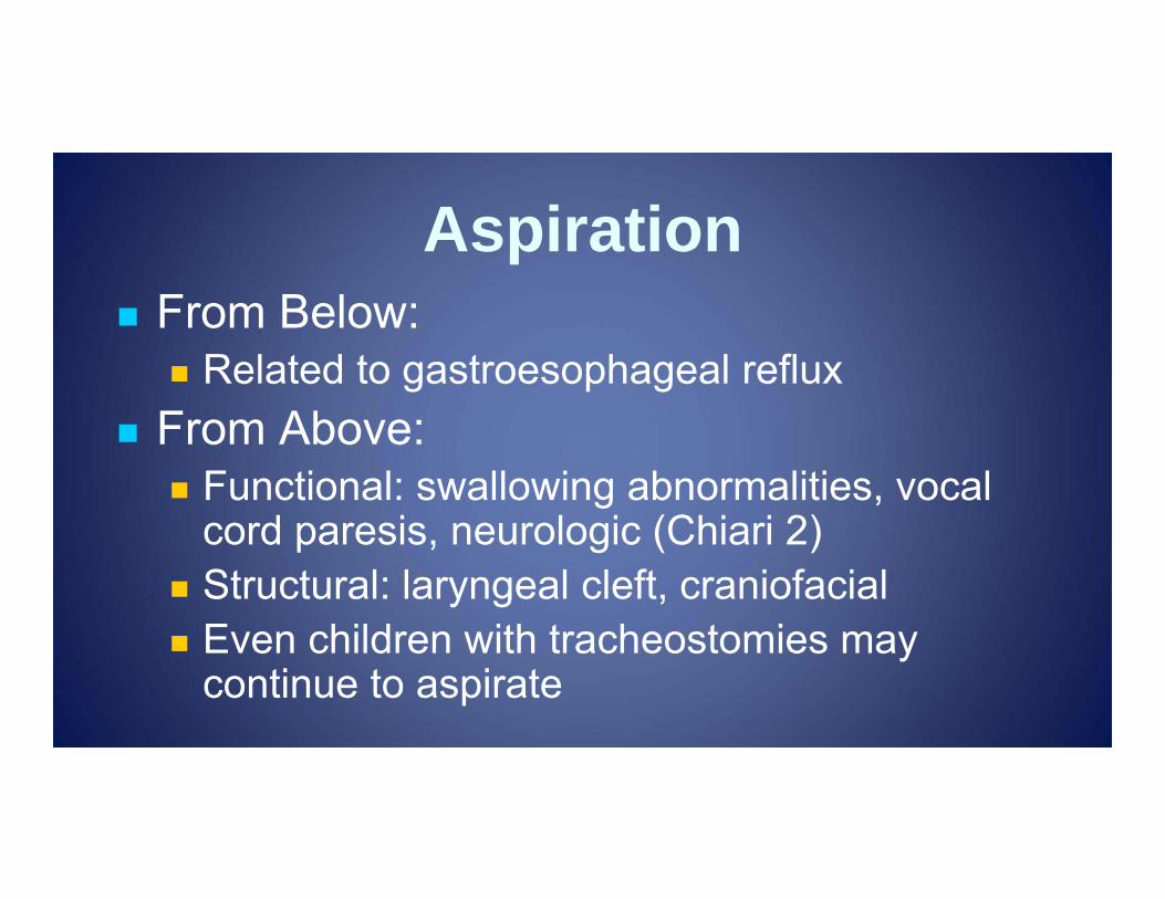
Aspiration From Below:
Related to gastroesophageal reflux From Above:
Functional: swallowing abnormalities, vocal cord paresis, neurologic (Chiari 2)
Structural: laryngeal cleft, craniofacial Even children with tracheostomies may
continue to aspirate
Chronic Aspiration- Evaluation
Radiographs may show chronic airway inflammation (non-specific)
Videofluoroscopic evaluation with feeding team is the gold standard
Bronchoscopy to look for indirect evidence of aspiration or structural etiologies
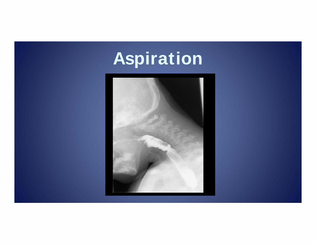
Aspiration
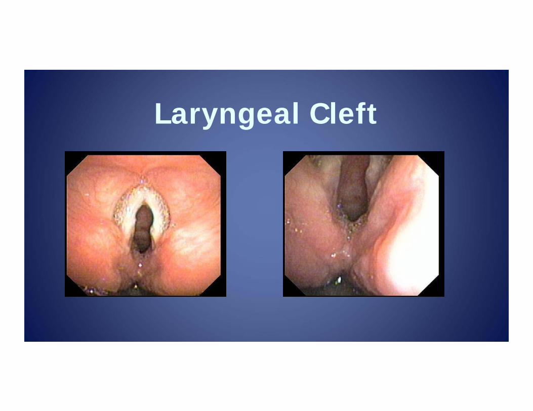
Laryngeal Cleft
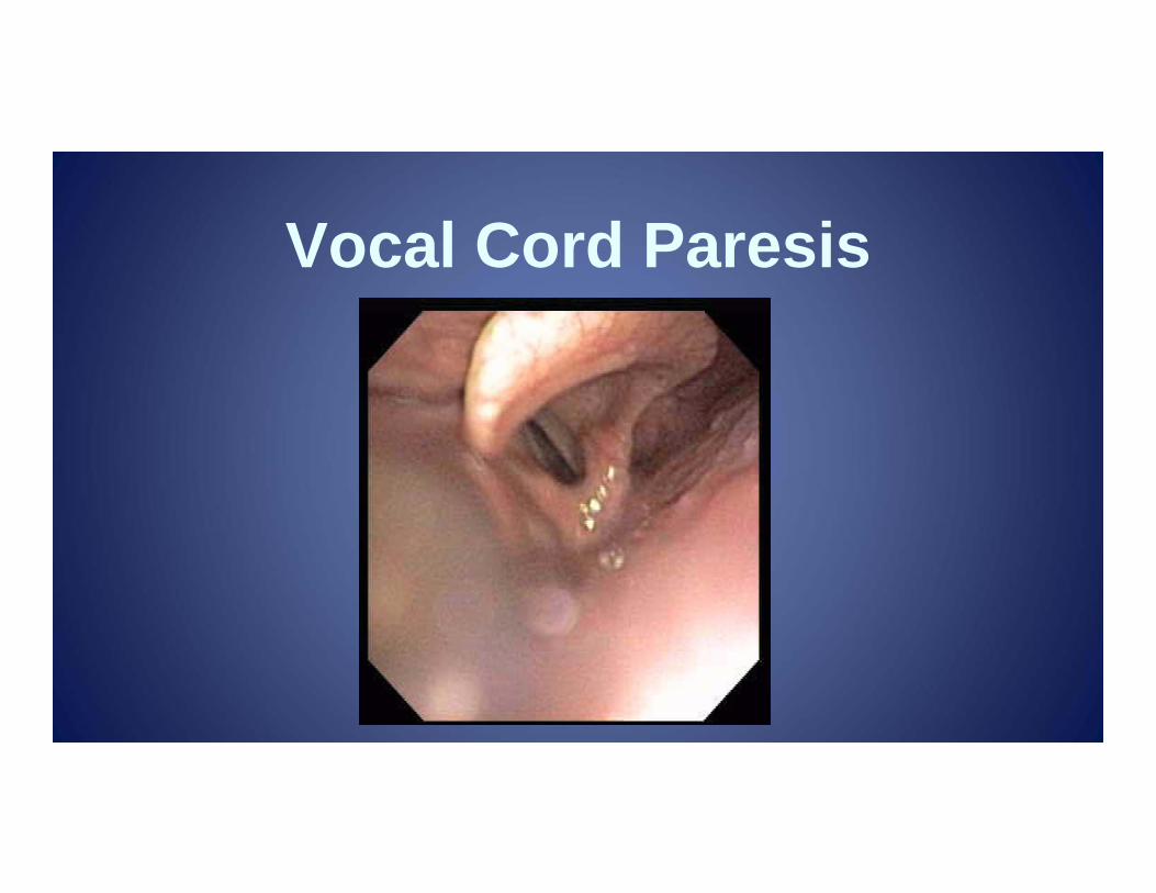
Vocal Cord Paresis
Sleep Apnea Obstructive
Most common Decreased airflow with chest wall movement
Central Lack of respiratory drive, no effort Congenital central hypoventilation syndrome
Mixed: Apnea of prematurity
Sleep Disordered Breathing Obstructive sleep apnea syndrome (OSAS)
Craniofacial anomalies Adenotonsillar hypertrophy Laryngomalacia Metabolic disorders Obesity
Hypoventilation Neuromuscular disorders Central apnea
Obstructive Sleep Apnea Snoring is common, but <50% have OSA Children may present with:
Witnessed apneic/gasping episodes Difficult to arouse from sleep Excessive daytime somnolence Deteriorating school performance Attention deficit hyperactivity Morning headaches
OSAS Witnessed apneic events with snoring,
abnormal physical exam findingsadenotonsillectomy
Polysomnography (sleep study) Different definitions for children vs. adults Apnea/hypopnea index (# total events/hour) AHI >1 is abnormal in children AHI <5 is normal in adults
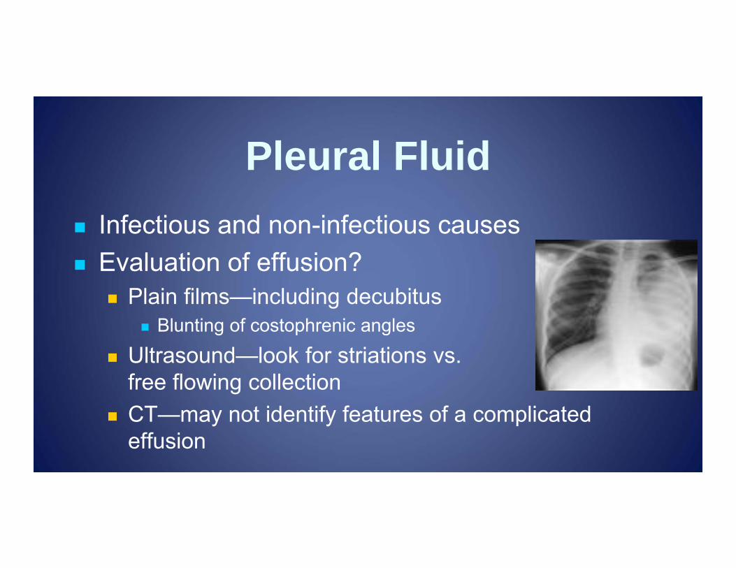
Pleural Fluid Infectious and non-infectious causes Evaluation of effusion?
Plain films—including decubitus Blunting of costophrenic angles
Ultrasound—look for striations vs. free flowing collection
CT—may not identify features of a complicated effusion
Pleural Fluid Increased hydrostatic pressure
Cardiogenic Responsive to diuretics, positive pressure
Decreased oncotic pressure Hypoalbuminemia Address with colloid, nutritional repletion
Capillary leak Seen in sepsis, SIRS/ARDS
Pleural Fluid Complicated parapneumonic effusion
Increased protein, low pH, high LDH Chylous effusion
Alkaline pH, milky, lymphocytes, chylomicrons Transudate
Plasma pH, straw colored, low protein/LDH
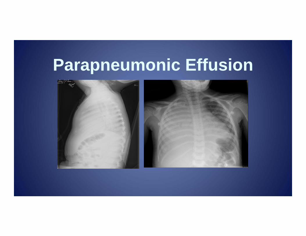
Parapneumonic Effusion
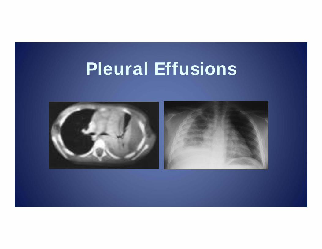
Pleural Effusions
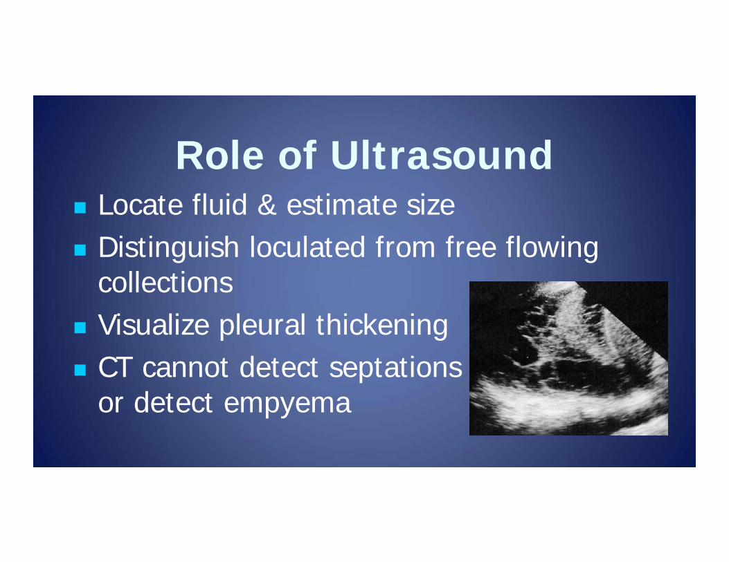
Role of Ultrasound Locate fluid & estimate size Distinguish loculated from free flowing
collections Visualize pleural thickening CT cannot detect septations
or detect empyema
Empyema Presence of pus in the pleural space Pleural fluid with positive Gram stain or
culture Tube drainage (usually ultrasound guided)
with parenteral antibiotics Adjunct treatment includes fibrinolytics
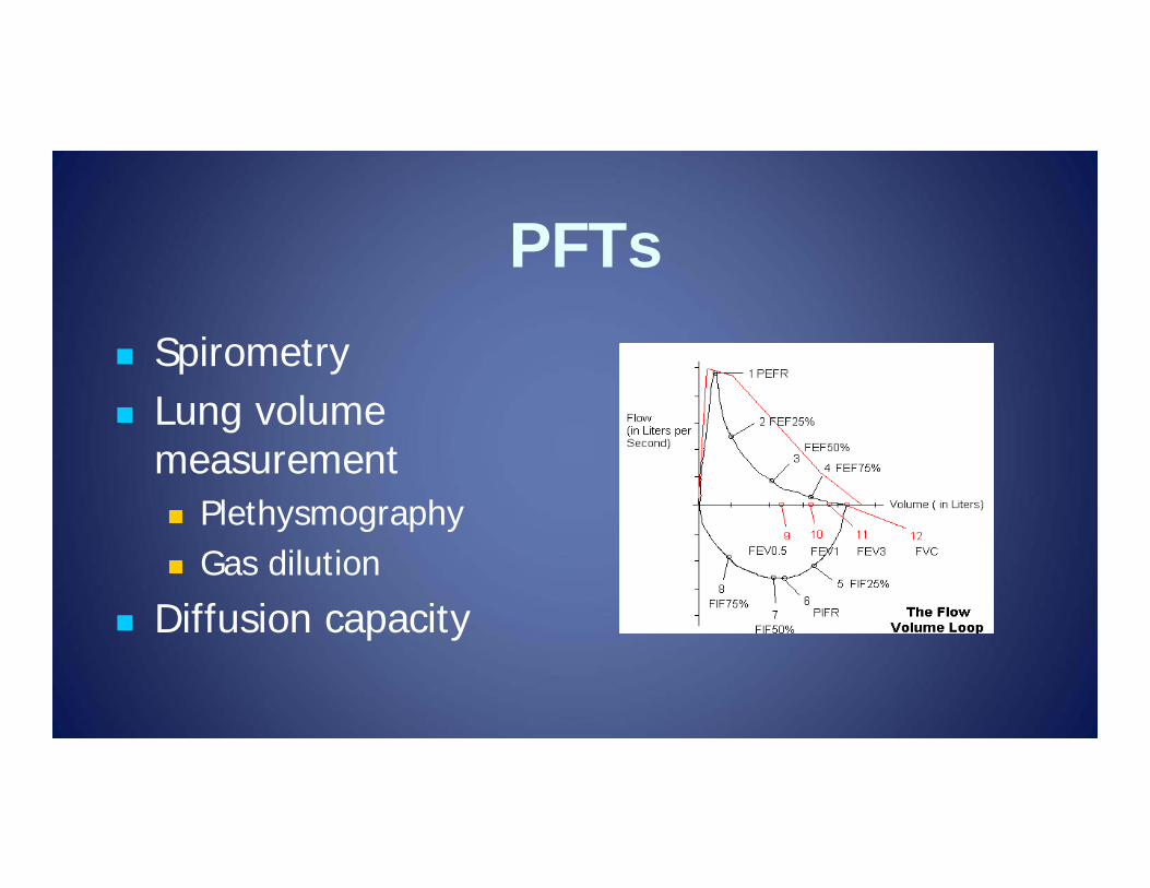
PFTs Spirometry Lung volume
measurement Plethysmography Gas dilution
Diffusion capacity
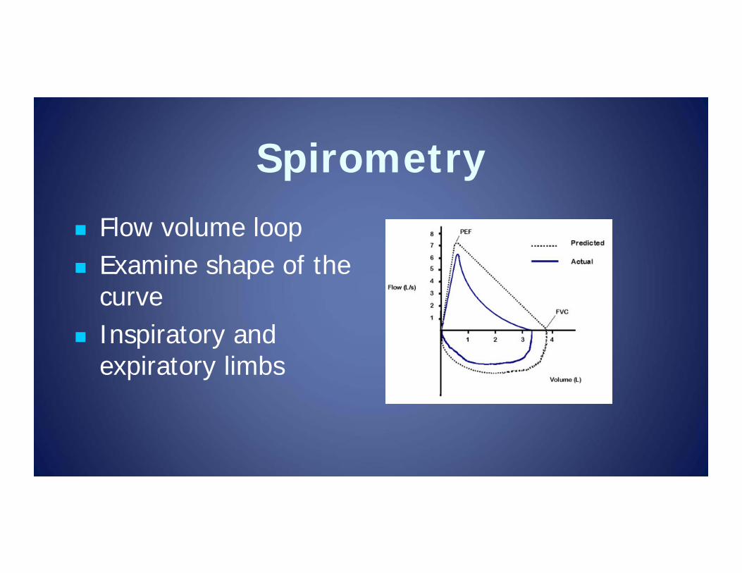
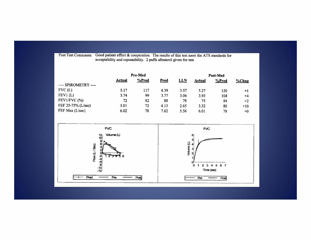
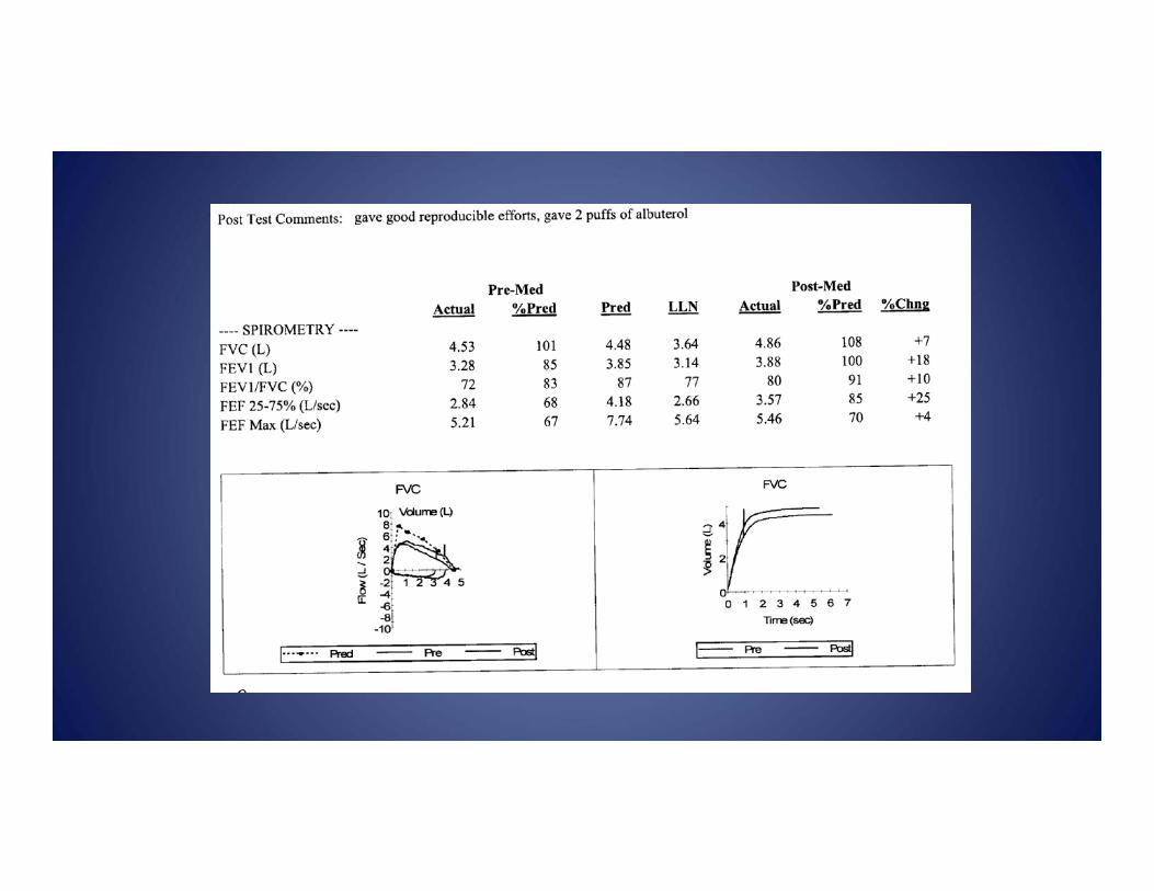
Spirometry Flow volume loop Examine shape of the
curve Inspiratory and
expiratory limbs
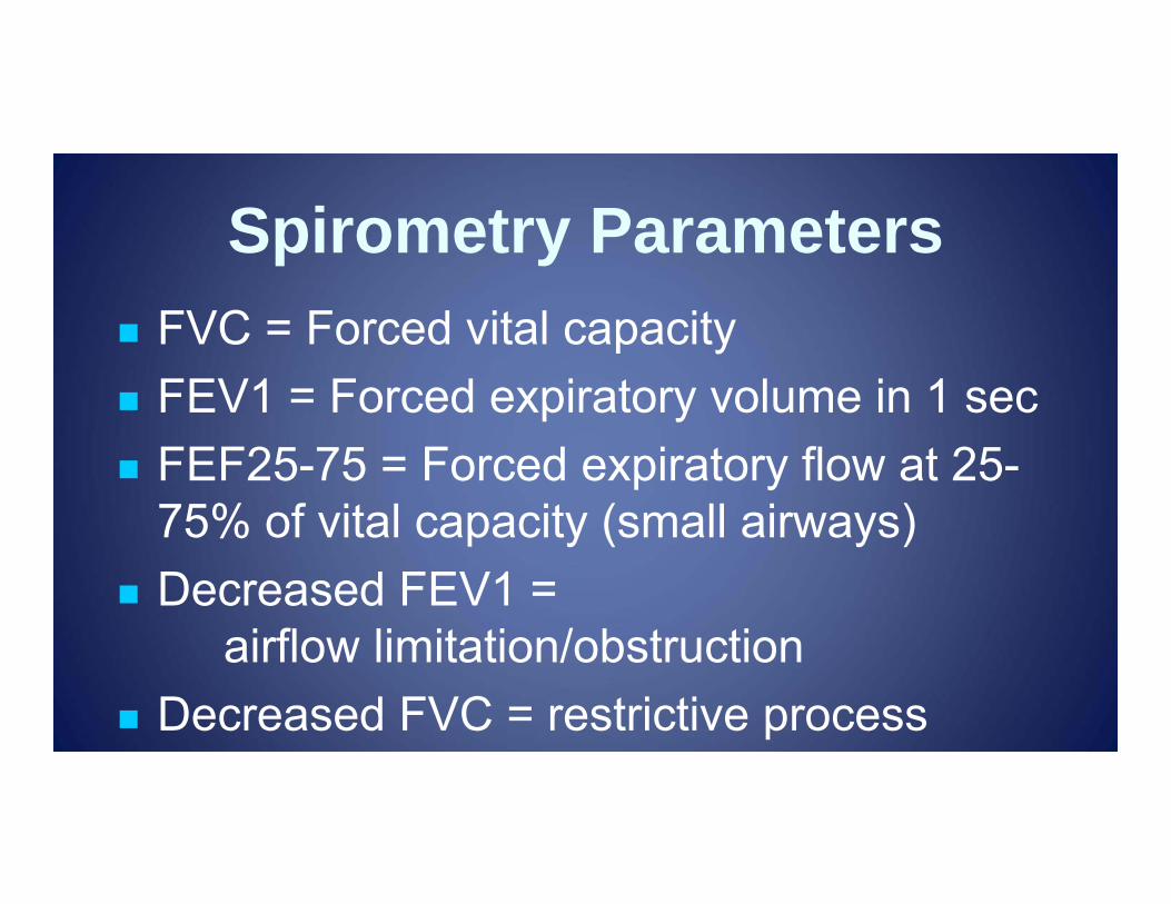
Spirometry Parameters FVC = Forced vital capacity FEV1 = Forced expiratory volume in 1 sec FEF25-75 = Forced expiratory flow at 25-
75% of vital capacity (small airways) Decreased FEV1 =
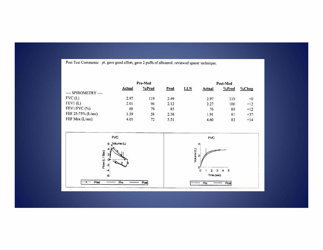
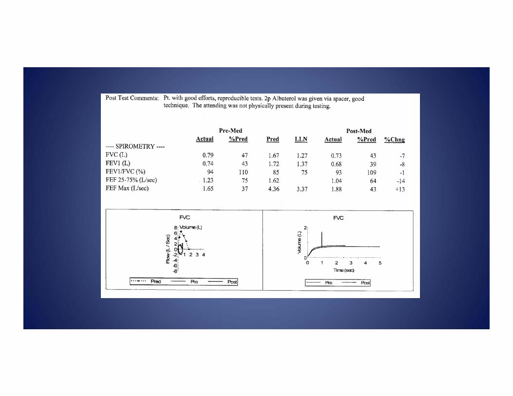
airflow limitation/obstruction Decreased FVC = restrictive process
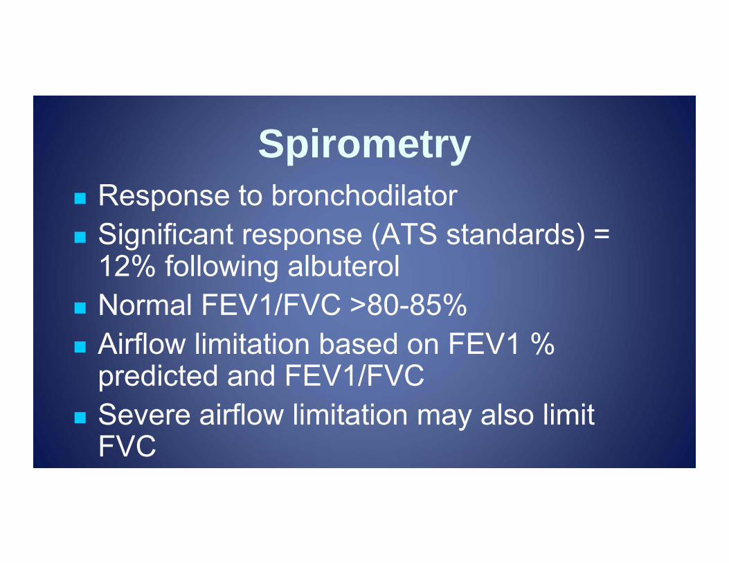
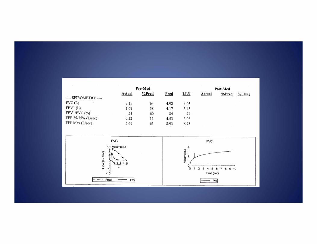
Spirometry Response to bronchodilator Significant response (ATS standards) =
12% following albuterol Normal FEV1/FVC >80-85% Airflow limitation based on FEV1 %
predicted and FEV1/FVC Severe airflow limitation may also limit
FVC

Lung Volume Parameters TLC = total lung capacity RV = residual volume FRC = functional residual capacity RV/TLC = marker of air trapping,
weakness or poor effort Normal RV/TLC is usually < 25%
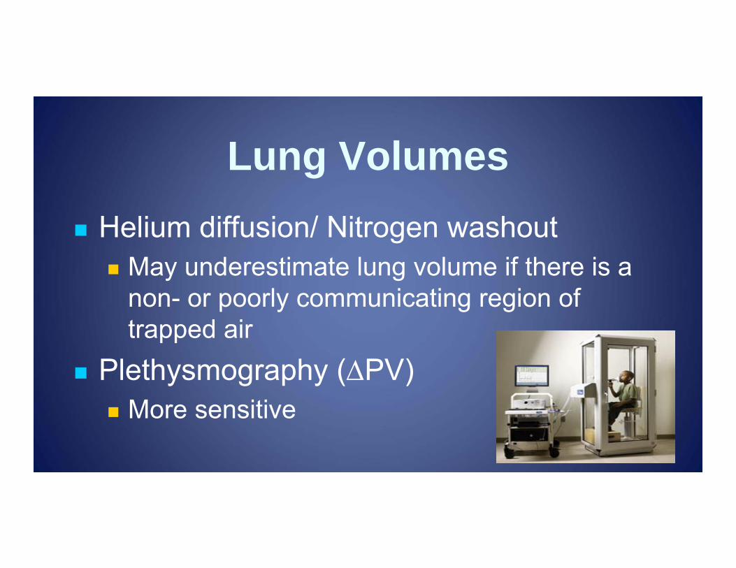
Lung Volumes Helium diffusion/ Nitrogen washout
May underestimate lung volume if there is a non- or poorly communicating region of trapped air
Plethysmography (PV) More sensitive

Diffusion Capacity Diffusing capacity of the lung for carbon
monoxide = DLCO Single breath technique Should be corrected for unit alveolar
volume (VA) and hemoglobin > 80% predicted is normal