research open access immunohistochemical localization of ... · research open access...
TRANSCRIPT

RESEARCH Open Access
Immunohistochemical localization ofhepatopancreatic phospholipase in gastropodsmollusc, Littorina littorea and Buccinum undatumdigestive cellsZied Zarai1, Nicholas Boulais2, Pascale Marcorelles3, Eric Gobin3, Sofiane Bezzine1, Hafedh Mejdoub1 andYoussef Gargouri1*
Abstract
Background: Among the digestive enzymes, phospholipase A2 (PLA2) hydrolyzes the essential dietaryphospholipids in marine fish and shellfish. However, we know little about the organs that produce PLA2, and theontogeny of the PLA2-cells. Accordingly, accurate localization of PLA2 in marine snails might afford a betterunderstanding permitting the control of the quality and composition of diets and the mode of digestion of lipidfood.
Results: We have previously producted an antiserum reacting specifically with mSDPLA2. It labeled zymogengranules of the hepatopancreatic acinar cells and the secretory materials of certain epithelial cells in the depths ofepithelial crypts in the hepatopancreas of snail. To confirm this localization a laser capture microdissection wasperformed targeting stained cells of hepatopancreas tissue sections. A Western blot analysis revealed a strongsignal at the expected size (30 kDa), probably corresponding to the PLA2.
Conclusions: The present results support the presence of two hepatopancreatic intracellular and extracellular PLA2in the prosobranchs gastropods molluscs, Littorina littorea and Buccinum undatum and bring insights on theirlocalizations.
Keywords: phospholipase A2, digestive enzyme, littorina littorea, Buccinum undatum hepatopancreas,immunolocalisation
BackgroundSnails require lipids for metabolic energy and for main-taining the structure and integrity of cell membranes incommon with other animals to tolerate environementalstrains [1]. The analyses of lipid composition of digestivegland and pedal muscle of two northern freshwater pul-monate snails Lymnaea stagnalis and Lymnaea ovataand three marine prosobranch gastropods Littorinaobtusata, Littorina littorea and Buccinum undatumfrom the White Sea, shown that the content of triacyl-glycerides both in digestive gland and pedal was higher
in littoral dwellers Littorina, the activity of whichdepends on the tide level. The presence of massive shellenhances demands in energy needed for supportingmovement and activity. Because the intensity of energymetabolism is related to quantity of total phospholipids,mitochondria and activity of their oxidizing ferments,the presence of thick shell in marine snails togetherwith motor activity costs more in terms of energy thanin freshwater snails with thin shell [1].In different molluscs, food is processed to varying
degrees as it passes through the alimentary tract. It isgenerally assumed that digestion of ingested materialtakes place in two phases, an extracellular process andintracellular digestion, where the prevalence of one overthe other depends on the type of diet of the animal.
* Correspondence: [email protected] de biochimie et de génie enzymatique des lipases, ENIS BPW1173 Université de Sfax-TunisiaFull list of author information is available at the end of the article
Zarai et al. Lipids in Health and Disease 2011, 10:219http://www.lipidworld.com/content/10/1/219
© 2011 Zarai et al; licensee BioMed Central Ltd. This is an Open Access article distributed under the terms of the Creative CommonsAttribution License (http://creativecommons.org/licenses/by/2.0), which permits unrestricted use, distribution, and reproduction inany medium, provided the original work is properly cited.

In general terms, the digestive glands of most molluscspresent a common organization and a single epitheliumcomprised by at least two cell types, namely, digestiveand basophilic cells found in the digestive diverticula[2]. Digestive cells are involved in the intracellular diges-tion of food and possess a highly developed endo-lysoso-mal system, whereas basophilic cells are secretory cellswith a highly developed rough endoplasmic reticulum[3].Although the digestive enzymes are well characterized,
including pepsin, trypsin, chymotrypsin, and amylase,little information is available on the lipid digestiveenzymes: lipases and phospholipases. This is mostly dueto difficulties in purification and histochemical analysisof the enzymes in fish [4].Among the lipid digestive enzymes, phospholipases A2
(PLA2; EC3.1.1.4) is potentially important in marinesnails, for hydrolysis of the essential dietary phospholi-pids. PLA2 catalyzes selective hydrolysis of the sn-2 acylester bond in 1,2-diacyl-sn-glycero-3 phospholipids,resulting in the formation of lysophospholipids and freefatty acids [5]. The occurrence, properties and physiolo-gical role of various PLA2 in aquatic organisms havebeen explained in several publications. Non-specificlipid acylhydrolases exhibiting combined action of var-ious lipases such as phospholipases have also beenrecovered and examined from aquatic organisms [6-8].This study describes immunohistochemically analysis
of PLA2 in the hepatopancreas organs of the adults’marine snail littorina littorea and Buccinum undatumusing an antiserum against Hexaplex trunculus hepato-pancreatic PLA2 [9].
ResultsMorphological analysis of digestive epitheliumThe digestive gland of the gastropod snail, Littorina lit-torea consists of blind ending tubules composed ofbasophilic and digestive cells (Figure 1) [10,11]. Thefunction of the digestive cells is the endocytosis and theintracellular digestion of food material, conveyed tothem from the stomach via the tubule lumina, and forthis purpose they have a well developed lysosomal-vacuolar system [12,13].Buccinum undatum, the northern whelk, is a common
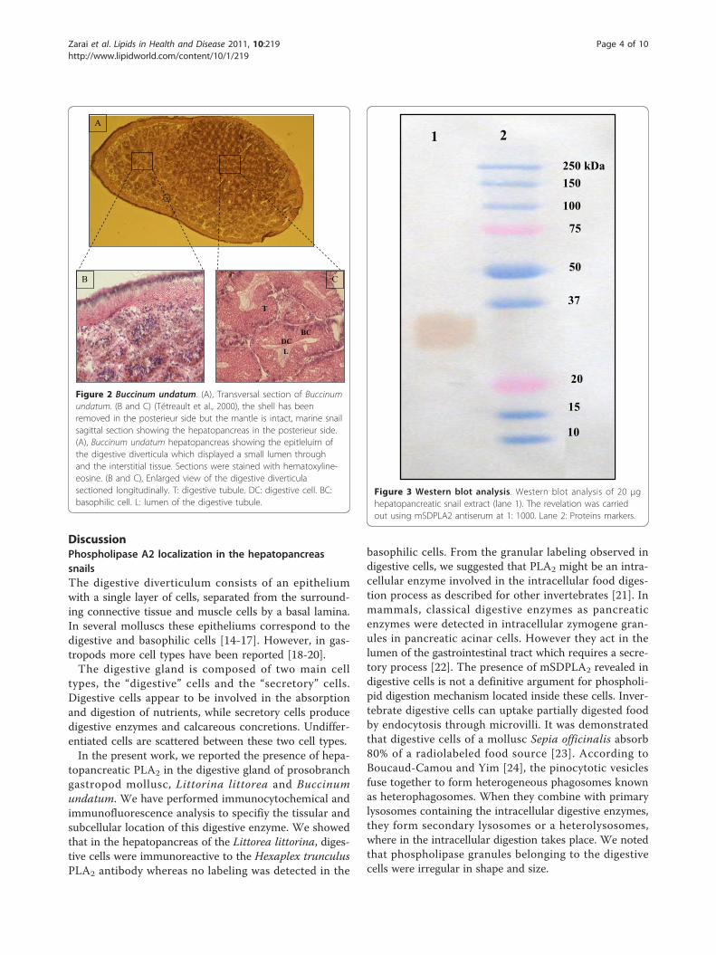
snail of moderate size (8 cm) on the northeastern coastof North America and in northern Europe. Several otherspecies occur in the Pacific Northwest. Buccinum unda-tum is commercially harvested for human consumptionin Europe.The digestive gland (Figure 2) is a vast pocket related
to stomach by the only one ciliate opening, it lacks welldifferentiated channels collectors. In the neighborhoodof stomach, it is partially divided up by folds of it wall.In the posterior zone towards the apex of the twist, the
partitions join to bound tubules. Every tubule of 30 μmapproximately, rest on a fine basal blade. Light tubulesare wide and cavities represent an important fraction ofthe volume of organ and composed of basophilic anddigestive cells.
Specificity of antiserum to Hexaplex trunculus PLA2
The supernatant of the marine snail hepatopancreashomogenate containing 100 μg of total proteins wassubjected to SDS-PAGE analysis followed by immuno-blotting. The anti-mSDPLA2 polyclonal antibody wasfound to react with a single band of 30 kDa correspond-ing to mSDPLA2 present in the crude extract (Figure 3).No other proteins react with anti-mSDPLA2 sera, sug-gesting a good specificity of our mSDPLA2 antiserum.Based on its specificity towards mSDPLA2, the polyclo-nal antibodies were used for immunocytolocalization ofPLA2 in the hepatopancreas tissue of the prosobranchsgastropods molluscs, Littorina littorea and Buccinumundatum.
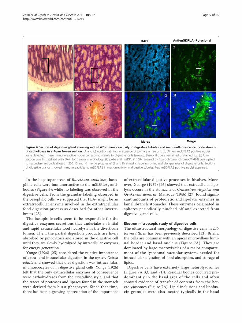
Histological, immunochemical and immunofluorescencestudiesThe location of the digestive PLA2 in marine snail Lit-torina littorea and Buccinum undatum was studiedimmunohistochemically by using an antiserum againstthe Hexaplex trunculus digestive mSDPLA2. The anti-serum efficiently react with hepatopancreatic cells ofmarine snail Littorina littorea and Buccinum undatum.Only digestive cells of Littorina littorea displayed a
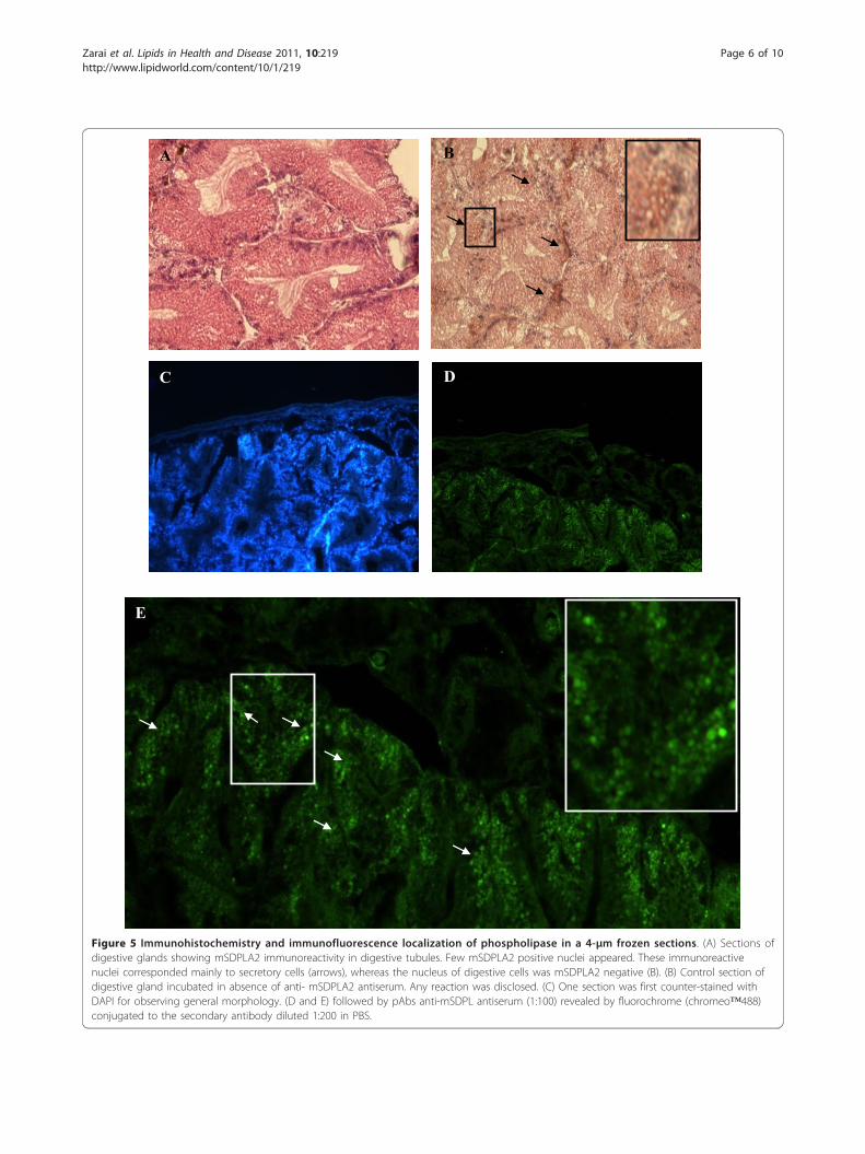
positive labeling for the presence of mSDPLA2. Conver-sely, secretory zymogene-like cells were not immunos-tained (Figure 4). Interestingly, we noticed that only fewintracellular granules on the digestive cells were immu-noreactive to anti-mSDPLA2. These granules with irre-gular in shape did not have a specific location in thedigestive diverticula; they were tentatively named phos-pholipase granules. However, the basophilic cells in thehepatopancreas of Buccinum undatum were immunor-eactive to the Hexaplex trunculus PLA2 antibodies. Nolabeling was detected in the digestive cells (Figure 5).The granular labeling observed in the basophilic cellsindicates that the PLA2 might be an extratracellularenzyme involved in the extratracellular food digestionprocess.To confirm the presence of PLA2 and the specificity of
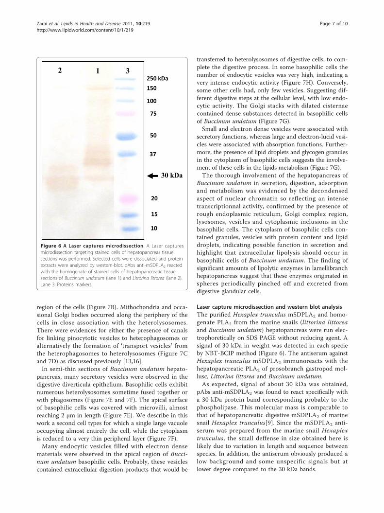
the mSDPLA2 polyclonal antibodies, we performed alaser capture microdissection targeting stained cells ofhepatopancreas tissue sections. Selected cells were disso-ciated and proteins were extracted for immunoblotinganalysis. Western blotting revealed a strong broad bandat around 30 kDa for both snails Littorina littorea andBuccinum undatum, corresponding probably to thePLA2 (Figure 6).
Zarai et al. Lipids in Health and Disease 2011, 10:219http://www.lipidworld.com/content/10/1/219
Page 2 of 10

T
DC BC
L
C
D E
Figure 1 Littorea. littorina. (A), Littorea. littorina fished in Franch Brittany coast. (B), the shell has been removed in the posterieur side taking careto keep the mantle is intact. The marine snail sagittal section exhibited the hepatopancreas in the posterieur side. (C), light microscopic view ofsections of digestive glands. Sections were stained with hematoxylin-eosin for observing the general morphology. (D and E), magnification ofthe digestive diverticula sectioned longitudinally. T: digestive tubule. DC: digestive cell. BC: basophilic cell. L: lumen of digestive tubule.
Zarai et al. Lipids in Health and Disease 2011, 10:219http://www.lipidworld.com/content/10/1/219
Page 3 of 10
DiscussionPhospholipase A2 localization in the hepatopancreassnailsThe digestive diverticulum consists of an epitheliumwith a single layer of cells, separated from the surround-ing connective tissue and muscle cells by a basal lamina.In several molluscs these epitheliums correspond to thedigestive and basophilic cells [14-17]. However, in gas-tropods more cell types have been reported [18-20].The digestive gland is composed of two main cell
types, the “digestive” cells and the “secretory” cells.Digestive cells appear to be involved in the absorptionand digestion of nutrients, while secretory cells producedigestive enzymes and calcareous concretions. Undiffer-entiated cells are scattered between these two cell types.In the present work, we reported the presence of hepa-
topancreatic PLA2 in the digestive gland of prosobranchgastropod mollusc, Littorina littorea and Buccinumundatum. We have performed immunocytochemical andimmunofluorescence analysis to specifiy the tissular andsubcellular location of this digestive enzyme. We showedthat in the hepatopancreas of the Littorea littorina, diges-tive cells were immunoreactive to the Hexaplex trunculusPLA2 antibody whereas no labeling was detected in the
basophilic cells. From the granular labeling observed indigestive cells, we suggested that PLA2 might be an intra-cellular enzyme involved in the intracellular food diges-tion process as described for other invertebrates [21]. Inmammals, classical digestive enzymes as pancreaticenzymes were detected in intracellular zymogene gran-ules in pancreatic acinar cells. However they act in thelumen of the gastrointestinal tract which requires a secre-tory process [22]. The presence of mSDPLA2 revealed indigestive cells is not a definitive argument for phospholi-pid digestion mechanism located inside these cells. Inver-tebrate digestive cells can uptake partially digested foodby endocytosis through microvilli. It was demonstratedthat digestive cells of a mollusc Sepia officinalis absorb80% of a radiolabeled food source [23]. According toBoucaud-Camou and Yim [24], the pinocytotic vesiclesfuse together to form heterogeneous phagosomes knownas heterophagosomes. When they combine with primarylysosomes containing the intracellular digestive enzymes,they form secondary lysosomes or a heterolysosomes,where in the intracellular digestion takes place. We notedthat phospholipase granules belonging to the digestivecells were irregular in shape and size.
A
B C
DC BC
L
T
Figure 2 Buccinum undatum. (A), Transversal section of Buccinumundatum. (B and C) (Tétreault et al., 2000), the shell has beenremoved in the posterieur side but the mantle is intact, marine snailsagittal section showing the hepatopancreas in the posterieur side.(A), Buccinum undatum hepatopancreas showing the epitleluim ofthe digestive diverticula which displayed a small lumen throughand the interstitial tissue. Sections were stained with hematoxyline-eosine. (B and C), Enlarged view of the digestive diverticulasectioned longitudinally. T: digestive tubule. DC: digestive cell. BC:basophilic cell. L: lumen of the digestive tubule.
250 kDa 150
100
75
50
37
20
10
15
2 1
Figure 3 Western blot analysis. Western blot analysis of 20 μghepatopancreatic snail extract (lane 1). The revelation was carriedout using mSDPLA2 antiserum at 1: 1000. Lane 2: Proteins markers.
Zarai et al. Lipids in Health and Disease 2011, 10:219http://www.lipidworld.com/content/10/1/219
Page 4 of 10
In the hepatopancreas of Buccinum undatum, baso-philic cells were immunoreactive to the mSDPLA2 anti-bodies (Figure 5); while no labeling was observed in thedigestive cells. From the granular labeling observed inthe basophilic cells, we suggested that PLA2 might be anextratracellular enzyme involved in the extratracellularfood digestion process as described for other inverte-brates [25].The basophilic cells seem to be responsible for the
digestive enzymes secretions that undertake an initialand rapid extracellular food hydrolysis in the diverticulalumen. Then, the partial digestion products are likelyabsorbed by pinocytosis and stored in the digestive celluntil they are slowly hydrolyzed by intracellular enzymesfor energy generation.Yonge (1926) [25] considered the relative importance
of extra- and intracellular digestion in the oyster, Ostreaedulis and showed that diet digestion was intracellular,in amoebocytes or in digestive gland cells. Yonge (1926)felt that the only extracellular enzymes of consequencewere carbohydrases from the crystalline style, and thatthe traces of proteases and lipases found in the stomachwere derived from burst phagocytes. Since that time,there has been a growing appreciation of the importance
of extracellular digestive processes in bivalves. More-over, George (1952) [26] showed that extracellular lipo-lysis occurs in the stomachs of Crassosirea virginica andGeukensia demissa. Mansour (1946) [27] found signifi-cant amounts of proteolytic and lipolytic enzymes inlamellibranch stomachs. These enzymes originated inspheres periodically pinched off and excreted fromdigestive gland cells.
Electron microscopic study of digestive cellsThe ultrastructural morphology of digestive cells in Lit-torina littirea has been previously described [13]. Briefly,the cells are columnar with an apical microvillous lumi-nal border and basal nucleus (Figure 7A). They aredominated by large macrovisicles of a major comparte-ment of the lysosomal-vacuolar system, needed forintracellular digestion of food absorption, and storage oflipids.Digestive cells have externely large heterolysosomes
(Figure 7A,B,C and 7D). Residual bodies occurred pre-dominantly in the basal area of the cells and oftenshowed evidence of transfer of contents from the het-erolysosomes (Figure 7A). Lipid inclusions and lipofus-cin granules were also located typically in the basal
DAPI
Merge Merge
Anti-mSDPLA2 Polyclonal
Figure 4 Section of digestive gland showing mSDPLA2 immunoreactivity in digestive tubules and immunofluorescence localization ofphospholipase in a 4-μm frozen section. (A and C) control satining in absence of primary antiserum. (B, D) Few mSDPLA2 positive nucleiwere detected. These immunoreactive nuclei correspond mainly to digestive cells (arrows). Basophilic cells remained unstained (D). (E) Onesection was first stained with DAPI for general morphology. (F) pAbs anti mSDPL (1:100) revealed by fluorochrome (chromeo™488) conjugatedto secondary antibody diluted 1:200. (G and H) merge pictures of (E and F), showing labeling of intracellular granules of digestive cells. Sectionsof digestive glands showed immunoreactivity to mSDPLA2 immunoreactivity in digestive tubules. Few mSDPLA2 positive nuclei appeared.
Zarai et al. Lipids in Health and Disease 2011, 10:219http://www.lipidworld.com/content/10/1/219
Page 5 of 10
A
C D
E
B
Figure 5 Immunohistochemistry and immunofluorescence localization of phospholipase in a 4-μm frozen sections. (A) Sections ofdigestive glands showing mSDPLA2 immunoreactivity in digestive tubules. Few mSDPLA2 positive nuclei appeared. These immunoreactivenuclei corresponded mainly to secretory cells (arrows), whereas the nucleus of digestive cells was mSDPLA2 negative (B). (B) Control section ofdigestive gland incubated in absence of anti- mSDPLA2 antiserum. Any reaction was disclosed. (C) One section was first counter-stained withDAPI for observing general morphology. (D and E) followed by pAbs anti-mSDPL antiserum (1:100) revealed by fluorochrome (chromeo™488)conjugated to the secondary antibody diluted 1:200 in PBS.
Zarai et al. Lipids in Health and Disease 2011, 10:219http://www.lipidworld.com/content/10/1/219
Page 6 of 10
region of the cells (Figure 7B). Mithochondria and occa-sional Golgi bodies occurred along the periphery of thecells in close association with the heterolysosomes.There were evidences for either the presence of canalsfor linking pinocytotic vesicles to heterophagosomes oralternatively the formation of ‘transport vesicles’ fromthe heterophagosomes to heterolysosomes (Figure 7Cand 7D) as discussed previously [13,16].In semi-thin sections of Buccinum undatum hepato-
pancreas, many secretory vesicles were observed in thedigestive diverticula epithelium. Basophilic cells exhibitnumerous heterolysosomes sometime fused together orwith phagosomes (Figure 7E and 7F). The apical surfaceof basophilic cells was covered with microvilli, almostreaching 2 μm in length (Figure 7E). We describe in thiswork a second cell types for which a single large vacuoleoccupying almost entirely the cell, while the cytoplasmis reduced to a very thin peripheral layer (Figure 7F).Many endocytic vesicles filled with electron dense
materials were observed in the apical region of Bucci-num undatum basophilic cells. Probably, these vesiclescontained extracellular digestion products that would be
transferred to heterolysosomes of digestive cells, to com-plete the digestive process. In some basophilic cells thenumber of endocytic vesicles was very high, indicating avery intense endocytic activity (Figure 7H). Conversely,some other cells had, only few vesicles. Suggesting dif-ferent digestive steps at the cellular level, with low endo-cytic activity. The Golgi stacks with dilated cisternaecontained dense substances detected in basophilic cellsof Buccinum undatum (Figure 7G).Small and electron dense vesicles were associated with
secretory functions, whereas large and electron-lucid vesi-cles were associated with absorption functions. Further-more, the presence of lipid droplets and glycogen granulesin the cytoplasm of basophilic cells suggests the involve-ment of these cells in the lipids metabolism (Figure 7G).The thorough involvement of the hepatopancreas of
Buccinum undatum in secretion, digestion, adsorptionand metabolism was evidenced by the decondensedaspect of nuclear chromatin so reflecting an intensetranscriptionnal activity, confirmed by the presence ofrough endoplasmic reticulum, Golgi complex region,lysosomes, vesicles and cytoplasmic inclusions in thebasophilic cells. The cytoplasm of basophilic cells con-tained granules, vesicles with protein content and lipiddroplets, indicating possible function in secretion andhighlight that extracellular lipolysis should occur inbasophilic cells of Buccinum undatum. The finding ofsignificant amounts of lipolytic enzymes in lamellibranchhepatopancreas suggest that these enzymes originated inspheres periodically pinched off and excreted fromdigestive glandular cells.
Laser capture microdissection and western blot analysisThe purified Hexaplex trunculus mSDPLA2 and homo-genate PLA2 from the marine snails (littorina littoreaand Buccinum undatum) hepatopancreas were run elec-trophoretically on SDS PAGE without reducing agent. Asignal of 30 kDa in weight was detected in each specieby NBT-BCIP method (Figure 6). The antiserum againstHexaplex trunculus mSDPLA2 immunoreacts with thehepatopancreatic PLA2 of prosobranch gastropod mol-lusc, Littorina littorea and Buccinum undatum.As expected, signal of about 30 kDa was obtained,
pAbs anti-mSDPLA2 was found to react specifically witha 30 kDa protein band corresponding probably to thephospholipase. This molecular mass is comparable tothat of hepatopancreatic digestive mSDPLA2 of marinesnail Hexaplex trunculus[9]. Since the mSDPLA2 anti-serum was prepared from the marine snail Hexaplextrunculus, the small deffense in size obtained here islikely due to variation in length and sequence betweenspecies. In addition, the antiserum obviously produced alow background and some unspecific signals but atlower degree compared to the 30 kDa bands.
2 1 250 kDa
150
100
75
50
37
20
10
15
30 kDa
3
Figure 6 A Laser captures microdissection. A Laser capturesmicrodissection targeting stained cells of hepatopancreas tissuesections was performed. Selected cells were dissociated and proteinextracts were analyzed by western-blot. pAbs anti-mSDPLA2 reactedwith the homogenate of stained cells of hepatopancreatic tissuesections of Buccinum undatum (lane 1) and Littorina littorea (lane 2).Lane 3: Proteins markers.
Zarai et al. Lipids in Health and Disease 2011, 10:219http://www.lipidworld.com/content/10/1/219
Page 7 of 10

Materials and methodsAnimals collectionMarine snails’ littorina littorea and Buccinum undatumwere collected from the Atlantic coasts of French Brittany.They were kept on ice until use. Only the hepatopan-creases were collected immediately and stored at -80°C.
Preparation of the antiserum to Hexaplex trunculus PLA2
Antibodies against mSDPLA2 were produced in rabbitsby three biweekly subcutaneous injections of 250 μg ofpurified mSDPLA2 emulsified with complete Freund’sadjuvant. Sera were obtained from blood samples twoweeks after the last injection. Reactivity of collected
A B
C D
F E
H G
100 m 10 m
3 m 10 m
10 m 10 m
50 m 10 m
G
Figure 7 Transmission Electron Microscopy. Transmission Electron Microscopy of the digestive cells of Littorina littirea (A, B, C and D) andbasophilic cells of Buccinum undatum (E, F, G and H).
Zarai et al. Lipids in Health and Disease 2011, 10:219http://www.lipidworld.com/content/10/1/219
Page 8 of 10

antiserum was determined by the microplate ELISAmethod using the Hexaplex trunculus mSDPLA2.
SDS-PAGE and immunoblotting techniqueAnalytical polyacrylamide gel electrophoresis of proteinsin the presence of sodium dodecyl sulfate (SDS-PAGE)was performed according to the Laemmli’s method [28].The specificity of pAbs anti-mSDPLA2 was establishedby protein blotting. Proteins from SDS gel were trans-ferred to nitrocellulose membranes. After the transfer,membranes were rinsed three times in PBS (10 mMphosphate, NaCl 150 mM, pH 7.2), with 3% half-fatmilk for 1 h at room temperature. Thereafter, mem-branes were incubated with pAbs anti-mSDPLA2 dilutedat 1:1000 in PBS containing 0.05% tween-20 (PBS-T) for1 h at room temperature. Afterward, they were washedthree times with PBS-T and incubated for 1 h at roomtemperature with a 1:2000 dilution of alkaline phospha-tase-conjugated anti-rabbit immunoglobulin (Sigma).Then, washing as above, the revelation were carried outusing a NBT-BCIP kit according to the supplier (Sigma).
Histology, immunochemistry and immunofluorescenceImmunohistochemistryTissues, with a size of around 1 mm3, were embedded inOptimal Cutting temperature (OCT) and cryopreservedin isopentane chilled on liquid nitrogen. Sections with athickness of 4 μm were cut. Slides were saturated with5% of normal goat serum in PBS with 0.05% Triton X-100 for 15 minutes and subsequently hybridized withprimary anti-mSDPLA2 polyclonal antibodies diluted at1:200 in Dako Diluent (S3022) for 2 h at 4°C. Slideswere rinsed twice and hybridized with secondary anti-rabbit biotinylated- antibodies diluted at 1:200 for twohours at room temperature.ImmunofluorescenceTissue sections were fixed in PBS with 4% paraformal-dehyde, permeabilized with 0.5% Triton X-100, saturatedin 5% normal goat serum in PBS-T and hybridized withanti-mSDPLA2 polyclonal antibodies diluted at 1:100 inDako Diluent (S3022) overnight at 4°C. After twowashes, cells were hybridized for 2 h with a chromeo™-488-conjugated secondary antibody from rabbit (Abcam,Cambridge, UK) diluted at 1:200 in PBS. The fluores-cence analysis were performed with BX41 Olympusupright microscope and pictures were taken with anOlympus C-5060 digital camera. Control experimentswere carried out in absence of anti-mSDPLA2 antibody.Electron microscopic studyFragments of tissues, with a size of about 1 mm3 werefixed in 2.5% glutaraldehyde for 2 hours, rinsed in sor-ensen’s phosphate buffer and post-fixed for 1 hour 30with 2% OsO4. Tissues were dehydrated in graded con-centrations of alcohol, and then in propylene oxide.
Thereafter, tissues were embedded in epoxy resin for 72h at 60°C. Finally, ultrathin sections were cut andobserved with a Jeol, Jem-1010 Electron Microscope.
ConclusionIn this study we found two different localizations forproduction and secretion of the hepatopancreatic diges-tive PLA2 in two species of marine snails Littorina lit-torea and Buccinum undatum. While the first producedmSDPLA2 in digested cells, the second produced it inbasophilic cells. Only these cells contained secretorymaterials exhibiting PLA2-like immunoreactivity. Thehepatopancreas is adapted to increase gut surface areaand it is a suitable compartment for lipid absorption.
AbbreviationsmSDPLA2: marine snail digestive phospholipase A2; SDS-PAGE: sodiumdodecyl sulfate-polyacrylamide gel electrophoresis; BSA: bovine serumalbumin; pAbs: polyclonal antibodies; PBS: phosphate buffer saline; DAPI:4’,6-diamidino-2-phenylindole; OCT: Optimal Cutting temperature.
AcknowledgementsWe are very grateful to Dr. Nicolas.lebonvallet., PhD. Jérémy.charet(Laboratoire de Neurobiologie Cutanée, CHU Morvan, Université de Brest,France) for their fruitful discussions and kind help during the preparation ofthis work. We thank Dr. Nacim Zouari (Ecole Nationale d’Igénieurs de Sfax(ENIS)) for his help during rabbits immunisation. This work is part of adoctoral thesis by Zied Zarai. This work received financial support from the «Ministry of Higher Education and Scientific Research, Tunisia».
Author details1Laboratoire de biochimie et de génie enzymatique des lipases, ENIS BPW1173 Université de Sfax-Tunisia. 2Laboratoire de Neurobiologie Cutanée, CHUMorvan, Université de Brest, 29609 BREST cedex France. 3Service d’AnatomiePathologique, Pôle de Biologie-Pathologie, CHU Morvan, Université de Brest,29609 BREST cedex France.
Authors’ contributionsZZ carried out all the studies, analyzed the data and drafted the manuscript.NB helped with the analysis, discussion of the data and correction of themanuscript. PM and EG helped with the electron microscopic analysis anddiscussion of the data. SB helped with the correction of the manuscript. TRhelped with the discussion of the data. HM and YG participated in the studydesign and helped to draft the manuscript. All authors have read andapproved the final manuscript.
Competing interestsThe authors declare that they have no competing interests.
Received: 21 October 2011 Accepted: 25 November 2011Published: 25 November 2011
References1. Arakelova ES: Lipids composition and speed of energy metabolism in
gastropods. Zh Obshch Biol 2008, 69:471-478.2. Morton B: Feeding and digestion in bivalves.Edited by: Saleuddin ASM,
Wilbur KM. The Mollusca, Academic Press, New York; 1983:563-586.3. Mason AZ: The uptake, accumulation and excretion of metals by the
marine prosobranch gastropod mollusc Littorina littorea (L). PhD Thesis,University of Wales, Gwyneed (UK); 1983, 575.
4. Henderson RJ, Tocher DR: The lipid composition and biochemistry offreshwater fish. Prog Lipid Res 1987, 26:281-347.
5. Burke JE, Dennis EA: Phospholipase A2 structure/function, mechanism,and signaling. J Lipid Res 2009, 50:237-242.
Zarai et al. Lipids in Health and Disease 2011, 10:219http://www.lipidworld.com/content/10/1/219
Page 9 of 10
6. Audrey MA, Shetty KJ, Kinsella JE: Isolation and properties ofphospholipase A from pollock muscle. J Food Sci 1978, 43:1771-1775.
7. Chawla P, Ablett RF: Detection of microsomal phospholipase activity onmyotomal tissue of Atlantic cod (Gadus morha. J Food Sci 1987,52:1194-1197.
8. Izquierdo MS, Henderson RJ: The determination of lipase andphospholipase activities in gut content of turbot (Scophthalmusmaximus) by fluorescence-based assays. Fish Physiol Biochem 1998,19:153-162.
9. Zarai Z, Ben Bacha A, Horchani H, Bezzine S, Zouari N, Gargouri Y,Mejdoub H: A novel hepatopancreatic phospholipase A2 from Hexaplextrunculus with digestive and toxic activities. Arch Biochem Biophys 2010,494:121-129.
10. Fretter V, Graham A: British Prosobranch molluscs:their functionalanatomy and ecology. Ray Society, London 1962, 755.
11. Farley J: Phasic activity in the digestive gland cells of the marineprosobranch gastropod, Littorina littorea (L.). Proc Malac Soc (London)1973, 40(4):73-482.
12. Halton DW: The cytochemical localization of lysosomal hydrolases in thedigestive cells of littorinids and changes induced by larval trematodeinfection. Z Parasitenk 1977, 53:115-122.
13. Pipe RK: Light and electron microscope localisation of β-glucuronidase inthe stomach and digestive gland of the marine gastropod Littorinalittorea. Histochem J 1986.
14. Pal SG: The fine structure of the digestive tubules of Mya arenaria L. I.Basiphilcell. Proc Malacol Soc London 1971, 39:303-309.
15. Pal SG: The fine structure of the digestive tubules of Mya arenaria L. II.Digestive cell. Proc Malacol Soc London 1972, 40:161-170.
16. Owen G: The fine structure and histochemistry of the digestivediverticula of the protobranchiate bivalve Nucula sulcata. Proc R SocLondon 1973, 183:249-264.
17. Lobo-da-Cunha A: The peroxisomes of the hepatopancreas in twospecies of chitons. Cell Tissue Res 1997, 290:655-664.
18. Nelson L, Morton JE: Cyclic activity and epithelial renewal in thedigestive gland tubules of the marine prosobranch Maoricryptamonoxyla (Lesson). J Moll Stud 1979, 45:262-283.
19. Franchini A, Ottaviani E: Histochemical and ultrastructural study of thedigestive gland of the freshwater snail Panorbarius corneus (L.)(Gastropoda, Pulmonata). Anim Biol 1993, 2:191-198.
20. Kress A, Schmekel L, Nott JA: Ultrastructure of the digestive gland in theopisthobranch mollusk, Runcina. Veliger 1994, 37:358-373.
21. Goyffon M, Martoja R: Cytophysiological aspects of digestion and storagein the liver of a scorpion. Cell Tissue Res 1983, 228:661-675.
22. Miled N, Canaan S, Dupuis L, Roussel A, Rivière M, Carrière F, de Caro A,Cambillau C, Verger R: Digestive lipases: from three-dimensional structureto physiology. Biochimie 2000, 82:973-986.
23. Boucaud-Camou E, Péquignant E: An experimental study of digestionabsorption in Sepia officinalis. Forma Functio 1973, 6:93-112.
24. Boucaud-Camou E, Yim M: Fine structure and function of the digestivecell of Sepia officinalis (Mollusca: Cephalopoda). J Zool (London) 1980,191:89-105.
25. Yonge GM: The digestive diverticula in the lamellibranchs. Trans R SocEdinb 1926, 54:703-718.
26. George WC: The digestion and absorption of fat in Iamellibranchs. BiolBull 1952, 102:118-127.
27. Mansour K: Food and digestive processes of the lamellibranchs. Nature,Land 1946, 157:482.
28. Laemmli UK: Cleavage of structural proteins during the assembly of thehead of bacteriophage T4. Nature 1970, 227:680-685.
doi:10.1186/1476-511X-10-219Cite this article as: Zarai et al.: Immunohistochemical localization ofhepatopancreatic phospholipase in gastropods mollusc, Littorina littoreaand Buccinum undatum digestive cells. Lipids in Health and Disease 201110:219.
Submit your next manuscript to BioMed Centraland take full advantage of:
• Convenient online submission
• Thorough peer review
• No space constraints or color figure charges
• Immediate publication on acceptance
• Inclusion in PubMed, CAS, Scopus and Google Scholar
• Research which is freely available for redistribution
Submit your manuscript at www.biomedcentral.com/submit
Zarai et al. Lipids in Health and Disease 2011, 10:219http://www.lipidworld.com/content/10/1/219
Page 10 of 10