research article predictive significance of serum level of
TRANSCRIPT

Research ArticlePredictive Significance of Serum Level of Vascular EndothelialGrowth Factor in Gastric Cancer Patients
Lu Wang, Yanli Chang, Jianjun Xu, and Qingyun Zhang
Department of Clinical Laboratory, Key Laboratory of Carcinogenesis and Translational Research, Ministry of Education,Peking University Cancer Hospital & Institute, Beijing 100142, China
Correspondence should be addressed to Qingyun Zhang; zhqy [email protected]
Received 2 June 2016; Revised 12 July 2016; Accepted 12 July 2016
Academic Editor: Mario Scartozzi
Copyright © 2016 Lu Wang et al.This is an open access article distributed under the Creative Commons Attribution License, whichpermits unrestricted use, distribution, and reproduction in any medium, provided the original work is properly cited.
The study aims to evaluate serumVEGF expression in gastric cancer patients and investigate its relationshipwith clinicopathologicalparameters. We also examined the serumVEGF levels in GC patients having received surgery or chemotherapy treatment to assessits predictive and prognostic value as a biomarker. We enrolled 154 GC patients having not received neoadjuvant treatment and 100healthy controls. In the treatment groups, 13 surgery patients and 15 chemotherapy patients were investigated. 42 chemotherapypatients with different chemotherapy efficacy were recruited as well. The serum VEGF was examined by ELISA. Serum VEGF levelwas remarkably upregulated in GC group compared with healthy group (𝑝 < 0.001). The serum VEGF level of GC group wassignificantly correlated with tumor cells differentiation degree, clinical stages, tumor infiltration depth, lymph nodemetastasis, andtumor size. The serum VEGF level of the 1 to 3 days after operation group was much lower than that of the preoperative group(𝑝 < 0.001) and the 7 days after operation group (𝑝 < 0.001). By contrast, serum VEGF level was decreased significantly afterchemotherapy (𝑝 = 0.001). Importantly, serum VEGF level in PD+SD group was significantly higher compared to the PR+CRgroup (𝑝 = 0.011). Therefore, serum VEGF was a valuable biomarker in clinically monitoring the condition of GC patients.
1. Introduction
Gastric cancer is one of the most common cancers and is thesecond cause of cancer-related death worldwide [1, 2]. Gastricadenocarcinoma accounts for about 95% of gastric cancercases, and the highest incidence countries are in Eastern Asia(e.g., Korea, Japan, and China) [3, 4]. Moreover, males areaffected twice as frequently as females, and the average age ofonset is between 60 and 70 years [5]. Conventional therapyoptions for gastric cancer include surgery, chemotherapy,radiation therapy, and combination treatments. In the earlystages, the disease can often be cured by complete surgicalremoval of the tumors. However, patients with advancedgastric cancer indicated a poor prognosis [6]. Thus, there isthe need for a better marker to identify the condition andprognosis of patients.
Angiogenesis is a process of new blood vessels formationthat plays a critical role in the growth and metastasis of many
solid tumors including gastric adenocarcinoma. Amongmul-tiple proangiogenic factors that participate in physiologicaland pathological angiogenesis, VEGF is the most important[7, 8]. VEGF can bind to VEGF receptor (VEGFR) to triggermultiple cellular signal pathways which inhibit apoptosisand stimulate survival of endothelial cells and recruit ofendothelial precursor cells from the bone marrow to the sitesof angiogenesis. It also plays an important role in increas-ing vascular permeability and inhibiting differentiation ofdendritic cells [9]. Notably, serum VEGF level is higherthan that of plasma. VEGF is mostly produced by tumorcells and transported by platelets. It is released into serumafter platelets degranulation during blood clotting.Therefore,serum VEGF levels can better reveal the amount of VEGFproduced by tumor cells and tumor burden [10].
The current study aimed to identify serum VEGF as abiomarker that may be used to monitor both surgery andchemotherapy patients during the course of the disease and
Hindawi Publishing CorporationBioMed Research InternationalVolume 2016, Article ID 8103019, 6 pageshttp://dx.doi.org/10.1155/2016/8103019

2 BioMed Research International
investigate the clinical significance of serum VEGF level topredict chemotherapy outcomes.
2. Materials and Methods
2.1. StudyCohort. Between September 2015 and January 2016,154 patients with histologically confirmed gastric adenocar-cinoma in Peking University Cancer Hospital were enrolledas the GC group. The GC patients included 112 males and42 females, with a mean age of 60 (range 27 to 82 years).None of the patients in this group have received neoadjuvanttreatment before. Tumor staging was based on clinical infor-mation, radiologic reports, operative findings, and pathologyreports. The staging was made in accordance with the TNMstaging system for gastric cancer and TNM staging wasdone according to the American Joint Committee on Cancer(AJCC). In addition, 100 healthy controls including 70 malesand 30 females, with a mean age of 40 years (range 22 to 61years), were chosen at the Medical Examination Center ofPeking University Cancer Hospital.
In order to evaluate the effects of treatment on the serumVEGF levels in gastric cancer patients, we selected 13 gastriccancer patients in the operation group and 15 patients inthe chemotherapy group.Their blood samples were collectedbefore and after treatment, respectively. No one in the oper-ation group received chemotherapy or radiotherapy beforeor after surgery. Moreover, 42 gastric patients who wereundergoing their chemotherapy cycles in Peking UniversityCancer Hospital were selected.
2.2. Detection of Serum VEGF. A total of 5mL peripheralvenous blood was obtained and then centrifuged to collectserum. Serum samples were stored at −80∘C for further use.The serum VEGF was detected using quantitative humanVEGF sandwich enzyme immunoassay kits (Jian Ping JinXing Biological Technology Co., Ltd., Beijing, China) follow-ing manufacturer’s instructions, under standard conditions.The ELISA readings weremeasured at 450 nm in amicroplatereader.
2.3. Evaluation of Chemotherapy Effect. All chemotherapypatients were assessed with CT scan after 2 chemotherapycycles. The treatment effect was assessed based on responseevaluation criteria in solid tumors (RECIST) guidelines [11]:complete response (CR): all known disease disappearing;partial response (PR): ≥30% reduction in the sum of lineartumor measurements; progressive disease (PD): at least 20%increase in the sum of target lesions and new lesions appear-ing; and stable disease (SD): neither enough shrinkage forPR nor enough increase for PD. CR and PR were defined aseffective response while SD and PD were labelled as invalidresponse.
2.4. Statistical Analysis. Statistical analysis was performedusing SPSS 19.0 for Windows. Statistical significance wasmeasured by 𝑝 value < 0.05. Measurement data with normaldistribution was expressed as mean ± standard deviation,while median (interquartile range, IQR) was used when
the data did not meet the normal distribution. Comparisonbetween the GC group and control group and parametersthat contained 2 varieties were evaluated with a Mann-Whitney 𝑈 test. Repeated measures ANOVA was appliedin continuous monitoring operation patients group whilepaired-samples 𝑡-test was used in chemotherapy patientsgroup. Two-independent-sample 𝑡-test was used to evaluatethe relationship between clinical progression and VEGFlevels.
3. Results
3.1. Characteristics of the Gastric Cancer Patients. There were112 (72.7%) males and 42 (27.3%) females in the GC groupand the mean age of GC group was 60 years (range 27 to82 years). The baseline parameters of gastric cancer patientswere shown in Table 1. The serum VEGF levels of gastriccancer patients showed no correlations with gender, age,distant metastasis, Lauren classification, Her2, and EGFRexpressions (𝑝 > 0.05, Table 1). The serum VEGF levelof patients with poorly differentiated carcinoma was signifi-cantly higher than that of moderately differentiated and well-differentiated carcinoma (𝑝 = 0.012). Serum level of patientswith early clinical stages (I+II) was significantly lower thanthat of advanced clinical stages (III+IV) (𝑝 = 0.004). Inaddition, serum VEGF level was markedly higher in patientswith infiltration depth of T3 and T4 compared with that ofthe T1 and T2 group (𝑝 = 0.004). Serum VEGF level ofpatients who had lymph node metastasis was higher thanthat of patients who had no lymph metastasis (𝑝 = 0.036).Meanwhile, compared to the group with tumor diameterwithin 4 cm, the serum VEGF level of the group with tumordiameter greater than or equal to 4 cm was significantlyhigher (𝑝 = 0.028).
3.2. VEGF Expression in the Sera of Gastric Cancer Patientsand Controls. In order to test the serum VEGF levels as thepotential biomarker for early prediction of gastric cancer, weselected 154 gastric cancer patients who had never receivedneoadjuvant treatment before and 100 healthy controls. TheELISA results showed that the serum VEGF levels weresignificantly different between the GC group and the controlgroup (145.812 (143.298) pg/mL versus 54.539 (67.355) pg/mL,𝑝 < 0.001, Table 2).
3.3. Evaluation of the Effects of Treatments on the SerumVEGFLevels in Gastric Cancer Patients
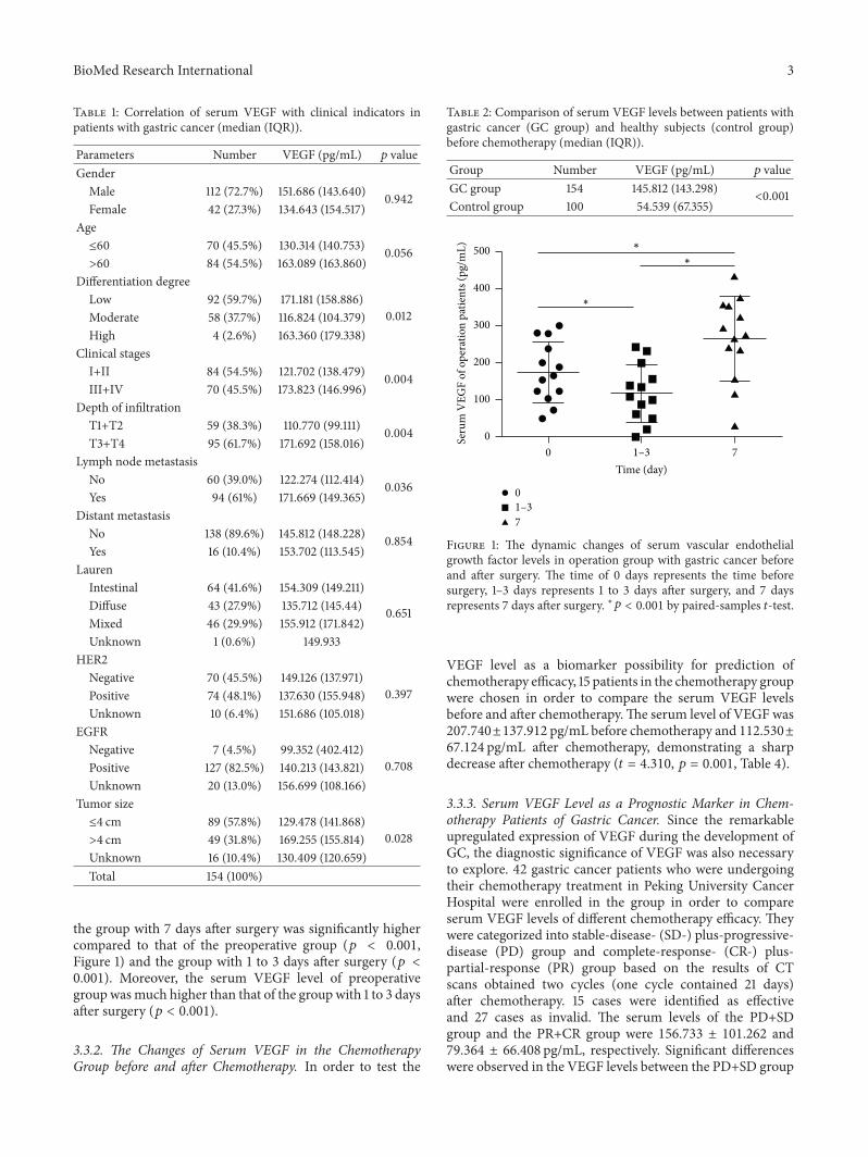
3.3.1. The Dynamic Changes of Serum VEGF Levels in theOperation Group before and after Surgery. In this research,13 gastric cancer patients were monitored to see the dynamicchanges of serumVEGF levels before and after surgerywithina short time.The serumVEGF levels of gastric cancer patientsbefore surgery and 1 to 3 days and 7 days after surgerywere 175.712 ± 81.329 pg/mL, 117.797 ± 76.022 pg/mL, and266.119 ± 112.218 pg/mL, respectively. The serum VEGFlevels differed significantly among the three groups (𝐹 =29.002, 𝑝 < 0.001, Table 3). The serum VEGF level of
BioMed Research International 3
Table 1: Correlation of serum VEGF with clinical indicators inpatients with gastric cancer (median (IQR)).
Parameters Number VEGF (pg/mL) 𝑝 valueGender
Male 112 (72.7%) 151.686 (143.640) 0.942Female 42 (27.3%) 134.643 (154.517)
Age≤60 70 (45.5%) 130.314 (140.753) 0.056>60 84 (54.5%) 163.089 (163.860)
Differentiation degreeLow 92 (59.7%) 171.181 (158.886)
0.012Moderate 58 (37.7%) 116.824 (104.379)High 4 (2.6%) 163.360 (179.338)
Clinical stagesI+II 84 (54.5%) 121.702 (138.479) 0.004III+IV 70 (45.5%) 173.823 (146.996)
Depth of infiltrationT1+T2 59 (38.3%) 110.770 (99.111) 0.004T3+T4 95 (61.7%) 171.692 (158.016)
Lymph node metastasisNo 60 (39.0%) 122.274 (112.414) 0.036Yes 94 (61%) 171.669 (149.365)
Distant metastasisNo 138 (89.6%) 145.812 (148.228) 0.854Yes 16 (10.4%) 153.702 (113.545)
LaurenIntestinal 64 (41.6%) 154.309 (149.211)
0.651Diffuse 43 (27.9%) 135.712 (145.44)Mixed 46 (29.9%) 155.912 (171.842)Unknown 1 (0.6%) 149.933
HER2Negative 70 (45.5%) 149.126 (137.971)
0.397Positive 74 (48.1%) 137.630 (155.948)Unknown 10 (6.4%) 151.686 (105.018)
EGFRNegative 7 (4.5%) 99.352 (402.412)
0.708Positive 127 (82.5%) 140.213 (143.821)Unknown 20 (13.0%) 156.699 (108.166)
Tumor size≤4 cm 89 (57.8%) 129.478 (141.868)
0.028>4 cm 49 (31.8%) 169.255 (155.814)Unknown 16 (10.4%) 130.409 (120.659)Total 154 (100%)
the group with 7 days after surgery was significantly highercompared to that of the preoperative group (𝑝 < 0.001,Figure 1) and the group with 1 to 3 days after surgery (𝑝 <0.001). Moreover, the serum VEGF level of preoperativegroup wasmuch higher than that of the group with 1 to 3 daysafter surgery (𝑝 < 0.001).
3.3.2. The Changes of Serum VEGF in the ChemotherapyGroup before and after Chemotherapy. In order to test the
Table 2: Comparison of serum VEGF levels between patients withgastric cancer (GC group) and healthy subjects (control group)before chemotherapy (median (IQR)).
Group Number VEGF (pg/mL) 𝑝 valueGC group 154 145.812 (143.298)
<0.001Control group 100 54.539 (67.355)
∗
∗
∗
7
1–30
1–3 70
Time (day)
0
100
200
300
400
500
Seru
m V
EGF
of o
pera
tion
patie
nts (
pg/m
L)
Figure 1: The dynamic changes of serum vascular endothelialgrowth factor levels in operation group with gastric cancer beforeand after surgery. The time of 0 days represents the time beforesurgery, 1–3 days represents 1 to 3 days after surgery, and 7 daysrepresents 7 days after surgery. ∗𝑝 < 0.001 by paired-samples 𝑡-test.
VEGF level as a biomarker possibility for prediction ofchemotherapy efficacy, 15 patients in the chemotherapy groupwere chosen in order to compare the serum VEGF levelsbefore and after chemotherapy.The serum level of VEGF was207.740±137.912 pg/mL before chemotherapy and 112.530±67.124 pg/mL after chemotherapy, demonstrating a sharpdecrease after chemotherapy (𝑡 = 4.310, 𝑝 = 0.001, Table 4).
3.3.3. Serum VEGF Level as a Prognostic Marker in Chem-otherapy Patients of Gastric Cancer. Since the remarkableupregulated expression of VEGF during the development ofGC, the diagnostic significance of VEGF was also necessaryto explore. 42 gastric cancer patients who were undergoingtheir chemotherapy treatment in Peking University CancerHospital were enrolled in the group in order to compareserum VEGF levels of different chemotherapy efficacy. Theywere categorized into stable-disease- (SD-) plus-progressive-disease (PD) group and complete-response- (CR-) plus-partial-response (PR) group based on the results of CTscans obtained two cycles (one cycle contained 21 days)after chemotherapy. 15 cases were identified as effectiveand 27 cases as invalid. The serum levels of the PD+SDgroup and the PR+CR group were 156.733 ± 101.262 and79.364 ± 66.408 pg/mL, respectively. Significant differenceswere observed in the VEGF levels between the PD+SD group

4 BioMed Research International
Table 3: Continuous monitoring of serum VEGF levels in patients with gastric cancer before and after operation (𝑥 ± 𝑠).
Group Number VEGF (pg/mL)𝐹 𝑝 value
0 days After 1–3 days After 7 daysOperative patients 13 175.712 ± 81.329 117.797 ± 76.022 266.119 ± 112.218 29.002 <0.001
Table 4: The changes of serum VEGF in patients with gastric cancer before and after chemotherapy (𝑥 ± 𝑠).
Group Number VEGF (pg/mL)𝑡 𝑝 value
Before chemotherapy After chemotherapyChemotherapy patients 15 207.740 ± 137.912 112.530 ± 67.124 4.310 0.001
Table 5: Comparison of serum VEGF levels in patients with gastric cancer between different chemotherapy efficacy groups (𝑥 ± 𝑠).
Efficacy Number VEGF (pg/mL) 𝑡 𝑝 valuePD+SD 27 156.733 ± 101.262 2.652 0.011PR+CR 15 79.364 ± 66.408PD: progressive disease, SD: stable disease, PR: partial response, and CR: complete response.
and the PR+CR group after chemotherapy (𝑡 = 2.652, 𝑝 =0.011, Table 5).
4. Discussion
In 1971, Folkman and coauthors suggested that tumor growthand metastatic spread may depend on the degree of vascular-ization [12]. Angiogenesis, the formation of new blood vesselsfrom existing vasculature, is an important process in manymalignancies including gastric cancer [13]. Many studieshave shown that VEGF played a key role in angiogenesis ingastric cancer among multiple proangiogenic factors [14–16].The serum assay for VEGF using ELISA can be frequentlyand easily performed, because it is a noninvasive methodcompared to surgically obtained tissue materials, whichmight make it useful in monitoring the course of disease orresponse to treatment.
In the present study, serum VEGF levels were higher inGC patients than healthy controls and high serum VEGFlevels were correlated with poorly differentiated tumors,advanced clinical stages, locally advanced T stages, lymphnode metastasis, and larger tumor sizes. The results wereconsistent with the findings of previous studies [17–19].However, Fushida et al. found serum VEGF levels werealso significantly correlated with peritoneal metastasis andmalignant ascites in gastric cancer [20]. In addition, Oh etal. reported a significant correlation between the serum levelof VEGF and Lauren’s classification [21]. So far, mostly singleangiogenic factor either in blood or in tumor tissue wasanalyzed in limited patient numbers. This may be one of thereasons why studies revealed inconclusive results.
Previous studies showed that serum VEGF levelsdecreased after the completion of treatment in patientswith resected tumors [19, 22]. However, dynamic changes ofserum VEGF levels in operation patients were monitoredin the present study. The serum VEGF levels decreased 1 to3 days after surgical removal but significantly increased 7days after surgery compared to preoperative levels. Radical
resection possibly resulted in a sharp decline of VEGF levelswithin a short time. However, serum VEGF levels appearedto increase maybe due to the healing of operative incisionor existence of a pathway that could produce VEGF but isnot dependent on tumor tissues. The same changes of serumVEGF levels were also found in patients with non-small celllung cancer [23].
In addition, serum VEGF levels were significantlydecreased after chemotherapy. Oh et al. also reported that themedian level of serum VEGF was decreased after FOLFOXchemotherapy [21]. With respect to medically treated gastriccancer patients, Kitamura et al. found a decrease in theserum VEGF levels after partial response by chemotherapy;the patients who had disease progression after chemotherapyshowed an increase in VEGF levels [24].We compared VEGFlevels in a different chemotherapy efficacy group, which couldpredict chemotherapy response of GC cancer patients. SerumVEGF levels of chemotherapy patients with PD and SD weremuch higher than those with PR and CR.
A recent meta-analysis of VEGF-A expression in gastriccancer showed that VEGF-A overexpression was associatedwith poor overall survival (OS) (hazard ratio [HR] = 1.57; 95%confidence interval [CI], 1.30–1.84) and disease-free survival(DFS) (HR = 1.85; 95% CI, 1.39–2.32) [13]. VEGF has becomea leading therapeutic target for antiangiogenic use in thetreatment of cancer [9, 25]. Various kinds of antiangiogenicagents which inhibit VEGF andVEGFR have been developedand discovered gradually, including antibodies, ribozymes,and small molecule inhibitors. Ramucirumab, an antibody tothe VEGF receptor 2 (VEGRF2), improvedOS of GC patientscompared with the best supportive care in a second-linesetting [26]. A recent study found that CRMP4 expressionmediated by the activation of VEGF signaling facilitatedgastric tumor growth andmetastasis, whichmay have clinicalimplications associated with a reduced survival rate in gastriccancer patients [27].
Interestingly, in the chemotherapy group we foundthat 8 patients were with peritoneal carcinomatosis and/or

BioMed Research International 5
retroperitoneal lymph node metastasis and 7 patients withhematogenous metastasis including 4 with metastasis tothe liver, 2 to the lung, and 1 to the bone. Althoughwith a few samples studied, serum VEGF levels were stillsignificantly different between the peritoneal carcinomatosisand the hematogenous metastasis (97.706 ± 44.225 pg/mLversus 273.921 ± 147.375 pg/mL, 𝑝 < 0.05). The patientswith hematogenous metastasis may present higher serumVEGF levels. Scartozzi et al. analyzed multiple SNPs inthe VEGFs, VEGF receptors, and integrins genes in pT4aresected gastric cancer patients. They demonstrated that theAC genotype of rs699947 (VEGFA) independently correlatedwith hematogenous metastases, while the AA genotype ofrs2269772 (ITGA) independently correlated with peritoneal-only diffusion [28]. In addition, VEGFA genotypingmay helpto determine clinical outcome in metastatic gastric cancerpatients receiving platinum-based first-line chemotherapy.VEGF-A rs25648 genotyping may indicate patients unlikelyto benefit from first-line chemotherapy and potential can-didates for alternative therapy choices [29]. Thus, detectionof genotyping for specific VEGFA and integrin genes mayhelp clinicians assess the risks ofmetastatic process and selectbetter therapeutic strategies for patients. In contrast, ELISAfor serum VEGF level is more suitable for diagnosis andmonitoring the progression of the disease. Combination ofthe two detection methods would allow clinicians to betterassess the status of the disease and improve the treatmentefficacy.We can also choose the proper methods tomatch thespecial goal in clinical practice.
However, there are some limitations in our study. Becauseof the small groups of patients enrolled into this study, furtherlarge collaborative studies are necessary to confirm ourresults. In addition, chemotherapy regimens of chemotherapypatients should be classified to better observe the response todifferent chemotherapy drugs. Amore in-depth researchmaybe needed to clarify the issue.
In conclusion, serum VEGF levels might be correlatedwith biological characteristics, such as differentiation degreeof tumor cells, clinical stages, depth of tumor infiltra-tion, lymph node metastasis, and tumor size. Moreover, inour current study, the serum VEGF levels of surgery andchemotherapy patients with gastric cancer were examined,respectively. In surgery patients, the dynamic changes ofserum VEGF levels decreased first and then increased withina short time after operation. We also suggest that the serumVEGF levels may be useful to predict the response of gastriccancer patients who receive chemotherapy treatment.
Ethical Approval
The study was approved by the Institutional Review Boardof the Peking University Cancer Hospital. The entire studyconformed to the guidelines and principles of Declaration ofHelsinki.
Competing Interests
The authors have no competing interests.
References
[1] F. Kamangar, G. M. Dores, and W. F. Anderson, “Patternsof cancer incidence, mortality, and prevalence across fivecontinents: defining priorities to reduce cancer disparities indifferent geographic regions of the world,” Journal of ClinicalOncology, vol. 24, no. 14, pp. 2137–2150, 2006.
[2] J. Ferlay, D. M. Parkin, and E. Steliarova-Foucher, “Estimates ofcancer incidence and mortality in Europe in 2008,” EuropeanJournal of Cancer, vol. 46, no. 4, pp. 765–781, 2010.
[3] J. Lee, K. Demissie, S.-E. Lu, and G. G. Rhoads, “Cancerincidence among Korean-American immigrants in the UnitedStates and native Koreans in South Korea,” Cancer Control, vol.14, no. 1, pp. 78–85, 2007.
[4] R. Rahman, A. W. Asombang, and J. A. Ibdah, “Characteristicsof gastric cancer in Asia,”World Journal of Gastroenterology, vol.20, no. 16, pp. 4483–4490, 2014.
[5] D. J. Park, N. J. Thomas, C. Yoon, and S. S. Yoon, “Vascularendothelial growth factor A inhibition in gastric cancer,”GastricCancer, vol. 18, no. 1, pp. 33–42, 2015.
[6] Y.-J. Bang, E. Van Cutsem, A. Feyereislova et al., “Trastuzumabin combination with chemotherapy versus chemotherapy alonefor treatment of HER2-positive advanced gastric or gastro-oesophageal junction cancer (ToGA): a phase 3, open-label,randomised controlled trial,”The Lancet, vol. 376, no. 9742, pp.687–697, 2010.
[7] N. Ferrara, H.-P. Gerber, and J. LeCouter, “The biology of VEGFand its receptors,” Nature Medicine, vol. 9, no. 6, pp. 669–676,2003.
[8] N. Ferrara, “VEGF and the quest for tumour angiogenesisfactors,”Nature Reviews Cancer, vol. 2, no. 10, pp. 795–803, 2002.
[9] V. P. Chekhonin, S. A. Shein, A. A. Korchagina, andO. I. Gurina,“VEGF in tumor progression and targeted therapy,” CurrentCancer Drug Targets, vol. 13, no. 4, pp. 423–443, 2013.
[10] K. Werther, I. J. Christensen, H. J. Nielsen et al., “Prognosticimpact of matched preoperative plasma and serum VEGF inpatients with primary colorectal carcinoma,” British Journal ofCancer, vol. 86, no. 3, pp. 417–423, 2002.
[11] E. A. Eisenhauer, P. Therasse, J. Bogaerts et al., “New responseevaluation criteria in solid tumours: revised RECIST guideline(version 1.1),” European Journal of Cancer, vol. 45, no. 2, pp. 228–247, 2009.
[12] J. Folkman, “Tumor angiogenesis: therapeutic implications,”New England Journal of Medicine, vol. 285, no. 21, pp. 1182–1186,1971.
[13] Y.-N. Ji, Q. Wang, Y. Li, and Z. Wang, “Prognostic valueof vascular endothelial growth factor A expression in gastriccancer: a meta-analysis,” Tumor Biology, vol. 35, no. 3, pp. 2787–2793, 2014.
[14] K. Maeda, S.-M. Kang, M. Ogawa et al., “Combined analysisof vascular endothelial growth factor and platelet—derivedendothelial cell growth factor expression in gastric carcinoma,”International Journal of Cancer, vol. 74, no. 5, pp. 545–550, 1997.
[15] H. Saito, S. Tsujitani, A. Kondo, M. Ikeguchi, M. Maeta, andN. Kaibara, “Expression of vascular endothelial growth factorcorrelates with hematogenous recurrence in gastric carcinoma,”Surgery, vol. 125, no. 2, pp. 195–201, 1999.
[16] X. Wang, X. Chen, J. Fang, and C. Yang, “Overexpression ofboth VEGF-A and VEGF-C in gastric cancer correlates withprognosis, and silencing of both is effective to inhibit cancergrowth,” International Journal of Clinical and ExperimentalPathology, vol. 6, no. 4, pp. 586–597, 2013.
6 BioMed Research International
[17] H. Y. Seo, J.M. Park, K.H. Park et al., “Prognostic significance ofserum vascular endothelial growth factor per platelet count inunresectable advanced gastric cancer patients,” Japanese Journalof Clinical Oncology, vol. 40, no. 12, Article ID hyq111, pp. 1147–1153, 2010.
[18] S. Blank, C.Deck, L.Dreikhausen et al., “Angiogenic and growthfactors in gastric cancer,” Journal of Surgical Research, vol. 194,no. 2, pp. 420–429, 2015.
[19] M. Li, F. Liu, P. Sun et al., “Correlations between serum levels ofvascular endothelial growth factor and endostatin with clinicalpathological characteristics of patients with gastrointestinalcancers,” Hepato-Gastroenterology, vol. 59, no. 118, pp. 1865–1868, 2012.
[20] S. Fushida, K. Oyama, J. Kinoshita et al., “VEGF is a targetmolecule for peritoneal metastasis and malignant ascites ingastric cancer: prognostic significance of VEGF in ascites andefficacy of anti-VEGF monoclonal antibody,” OncoTargets andTherapy, vol. 6, pp. 1445–1451, 2013.
[21] S. Y. Oh, H.-C. Kwon, S. H. Kim et al., “Prognostic significanceof serum levels of vascular endothelial growth factor andinsulin-like growth factor-1 in advanced gastric cancer patientstreated with FOLFOX chemotherapy,” Chemotherapy, vol. 58,no. 6, pp. 426–434, 2012.
[22] P. Villarejo-Campos, D. Padilla-Valverde, R. M. Martin etal., “Serum VEGF and VEGF-C values before surgery andafter postoperative treatment in gastric cancer,” Clinical andTranslational Oncology, vol. 15, no. 4, pp. 265–270, 2013.
[23] Y. Liu, Q. Zhou, Y. Lu et al., “Serum levels of endostatin andVEGF in non-small cell lung cancer patients and the rela-tionship with the clinical pathophysiological characteristics,”Chinese Journal of Lung Cancer, vol. 7, no. 1, pp. 50–54, 2004.
[24] M. Kitamura, M. Toi, K. Arai, Y. Iwasaki, H. Suzuki, and K.Matsuo, “Concentrations of vascular endothelial growth factorin the sera of gastric cancer patients,” Oncology Reports, vol. 5,no. 6, pp. 1419–1424, 1998.
[25] X. Liang, F. Xu, X. Li, C.Ma, Y. Zhang, andW. Xu, “VEGF signalsystem: the application of antiangiogenesis,” Current MedicinalChemistry, vol. 21, no. 7, pp. 894–910, 2014.
[26] C. S. Fuchs, J. Tomasek, C. J. Yong et al., “Ramucirumabmonotherapy for previously treated advanced gastric or gastro-oesophageal junction adenocarcinoma (REGARD): an inter-national, randomised, multicentre, placebo-controlled, phase 3trial,”The Lancet, vol. 383, no. 9911, pp. 31–39, 2014.
[27] S. Chen, X. Zhang, J. Peng et al., “VEGF promotes gastric cancerdevelopment by upregulatingCRMP4,”Oncotarget, vol. 7, no. 13,pp. 17074–17086, 2016.
[28] M. Scartozzi, C. Loretelli, E. Galizia et al., “Role of vascularendothelial growth factor (VEGF) and VEGF-R genotyping inguiding the metastatic process in pT4a resected gastric cancerpatients,” PLoS ONE, vol. 7, no. 7, Article ID e38192, 2012.
[29] M. Scartozzi, R. Giampieri, C. Loretelli et al., “Tumor angio-genesis genotyping and efficacy of first-line chemotherapy inmetastatic gastric cancer patients,” Pharmacogenomics, vol. 14,no. 16, pp. 1991–1998, 2013.

Submit your manuscripts athttp://www.hindawi.com
Stem CellsInternational
Hindawi Publishing Corporationhttp://www.hindawi.com Volume 2014
Hindawi Publishing Corporationhttp://www.hindawi.com Volume 2014
MEDIATORSINFLAMMATION
of
Hindawi Publishing Corporationhttp://www.hindawi.com Volume 2014
Behavioural Neurology
EndocrinologyInternational Journal of
Hindawi Publishing Corporationhttp://www.hindawi.com Volume 2014
Hindawi Publishing Corporationhttp://www.hindawi.com Volume 2014
Disease Markers
Hindawi Publishing Corporationhttp://www.hindawi.com Volume 2014
BioMed Research International
OncologyJournal of
Hindawi Publishing Corporationhttp://www.hindawi.com Volume 2014
Hindawi Publishing Corporationhttp://www.hindawi.com Volume 2014
Oxidative Medicine and Cellular Longevity
Hindawi Publishing Corporationhttp://www.hindawi.com Volume 2014
PPAR Research
The Scientific World JournalHindawi Publishing Corporation http://www.hindawi.com Volume 2014
Immunology ResearchHindawi Publishing Corporationhttp://www.hindawi.com Volume 2014
Journal of
ObesityJournal of
Hindawi Publishing Corporationhttp://www.hindawi.com Volume 2014
Hindawi Publishing Corporationhttp://www.hindawi.com Volume 2014
Computational and Mathematical Methods in Medicine
OphthalmologyJournal of
Hindawi Publishing Corporationhttp://www.hindawi.com Volume 2014
Diabetes ResearchJournal of
Hindawi Publishing Corporationhttp://www.hindawi.com Volume 2014
Hindawi Publishing Corporationhttp://www.hindawi.com Volume 2014
Research and TreatmentAIDS
Hindawi Publishing Corporationhttp://www.hindawi.com Volume 2014
Gastroenterology Research and Practice
Hindawi Publishing Corporationhttp://www.hindawi.com Volume 2014
Parkinson’s Disease
Evidence-Based Complementary and Alternative Medicine
Volume 2014Hindawi Publishing Corporationhttp://www.hindawi.com