research article open access totally biological composite
TRANSCRIPT

RESEARCH ARTICLE Open Access
Totally biological composite aortic stentlessvalved conduit for aortic root replacement:10-year experienceManuel Galiñanes1,2*, Ayo Meduoye1, Ignacio Ferreira3 and Andrzej Sosnowski1
Abstract
Objectives: To retrospectively analyze the clinical outcome of a totally biological composite stentless aortic valvedconduit (No-React® BioConduit) implanted using the Bentall procedure over ten years in a single centre.
Methods: Between 27/10/99 and 19/01/08, the No-React® BioConduit composite graft was implanted in 67patients. Data on these patients were collected from the in-hospital database, from patient notes and fromquestionnaires. A cohort of patients had 2D-echocardiogram with an average of 4.3 ± 0.45 years post-operativelyto evaluate valve function, calcification, and the diameter of the conduit.
Results: Implantation in 67 patients represented a follow-up of 371.3 patient-year. Males were 60% of the operatedpopulation, with a mean age of 67.9 ± 1.3 years (range 34.1-83.8 years), 21 of them below the age of 65. After amean follow-up of 7.1 ± 0.3 years (range of 2.2-10.5 years), more than 50% of the survivors were in NYHA I/II andmore than 60% of the survivors were angina-free (CCS 0). The overall 10-year survival following replacement of theaortic valve and root was 51%. During this period, 88% of patients were free from valved-conduit relatedcomplications leading to mortality. Post-operative echocardiography studies showed no evidence of stenosis,dilatation, calcification or thrombosis. Importantly, during the 10-year follow-up period no failures of the valvedconduit were reported, suggesting that the tissue of the conduit does not structurally change (histology of oneexplant showed normal cusp and conduit).
Conclusions: The No-React® BioConduit composite stentless aortic valved conduit provides excellent long-termclinical results for aortic root replacement with few prosthesis-related complications in the first post-operativedecade.
Keywords: aorta and aortic valve disease, aortic valved conduit, aortic root replacement, Bentall?’?s operation, Bio-Conduit, detoxified tissue, homografts, autografts
IntroductionThe aortic root replacement by the Bentall’s procedure,described in 1968 [1], has been refined over time [2-4] andstill represents the preferred treatment for patients withascending aortic aneurysm and aortic valve disease inwhom the David’s or Yacoub’s operation cannot be per-formed. Initially, the use of composite grafts with mostlymechanical valves was considered a good treatment, how-ever the 10-year results are less than desirable [2,5]. Thisis mostly because of the complications of anticoagulation
and low, but consistent, rates of infection that requireremoval of the Dacron graft, which carries a high rate ofmortality [4,6]. In the presence of infection, replacementof the synthetic graft with a biological conduit is needed[6,7]. Therefore, there is a need to determine the “ideal”valved conduit, preferably totally biological, not requiringanticoagulation, and durable for all patient ages.Among the biological conduits, allografts (also known
as homografts) and pulmonary autografts (Ross proce-dure) have been considered for aortic root replacement,but the former are not always available and the latter isnot always possible. However, short-term follow-upstudies have clearly shown that, when compared to the
* Correspondence: [email protected] of Cardiac Surgery, The Glenfield Hospital, Leicester, UKFull list of author information is available at the end of the article
Galiñanes et al. Journal of Cardiothoracic Surgery 2011, 6:86http://www.cardiothoracicsurgery.org/content/6/1/86
© 2011 Galiñanes et al; licensee BioMed Central Ltd. This is an Open Access article distributed under the terms of the CreativeCommons Attribution License (http://creativecommons.org/licenses/by/2.0), which permits unrestricted use, distribution, andreproduction in any medium, provided the original work is properly cited.
brought to you by COREView metadata, citation and similar papers at core.ac.uk
provided by Springer - Publisher Connector

composite mechanical valve conduits, allografts and pul-monary autografts have advantages only early afterimplantation [5], because allografts seems to have age-related limited durability [8] and after eight years fol-low-up the pulmonary autografts’ freedom from moder-ate or severe regurgitation is below 75%, and freedomfrom dilatation is between 10-15% [9]. Furthermore,Pasquali et al. have shown that pulmonary autografts inthe aortic position dilate for up to 60% of patients at 6years follow-up [10]. Such a high rate of dilatationwould lead to progressive rates of aortic valve dysfunc-tion, a process that appears to start only after 3 years[9-11]. The causes of these detrimental changes are notfully elucidated but Schoof et al. [11] performing histo-logical analysis on explanted pulmonary autografts fromthe Ross procedure demonstrated that the elastic tissueof the autograft had been slowly substituted with fibrousmaterial, including both the conduit wall and the valvecusps, a “degeneration” considered as a negative “remo-delling” process. It is clear that biological valved con-duits alternative to allografts and pulmonary autograftsare much needed, having in mind that the “ideal” bio-conduit should require no anticoagulation, can beimplanted in patients of all ages, and should have nostructural changes over the years. In addition, the idealconduit should resist infection.The No-React® BioConduit is a valved conduit made
from bovine pericardium made with the aim to resistforeign body reaction and degeneration without needinganticoagulation. Previous experimental animal studieshave shown that No-React® tissue causes no foreignbody reaction, leading to a resistance to calcificationand degeneration [12]. Further studies using No-React®
tissue as a patch for the Norwood procedure caused noanti-HLA antibodies whereas allografts induced the pro-duction of antibodies [13]. Indeed, clinical studies withNo-React® valves, receiving an identical treatment tothe No-React® BioConduit, have shown a high resis-tance to infection [14,15]. However, a recent report haswarned on the possibility that this valved conduit mayundergo degeneration [16]. For a number of years, theNo-React® BioConduit has been used in our institutionin patients with low life expectancy and advanced dis-ease of the aortic valve and root in whom the implanta-tion of other valved conduits was not advisable orpossible. Therefore, in this study, we have investigatedthe long-term clinical results with the Bentall procedureusing the No-React® BioConduit (BioIntegral Surgical,Inc., Canada, formerly manufactured by Shelhigh) over a10-year period in high risk patients in a single centre.
Patients and MethodsBetween October 1999 and January 2008, 67 patientswith significant aortic valve and root pathology received
a No-React® BioConduit. The preoperative characteris-tics of the study population are shown in Table 1. Ofthem, 40 were males (60%) and 27 females (40%) with amean age of 67.9 ± 1.3 years (range 34.1-83.8 years),with 21 being below the age of 65. This also shows thatapproximately 40% of the patients suffered from angina
Table 1 Patients’ characteristics
N valid
Age at surgery (mean; SD) 67 67.9 (10)Range: 34-84
Females 67 27 (40.3%)
BMI (mean; SD) 62 26.2 (4.6)Range: 14-38
Associated conditions
DM 67 1 (1.5%)
Hypertension 67 34 (50.7%)
Smoking status 67
Never 29 (43.3%)
Ex -smoker 31 (46.3%)
Current 7 (10.4%)
Renal dysfunction 67 3 (4.5%)
Chronic Pulmonary Disease 67 11 (16.4%)
Cerebrovascular disease 67 5 (7.5%)
Peripheral vascular disease 67 4 (6%)
Previous Q wave MI 67 2 (3%)
Preoperative clinical status
Angina status (CCS class) 67
0 39 (58.2%)
1 8 (11.9%)
2 11 (16.4%)
3 9 (13.4%)
4 0
Dyspnea status (NYHA class) 67
1 14 (19.9%)
2 23 (34.3%)
3 23 (34.3%)
4 7 (10.4%)
Congestive heart failure 67
Never 42 (62.7%)
Now 18 (26.9%)
Past 7 (10.4%)
Neurological dysfunction 67 2 (3%)
Preop arrythmias (AF/Flutter) 67 8 (11.9)
Left ventricular ejection fraction 67
> 50% 52 (77.6%)
30-50% 9 (13.4%)
< 30% 6 (9%)
Critical preoperative state 67 13 (19.4%)
Preoperative pacemaker 67 2 (3%)
Galiñanes et al. Journal of Cardiothoracic Surgery 2011, 6:86http://www.cardiothoracicsurgery.org/content/6/1/86
Page 2 of 9

and 80% were in NYHA class ≥ 2. Importantly, approxi-mately 40% were or had previously experienced conges-tive heart failure with 20% of them being in a criticalpreoperative state. The advanced cardiac disease pre-sented at the time of surgery was reflected by a highlogistic EuroSCORE with a mean of 46.8.
Surgical TechniqueSurgical procedures were performed under standardanesthetic protocol, operative techniques and post-operative care. Briefly, patients were given Temazepam20 mg and ranitidine 150 mg as premedication 2 hoursbefore their scheduled operation. Intravenous accesswas established in the induction room, before thepatients were preoxygenated and monitored with ECG,pulse oximetry and arterial line pressure tracing.Anesthesia was then induced with fentanyl 5-10 μg/kg,midazolam 0.05-0.1 mg/kg and rocuronium 1 mg/kg,and maintained with O2/air mixture and isoflurane toachieve a Bispectral Index System reading of less than50. Patients were then intubated and a central venouscatheter was inserted. All operations were performedthrough a median sternotomy using standard techniqueswith cardiopulmonary bypass (CPB) under full heparini-zation (3-4 mg/kg intravenously), and regular doses ofcold blood cardioplegia (ratio of blood to St. Thomas’cardioplegic solution No1 of 4:1. 1000 ml was givenduring the first dose, subsequently 500 ml was given at20-30 minutes interval). Following the opening of theaorta the aortic valve was excised. The dilated aorticroot and ascending aorta were removed and the coron-ary ostia were dissected free. Following this, the appro-priate No-React® BioConduit size was anastomosed tothe aortic annulus with a continuous or interruptedsutures, then the coronary buttons were attached to thegraft and finally the distal end of the conduit was ana-stomosed to the distal ascending aorta. In some cases inwhich the distal aorta was aneurysmatic, a wovenDacron graft with a side branch for reinstating arterialflow was inserted first using circulatory arrest at 17°C(oesophagus temperature) without utilizing cerebralperfusion.
Data Collection and Postoperative Follow-upClinical outcomes were investigated for a mean follow-up of 7.1 ± 0.3 years (range of 2.2-10.5 years) andreported following the AATS/STS/EACTS 2008 guide-lines [17]. Patients’ data were obtained from hospitalrecords, telephone interview and mailed questionnaire.The occurrence of death was obtained by reviewing thedata from the Office of National Statistics Registry andcontacting relatives or their general practitioners. Thestudy, as well as the use of patient’s data for researchpurposes and publication, was approved by the localEthics Committee and, because this was a retrospectiveanalysis of a well established surgical procedure and theinvestigations were performed as part of the standardcare, patient’s consent was not required.The echocardiographic findings were collected from
the existing records and the aortic valve pathology wasqualitatively graded according to American Society ofEchocardiography guidelines.
Statistics and Expression of ResultsDiscrete variables are presented as number and as per-centage. Continuous variables are presented as mean ±standard deviation (SD) or interquartile range dependingon the normality deviation of the underlying distribu-tion. Mean survival was estimated using Kaplan-Meiermethod. Three events were considered for the analyses:total mortality, cardiac mortality, and valve conduitrelated mortality. To estimate mean survival free fromcardiac related fatal events those patients who died fromnon-cardiac causes were censored at the time of death.To estimate mean survival free from valve-conduitrelated fatal events, those patients who died from othercauses considered non related with valve-conduit com-plications were also censored at the time of death.
ResultsTable 2 shows the surgical data. Up to 1/3 of thepatients were operated as urgent, emergent or as a sal-vage procedure. 80.6% of the patients had an aneurysmof the aortic root and ascending aorta, 13.4% presentedwith acute dissection type A and 6% infection. The mostused No-React® BioConduit graft sizes were the 25 and27 mm diameter. Also up to 1/3 of the patients receivedan associated surgical procedure with 17 requiring cor-onary artery bypass grafting (CABG) with the left inter-nal mammary artery (IMA) or saphenous vein grafts(SVGs). In those with vein grafts, the proximal end ofthe SVG was attached to the ascending aorta throughan opening made in the No-React® BioConduit graft.The operative times (cardiopulmonary bypass, aorticcross-clamp and circulatory arrest) are also shown inTable 2. None of the patients were anticoagulated withwarfarin but they received aspirin (75 mg/day) for life.
Table 1 Patients’ characteristics (Continued)
Extent of coronary disease 67
Grossly normal 55 (82.1%)
Single vessel 5 (7.5%)
Double vessel 4 (6%)
Triple vessel 3 (4.5%)
Left main stem disease 2 (3%)
Logistic EuroSCORE (mean;SD) 67 46.8 (19.3)
Logistic EuroSCORE (P25, P50, P75) 67 31.2; 45.3; 58.8
Galiñanes et al. Journal of Cardiothoracic Surgery 2011, 6:86http://www.cardiothoracicsurgery.org/content/6/1/86
Page 3 of 9

Early resultsEight patients (11.9%) died within the 30-day postopera-tive period. All of these patients were at an advancedstate of disease: 5 patients were in cardiogenic shock, 1patient had active aortic endocarditis, and 2 patientshad complicated type A dissection of the aorta.Although the case with endocarditis is considered valve-related, the patient was in septic shock at the time ofsurgery and died the same operative day.As shown in Table 2 atrial fibrillation was the com-
monest postoperative complication followed by develop-ment of low cardiac output. Three patients sufferedfrom cerebral ischemic attacks, all of them resolvingwithout permanent neurological deficit within a weekand only 2 patients were re-operated for surgical bleed-ing non-related to the valved conduit.
Late resultsFigure 1 shows that the overall 10-year survival followingreplacement of the aortic valve and root was 51% (meansurvival time: 6.6 years; 95% CI 5.5-7.7). It also showsthat the actuarial freedom from cardiac death was 65% at10 years including operative mortality (mean survival freefrom cardiac related mortality: 7.6 years; 95% CI 6.5-8.7)whilst the actuarial freedom from device-related mortal-ity was 88% for the same period (mean survival free fromvalve-conduit related mortality: 9.4 years; 95% CI 8.7-10.2), this including operative mortality.
Table 2 Surgical data and postoperative complications
N valid
Operative priority 67
Elective 45 (67.2%)
Urgent 15 (22.4%)
Emergency 4 (6%)
Salvage 3 (4.5%)
Number of operations 67
First 56 (83.6%)
Second 10 (14.9%)
Third 1 (1.5%)
Type of aortic valve lesion 67
Stenotic 18 (26.9%)
Regurgitant 35 (52.2%)
Mixed 14 (20.9%)
Explanted valve 67
Native 52 (77.6%)
Bioprosthesis 7 (10.4%)
Mechanical 7 (10.4%)
Homograf 1 (1.5%)
Aortic valve pathology 67
Calcific degeneration 29 (43.3%)
Myxomatous degeneration 11 (16.4%)
Prosthetic valve failure 9 (13.4%)
Congenital 5 (7.5%)
Annuloaortic ectasia 3 (4.5%)
Dissection 3 (4.5%)
Infection 2 (3%)
Rheumatic 1 (1.5%)
Other degenerative 1 (1.5%)
Unknown 3 (4.5%)
Pathology of the aorta 67
Aneurysm 54 (80.6%)
Dissection 9 (13.4%)
Infection 4 (6%)
No-React® BioConduit size 67
20 1 (1.5%)
21 5 (7.5%)
23 7 (10.4%)
25 23 (34.3%)
27 24 (35.8%)
29 4 (6%)
31 1 (1.5%)
Unknown 2 (3%)
Other cardiac procedures 67
CABG 17 (24.4%)
Replacement/repair of other valves 3 (4.5%)
LV Aneurysmectomy 1 (1.5%)
Other procedures 5 (7.5%)
Table 2 Surgical data and postoperative complications(Continued)
CBP time (min); mean (SD) 65 183 (86)Range: 96-554
CPB time (P25, P50, P75) 135; 155; 207
Aortic XC time (min); mean (SD) 65 130 (45)Range: 63-275
Aortic XC time (min) (P25, P50, P75) 99.5; 117; 146.5
Circulatory arrest time; mean (SD) 28 33.71 (17)Range: 4-69
Circulatory arrest time (P25, P50, P75) 24.2; 28.5; 43.7
Postoperative complications
Atrial fibrillation 67 17 (25.4%)
Low cardiac ouput 67 11 (16.4%)
Renal complication 67 5 (7.5%)
Neurological 67 3 (4.5%)
Implantation of PPM 67 3 (4.5%)
Pulmonary 67 2 (3%)
Infective complications 67 2 (3%)
G.I. complications 67 2 (3%)
Re-sternotomy for bleeding 67 2 (3%)
Sternal resuturing 67 1 (1.5%)
Readmitted to ITU 67 1 (1.5%)
Galiñanes et al. Journal of Cardiothoracic Surgery 2011, 6:86http://www.cardiothoracicsurgery.org/content/6/1/86
Page 4 of 9

After 7.1 ± 0.3 years (range of 2.2-10.5 years) follow-up, more than 50% of the survivors were in NYHA I/IIand more than 60% of the survivors were angina-free(CCS 0). During this period, no thromboembollic eventswere recorded neither structural deterioration of thevalved conduit. However, there were two cases withmild aortic regurgitation (+1 regurgitation only). In oneof them, fever was developed 9 months prior beingadmitted to hospital in severe cardiogenic shock withboth stenosis and regurgitation and endocarditis wasdiagnosed. She was urgently taken to the OR where anabscess outside the conduit was compressing and dis-torting and causing both stenosis and regurgitation.Upon explantation the valve and the conduit lookednormal. Figure 2 shows the outflow and inflow of theexplanted graft, as well as histological analysis of thevalve. The pericardium of the conduit and the valvewere unaffected by the infection that was exclusivelylocated to the outside of the graft. Interestingly, thevalve cusps and the pericardium of the prosthesisstained positive for Factor VIII immunoassay indicatingthe covering of the graft by a monolayer of endothelialcells. The source (human versus porcine) of theendothelial cells on the graft was not investigated but itwill be expected that they are coming from the samepatient since the process of graft preparation eliminates
any endothelium of porcine origin. This case was theonly one reoperated in this series.
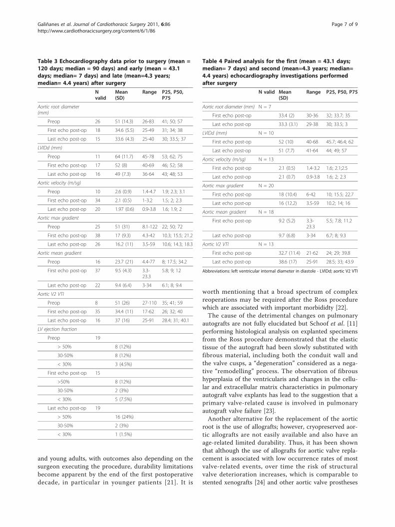
Echocardiographic findingsThe echocardiografic findings (Tables 3 and 4) at lastfollow-up (mean = 4.3 years) showed that, in the studiedpatients, the valve function did not deteriorate and thediameter of the conduit was not increased, with no evi-dence of stenosis, dilatation, calcification or thrombosisof the graft.
DiscussionThe present study shows for the first time that thetotally biological No-React® BioConduit graft affordsexcellent long-term clinical results, with few graft-related complications, in a high risk group of patients(eg, elevated EuroScore) with short life expectancy. Dur-ing the study period, no deterioration of the valve (eg,calcification, rupture) and conduit (eg, dilatation, calcifi-cation) were detected. These results are of clinicalimportance for the surgery of the aortic root and insome patients might represent a better alternative thanother synthetic or biological valved conduits.The 10-year results of aortic root replacement using
composite grafts with mostly mechanical valves are lessthan desirable [2,5], mainly due to complications with
0%
10%
20%
30%
40%
50%
60%
70%
80%
90%
100%
00 02 04 06 08 10 12Post-operative years
Freedom from all mortalityFreedom from cardiac mortalityFreedom from Valve-conduit mortality
Years from intervention 0 1 2 3 4 5 6 7 8 9 10Patients at risk 66 47 43 40 39 36 30 25 15 10 3
Figure 1 Kaplan-Meier curves of mortality.
Galiñanes et al. Journal of Cardiothoracic Surgery 2011, 6:86http://www.cardiothoracicsurgery.org/content/6/1/86
Page 5 of 9

anticoagulation and low, but consistent, rates of infec-tion. There are several aortic valved conduits as alterna-tive to a synthetic prosthesis with a mechanical aorticvalve prosthesis but they also present their own set ofspecific issues that are discussed below. In biologicalaortic valved conduits, preservation of the graft collagenstructure appears to be critical to avoid dilatation of theconduit and incompetence of the prosthetic valve. Pul-monary autografts have been shown to develop a highincidence of both dilatation of the conduit and incom-petence of the valve [9-11] and they may not be suitableas substitute of the aortic root because their collagenstructure and content differs form that of the aorta. Inaddition, in patients with cystic medial degeneration ofthe aorta, particularly those with bileaflet aortic valveand those with Marfan’s syndrome, the pulmonaryartery may also present with degenerative changes ofthe wall [18] that may make the autograft unsuitable foruse as a substitute of the aortic root. Indeed, dilatation
and regurgitation of the pulmonary autograft constitutethe primary cause of failure and the principal reason forreoperation after the Ross procedure [9-11]. To over-come this problem, it has been proposed that in patientswith bileaflet aortic valve the pulmonary autograftshould be implanted with the use of the aortic rootinclusion technique instead of aortic root replacementand that both the aortic annulus and the sinotubularjunction should be fixed with a strip of Dacron fabric[19]. When an inclusion technique is not feasible, pul-monary autograft reinforcement with a Valsalva Gel-weave Dacron tube (Terumo Cardiovascular SystemsInc, Ann Arbor, Mich) has been recommended as anoption [20]. However, even with the use of the aorticroot inclusion techniques, valve prolapse still remainsthe main cause of failure of the pulmonary autograft[21]. A systematic review of evidence on outcome afterthe Ross procedure has shown that, although the Rossprocedure provides satisfactory results for both children
Figure 2 Outflow and inflow of the explanted graft, as well as histological analysis of the valve. Panel A shows the outflow of the valve,with totally normal looking, coapting cusps; Panel B shows the outflow, and the inflammatory reaction to the abscess which was responsible forvalvular distortion is seen in the upper right hand corner; Panel C shows the H+E histological slide, showing normal looking pericardium of theconduit and the inflammatory cells of the abscess cavity; Panel D shows the Factor VIII immunoassay of one of the cusps, positive formonolayered endothelial cells.
Galiñanes et al. Journal of Cardiothoracic Surgery 2011, 6:86http://www.cardiothoracicsurgery.org/content/6/1/86
Page 6 of 9
and young adults, with outcomes also depending on thesurgeon executing the procedure, durability limitationsbecome apparent by the end of the first postoperativedecade, in particular in younger patients [21]. It is
worth mentioning that a broad spectrum of complexreoperations may be required after the Ross procedurewhich are associated with important morbididty [22].The cause of the detrimental changes on pulmonary
autografts are not fully elucidated but Schoof et al. [11]performing histological analysis on explanted specimensfrom the Ross procedure demonstrated that the elastictissue of the autograft had been slowly substituted withfibrous material, including both the conduit wall andthe valve cusps, a “degeneration” considered as a nega-tive “remodelling” process. The observation of fibroushyperplasia of the ventricularis and changes in the cellu-lar and extracellular matrix characteristics in pulmonaryautograft valve explants has lead to the suggestion that aprimary valve-related cause is involved in pulmonaryautograft valve failure [23].Another alternative for the replacement of the aortic
root is the use of allografts; however, cryopreserved aor-tic allografts are not easily available and also have anage-related limited durability. Thus, it has been shownthat although the use of allografts for aortic valve repla-cement is associated with low occurrence rates of mostvalve-related events, over time the risk of structuralvalve deterioration increases, which is comparable tostented xenografts [24] and other aortic valve prostheses
Table 3 Echocardiography data prior to surgery (mean =120 days; median = 90 days) and early (mean = 43.1days; median= 7 days) and late (mean=4.3 years;median= 4.4 years) after surgery
Nvalid
Mean(SD)
Range P25, P50,P75
Aortic root diameter(mm)
Preop 26 51 (14.3) 26-83 41; 50; 57
First echo post-op 18 34.6 (5.5) 25-49 31; 34; 38
Last echo post-op 15 33.6 (4.3) 25-40 30; 33.5; 37
LVIDd (mm)
Preop 11 64 (11.7) 45-78 53; 62; 75
First echo post-op 17 52 (8) 40-69 46; 52; 58
Last echo post-op 16 49 (7.3) 36-64 43; 48; 53
Aortic velocity (m/sg)
Preop 10 2.6 (0.9) 1.4-4.7 1.9; 2.3; 3.1
First echo post-op 34 2.1 (0.5) 1-3.2 1.5; 2; 2.3
Last echo post-op 20 1.97 (0.6) 0.9-3.8 1.6; 1.9; 2
Aortic max gradient
Preop 25 51 (31) 8.1-122 22; 50; 72
First echo post-op 38 17 (9.3) 4.3-42 10.3; 15.5; 21.2
Last echo post-op 26 16.2 (11) 3.5-59 10.6; 14.3; 18.3
Aortic mean gradient
Preop 16 23.7 (21) 4.4-77 8; 17.5; 34.2
First echo post-op 37 9.5 (4.3) 3.3-23.3
5.8; 9; 12
Last echo post-op 22 9.4 (6.4) 3-34 6.1; 8; 9.4
Aortic V2 VTI
Preop 8 51 (26) 27-110 35; 41; 59
First echo post-op 35 34.4 (11) 17-62 26; 32; 40
Last echo post-op 16 37 (16) 25-91 28.4; 31; 40.1
LV ejection fraction
Preop 19
> 50% 8 (12%)
30-50% 8 (12%)
< 30% 3 (4.5%)
First echo post-op 15
>50% 8 (12%)
30-50% 2 (3%)
< 30% 5 (7.5%)
Last echo post-op 19
> 50% 16 (24%)
30-50% 2 (3%)
< 30% 1 (1.5%)
Table 4 Paired analysis for the first (mean = 43.1 days;median= 7 days) and second (mean=4.3 years; median=4.4 years) echocardiography investigations performedafter surgery
N valid Mean(SD)
Range P25, P50, P75
Aortic root diameter (mm) N = 7
First echo post-op 33.4 (2) 30-36 32; 33.7; 35
Last echo post-op 33.3 (3.1) 29-38 30; 33.5; 3
LVIDd (mm) N = 10
First echo post-op 52 (10) 40-68 45.7; 46.4; 62
Last echo post-op 51 (7.7) 41-64 44; 49; 57
Aortic velocity (m/sg) N = 13
First echo post-op 2.1 (0.5) 1.4-3.2 1.6; 2.1;2.5
Last echo post-op 2.1 (0.7) 0.9-3.8 1.6; 2; 2.3
Aortic max gradient N = 20
First echo post-op 18 (10.4) 6-42 10; 15.5; 22.7
Last echo post-op 16 (12.2) 3.5-59 10.2; 14; 16
Aortic mean gradient N = 18
First echo post-op 9.2 (5.2) 3.3-23.3
5.5; 7.8; 11.2
Last echo post-op 9.7 (6.8) 3-34 6.7; 8; 9.3
Aortic V2 VTI N = 13
First echo post-op 32.7 (11.4) 21-62 24; 29; 39.8
Last echo post-op 38.6 (17) 25-91 28.5; 33; 43.9
Abbreviations: left ventricular internal diameter in diastole - LVIDd; aortic V2 VTI
Galiñanes et al. Journal of Cardiothoracic Surgery 2011, 6:86http://www.cardiothoracicsurgery.org/content/6/1/86
Page 7 of 9

(Carpentier-Edwards pericardial and supra-annularvalve, Medtronic Freestyle valve) [25]. Furthermore,Smedira et al. reported the explantation of 46 allograftsafter 5.6 ± 3.1 years follow-up in 744 patients whomhave received cryopreserved allografts with a mean ageof 49 ± 12 years. In this study, structural valve dete-rioration was the most frequent cause (59% of the cases)of valve-related reoperation after allograft aortic valvereplacement [26]. Therefore there is a considerable life-time risk of reoperation, especially in young patients,and, because of this, at some institutions the use of allo-grafts only remains the preferred valve substitute forpatients with active aortic root endocarditis and forpatients in whom anticoagulation should be avoided.The No-React® BioConduit, being a completely biolo-
gical and readily available graft, is an excellent alterna-tive to pulmonary autografts and allografts. TheBioConduit is easily handled facilitating its technicallyinsertion. The absence of clinical evidence for degenera-tive changes of the No-React® BioConduit graft seen inour study may be explained by the manufacturing pro-cess used. Thus, the recognition that glutaraldehyde andformaldehyde are prerequisites for limiting calcificationand the importance of preservation of cross-linked col-lagen for the durability of biological tissue [27] was fun-damental to develop the method used in the No-React®
BioConduit graft. In this process, heparin is used to lockthe glutaraldehyde residue, so that glutaraldehyde leach-ing is abolished and its potential immunological reactiv-ity is prevented [12], hence keeping all the advantages ofglutaraldehyde but abolishing its side effects.One important finding of our study was the rare
occurrence of infection of the No-React® BioConduitgraft. One case with aortic endocarditis was in septicshock at the time of surgery dying the following day;therefore the lost of this patient cannot be attributed toinfection of the newly implanted graft. The only othercase presenting with late endocarditis was in fact a peri-prosthetic abscess without affecting the graft. Clinicalstudies with No-React® valves, receiving an identicaltreatment to the No-React® BioConduit, have alsoshown a high resistance to infection [14,15]. By contrast,synthetic aortic valved conduits [7], pulmonary auto-grafts [28] and allografts [29] have an important rate offailure because of endocarditis. In the active phase ofallografts with endocarditis the operative mortality andlong-term prognosis are similar to those reported withconventional prostheses [30]. The reason for the resis-tance of No-React® BioConduit and valve to infection isnot fully understood but the presence of endotheliumwith No-React® tissue on blood contacting surfaces hasbeen suggested as a potential explanation [31]. Ourresults contrast with the recently reported degenerationof the No-React® BioConduit in 7 of the 115 cases
implanted with the prosthesis more than 1 year aftersurgery [16]. Endocarditis was identified as the mostlikely cause, although extensive microbiological exami-nations did not reveal a causative organism [16]. Duringthe follow-up study period, we did not observed thiscomplication in none of the patients implanted with thisprosthesis. However, our study is up to 10 years and webelieve it would be required at least 15 to 20 years toconfirm whether the No-React® BioConduit is reallyresistant to degeneration and infection, a question thatprobably should also be explored in a larger populationin prospective and randomized studies comparing theNo-React® BioConduit with other biological conduits.In conclusion, the present study has demonstrated
that the No-React® BioConduit does not dilate or dete-riorate and resists infection after 10-year follow-up.Therefore, the No-React® BioConduit may be a goodalternative to other conduits for surgery of the aorticroot in all age range.
Author details1Department of Cardiac Surgery, The Glenfield Hospital, Leicester, UK.2Department of Cardiac Surgery, Research Institute, University Hospital Valld’Hebron, Universitat Autònoma de Barcelona, Barcelona, Spain. 3Departmentof Cardiology, Reparative Therapy of the Heart, Area del Cor (ACOR) andResearch Institute, University Hospital Vall d’Hebron, Universitat Autònomade Barcelona, Barcelona, Spain.
Authors’ contributionsMG and AS performed the surgery, designed the study, analysed the resultsand participated in the writing of the manuscript. AM contributed to thecollection of data. IF carried out the statistical analyses and also participatedin the writing of the manuscript. All authors read and approved the finalmanuscript.
Competing interestsThe authors declare that they have no competing interests.
Received: 22 February 2011 Accepted: 23 June 2011Published: 23 June 2011
References1. Bentall H, De Bono A: A technique for complete replacement of the
ascending aorta. Thorax 1968, 23:338-339.2. Gott VL, Gillinov AM, Pyeritz RE, Cameron DE, Reitz BE, Greene PS,
Stone CD, Ferris RL, Alejo DE, McKusick VA: Aortic Root Replacement: RiskFactor analysis of a seventeen-year experience with 270 patients.J Thorac Cardiovasc Surg 1995, 109:536-545.
3. Bachet J, Termignon JL, Goudot B, Dreyfus G, Piqouis A, Brodaty D,Dubois C, Delentdecker P, Guilmet D: Aortic root replacement with acomposite graft. Factors influencing immediate and long-term results.Eur J Cardiothoracic Surg 1996, 10:207-213.
4. Kouchoukos NT, Wareing TH, Murphy S, Perrillo JBRN: Sixteen-yearexperience with aortic root replacement: results of 172 operations.Ann Surg 1991, 214:308-318.
5. Luciani GB, Casali G, Santini F, Mazzuco A: Aortic root replacement inadolescents and young adults: composite graft versus homograft orautograft. Ann Thorac Surg 1998, 66:S189-S193.
6. Mahesh B, Caputo M, Angelini GD, Bryan AJ: Treatment of an aortic fungalfalse aneurysm by composite stentless porcine/pericardial conduit: acase report. Cardiovasc Surg 2003, 11:93-95.
7. Mahesh B, Angelini G, Caputo M, Jin XY, Bryan A: Prosthetic valveendocarditis. Ann Thorac Surg 2005, 80:1151-1158.
Galiñanes et al. Journal of Cardiothoracic Surgery 2011, 6:86http://www.cardiothoracicsurgery.org/content/6/1/86
Page 8 of 9

8. Takkenberg JJM, Eijkemans MJC, van Herwerden LA, Steyerberg EW,Lane MM, Elkins RC, Habbema JDF, Bogers AJJC: Prognosis after aorticroot replacement with cryopreserved allografts in adults. Ann ThoracSurg 2003, 75:1482-1489.
9. Luciani GB, Casali G, Favaro A, Prioli MA, Barozzi L, Santini F, et al: Fate ofthe aortic root late after Ross operation. Circulation 2003, 108(suppl II):II-61-II -67.
10. Pasquali S, Cohen MS, Shera D, Wernovsky G, Spray TL, Marino BS: Therelationship between neo-aortic root dilatation, insufficiency, andreintervention following the Ross procedure in infants, children, andyoung adults. J Am Coll Cardiol 2007, 49:1806-1812.
11. Takkenberg JJM, van Suylen RJ, Zondervan PE, Hazekamp MG, Dion RAE,Bogers AJJC: Degeneration of the pulmonary autograft: An explantstudy. J Thorac Cardiovasc Surg 2006, 132:1426-1432.
12. Abolhoda A, Yu S, Oyarzun JR, Allen KR, McCormick JR, Han S, Kemp FW,Bogden JD, Lu Q, Gabbay S: No-React detoxification process: A superioranticalcification method for bioprostheses. Ann Thorac Surg 1996,62:1724-1730.
13. Morell VO, Wearden PA: Experience with bovine pericardium for thereconstruction of the aortic arch in patients undergoing a Norwoodprocedure. Ann Thorac Surg 2007, 84:1312-1315.
14. Musci M, Siniawski H, Pasic M, Weng Y, Loforte A, Kosky S, Yankah C,Hetzer R: Surgical therapy with patients with active infectiveendocarditis: seven-year single centre experience in a subgroup of 255patients treated with the Shelhigh® stentless bioprosthesis. Eur JCardiothoracic Surg 2008, 34:410-417.
15. Siniawski H, Grauhan O, Hofmann M, Pasic M, Weng Y, Yankah C,Lehmkuhl H, Hetzer R: Factors influencing the results of double-valvesurgery in patients with fulminant endocarditis: the importance of valveselection. Heart Surg Forum 2004, 7:1-6.
16. Carrel TP, Schoenhoff FS, Schmidli J, Stalder M, Eckstein FS, Englberger L:Deleterious outcome of No-React-treated stentless valved conduits afteraortic root replacement: why were warnings ignored? J Thorac CardiovascSurg 2008, 136:52-57.
17. Akins CW, Miller DC, Turina MI, Kouchouckos NT, Blackstone EH,Grunkemeier GL, Takkenberg JJM, David TE, Butchard EG, Adams DH,Shahian DM, Hagl S, Mayer JE, Lytle BW: Guidelines for reporting mortalityafter cardiac surgery interventions. Ann Thorac Surg 2008, 85:1490-1495.
18. de Sa M, Moshkovitz Y, Butany J, David TE: Histologic abnormalities of theascending aorta and pulmonary trunk in patients with bicuspid aorticvalve disease: clinical relevance to the Ross procedure. J ThoracCardiovasc Surg 1999, 118:588-596.
19. Juthier F, Carlo Banfi C, Vincentelli A, Ennezat PV, Le Tourneau T, Pinçon C,Prat A: Modified Ross operation with reinforcement of the pulmonaryautograft: six-year results. J Thorac Cardiovasc Surg 2010, 139:1420-1423.
20. de Kerchove L, Rubay J, Pasquet A, Poncelet A, Ovaert C, Pirotte M,Buche M, D’Hoore W, Noirhomme P, El Khoury G: Ross operation in theadult: long-term outcomes after root replacement and inclusiontechniques. Ann Thorac Surg 2009, 87:95-102.
21. Takkenberg JJM, Klieverik LMA, Schoof PH, van Suylen RJ, vanHerwerden LA, Zondervan PE, Roos-Hesselink JW, Eijkemans MJC,Yacoub MH, Bogers AJJC: The Ross procedure: a systematic review andmeta-analysis. Circulation 2009, 119:222-228.
22. Stulak JM, Burkhart HM, Sundt III TM, Connolly HM, Suri RM, Schaff HV,Dearani JA: Spectrum and outcome of reoperations after the Rossprocedure. Circulation 2010, 122:1153-1158.
23. Mookhoek A, de Heer E, Bogers AJJC, Takkenberg JJM, Schoof PH:Pulmonary autograft valve explants show typical degeneration. J ThoracCardiovasc Surg 2010, 139:1416-1419.
24. Takkenberg JJM, Klieverik LMA, Bekkers JA, Kappetein AP, Roos JW,Eijkemans MJC, Bogers AJJC: Allografts for aortic valve or rootreplacement: insights from an 18-year single-center prospective follow-up study. Eur J Cardiothorac Surg 2007, 31:851-859.
25. Kappetein AP, Takkenberg JJM, Puvimanasinghe JPA, Jamieson WRE,Eijkemans M, Bogers AJJC: Does the type of biological valve affect patientoutcome? Interact CardioVasc Thorac Surg 2006, 5:398-402.
26. Smedira NG, Blackstone EH, Roselli EE, Laffey CC, Cosgrove DM: Areallografts the biologic valve of choice for aortic valve replacement innonelderly patients? Comparison of explantation for structural valvedeterioration of allograft and pericardial prostheses. J Thorac CardiovascSurg 2006, 131:558-564.
27. Levy RJ, Schoen FJ, Sherman FS, Nichols J, Hawley MA, Lund SA:Calcification of subcutaneously implanted type I collagen sponges:effects of formaldehyde and glutaraldehyde pretreatments. Am J Pathol1986, 122:71-82.
28. Chambers JC, Somerville J, Stone S, Ross DN: Pulmonary autograftprocedure for aortic valve disease: long-term results of the pioneerseries. Circulation 1997, 96:2206-2214.
29. Clarkson PM, Barratt-Boyes BG: Bacterial endocarditis following homograftreplacement of the aortic valve. Circulation 1970, 42:987-991.
30. Avierinos JF, Thuny F, Chalvignac V, Giorgi R, Tafanelli L, Casalta JP,Raoult D, Mesana T, Collart F, Metras D, Habib G, Riberi A: Surgicaltreatment of active aortic endocarditis: homografts are not thecornerstone of outcome. Ann Thorac Surg 2007, 84:1935-1942.
31. Dohmen PM, Gabbieri D, Lembcke A, Konertz W: Endothelial cell-seededbovine internal mammary artery for complete revascularization.Ann Thorac Surg 2007, 83:1168-1169.
doi:10.1186/1749-8090-6-86Cite this article as: Galiñanes et al.: Totally biological composite aorticstentless valved conduit for aortic root replacement: 10-yearexperience. Journal of Cardiothoracic Surgery 2011 6:86.
Submit your next manuscript to BioMed Centraland take full advantage of:
• Convenient online submission
• Thorough peer review
• No space constraints or color figure charges
• Immediate publication on acceptance
• Inclusion in PubMed, CAS, Scopus and Google Scholar
• Research which is freely available for redistribution
Submit your manuscript at www.biomedcentral.com/submit
Galiñanes et al. Journal of Cardiothoracic Surgery 2011, 6:86http://www.cardiothoracicsurgery.org/content/6/1/86
Page 9 of 9