research article open access in vivo activity of sapindus
TRANSCRIPT
RESEARCH ARTICLE Open Access
In vivo activity of Sapindus saponaria againstazole-susceptible and -resistant human vaginalCandida speciesEdílson Damke1, Joyce K Tsuzuki2, Diógenes AG Cortez2, Izabel CP Ferreira2, Thâmara A Bertoni1, Márcia R Batista1,Lucélia Donati3, Terezinha IE Svidzinski1 and Márcia EL Consolaro1*
Abstract
Background: Study of in vivo antifungal activity of the hydroalcoholic extract (HE) and n-BuOH extract (BUTE) ofSapindus saponaria against azole-susceptible and -resistant human vaginal Candida spp.
Methods: The in vitro antifungal activity of HE, BUTE, fluconazole (FLU), and itraconazole (ITRA) was determined bythe broth microdilution method. We obtained values of minimal inhibitory concentration (MIC) and minimumfungicide concentration (MFC) for 46 strains of C. albicans and 10 of C. glabrata isolated from patients withvulvovaginal candidiasis (VVC). VVC was induced in hyperestrogenic Wistar rats with azole-susceptible C. albicans(SCA), azole-resistant C. albicans (RCA), and azole-resistant C. glabrata (RCG). The rats were treated intravaginallywith 0.1 mL of HE or BUTE at concentrations of 1%, 2.5% and 5%; 100 μg/mL of FLU (treatment positive control);or distilled water (negative control) at 1, 24, and 48 h after induction of the infection, and the progress of VVC wasmonitored by culturing and scanning electron microscopy (SEM). The toxicity was evaluated in cervical cells of theHeLa cell line.
Results: The extracts showed in vitro inhibitory and fungicidal activity against all the isolates, and the MIC and MFCvalues for the C. glabrata isolates were slightly higher. In vivo, the SCA, RCA, and RCG infections were eliminated by21 days post-infection, with up to 5% HE and BUTE, comparable to the activity of FLU. No cytotoxic action wasobserved for either extract.
Conclusions: Our results demonstrated that HE and BUTE from S. saponaria show inhibitory and fungicidal activityin vitro, in addition to in vivo activity against azole-resistant vaginal isolates of C. glabrata and azole-susceptible andresistant isolates of C. albicans. Also considering the lack of cytotoxicity and the low concentrations of the extractsnecessary to eliminate the infection in vivo, HE and BUTE show promise for continued studies with purifiedantifungal substances in VVC yeast isolates.
Keywords: Sapindus saponaria vaginal yeasts, antifungal activity, in vivo
BackgroundNatural products have been traditionally used in thecontrol of various diseases, because they are a source ofmany active compounds that show multiple therapeuticeffects, in addition to constituting models for the synth-esis of a large number of pharmaceuticals [1]. The fruitof Sapindus saponaria L. (Sapindaceae), a medium-sized
tropical tree found principally in America and India, hasshown antimicrobial activity [2-4], but few studies havebeen carried out with this plant.In a recent study, members of our research group iso-
lated and identified the principal constituents of the n-BuOH extract (BUTE) of the pericarps of S. saponaria:two acetylated triterpene saponins, S1 and S2, and alsoan acyclic oligoglycoside. The same group also demon-strated excellent inhibitory action in vitro of the water-alcohol extract (HE) and BUTE against the yeasts Can-dida albicans and non- C. albicans isolated from
* Correspondence: [email protected] of Clinical Analysis and Biomedicine, State University ofMaringá, Paraná, BrazilFull list of author information is available at the end of the article
Damke et al. BMC Complementary and Alternative Medicine 2011, 11:35http://www.biomedcentral.com/1472-6882/11/35
© 2011 Damke et al; licensee BioMed Central Ltd. This is an Open Access article distributed under the terms of the Creative CommonsAttribution License (http://creativecommons.org/licenses/by/2.0), which permits unrestricted use, distribution, and reproduction inany medium, provided the original work is properly cited.
patients with vulvovaginal candidiasis (VVC)[5], signal-ing the possibility of using this plant as an antifungalagent in this pathology. In spite of these recent investi-gations of the constituents and biological properties ofS. saponaria, few in vivo studies have yet been carriedout to establish a correlation with the in vitro results.VVC is caused by abnormal growth of these yeast-like
fungi in the mucosa of the female genital system [6]. Itaffects millions of women annually and can cause greatdiscomfort, affecting sexual and affective relations andjob performance, and is considered an important pro-blem of world public health [7]. Management of patientswith VVC is often difficult because of the few availabletherapeutic options; and furthermore, cross-resistance ofvaginal C. albicans has been detected to itraconazoleand fluconazole, which are the antifungal agents ofchoice for treatment of this pathology [7,8]. In view ofthe need for new therapeutic options for VVC and thepromising in vitro inhibitory activity of S. saponaria L.against yeasts, we conducted a study of antifungal activ-ity in vivo of HE and BUTE against azole-susceptibleand -resistant human vaginal Candida spp., and also ofthe cell toxicity of these extracts.
MethodsPlant and componentsDry pericarps of the fruits of S. saponaria were collectedon the campus of the State University of Maringá,Paraná, Brazil (UEM). The plant was identified by staffmembers of the UEM Department of Botany, and anexsiccate was deposited in the Herbarium of this institu-tion (HUM 11710). The dried pericarps of the fruits(450.0 g) of S. saponaria were ground and extractedwith EtOH:H2O (9:1) at room temperature, by a processof dynamic maceration with constant mechanical stir-ring. The extraction was carried out in an amber flask,maintained at ambient temperature, for six consecutivedays, for 6 h per day. The extract was concentratedunder low pressure in a rotary evaporator, at a tempera-ture of 40°C. After elimination of the solvent, the extractwas frozen in liquid nitrogen and lyophilized in a MartinChrist Alpha 1-2 freeze dryer. The lyophilized extractwas stored in a closed plastic flask and kept frozen.The HE of the pericarp (50.15 g) was chromato-
graphed in a column (ji = 4.0 cm) of silica gel 60(Merck, Darmstadt, Germany), and eluted with solventsof increasing polarity including hexane, dichloro-methane, ethyl acetate, and methanol (Merck, Darm-stadt, Germany). The solvents were evaporated at atemperature of 40°C, frozen in liquid nitrogen, and lyo-philized in a Martin Christ Alpha 1-2 freeze dryer. Thelyophilized dichloromethane, hexane, ethyl acetate, andmethanol fractions were stored in closed containers andkept frozen. The methanol extract was suspended in
H2O and extracted with n-butanol, which after evapora-tion gave a solid residue (28.9 g) (BUTE), which wasalso lyophilized. The structures were established withthe use of spectroscopic methods (1H and 13C NMR,HSQC, HMBC, and ESI/MS) and by comparing themwith literature data [2,4].
Yeast isolatesFor the experiments on in vitro susceptibility, 56 vaginalisolates obtained from patients with VVC were tested,including 46 isolates of C. albicans and 10 of C. glab-rata, which are part of a bank of yeasts at the MedicalMycology Laboratory/UEM. In this yeast bank, aliquotsof the yeasts are stored after their identification in 10%glycerinated water at -20°C. The yeasts used in thisstudy were isolated and identified in 2008, by classicalmethods [9-11] and also by rDNA sequencing [12].Prior to each experiment, the isolates were reactivatedin Sabouraud Dextrose Broth (SDB) (Difco, Detroit,USA) at 25°C for 24/48 h, seeded on Sabouraud Dex-trose Agar (SDA) (Difco, Detroit, USA) with chloram-phenicol (2.0 mg/mL), and incubated again under theprevious conditions. A new subculture was made inCHROMágar Candida® (Probac, France) to assure thepurity of the isolates.
Antifungal agentsStock solutions of fluconazole (FLU) (5000 mg/mL; Pfi-zer Inc., NY, USA) and itraconazole (ITRA) (1000 mg/mL; Janssen Pharmaceutica, Titusville, NJ, USA) wereprepared. From the first prepared solution, new stocksolutions of FLU and ITRA were prepared at a concen-tration of 10 times that of the final test concentration,and diluted in bicarbonate-free RPMI-1640 with L-gluta-mine, supplemented with 2% dextrose and buffered topH 7.0 with 0.165 M morpholinopropanesulfonic acid(MOPS) (Sigma, Steinheim, Germany). The lyophilizedHE and BUTE extracts were dissolved in sterile distilledwater to obtain a 10 mg/mL solution of each extract.
Determination of minimum inhibitory (MIC) andfungicidal concentrations (MFC)The tests of susceptibility of Candida spp. to FLU andITRA were carried out according to the broth microdi-lution method recommended by the CLSI (ClinicalLaboratory Standards Institute, 2002), and of susceptibil-ity to HE and BUTE according to the same document,with adaptations for natural products [13,14].A suspension of yeast compatible with 1.0 to 5.0 × 10⁶
colony-forming units per mL (CFU/mL) was prepared insterile saline, adjusting the cell density by means of aspectrophotometer (Spectronic 70, Bausch & Lomb,USA) at 530 nm with 90 ± 2% transmittance. From thissuspension, new dilutions were made: 1:50 in sterile
Damke et al. BMC Complementary and Alternative Medicine 2011, 11:35http://www.biomedcentral.com/1472-6882/11/35
Page 2 of 9

saline, and then 1:20 in RPMI (Sigma, Steinheim, Ger-many), thus obtaining the desired final inoculum of 0.5to 2.5 × 10³ CFU/mL. The tests were carried out insterilized plastic microplates (TPP Zellkultur Test Plate96F, Switzerland) containing 96 wells arranged in 8rows labeled A to H, each row with 12 wells numbered1 to 12. Each row (A-H) corresponded to one isolate,and each well received 100 μL of the measured inocu-lum, except for the 12th well which was the negativecontrol. Aliquots of 100 μL of RPMI (Gibco, NY, USA)were distributed from columns 2 to 11. Aliquots of100 μL of FLU, ITRA, HE, or BUTE, prepared as pre-viously described, were added to the columns of themicroplates, and from column 2 a 2-fold serial dilutionwas made up to the 10th well (diluitions between0.125 and 64.0 μg/mL for FLU, 0.03 and 16.0 μg/mLfor ITRA, and 9.0 and 5000.0 μg/mL for HE andBUTE).For each isolate tested, negative controls (only RPMI)
and positive (RPMI plus inoculum, with no antifungalsadded) were included, for growth and for the possibleaction of the diluent of the extracts or drugs (only buta-nol, ethanol, or polyethyleneglycol 400 with the inocu-lum). On each microplate a strain of Candidaparapsilosis (ATCC 22019) was included as the refer-ence yeast. The plates thus set up were incubated in anoven at 35°C with daily monitoring. After 48 h theywere read for FLU and ITRA in a microplate reader(Asys Hitech GmbH, Eugendorf, Austria) and after 72 hfor the extracts, by visual comparison of the reflectionin a mirror.The MIC for FLU/ITRA was determined as the lowest
concentration of the drug that was capable of inhibiting50% of the growth of each yeast, with reference to itsrespective positive control [13]. The criteria for defini-tion of susceptibility/resistance to FLU/ITRA were thoseestablished by CSLI [13]. For HE and BUTE, the MICwas considered to be the smallest concentration of theextract that was capable of inhibiting 100% of the inocu-lum compared to its respective positive control [14].The MIC50 and MIC90 for the drugs and extracts weredefined as the MICs capable of inhibiting 50% and 90%of the isolates, respectively [13].To determine the MFC, subcultures from all wells
showing growth inhibition were made by seeding 5.0 μLin SDA at 25°C. After 48 h, the CFU were counted todetermine the viability. All the assays for determinationof the MIC and MFC were carried out in duplicate,independently. The MFC for HE and BUTE was consid-ered as the lowest concentration that impeded thegrowth of 100% of the inoculum. The MFC50 andMFC90 for the extracts were defined as the MFCs cap-able of inhibiting 50% and 90% of the isolates,respectively.
Experimental vaginal infectionA rat model was used as previously described [15], withsome adaptations. The experiments were carried outwith three yeast isolates, which were selected accordingto the results of the in vitro tests: C. albicans susceptibleto FLU and ITRA (SCA), C. albicans resistant to ITRA(RCA), and C. glabrata resistant to both antifungals(RCG).The experiments were carried out with groups of five
rats for each isolate, in duplicate and on two differentdays. Non-oophorectomized Wistar rats (Rattus norvegi-cus) weighing from 200 to 300 g and 70 days old (fromthe UEM Central Animal Facility) were used. The ratsreceived subcutaneous injections of estradiol valerate(Sigma, Steinheim, Germany) at a concentration of 0.2mg/week/rat. Six days after the first injection of the hor-mone, the animals were inoculated intravaginally with108 yeast cells/mL of each isolate tested, prepared in 0.1mL of sterile saline and counted in a Neubauerchamber.For the treatment, HE and BUTE were administered
intravaginally (0.1 mL at 1%, 2.5%, and 5.0% in distilledwater) at 1, 24, and 48 h after the induction of the vagi-nal infection. Rats receiving FLU (3 doses of 100 μgintravaginally over the same time periods as theextracts) or distilled water served as positive and nega-tive treatment controls, respectively. The kinetics of theCandida vaginal infection in the treated and control ratswas monitored in each animal by means of the numberof CFU/mL in the vaginal fluid at 24 and 48 h afterinduction of the infection, and on days 5, 7, 14, and 21.The animal experimentation carried out in this investi-gation was approved by the UEM Committee on EthicalConduct in Animal Use (Protocol No. 013/2006, Opi-nion No. 050/2006).
Toxicity in cervical cellsCells from the HeLa human cervical line were pre-viously cultured in Eagle’s minimum essential medium(MEM, PPA Laboratories, Germany) supplemented with10% fetal bovine serum (FBS, Laborclin, Brazil), 0.1 mMnon-essential amino acids, and 1 mM sodium pyruvate,at 37°C in a humid oven with 5% CO2. In the exponen-tial growth stage, the cells were diluted in the samemedium and plated in volumes of 0.2 mL of a suspen-sion of 2.5 × 10⁵ cells per well in a 24-well plate (Corn-ing Glass, New York, USA), and incubated in the sameconditions overnight to allow them to form a cell mono-layer. The culture medium was replaced by serial dilu-tions of the HE or BUTE extracts at 1%, 2.5%, 5%, and10% concentrations, in triplicate. The control wells con-tained only cells and culture medium. The microplatewas incubated again at 37 °C for 24 h, and the extractswere replaced by a trypsin-EDTA solution to undo cell
Damke et al. BMC Complementary and Alternative Medicine 2011, 11:35http://www.biomedcentral.com/1472-6882/11/35
Page 3 of 9
adhesion, followed by addition of 0.2 ml of PBS with50% trypan blue. Live and dead cells in each well werecounted with the aid of light microscopy.
Scanning electron microscopy (SEM)The vaginal epithelium of rats infected with CAS, CGR,and CAR before and after treatment with HE and BUTEwas observed by SEM. After 48 h of infection and alsoat the end of treatment, the rats to be analyzed by SEMwere killed with an overdose of anesthetics (Ketamineand Xylazine, Parke-Davis Co, Morris Plains, NJ, USA).The vagina was removed, washed, fixed in a solution of2.5% glutaraldehyde in 0.1 M cacodylate buffer (SigmaChemical, St. Louis, MO, USA), and dehydrated in anascending ethanol series. The critical point was obtainedin a Balzers CPD-010 (Balzers Instruments, Balzers,Liechtenstein) with carbonic gas. Metallization in goldwas carried out in a Balzers SCD-030 (Balzers Instru-ments, Balzers, Liechtenstein). The vagina, uterine cer-vix, and tissue sections of all rats were observed andphotographed with a JEOL-JSM 6360 LV scanning elec-tron microscope (JEOL Ltd, Tokyo, Japan) at the Elec-tron Microscopy Center, Federal University of Paraná/Curitiba/Brazil.
Statistical analysisThe results were analyzed using Student’s t test andTukey’s test for multiple comparisons of the different invivo experimental treatment situations. The significancelevel was set at 5%. The tests were carried out by meansof the program Graph Pad Prismâ version 3.0 (GraphPad Software Inc.).
ResultsPlants and componentsThe presence of two acetylated triterpene saponins wasconfirmed: saponin S1, hederagenin-3-O- (3,4-di-O-acetyl-b-D-xylopyranosyl)- (1®3)-a-L-ramnopyranosyl-(1®2)-a-L-arabinopyranoside; and saponin S2, hedera-genin-3-O-(4-O-acetyl-b-D-xylopyranosyl)-(1®3)-a-L-ramnopyranosyl-(1®2)-a-L-rabinopyranoside; and alsoan acyclic oligoglycoside-1 (OGSA-1) in HE and BUTE,as previously described [5].
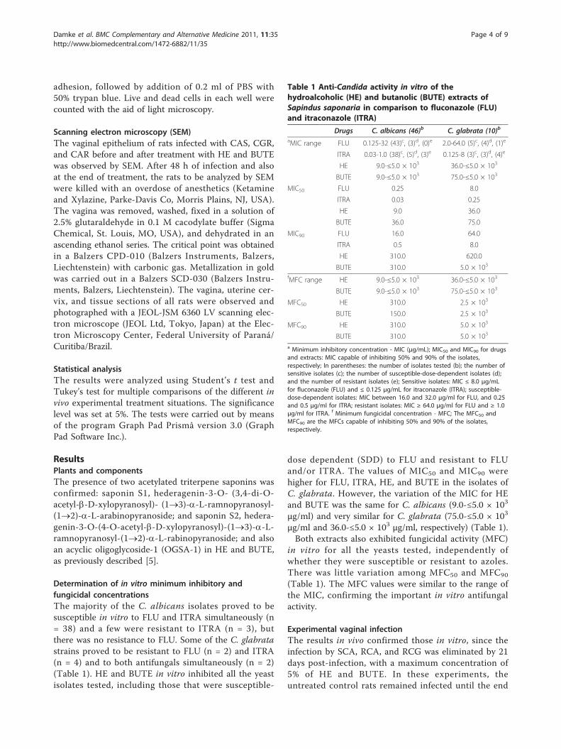
Determination of in vitro minimum inhibitory andfungicidal concentrationsThe majority of the C. albicans isolates proved to besusceptible in vitro to FLU and ITRA simultaneously (n= 38) and a few were resistant to ITRA (n = 3), butthere was no resistance to FLU. Some of the C. glabratastrains proved to be resistant to FLU (n = 2) and ITRA(n = 4) and to both antifungals simultaneously (n = 2)(Table 1). HE and BUTE in vitro inhibited all the yeastisolates tested, including those that were susceptible-
dose dependent (SDD) to FLU and resistant to FLUand/or ITRA. The values of MIC50 and MIC90 werehigher for FLU, ITRA, HE, and BUTE in the isolates ofC. glabrata. However, the variation of the MIC for HEand BUTE was the same for C. albicans (9.0-≤5.0 × 10³
μg/ml) and very similar for C. glabrata (75.0-≤5.0 × 10³
μg/ml and 36.0-≤5.0 × 10³ μg/ml, respectively) (Table 1).Both extracts also exhibited fungicidal activity (MFC)
in vitro for all the yeasts tested, independently ofwhether they were susceptible or resistant to azoles.There was little variation among MFC50 and MFC90
(Table 1). The MFC values were similar to the range ofthe MIC, confirming the important in vitro antifungalactivity.
Experimental vaginal infectionThe results in vivo confirmed those in vitro, since theinfection by SCA, RCA, and RCG was eliminated by 21days post-infection, with a maximum concentration of5% of HE and BUTE. In these experiments, theuntreated control rats remained infected until the end
Table 1 Anti-Candida activity in vitro of thehydroalcoholic (HE) and butanolic (BUTE) extracts ofSapindus saponaria in comparison to fluconazole (FLU)and itraconazole (ITRA)
Drugs C. albicans (46)b C. glabrata (10)b
aMIC range FLU 0.125-32 (43)c, (3)d, (0)e 2.0-64.0 (5)c, (4)d, (1)e
ITRA 0.03-1.0 (38)c, (5)d, (3)e 0.125-8 (3)c, (3)d, (4)e
HE 9.0-≤5.0 × 10³ 36.0-≤5.0 × 10³
BUTE 9.0-≤5.0 × 10³ 75.0-≤5.0 × 10³
MIC50 FLU 0.25 8.0
ITRA 0.03 0.25
HE 9.0 36.0
BUTE 36.0 75.0
MIC90 FLU 16.0 64.0
ITRA 0.5 8.0
HE 310.0 620.0
BUTE 310.0 5.0 × 10³
fMFC range HE 9.0-≤5.0 × 10³ 36.0-≤5.0 × 10³
BUTE 9.0-≤5.0 × 10³ 75.0-≤5.0 × 10³
MFC50 HE 310.0 2.5 × 10³
BUTE 150.0 2.5 × 10³
MFC90 HE 310.0 5.0 × 10³
BUTE 310.0 5.0 × 10³
a Minimum inhibitory concentration - MIC (μg/mL); MIC50 and MIC90 for drugsand extracts: MIC capable of inhibiting 50% and 90% of the isolates,respectively; In parentheses: the number of isolates tested (b); the number ofsensitive isolates (c); the number of susceptible-dose-dependent isolates (d);and the number of resistant isolates (e); Sensitive isolates: MIC ≤ 8.0 μg/mLfor fluconazole (FLU) and ≤ 0.125 μg/mL for itraconazole (ITRA); susceptible-dose-dependent isolates: MIC between 16.0 and 32.0 μg/ml for FLU, and 0.25and 0.5 μg/ml for ITRA; resistant isolates: MIC ≥ 64.0 μg/ml for FLU and ≥ 1.0μg/ml for ITRA. f Minimum fungicidal concentration - MFC; The MFC50 andMFC90 are the MFCs capable of inhibiting 50% and 90% of the isolates,respectively.
Damke et al. BMC Complementary and Alternative Medicine 2011, 11:35http://www.biomedcentral.com/1472-6882/11/35
Page 4 of 9

of all the assays (CFU/mL between 96 and 1.0 × 10³)(Figures 1, 2, 3).In the infection by SCA, the inhibitory activity of FLU
was superior only to that of HE and BUTE at 1% (p <0.001), which at this concentration showed similar beha-vior (p > 0.05) and inhibited fungal growth compared tothe negative control, but did not lead to elimination ofthe infection. The treatments with 2.5% and 5.0% HEand BUTE showed similar profiles of inhibition of infec-tion, comparable to FLU (p > 0.05) (Figure 1).In the RCA infection, FLU showed better inhibitory
activity than 1% and 2.5% HE (p < 0.001) and 1% BUTE(p < 0.001). The HE at 1% and 2.5%, and 1.0% BUTEdid not eliminate the infection, showing similar behaviorto that of the untreated control (p > 0.05). For HE, the5% concentration showed the best inhibitory activity (p< 0.001), as did the 2.5% and 5.0% concentrations ofBUTE. There was no difference in the action betweenthese last two concentrations, and also between the HE
and BUTE at the 5% concentration (p > 0.05) (Figure 2).The RCA was resistant in vitro only to ITRA, and thepositive control of the treatment was carried out withFLU for all yeasts tested, which although it is also anazole, showed excellent activity in vivo. No treatmentwas carried out with ITRA itself, because no vaginal for-mulations of this antifungal exist.For RCG, the inhibitory activity of HE and BUTE at
all concentrations tested was excellent and similar tothat of FLU (p> 0.05). At the 1% concentration of bothextracts, there was a significant decrease in the CFUcount in the first days of infection (p < 0.05), and theinfection was eliminated on day 14 in the experimentfor HE, and on day 21 for BUTE. At 2.5% and 5.0%,both extracts showed the same activity, with eliminationof the infection on day 5 (p > 0.05); while the FLU treat-ment eliminated the infection on day 7 (Figure 3). Ingeneral, HE in a concentration of 5% and BUTE in con-centrations of 2.5% and 5% were capable of eliminatingthe infection induced by the different yeasts tested.
0
10
20
30
40
50
60
70
80
90
100
110
0 1 2 5 7 14 21
Days
FLU 1.0 HE 2.5 HE 5.0 HE1.0 BUTE 2.5 BUTE 5.0 BUTE Water
CFU/mL x 103
Figure 1 Colony-forming units (CFU/mL) of yeasts in thevaginal exudate of Wistar rats treated intravaginally withhydroalcoholic (HE) and butanolic (BUTE) extracts of Sapindussaponaria at 1.0%, 2.5%, and 5.0%, 100 μg of fluconazole, orsterile distilled water at 1, 24, and 48 h after induction of thevaginal infection, followed for up to 21 days. Each curverepresents the mean (± standard deviation) of the CFU of five rats,in two independent experiments. Experimental vaginal infection byCandida albicans susceptible to itraconazole and fluconazole (SCA).
0
10
20
30
40
50
60
70
80
90
100
110
0 1 2 5 7 14 21Days
FLU 1.0 HE 2.5 HE 5.0 HE1.0 BUTE 2.5 BUTE 5.0 BUTE Water
CFU/mL x 103
Figure 2 Colony-forming units (CFU/mL) of yeasts in thevaginal exudate of Wistar rats treated intravaginally withhydroalcoholic (HE) and butanolic (BUTE) extracts of Sapindussaponaria at 1.0%, 2.5%, and 5.0%, 100 μg of fluconazole, orsterile distilled water at 1, 24, and 48 h after induction of thevaginal infection, followed for up to 21 days. Each curverepresents the mean (± standard deviation) of the CFU of five rats,in two independent experiments. Experimental vaginal infection byC. albicans resistant to itraconazole (RCA).
Damke et al. BMC Complementary and Alternative Medicine 2011, 11:35http://www.biomedcentral.com/1472-6882/11/35
Page 5 of 9

Toxicity in cervical cellsThe percentage of live HeLa cells did not vary betweenthe controls and tests, and between HeLa cells exposedto different concentrations of HE and BUTE (p > 0.05).The mean of live cells in the different HE concentra-tions was 94.22 ± 0.1555, and in the BUTE concentra-tions was 94.41 ± 0.1131 (IC 95% = -0.6295 to 0.2575)(Figure 4).
SEMThe morphology of the vaginal epithelium of the hyper-estrogenic rats infected by SCA, RCA, and RCG wasindistinguishable upon examination by SEM images. As aresult of this, we selected figures of RCA, which showeda profile of elimination of infection with slightly higherconcentrations of the extracts than for SCA and RCG.Figure 5 shows several yeast cells of C. albicans adheredto the epithelium (A); greater detail of the adhesion of C.albicans to the anucleate cells of the vaginal epithelium,characteristic of the state of pseudo-estrus (B); andepithelium constituted only by anucleate cells, withoutyeasts (C), before and after treatment with FLU or with
the extracts of S. saponaria. Thus, the SEM confirmedthe results of the yeast cultures, with respect to the devel-opment of the experimental infection as well as its post-treatment elimination in the conditions tested.
DiscussionPhytochemical analyses of some species of the genusSapindus have shown that they are rich in triterpenoidsaponins, containing oleanoic acid and hederagenin withaglycones [4]. The presence of these compounds wasalso confirmed in S. saponaria. In general, saponinshave shown antifungal activity against C. glabrata, C.albicans, Trichosporon beigeli, Penicillum avelaneum,Pyriculata oryzae, Cryptococcus neoformans, Coccidioidisimmitis, and Saccharomyces cerevisiae, as well as againstthe dermatophytes Microsporum canis and Trichophytonmentagrophytes [16-18]. Hederagenin isolated from thepericarps of Sapindus mukurossi exhibits potent antifun-gal activity against Epidermophyton floccosum, Tricho-phyton mentagrophytes, T. rubrum, Sabouradites canis,and C. albicans[18].The constituents previously identified in the HE and
BUTE in S. saponaria and confirmed by us, that is, S1, S2,and OGASA-01, are very possibly the same substances thatare responsible for its antifungal action. Because of theirantimicrobial activities, the saponins have been the targetof many studies for the purpose of developing phytothera-peutic options for treatment of infections, that are possiblyless toxic, more efficaceous, and economically accessible[19-21]. According to Francis et al. [22] the principal
0
10
20
30
40
50
60
70
80
90
100
110
0 1 2 5 7 14 21
Days
FLU 1.0 HE 2.5 HE 5.0 HE1.0 BUTE 2.5 BUTE 5.0 BUTE Water
CFU/mL x 103
Figure 3 Colony-forming units (CFU/mL) of yeasts in thevaginal exudate of Wistar rats treated intravaginally withhydroalcoholic (HE) and butanolic (BUTE) extracts of Sapindussaponaria at 1.0%, 2.5%, and 5.0%, 100 μg of fluconazole, orsterile distilled water at 1, 24, and 48 h after induction of thevaginal infection, followed for up to 21 days. Each curverepresents the mean (± standard deviation) of the CFU of five rats,in two independent experiments. Experimental vaginal infection byC. glabrata resistant to itraconazole and fluconazole (RCG).
Figure 4 Percentage of live HeLa cells after contact withdifferent concentrations of the hydroalcoholic (HE) andbutanolic (BUTE) extracts from S. saponaria, with no significantdifference between the controls and extracts at allconcentrations tested (p > 0.05). Mean of live cells in theconcentrations of HE = 94.22 ± 0.1555; Mean of live cells in theconcentrations of BUTE = 94.41 ± 0.1131 (IC 95% = -0.6295 to0.2575).
Damke et al. BMC Complementary and Alternative Medicine 2011, 11:35http://www.biomedcentral.com/1472-6882/11/35
Page 6 of 9

mechanism for the antifungal activity of the saponins istheir interaction with steroids of the fungal membrane.This same study mentioned plants containing saponinswith proven antifungal activity, among them Kalopanaxpinctus against C. albicans and Cryptococcus neoformans,and Aspargus officinalis against different types of fungi.The in vitro susceptibility tests of the FLU, ITRA, HE,
and BUTE against vaginal yeasts were conducted withthe goal of screening isolates for in vivo activity, toenable comparisons between the activities in vitro andin vivo and of the degree of antifungal activity forextracts. With respect to the in vitro susceptibility testfor antifungal azoles, a few C. abicans were resistant toITRA, but not to FLU, in concordance with other stu-dies that also recently demonstrated resistance to azolesamong vaginal isolates of this yeast [9,14]. Some isolatesof C. glabrata were resistant to ITRA and FLU and alsoto both antifungals simultaneously, also in concordancewith studies that demonstrated that vaginal isolates ofnon- C. albicans, principally C. glabrata, are less suscep-tible to azoles than is C. albicans [9,23].Duarte et al. [24] proposed a classification for the
inhibitory activity of plant extracts based on MIC values,so that MICs below 500 μg/mL represent strong inhibi-tion, MICs between 600 and 1500 μg/mL moderate inhi-bition, and MICs above 1600 μg/mL weak inhibition.According to this classification and from the values ofMIC50 and MIC90 obtained for the isolates of C. albi-cans, HE and BUTE demonstrated strong inhibitoryactivity, and moderate to strong activity against C. glab-rata (Table 1). Tsuzuki et al. [5] have also demonstratedin vitro inhibitory and fungicidal activities of extracts ofS. saponaria against some vaginal isolates of C. albicansand non- C. albicans. However, the degree of inhibitoryactivity was not determined.In the in vivo tests, FLU and HE in a concentration of
5% and BUTE in concentrations of 2.5% and 5% werecapable of eliminating the infection induced by the dif-ferent yeasts tested (SCA, RCA, and RCG), includingthose that were resistant to in vitro tests. These resultsfor in vitro resistant C. albicans and principally C. glab-rata are important because there are few treatmentoptions available for management of patients with VVCcaused by these resistant yeasts [9,14]. The polyene deri-vatives nystatin and amphotericin B are the only pre-sently available fungicidal drugs. The use of thesemedications is limited, principally because of their toxi-city, and some isolates with dose-dependent susceptibil-ity or resistance to these antifungals have been found[9,14]. Notably, FLU, the antifungal that is most fre-quently used to treat VVC, is only fungistatic. Therefore,even though the fungicidal activity (MFC) of the extractsof S. saponaria is not exceptional, the inhibitory activity
(A)
(B)
(C)
Figure 5 MEV of the vaginal epithelium of hyperestrogenicWistar rats after infection with Candida albicans. In (A) manyyeasts adhered to the epithelium. In (B) larger detail of the adhesionof C. albicans to the anucleate cells of the vaginal epithelium,characteristic for the state of pseudo-estrus. In (C) epitheliumcomposed only of anucleate scales, without yeasts, after treatmentwith fluconazole and with the extracts from S. saponaria.
Damke et al. BMC Complementary and Alternative Medicine 2011, 11:35http://www.biomedcentral.com/1472-6882/11/35
Page 7 of 9
(MIC) appears to us to be promising. However, ourexperiments did not use a previously purified antifungalsubstance, which would be necessary for more definiteconclusions about antifungal activity.Furthermore, it must be considered that C. glabrata is
the second most frequently isolated species in cases ofVVC, preceded only by C. albicans; and that in somehuman populations the rate of isolation of non- C. albi-cans yeasts has increased [25,26], emphasizing theimportance of the antifungal activity of S. saponaria andof continuing studies with this plant.In general, HE in a concentration of 5% and BUTE in
concentrations of 2.5% and 5% were capable of eliminat-ing the infection induced by the different yeasts tested.The results also evidenced the importance of correctidentification of the yeasts in cases of VVC, as well asthe determination of their in vitro profile of susceptibil-ity to commercially available antifungals, because therewere clear differences among the different isolates in thesusceptibility profile, both in vitro and in vivo.The results for cell toxicity indicate an absence of toxi-
city of the extracts to the cervical cells, a positive sign forthe continuity of studies with S. saponaria. However, theanimal toxicity is yet to be determined. Jacobs [27] haspreviously demonstrated the absence of cellular toxicity ofS. saponaria. Interestingly, in tumor cells the toxicityappears to change, since Quetin-Leclerq et al. [28] demon-strated cytotoxic activity of saponins isolated from certainplant species, among them S. mukorossi, in B16 melanomacells and HeLa human tumor cells, and Meyer et al. [29]demonstrated cytotoxic activity of the ethanol extract of S.saponaria on cells from an ascitic tumor.
ConclusionsOur results demonstrated that HE and BUTE from S.saponaria show inhibitory and fungicidal activity in vitro,in addition to in vivo activity against azole-resistant vaginalisolates of C. glabrata and azole-susceptible and resistantisolates of C. albicans. Also considering the absence ofcytotoxicity and the low concentrations of the extractsnecessary to eliminate the infection in vivo, HE and BUTEconstitute a promising source to continue studies withpurified antifungal substance in VVC yeast isolates. Thereis still a need to determine the mechanisms of antifungalactivity in order to validate the use of S. saponaria as anantifungal phytotherapeutic product.
AbbreviationsHE: hydroalcoholic extracts of Sapindus saponaria; BUTE: n-BuOH extract ofSapindus saponaria; FLU: fluconazole; ITRA: itraconazole; MIC: minimalinhibitory concentration; MFC: minimum fungicide concentration; VVC:vulvovaginal candidiasis; SCA: azole-susceptible C. albicans; RCA: azole-resistant C. albicans; RCG: azole-resistant C. glabrata; UEM: State University ofMaringá, Paraná, Brazil; SDB: Sabouraud Dextrose Broth; SDA: Sabouraud
Dextrose Agar; CFU/mL: colony-forming units per mL; CLSI: ClinicalLaboratory Standards Institute; SEM: Scanning electron microscopy
AcknowledgementsFinancial support. This study was financial supported of FundaçãoAraucária (Paraná State, Brazil), Proc. 15.025, 421/09.
Author details1Department of Clinical Analysis and Biomedicine, State University ofMaringá, Paraná, Brazil. 2Department of Pharmacy and Pharmacology, StateUniversity of Maringá, Paraná, Brazil. 3Department of Cell Biology, FederalUniversity of Paraná, Curitiba, Paraná, Brazil.
Authors’ contributionsED conceived the study, participated in its design and coordination, andhelped to draft the manuscript. JKT prepared the extracts. DAGC and ICPFanalyzed the plant components and helped to draft the manuscript. TABcarried out the in vitro susceptibility tests. MRB performed the statisticalanalyses and helped to draft the manuscript. LD carried out the scanningelectron microscopy and helped to draft the manuscript. TIES conceived thestudy, participated in its design and coordination, and helped to draft themanuscript. MELC carried out the cell toxicity analyses, participated in thestudy coordination, and helped to draft the manuscript. All authors read andapproved the final manuscript.
Competing interestsThe authors declare that they have no competing interests.
Received: 30 July 2010 Accepted: 4 May 2011 Published: 4 May 2011
References1. Matos FJA: Farmácias Vivas: sistema de utilização de plantas medicinais
projetado para pequenas comunidades. Fortaleza: Ed. UFC; 2002.2. Ribeiro A, Zani CL, Alves TMA, Mendes NM, Hamburger M, Hostettmann K:
Molluscicidal saponins from the pericarp of Sapindus saponaria. Int JPharmacognosy 1995, 33:177-180.
3. Albiero ALM, Sertié JAA, Bacchi EM: Antiulcer activity of Sapindussaponaria L. in the rat. J Ethnopharmacol 2002, 82:41-44.
4. Murgu M, Rodrigues-Filho E: Hydroxilation of a hederagenin derivedsaponin by a Xylareaceous fungus found in fruits of Sapindus saponaria.J Braz Chem Soc 2006, 17:1281-1290.
5. Tzuzuki JK, Svidzinski TIE, Shinobu CS, Silva LFA, Rodrigues-Filho E,Cortez DAG, Ferreira ICP: Antifungal activity of the extracts and saponinsfrom Sapindus saponaria L. Anais Ac Bras Ciências 2007, 79:577-583.
6. Sobel JD: Vulvovaginal candidosis. Lancet 2007, 369:1961-1971.7. Corsello S, Spinillo A, Osnengo G, Penna C, Guaschino S: Beltrame: An
epidemiological survey of vulvovaginal candidiasis in Italy. Eur J ObstetGynecol Reprod Biol 2003, 110:66-72.
8. Sojakova M, Liptajova D, Borovsky M, Subik J: Fluconazole and itraconazolesusceptibility of vaginal yeasts isolated from Slovakia. Mycopathologia2004, 157:163-169.
9. Dota KFD, Shinobu CS, Patussi EV, Consolaro MEL, Svidzinski TIE:Susceptibility to vaginal yeast in most used antifungal in Maringá,Paraná, Brazil. Acta Bioquim Clin Latinoam 2008, 110:66-72.
10. Larone DH: Medically important fungi. A guide to identification.Washington: ASM Press; 2005.
11. Kurtzmann CP, Fell FW: The yeast. A taxonomy study. Amsterdam: Elsevier;1998.
12. Sugita T, Kurosaka S, Yajitate M, Sato H, Nishikawa A: Extracellularproteinase and phospolipase activity of three genotypic strains of ahuman pathogenic yeast, Candida albicans. Microbiol Immunol 2002,46:881-883.
13. Clinical Laboratory Standard Institute: Reference method for brothdillution antifungal susceptibility testing for yeasts: approved standardM27-A2. CLSI, Wayne, PA, USA; 2002.
14. Dota KFD, Faria MGI, Bruschi ML, Pelloso SM, Consolaro MEL, Svidzinski TIE:Antifungal activity of propolis extract against yeasts isolated fromvaginal exudates. J Alt Compl Med 2010, 16(3):285-290.
15. De Bernardis F, Lucciarini R, Boccanera M, Amantini C, Arancia S, Morrone S,Mosca M, Cassone A, Santoni G: Phenotypic and functional
Damke et al. BMC Complementary and Alternative Medicine 2011, 11:35http://www.biomedcentral.com/1472-6882/11/35
Page 8 of 9

characterization of vaginal dendritic cells in a rat model of Candidaalbicans vaginitis. Infec Immun 2006, 74:4282-4294.
16. Lee MW, Kim S, Han DR: Antifungal activity of modified hederageninfrom the leaves of Kalopanax pictum var. Chinese. Biol Pharm Bull 2001,24:718-719.
17. Du Z, Zhu N, Ze-Ren-Wang-Mu N, Shen Y: Two new antifungal saponinsfrom the Tibetan herbal medicine Clematis tangutica. Planta Med 2003,69:547-551.
18. Tamura Y, Mizutani K, Ikeda T, OHTANI K, KASAI R, YAMASAKI K, TANAKA O:Antimicrobial activies of saponins of pericarps of Sapindus mukurossi ondermatophytes. Nat Med 2001, 55(1):11-16.
19. Barile E, Bonanomi G, Antignani V, Zolfaghari B, Sajjadi SE, Scala F,Lanzotti V: Saponins from Allium minutiflorum with antifungal activity.Phitochem 2007, 68(5):596-603.
20. Kuete V, Tangmouo JG, PenlapBeng V, Ngounou FN, Lontsi D:Antimicrobial activity of the methanolic extract from from the stem barkof Tridesmostemon omphalocarpoides (Sapotaceae). J Ethn 2006, 104:8-11.
21. Mandal P, Sinha Babu SP, Mandal NC: Antimicrobial activity of saponinsfrom Acacia auriculiformis. Fitoter 2005, 76:462-465.
22. Francis G, Kerem Z, Makkar HPS, Becker K: The biological action saponinsin animal systems: a review. British J Nutr 2002, 88:587-605.
23. Ferrer J: Vaginal candidosis: epidemiological and etiological factors. I JGynaecol Obstet 2000, 71(1):21-27.
24. Duarte MCT, Figueira M, Sartorato A, Rehder VLG, Delarmelina C: Anti-Candida activity of brazilian medicinal plants. J Ethnoph 2005, 97:305-311.
25. Ferraza MHSH, Maluf MLF, Consolaro MEL, Shinobu CS, Svidzinski TIE:Caracterização de leveduras isoladas da vagina e sua associação comcandidíase vulvovaginal em duas cidades do sul do Brasil. Rev BrasGinecol obstet 2005, 27(2):58-63.
26. Consolaro MEL, Albertoni TA, Yoshida CS, Mazucheli J, Peralta RM,Svidzinski TIE: Correlation of Candida species and symptoms amongpatients with vulvovaginal candidiasis in Maringa, Parana, Brazil. RevIberoam Micol 2004, 21:202-205.
27. Jacobs WA: The saponin occurring in Sapindus saponaria L. and Sapindusmukorossi utilis. J Biol Chem 1925, 64:379-381.
28. Quetin-Leclercq J, Elias R, Balansard G, Bassleer R, Angenot L: Cytotoxicactivity of some triterpenoid saponins. Planta Med 1992, 58(3):279-281.
29. Meyer ALA, Sarragiotto MH, Fujimura A, Bacchi EM: Cytotoxic activity ofSapindus saponaria L. fruits on Ehrlich ascitic tumor cells. Acta FarmacBonaer 2001, 20(3):169-171.
Pre-publication historyThe pre-publication history for this paper can be accessed here:http://www.biomedcentral.com/1472-6882/11/35/prepub
doi:10.1186/1472-6882-11-35Cite this article as: Damke et al.: In vivo activity of Sapindus saponariaagainst azole-susceptible and -resistant human vaginal Candida species.BMC Complementary and Alternative Medicine 2011 11:35.
Submit your next manuscript to BioMed Centraland take full advantage of:
• Convenient online submission
• Thorough peer review
• No space constraints or color figure charges
• Immediate publication on acceptance
• Inclusion in PubMed, CAS, Scopus and Google Scholar
• Research which is freely available for redistribution
Submit your manuscript at www.biomedcentral.com/submit
Damke et al. BMC Complementary and Alternative Medicine 2011, 11:35http://www.biomedcentral.com/1472-6882/11/35
Page 9 of 9