research article open access functional redundancy …
TRANSCRIPT
Kim and Bassham BMC Biochemistry 2013, 14:22http://www.biomedcentral.com/1471-2091/14/22
RESEARCH ARTICLE Open Access
Functional redundancy between trans-Golginetwork SNARE family members in ArabidopsisthalianaSang-Jin Kim1,2,4 and Diane C Bassham1,2,3*
Abstract
Background: Vesicle fusion is an essential process for maintaining the structure and function of theendomembrane system. Fusion is mediated by t-SNARE (soluble N-ethylmaleimide-sensitive factor attachmentprotein receptor) fusion proteins on the target membrane and v-SNAREs on the vesicle membrane; v-and t-SNAREsinteract with each other, driving vesicle fusion with the target membrane. The Arabidopsis thaliana trans-Golginetwork resident SNAREs SYP41 and VTI12, along with YKT61/62, have been shown to function in vesicle fusionin vitro, consistent with immunoprecipitation results showing their interaction in Arabidopsis cell extracts.Conflicting published results have indicated that SYP4 family members are either functionally redundant or havedistinct and essential functions; the reason for this discrepancy is unclear.
Results: Here we used a proteoliposome fusion assay to demonstrate that SYP42 and SYP43 can substitute forSYP41 in driving lipid mixing, providing support for functional overlap between family members. Previous reportshave also suggested that VTI11 and VTI12 SNAREs show partial overlap in function, despite having mostly distinctlocalizations and binding partners. We show that VTI11 can substitute for VTI12 in in vitro lipid mixing reactions,providing molecular support for the genetic evidence for partial functional redundancy in vivo.
Conclusions: Our data provide biochemical evidence for functional overlap in membrane fusion betweenmembers of the SYP4 or VTI1 SNARE groups, supporting previous genetic data suggesting redundancy.
Keywords: Membrane fusion, SNARE, Trans-Golgi network, Vesicle trafficking
BackgroundThe endomembrane system in plants, consisting of theendoplasmic reticulum, Golgi apparatus, trans-Golgi net-work (TGN), prevacuolar compartment (PVC), vacuoleand endosomes, has important roles throughout develop-ment, in responses to stress conditions and in defenseresponses [1-3]. Transport between organelles of theendomembrane system is mediated by transport vesiclesdelivering appropriate proteins, lipids and polysaccharides.The correct trafficking of vesicles requires a number ofproteins that function in processes from vesicle buddingto vesicle fusion [4-6].
* Correspondence: [email protected] of Genetics, Development and Cell Biology, Iowa StateUniversity, Ames, IA 50011, USA2Interdepartmental Genetics Program, Iowa State University, Ames, IA 50011,USAFull list of author information is available at the end of the article
© 2013 Kim and Bassham; licensee BioMed CeCreative Commons Attribution License (http:/distribution, and reproduction in any medium
Soluble N-ethylmaleimide-sensitive factor attachmentprotein receptor (SNARE) proteins have a central role invesicle trafficking in the recognition and fusion betweenvesicle and target membranes [7,8]. SNAREs have acoiled-coil domain which interacts with other SNAREsand typically have a C-terminal integral membrane do-main for anchoring into the membrane. There are twofunctional types of SNAREs; v-SNAREs are inserted intothe vesicular membrane, while t-SNAREs are located onthe target membrane. SNAREs can be divided into 4classes, Qa, Qb, Qc and R, depending on the presence ofa conserved Q or R residue in their coiled-coil domain.Q-type SNAREs are usually located on the target mem-brane and R-type SNAREs are typically on the vesicularmembrane [9]. In general, two or three t-SNARE poly-peptides form a cis-SNARE complex on the target mem-brane, which interacts with a v-SNARE on an incomingvesicle via their coiled-coil domains, forming a four-
ntral Ltd. This is an Open Access article distributed under the terms of the/creativecommons.org/licenses/by/2.0), which permits unrestricted use,, provided the original work is properly cited.
Kim and Bassham BMC Biochemistry 2013, 14:22 Page 2 of 10http://www.biomedcentral.com/1471-2091/14/22
helix trans-SNARE complex [10]. This trans-SNAREcomplex allows the vesicle to fuse with its target mem-brane and release its cargo. The trans-SNARE complexis typically formed only by the correct combination of v-and t-SNAREs, which is one mechanism for providingfusion specificity [11]. The requirements for vesicle fu-sion have been studied extensively, and it has been dem-onstrated that SNARE complex formation is sufficient todrive membrane fusion in an in vitro proteoliposome fu-sion assay, suggesting that the SNAREs themselves formthe core of the membrane fusion machinery [12-17].The Arabidopsis TGN contains the SYP4 family of
closely-related SNAREs, which has 3 members, SYP41,SYP42 and SYP43 [18,19]. SYP41 and SYP42 each inter-act with the t-SNARE SYP61 and v-SNARE VTI12 inaddition to the SM (Sec1/Munc18) protein VPS45, a po-tential regulator of vesicle fusion [18,20,21]. There havebeen conflicting reports regarding the possible func-tional redundancy of the SYP4 family members. SYP41and SYP42 were originally reported to be essential pro-teins in Arabidopsis, with syp41 and syp42 knockout sin-gle mutations being gametophyte lethal [22], suggestingthat each SYP4 family member had a distinct function.However, a more recent study found that members ofthis family had redundant or overlapping functions, withno or only subtle phenotypes for the single mutants butlethality for the syp41syp42syp43 triple mutant. Vacuolarand secretory trafficking was defective in a syp42syp43double mutant, suggesting a function of the SYP4 familyin multiple trafficking pathways [23]. The reason for thediscrepancies between these two studies is not clear.The VTI1 SNARE family consists of four genes (VTI11,
VTI12, VTI13 and VTI14) in Arabidopsis but only VTI11and VTI12 are expressed at significant levels [19,24-26].VTI11 and VTI12 have high amino acid sequence identity(60%), but they function in different trafficking pathways;VTI11 is involved in trafficking to the lytic vacuole, whileVTI12 is involved in trafficking of storage proteins [27].Mutant phenotypes of VTI11 and VTI12 are also distinct.A vti11 mutant shows defects in shoot gravitropism,whereas a vti12 mutant is defective in the autophagy path-way and a vti11/vti12 double mutant is lethal [26,28].Although VTI11 and VTI12 have different functions invacuolar trafficking, overexpressed VTI12 or a mutant ofVTI12 that changes its specificity can substitute for VTI11in the vti11 mutant [29], and VTI11 is also able to interactwith SYP41 and SYP42 when VTI12 is not available [26].These genetic studies suggest that VTI11 may be able tofunctionally substitute for VTI12 in the SYP4 SNAREcomplex to drive vesicle fusion, although this has not beenaddressed directly.Previously, either SYP41 or SYP61, together with VTI12
and the multifunctional SNARE YKT61/62, were found tobe sufficient to drive lipid mixing of liposomes in vitro
[15]. In yeast and mammalian cells, Ykt6 is involved invacuolar trafficking and recycling from endosomes to theTGN by interacting with Vti1p [30-33]. In Arabidopsis,there are two proteins related to yeast Ykt6, and both ofthem are able to drive membrane fusion with SYP41 andVTI12 in vitro, suggesting that they may be functionallyredundant [15].The requirement for only three SNAREproteins in the fusion reaction suggests that either a three-helix bundle may be formed at the Arabidopsis TGN or,more likely, two molecules of one component may berequired to make a four-helix bundle.Here, we use in vitro liposome lipid mixing assays to ad-
dress two questions relating to TGN SNARE redundancy:can SYP42 and/or SYP43 substitute for SYP41 in thein vitro lipid mixing assays, thus providing further evidenceto distinguish between overlapping vs. distinct functions ofthese SNAREs?; and can VTI11 also drive fusion in com-bination with SYP4 SNAREs and YKT61/62, providing bio-chemical evidence that VTI11 may be able to functionallysubstitute for VTI12 in the SYP4 SNARE complex whenVTI12 is absent, thus supporting the genetic studies [29]?We show that SYP42 or SYP43 reconstituted into vesiclesare also able to drive lipid mixing with VTI12-containingvesicles, supporting the idea of functional redundancybetween these SNAREs. In addition, VTI11-containing ves-icles are able to fuse with vesicles containing a SYP4 familymember, indicating that functional overlap between VTI11and VTI12 is mediated by interaction and fusion activitywith SYP4 family proteins.
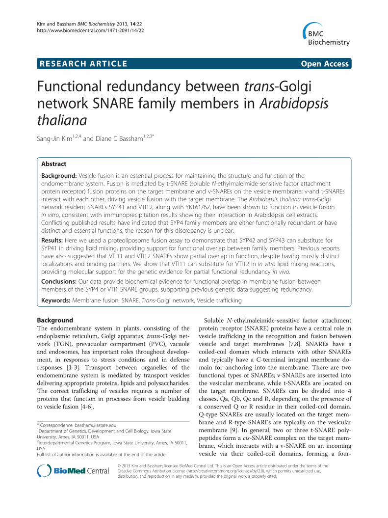
ResultsExpression of recombinant SNAREs in Escherichia coliSNAREs used in this study are shown schematically inFigure 1A. Recombinant proteins were expressed in E. coliand purified via their N-terminal His6 tag using Ni-NTAresin. Purified proteins were separated by SDS-PAGE andstained with Coomassie Blue. The purified proteins mi-grated at their expected MW with the exception of SYP42(Figure 1B). The predicted size of SYP42 is approximately36 kDa, but recombinant SYP42 migrated at around 45kDa, presumably due to structural hindrance during mi-gration in the gel (Figure 1B). This size difference was alsoobserved in a previous study of epitope-tagged SYP42 intransgenic Arabidopsis plants [18].
SYP42 and SYP43 can also drive liposome fusionPreviously, proteoliposome fusion assays using SYP41,VTI12 and YKT62 demonstrated that these proteins weresufficient to drive full lipid mixing between liposomes, as aproxy for vesicle fusion [15]. Conflicting reports in the lit-erature have suggested that the SYP4 family SNAREs haveeither distinct [22] or redundant [23] functions. To furtheraddress this issue, we tested whether other SYP4 familyproteins can substitute for SYP41 in driving lipid mixing
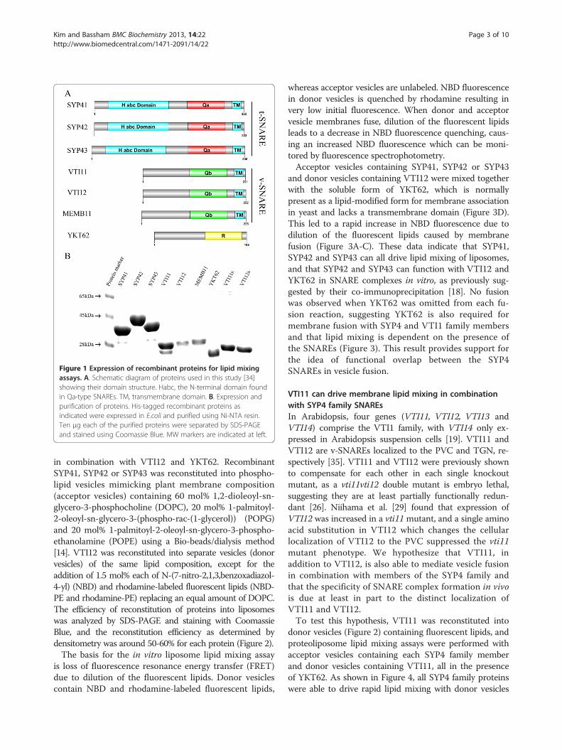
Figure 1 Expression of recombinant proteins for lipid mixingassays. A. Schematic diagram of proteins used in this study [34]showing their domain structure. Habc, the N-terminal domain foundin Qa-type SNAREs. TM, transmembrane domain. B. Expression andpurification of proteins. His-tagged recombinant proteins asindicated were expressed in E.coli and purified using Ni-NTA resin.Ten μg each of the purified proteins were separated by SDS-PAGEand stained using Coomassie Blue. MW markers are indicated at left.
Kim and Bassham BMC Biochemistry 2013, 14:22 Page 3 of 10http://www.biomedcentral.com/1471-2091/14/22
in combination with VTI12 and YKT62. RecombinantSYP41, SYP42 or SYP43 was reconstituted into phospho-lipid vesicles mimicking plant membrane composition(acceptor vesicles) containing 60 mol% 1,2-dioleoyl-sn-glycero-3-phosphocholine (DOPC), 20 mol% 1-palmitoyl-2-oleoyl-sn-glycero-3-(phospho-rac-(1-glycerol)) (POPG)and 20 mol% 1-palmitoyl-2-oleoyl-sn-glycero-3-phospho-ethanolamine (POPE) using a Bio-beads/dialysis method[14]. VTI12 was reconstituted into separate vesicles (donorvesicles) of the same lipid composition, except for theaddition of 1.5 mol% each of N-(7-nitro-2,1,3,benzoxadiazol-4-yl) (NBD) and rhodamine-labeled fluorescent lipids (NBD-PE and rhodamine-PE) replacing an equal amount of DOPC.The efficiency of reconstitution of proteins into liposomeswas analyzed by SDS-PAGE and staining with CoomassieBlue, and the reconstitution efficiency as determined bydensitometry was around 50-60% for each protein (Figure 2).The basis for the in vitro liposome lipid mixing assay
is loss of fluorescence resonance energy transfer (FRET)due to dilution of the fluorescent lipids. Donor vesiclescontain NBD and rhodamine-labeled fluorescent lipids,
whereas acceptor vesicles are unlabeled. NBD fluorescencein donor vesicles is quenched by rhodamine resulting invery low initial fluorescence. When donor and acceptorvesicle membranes fuse, dilution of the fluorescent lipidsleads to a decrease in NBD fluorescence quenching, caus-ing an increased NBD fluorescence which can be moni-tored by fluorescence spectrophotometry.Acceptor vesicles containing SYP41, SYP42 or SYP43
and donor vesicles containing VTI12 were mixed togetherwith the soluble form of YKT62, which is normallypresent as a lipid-modified form for membrane associationin yeast and lacks a transmembrane domain (Figure 3D).This led to a rapid increase in NBD fluorescence due todilution of the fluorescent lipids caused by membranefusion (Figure 3A-C). These data indicate that SYP41,SYP42 and SYP43 can all drive lipid mixing of liposomes,and that SYP42 and SYP43 can function with VTI12 andYKT62 in SNARE complexes in vitro, as previously sug-gested by their co-immunoprecipitation [18]. No fusionwas observed when YKT62 was omitted from each fu-sion reaction, suggesting YKT62 is also required formembrane fusion with SYP4 and VTI1 family membersand that lipid mixing is dependent on the presence ofthe SNAREs (Figure 3). This result provides support forthe idea of functional overlap between the SYP4SNAREs in vesicle fusion.
VTI11 can drive membrane lipid mixing in combinationwith SYP4 family SNAREsIn Arabidopsis, four genes (VTI11, VTI12, VTI13 andVTI14) comprise the VTI1 family, with VTI14 only ex-pressed in Arabidopsis suspension cells [19]. VTI11 andVTI12 are v-SNAREs localized to the PVC and TGN, re-spectively [35]. VTI11 and VTI12 were previously shownto compensate for each other in each single knockoutmutant, as a vti11vti12 double mutant is embryo lethal,suggesting they are at least partially functionally redun-dant [26]. Niihama et al. [29] found that expression ofVTI12 was increased in a vti11 mutant, and a single aminoacid substitution in VTI12 which changes the cellularlocalization of VTI12 to the PVC suppressed the vti11mutant phenotype. We hypothesize that VTI11, inaddition to VTI12, is also able to mediate vesicle fusionin combination with members of the SYP4 family andthat the specificity of SNARE complex formation in vivois due at least in part to the distinct localization ofVTI11 and VTI12.To test this hypothesis, VTI11 was reconstituted into
donor vesicles (Figure 2) containing fluorescent lipids, andproteoliposome lipid mixing assays were performed withacceptor vesicles containing each SYP4 family memberand donor vesicles containing VTI11, all in the presenceof YKT62. As shown in Figure 4, all SYP4 family proteinswere able to drive rapid lipid mixing with donor vesicles
Prot
ein
mar
ker
SYP4
1
SYP4
3
VTI
11
VTI
12
MEM
B11
R-M
EMB
11
R-V
TI11
R-V
TI12
SYP4
2
R-S
YP4
1
R-S
YP4
3
R-S
YP4
2
45kDa
A
B
28kDa
Figure 2 Reconstitution of recombinant proteins into liposomes. A. Recombinant proteins were reconstituted into liposomes in thepresence of detergent (Triton X-100 or n-octylglucoside), followed by dialysis to remove detergents after reconstitution. The amount of eachprotein before and after reconstitution was analyzed by SDS-PAGE and Coomassie Blue staining to determine the reconstitution efficiency. PrefixR- stands for reconstituted proteins. B. The average reconstitution efficiency was obtained from three independent experiments by densitometryof gels. The average reconstitution efficiency was ~50% for all proteins. Error bars represent standard deviation (n = 3).
Kim and Bassham BMC Biochemistry 2013, 14:22 Page 4 of 10http://www.biomedcentral.com/1471-2091/14/22
containing VTI11, suggesting that the SYP4 family is alsoable to form functional SNARE complexes with VTI11.When the maximum percentage of lipid mixing (mea-sured as NBD fluorescence) containing one SYP4 familymember with vesicles containing either VTI11 or VTI12was compared, SYP41 and SYP43 showed similar max-imum percentages. However, SYP42 showed more lipidmixing with VTI11 than VTI12 by 5-10%. This differencewas found to be statistically significant (t-test, p < 0.05),suggesting that VTI11 functions with SYP42 better thanVTI12 in vitro (Figure 4B,D), although whether this hasbiological significance in vivo is unknown.We have shown that both VTI11 and VTI12 can drive
lipid mixing in combination with SYP4 family members(Figure 3 and 4). To test whether the lipid mixing is
driven by the specific interaction between individual SYP4and VTI1 family members rather than a general require-ment that any SNARE can fulfill, MEMB11, a v-SNAREinvolved in endoplasmic reticulum to Golgi anterogradetrafficking and fusion at the Arabidopsis cis-Golgi, wassynthesized and incorporated into donor vesicles [36]. Lit-tle lipid mixing was seen between vesicles containingMEMB11 and vesicles containing SYP4 family SNAREs inthe presence of YKT62, indicating that the reactions arespecific for the tested SNAREs (Figure 4).As a further confirmation that the observed lipid mixing
is specifically dependent on the VTI1 family, soluble frag-ments of VTI11 and VTI12 (VTI11s and VTI12s) weresynthesized lacking the transmembrane domain. These sol-uble VTI11 or VTI12 fragments were mixed with YKT62
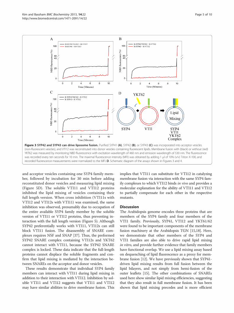
Figure 3 SYP42 and SYP43 can drive liposome fusion. Purified SYP41 (A), SYP42 (B), or SYP43 (C) was incorporated into acceptor vesicles(non-fluorescent vesicles), and VTI12 was reconstituted into donor vesicles containing fluorescent lipids. Membrane fusion with (black) or without (red)YKT62 was measured by monitoring NBD fluorescence with excitation wavelength of 460 nm and emission wavelength of 530 nm. The fluorescencewas recorded every ten seconds for 10 min. The maximal fluorescence intensity (MFI) was obtained by adding 1 μl of 10% (v/v) Triton X-100, andrecorded fluorescence measurements were normalized to the MFI. D. Schematic diagram of the assays shown in Figures 3 and 4.
Kim and Bassham BMC Biochemistry 2013, 14:22 Page 5 of 10http://www.biomedcentral.com/1471-2091/14/22
and acceptor vesicles containing one SYP4 family mem-ber, followed by incubation for 30 min before addingreconstituted donor vesicles and measuring lipid mixing(Figure 5D). The soluble VTI11 and VTI12 proteinsinhibited the lipid mixing of vesicles containing theirfull length version. When cross inhibition (VTI11s withVTI12 and VTI12s with VTI11) was examined, the sameinhibition was observed, presumably due to occupation ofthe entire available SYP4 family member by the solubleversion of VTI11 or VTI12 proteins, thus preventing in-teraction with the full length version (Figure 5). AlthoughSYP42 preferentially works with VTI11, VTI12s can stillblock VTI11 fusion. The disassembly of SNARE com-plexes requires NSF and SNAP [37]. Thus, the preformedSYP42 SNARE complex containing VTI12s and YKT62cannot interact with VTI11, because the SYP42 SNAREcomplex is locked. These data indicate that the full-lengthproteins cannot displace the soluble fragments and con-firm that lipid mixing is mediated by the interaction be-tween SNAREs on the acceptor and donor vesicles.These results demonstrate that individual SYP4 family
members can interact with VTI11 during lipid mixing inaddition to their interaction with VTI12. Inhibition by sol-uble VTI11 and VTI12 suggests that VTI11 and VTI12may have similar abilities to drive membrane fusion. This
implies that VTI11 can substitute for VTI12 in catalyzingmembrane fusion via interaction with the same SYP4 fam-ily complexes to which VTI12 binds in vivo and provides amolecular explanation for the ability of VTI11 and VTI12to partially compensate for each other in the respectivemutants.
DiscussionThe Arabidopsis genome encodes three proteins that aremembers of the SYP4 family and four members of theVTI1 family. Previously, SYP41, VTI12 and YKT61/62were found to be important components of the membranefusion machinery at the Arabidopsis TGN [15,18]. Here,we demonstrate that other members of the SYP4 andVTI1 families are also able to drive rapid lipid mixingin vitro, and provide further evidence that family membershave functional overlap. We use a lipid mixing assay basedon dequenching of lipid fluorescence as a proxy for mem-brane fusion [12]. We have previously shown that SYP41-driven lipid mixing results from full fusion between thelipid bilayers, and not simply from hemi-fusion of theouter leaflets [15]. The other combinations of SNAREsused here show similar lipid mixing efficiencies, suggestingthat they also result in full membrane fusion. It has beenshown that lipid mixing precedes and is more efficient
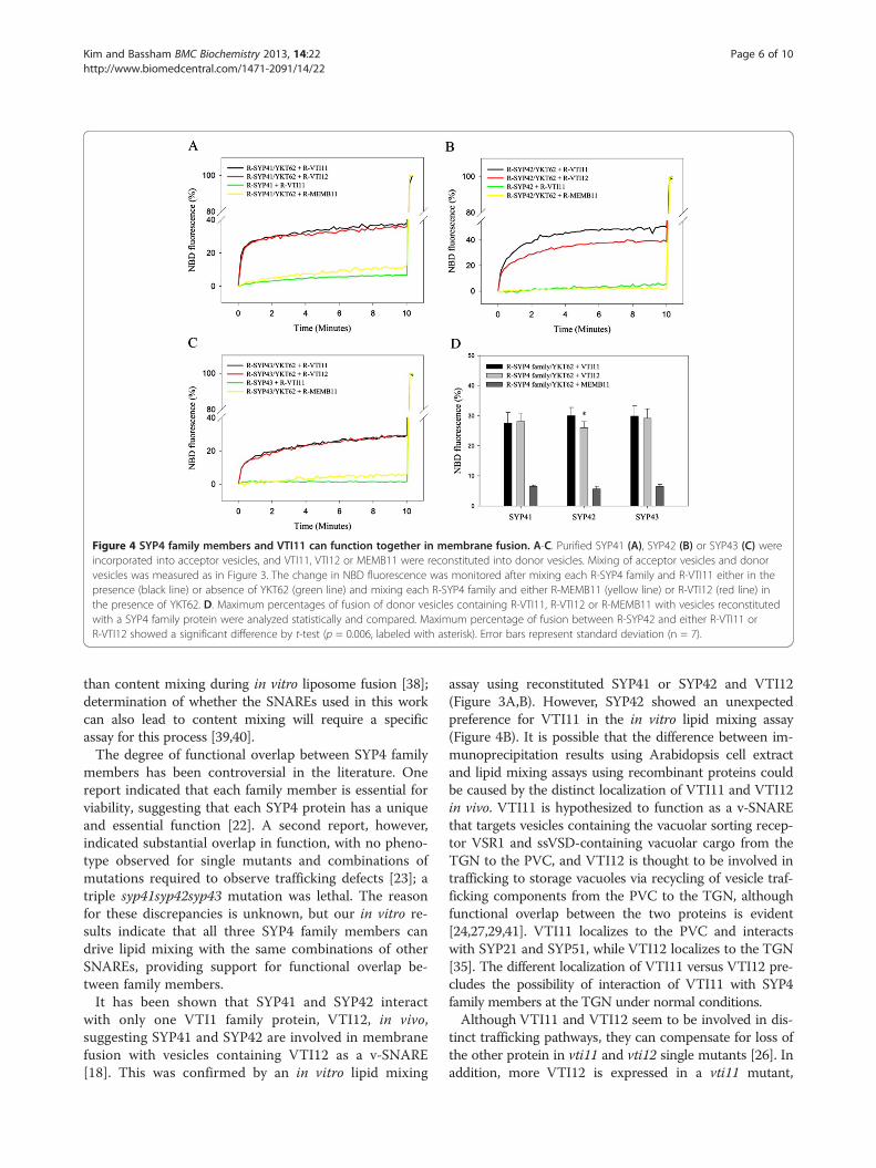
Figure 4 SYP4 family members and VTI11 can function together in membrane fusion. A-C. Purified SYP41 (A), SYP42 (B) or SYP43 (C) wereincorporated into acceptor vesicles, and VTI11, VTI12 or MEMB11 were reconstituted into donor vesicles. Mixing of acceptor vesicles and donorvesicles was measured as in Figure 3. The change in NBD fluorescence was monitored after mixing each R-SYP4 family and R-VTI11 either in thepresence (black line) or absence of YKT62 (green line) and mixing each R-SYP4 family and either R-MEMB11 (yellow line) or R-VTI12 (red line) inthe presence of YKT62. D. Maximum percentages of fusion of donor vesicles containing R-VTI11, R-VTI12 or R-MEMB11 with vesicles reconstitutedwith a SYP4 family protein were analyzed statistically and compared. Maximum percentage of fusion between R-SYP42 and either R-VTI11 orR-VTI12 showed a significant difference by t-test (p = 0.006, labeled with asterisk). Error bars represent standard deviation (n = 7).
Kim and Bassham BMC Biochemistry 2013, 14:22 Page 6 of 10http://www.biomedcentral.com/1471-2091/14/22
than content mixing during in vitro liposome fusion [38];determination of whether the SNAREs used in this workcan also lead to content mixing will require a specificassay for this process [39,40].The degree of functional overlap between SYP4 family
members has been controversial in the literature. Onereport indicated that each family member is essential forviability, suggesting that each SYP4 protein has a uniqueand essential function [22]. A second report, however,indicated substantial overlap in function, with no pheno-type observed for single mutants and combinations ofmutations required to observe trafficking defects [23]; atriple syp41syp42syp43 mutation was lethal. The reasonfor these discrepancies is unknown, but our in vitro re-sults indicate that all three SYP4 family members candrive lipid mixing with the same combinations of otherSNAREs, providing support for functional overlap be-tween family members.It has been shown that SYP41 and SYP42 interact
with only one VTI1 family protein, VTI12, in vivo,suggesting SYP41 and SYP42 are involved in membranefusion with vesicles containing VTI12 as a v-SNARE[18]. This was confirmed by an in vitro lipid mixing
assay using reconstituted SYP41 or SYP42 and VTI12(Figure 3A,B). However, SYP42 showed an unexpectedpreference for VTI11 in the in vitro lipid mixing assay(Figure 4B). It is possible that the difference between im-munoprecipitation results using Arabidopsis cell extractand lipid mixing assays using recombinant proteins couldbe caused by the distinct localization of VTI11 and VTI12in vivo. VTI11 is hypothesized to function as a v-SNAREthat targets vesicles containing the vacuolar sorting recep-tor VSR1 and ssVSD-containing vacuolar cargo from theTGN to the PVC, and VTI12 is thought to be involved intrafficking to storage vacuoles via recycling of vesicle traf-ficking components from the PVC to the TGN, althoughfunctional overlap between the two proteins is evident[24,27,29,41]. VTI11 localizes to the PVC and interactswith SYP21 and SYP51, while VTI12 localizes to the TGN[35]. The different localization of VTI11 versus VTI12 pre-cludes the possibility of interaction of VTI11 with SYP4family members at the TGN under normal conditions.Although VTI11 and VTI12 seem to be involved in dis-
tinct trafficking pathways, they can compensate for loss ofthe other protein in vti11 and vti12 single mutants [26]. Inaddition, more VTI12 is expressed in a vti11 mutant,
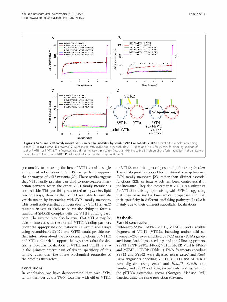
Figure 5 SYP4 and VTI1 family-mediated fusion can be inhibited by soluble VTI11 or soluble VTI12. Reconstituted vesicles containingeither SYP41 (A), SYP42 (B) or SYP43 (C) were mixed with YKT62 and either soluble VTI11 or soluble VTI12 for 30 min, followed by addition ofeither R-VTI11 or R-VTI12. The fluorescence did not increase significantly (less than 4%), indicating inhibition of the fusion reaction in the presenceof soluble VTI11 or soluble VTI12. D. Schematic diagram of the assays in Figure 5.
Kim and Bassham BMC Biochemistry 2013, 14:22 Page 7 of 10http://www.biomedcentral.com/1471-2091/14/22
presumably to make up for loss of VTI11, and a singleamino acid substitution in VTI12 can partially suppressthe phenotype of vti11 mutants [29]. These results suggestthat VTI1 family proteins can bind to non-cognate inter-action partners when the other VTI1 family member isnot available. This possibility was tested using in vitro lipidmixing assays, showing that VTI11 was able to mediatevesicle fusion by interacting with SYP4 family members.This result indicates that compensation by VTI11 in vti12mutants in vivo is likely to be via the ability to form afunctional SNARE complex with the VTI12 binding part-ners. The inverse may also be true, that VTI12 may beable to interact with the normal VTI11 binding partnersunder the appropriate circumstances. In vitro fusion assaysusing recombinant SYP21 and SYP51 could provide fur-ther information about the redundant functions of VTI12and VTI11. Our data support the hypothesis that the dis-tinct subcellular localization of VTI11 and VTI12 in vivois the primary determinant of fusion specificity of thisfamily, rather than the innate biochemical properties ofthe proteins themselves.
ConclusionsIn conclusion, we have demonstrated that each SYP4family member at the TGN, together with either VTI11
or VTI12, can drive proteoliposome lipid mixing in vitro.These data provide support for functional overlap betweenSYP4 family members [23] rather than distinct essentialfunctions [22], an issue which has been controversial inthe literature. They also indicate that VTI11 can substitutefor VTI12 in driving lipid mixing with SYP41, suggestingthat they have similar biochemical properties and thattheir specificity in different trafficking pathways in vivo ismainly due to their different subcellular localizations.
MethodsPlasmid constructionFull-length SYP42, SYP43, VTI11, MEMB11 and a solublefragment of VTI11 (VTI11s, including amino acid se-quence 1–200) were amplified by PCR using cDNAs gener-ated from Arabidopsis seedlings and the following primers;SYP42 FP/RP, SYP43 FP/RP, VTI11 FP/RP, VTI11s FP/RPand MEMB11 FP/RP (Table 1). DNA fragments encodingSYP42 and SYP43 were digested using EcoRI and XhoI.DNA fragments encoding VTI11, VTI11s and MEMB11were digested using EcoRI and HindIII, BamHI andHindIII, and EcoRI and XhoI, respectively, and ligated intothe pET28a expression vector (Novagen, Madison, WI)digested using the same restriction enzymes.
Table 1 Primer sequences
Gene Direction Sequence
SYP42 Forward 5′-GGAATTCCATATGGCGACGAGGAATCGAACGACGGTG-3′
Reverse 5′-CGCCTCGAGCTAAAACAAAATATTCTTAAGAATTAA-3′
SYP43 Forward 5′-GGAATTCCATATGGCGACTAGGAATCGTACGCTGTTG-3′
Reverse 5′-CGCCTCGAGTCACAACAGAATCTCCTTGAGGATTAAGA-3′
VTI11 Forward 5′-GGAATTCCATATGAGTGACGTGTTTGATGGATATGAG-3′
Reverse 5′-CCCAAGCTTTTACTTGGTGAGTTTGAAGTACAAGATG-3′
VTI11s Forward 5′-GGATCCATGAGTGACGTGTTTGATGG-3′
Reverse 5′-AAGCTTTTAGGTCCATTTGTTCTTGT-3′
MEMB11 Forward 5′-GAATTCATGGCGTCTGGTATCGTCGA-3′
Reverse 5′- CTCGAGGGTTAGCGTGTCCATCTTATGA-3′
The restriction sites used for cloning are underlined.
Kim and Bassham BMC Biochemistry 2013, 14:22 Page 8 of 10http://www.biomedcentral.com/1471-2091/14/22
Protein expression and purificationProtein expression was performed as described in Chenet al. [15] with minor changes. SYP41, SYP42, SYP43,VTI11, VTI12, VTI11s, VTI12s, YKT62 and MEMB11were expressed in E. coli strain BL21 (DE3) as N-terminalHis6-tagged proteins. Ten ml of an overnight culture weretransferred to 500 ml Luria-Burtani media with 50 μg/mlkanamycin and 2 mg/ml glucose. Cells were grown at 37°Cuntil the OD600 reached 0.6. Isopropyl-β-D-thiogalactopy-ranoside was added to 0.5 mM final concentration to in-duce expression, and cells were incubated for 5 h at 16°C.SYP41, VTI12s and YKT62 were purified according to
Chen et al. [15], and SYP42, SYP43, VTI11, VTI12, VTI11sand MEMB11 were purified with minor changes in thewashing and elution steps. SYP42 and SYP43 were elutedin elution buffer with 0.2% (v/v) Triton X-100 and VTI11swas purified in the same manner as VTI12s. VTI11, VTI12and MEMB11 were washed sequentially using washing buf-fer I (50 mM NaH2PO4 (pH 8.0), 200 mM NaCl, 50 mMimidazole, 0.2% (v/v) Triton X-100), washing buffer II (50mM Tris–HCl (pH 8.0), 300 mM NaCl, 50 mM imidazole,0.2% (v/v) Triton X-100), washing buffer III (50 mM Tris–HCl (pH 8.0), 300 mM NaCl, 50 mM imidazole) and wash-ing buffer IV (50 mM Tris–HCl (pH 8.0), 300 mM NaCl,50 mM imidazole, 0.8% (w/v) n-octylglucoside), followed byelution using elution buffer (50 mM Tris–HCl (pH 8.0),300 mM NaCl, 300 mM imidazole, 0.8% (w/v) n-octylglucoside).
Preparation of lipid vesicles and membrane reconstitutionPreparation of lipid vesicles and reconstitution of pro-teins into vesicles were performed as described in Chenet al. [15] except for proteins inserted into donor vesi-cles (VTI11, VTI12 and MEMB11). Donor vesiclescontaining fluorescent dyes were mixed with VTI11,VTI12 or MEMB11 at a 1:200 protein-to-lipid molar ra-tio and incubated at 4°C for 30 minutes. The same vol-ume of fusion assay buffer was added to the mixture,
which was dialyzed overnight against fusion assay bufferwith 4% (v/v) glycerol containing 1 g/l of Bio-beads SM-2 (Bio-Rad, Hercules, CA) to remove the trace amount ofdetergent. The reconstitution efficiency was analyzed bySDS-PAGE and staining with Coomassie Blue. The amountof protein in vesicles was compared with a known concen-tration of protein before reconstitution using densitometry(GS-800 Calibrated Densitometer, Bio-Rad, Hercules, CA)and Quantity one software (Bio-Rad, Hercules, CA).
Removal of detergent from YKT62The detergent in YKT62 samples was removed by adding20 mg Bio-beads SM-2, followed by rocking at 4°C for 30min. Removal of detergent was repeated three times,followed by dialysis as described above.
Total lipid mixing assayThe donor and acceptor vesicles were mixed in a molar ra-tio of 1:9, and the same molar amount of YKT62 to SYP41,SYP42 or SYP43 was added to the mixture. The total lipidconcentration was 0.5 mM and the total volume of themixture was set to 100 μl. Fusion of donor vesicles with ac-ceptor vesicles decreases quenching between rhodamineand NBD, measured as an increase in NBD fluorescence.Fluorescence was measured at excitation and emission
wavelengths of 465 and 530 nm, respectively. Fluorescencechanges were recorded every 10 sec with a Varian CaryEclipse model fluorescence spectrophotometer (Varian,Palo Alto, CA) with 2 mm path length at 25°C for 10 min.The maximum fluorescence intensity (MFI) was achievedby adding 1 μl of 10% (v/v) Triton X-100.
Lipid mixing with VTI11s or VTI12sTo inhibit fusion between donor and acceptor vesicles,a soluble version of VTI11 or VTI12 was added to theacceptor vesicles and incubated for 30 min at roomtemperature. The incubated acceptor vesicles were usedfor the lipid mixing assay as described above.
Kim and Bassham BMC Biochemistry 2013, 14:22 Page 9 of 10http://www.biomedcentral.com/1471-2091/14/22
AbbreviationsSNARE: Soluble N-ethylmaleimide-sensitive factor attachment proteinreceptor; TGN: Trans-Golgi network; PVC: Prevacuolar compartment;SM: Sec1/Munc18; DOPC: 1,2-dioleoyl-sn-glycero-3-phosphocholine; POPG: 1-palmitoyl-2-oleoyl-sn-glycero-3-(phospho-rac-(1-glycerol)); POPE: 1-palmitoyl-2-oleoyl-sn-glycero-3-phospho-ethanolamine; NBD: N-(7-nitro-2,1,3,benzoxadiazol-4-yl); FRET: Fluorescence resonance energy transfer.
Competing interestsThe authors declare that they have no competing interests.
Authors’ contributionsSJK performed the experiments, analyzed the results and drafted themanuscript. DCB conceived of the study, designed the experiments, analyzedthe results and revised the manuscript. Both authors read and approved thefinal manuscript.
AcknowledgementsWe thank Drs Yeon-Kyun Shin, Zhengliu Su and Yong Chen for providingconstructs, equipment and valuable assistance and expertise in the in vitrofusion assay. This work was supported by a grant from the NationalAeronautics and Space Administration (grant no. NNX09AK78G) to DCB.
Author details1Department of Genetics, Development and Cell Biology, Iowa StateUniversity, Ames, IA 50011, USA. 2Interdepartmental Genetics Program, IowaState University, Ames, IA 50011, USA. 3Plant Sciences Institute, Iowa StateUniversity, Ames, IA 50011, USA. 4Current address: Great Lakes BioenergyResearch Center, Michigan State University, East Lansing, MI 48824, USA.
Received: 14 June 2013 Accepted: 6 September 2013Published: 11 September 2013
References1. Robatzek S: Vesicle trafficking in plant immune responses. Cell Microbiol
2007, 9(1):1–8.2. Saito C, Ueda T: Chapter 4: functions of RAB and SNARE proteins in plant
life. Int Rev Cell Mol Biol 2009, 274:183–233.3. Fujimoto M, Ueda T: Conserved and plant-unique mechanisms regulating
plant post-Golgi traffic. Front Plant Sci 2012, 3:197.4. Lipka V, Kwon C, Panstruga R: SNARE-ware: the role of SNARE-domain
proteins in plant biology. Annu Rev Cell Dev Biol 2007, 23:147–174.5. Bassham D, Blatt M: SNAREs: cogs and coordinators in signaling and
development. Plant Physiol 2008, 147(4):1504–1515.6. Hwang I, Robinson DG: Transport vesicle formation in plant cells.
Curr Opin Plant Biol 2009, 12(6):660–669.7. Söllner T, Whiteheart S, Brunner M, Erdjument-Bromage H, Geromanos S,
Tempst P, Rothman J: SNAP receptors implicated in vesicle targeting andfusion. Nature 1993, 362(6418):318–324.
8. Sudhof TC, Rothman JE: Membrane fusion: grappling with SNARE and SMproteins. Science 2009, 323(5913):474–477.
9. Bock JB, Matern HT, Peden AA, Scheller RH: A genomic perspective onmembrane compartment organization. Nature 2001, 409(6822):839–841.
10. Poirier MA, Xiao W, Macosko JC, Chan C, Shin YK, Bennett MK: The synapticSNARE complex is a parallel four-stranded helical bundle. Nat Struct Biol1998, 5(9):765–769.
11. Paumet F, Rahimian V, Rothman JE: The specificity of SNARE-dependentfusion is encoded in the SNARE motif. Proc Natl Acad Sci USA 2004,101(10):3376–3380.
12. Weber T, Zemelman B, McNew J, Westermann B, Gmachl M, Parlati F,Söllner T, Rothman J: SNAREpins: minimal machinery for membranefusion. Cell 1998, 92(6):759–772.
13. McNew JA, Parlati F, Fukuda R, Johnston RJ, Paz K, Paumet F, Sollner TH,Rothman JE: Compartmental specificity of cellular membrane fusionencoded in SNARE proteins. Nature 2000, 407(6801):153–159.
14. Chen Y, Xu Y, Zhang F, Shin YK: Constitutive versus regulated SNAREassembly: a structural basis. EMBO J 2004, 23(4):681–689.
15. Chen Y, Shin YK, Bassham DC: YKT6 is a core constituent of membranefusion machineries at the Arabidopsis trans-Golgi network. J Mol Biol2005, 350(1):92–101.
16. Cho WJ, Lee JS, Zhang L, Ren G, Shin L, Manke CW, Potoff J, Kotaria N,Zhvania MG, Jena BP: Membrane-directed molecular assembly of theneuronal SNARE complex. J Cell Mol Med 2011, 15(1):31–37.
17. Shi L, Shen QT, Kiel A, Wang J, Wang HW, Melia TJ, Rothman JE, Pincet F:SNARE proteins: one to fuse and three to keep the nascent fusion poreopen. Science 2012, 335(6074):1355–1359.
18. Bassham DC, Sanderfoot AA, Kovaleva V, Zheng H, Raikhel NV: AtVPS45complex formation at the trans-Golgi network. Mol Biol Cell 2000,11(7):2251–2265.
19. Uemura T, Ueda T, Ohniwa RL, Nakano A, Takeyasu K, Sato MH: Systematicanalysis of SNARE molecules in Arabidopsis: dissection of the post-Golginetwork in plant cells. Cell Struct Funct 2004, 29(2):49–65.
20. Zouhar J, Rojo E, Bassham DC: AtVPS45 is a positive regulator of theSYP41/SYP61/VTI12 SNARE complex involved in trafficking of vacuolarcargo. Plant Physiol 2009, 149(4):1668–1678.
21. Bassham DC, Raikhel NV: An Arabidopsis VPS45p homolog implicated inprotein transport to the vacuole. Plant Physiol 1998, 117(2):407–415.
22. Sanderfoot AA, Pilgrim M, Adam L, Raikhel NV: Disruption of individualmembers of Arabidopsis syntaxin gene families indicates each hasessential functions. Plant Cell 2001, 13(3):659–666.
23. Uemura T, Kim H, Saito C, Ebine K, Ueda T, Schulze-Lefert P, Nakano A: Qa-SNAREs localized to the trans-Golgi network regulate multiple transport.Proc Natl Acad Sci U S A 2012, 109(5):1784–1789.
24. Zheng H, Von Mollard G, Kovaleva V, Stevens T, Raikhel N: The plantvesicle-associated SNARE AtVTI1a likely mediates vesicle transport fromthe trans-Golgi network to the prevacuolar compartment. Mol Biol Cell1999, 10(7):2251–2264.
25. Sanderfoot AA, Assaad FF, Raikhel NV: The Arabidopsis genome: anabundance of soluble N-ethylmaleimide-sensitive factor adaptor proteinreceptors. Plant Physiol 2000, 124(4):1558–1569.
26. Surpin M, Zheng HJ, Morita MT, Saito C, Avila E, Blakeslee JJ,Bandyopadhyay A, Kovaleva V, Carter D, Murphy A, et al: The VTI family ofSNARE proteins is necessary for plant viability and mediates differentprotein transport pathways. Plant Cell 2003, 15(12):2885–2899.
27. Sanmartin M, Ordonez A, Sohn EJ, Robert S, Sanchez-Serrano JJ, Surpin MA,Raikhel NV, Rojo E: Divergent functions of VTI12 and VTI11 in traffickingto storage and lytic vacuoles in Arabidopsis. Proc Natl Acad Sci U S A2007, 104(9):3645–3650.
28. Yano D, Sato M, Saito C, Sato MH, Morita MT, Tasaka M: A SNARE complexcontaining SGR3/AtVAM3 and ZIG/VTI11 in gravity-sensing cells isimportant for Arabidopsis shoot gravitropism. Proc Natl Acad Sci U S A2003, 100(14):8589–8594.
29. Niihama M, Uemura T, Saito C, Nakano A, Sato MH, Tasaka M, Morita MT:Conversion of functional specificity in Qb-SNARE VTI1 homologues ofArabidopsis. Curr Biol 2005, 15(6):555–560.
30. Dilcher M, Kohler B, Von Mollard GF: Genetic interactions with the yeastQ-SNARE VTI1 reveal novel functions for the R-SNARE YKT6. J Biol Chem2001, 276(37):34537–34544.
31. Kweon Y, Rothe A, Conibear E, Stevens T: Ykt6p is a multifunctional yeastR-SNARE that is required for multiple membrane transport pathways tothe vacuole. Mol Biol Cell 2003, 14(5):1868–1881.
32. Fukasawa M, Varlamov O, Eng WS, Sollner TH, Rothman JE: Localization andactivity of the SNARE Ykt6 determined by its regulatory domain andpalmitoylation. Proc Natl Acad Sci U S A 2004, 101(14):4815–4820.
33. Tai G, Lu L, Wang T, Tang B, Goud B, Johannes L, Hong W: Participation ofthe syntaxin 5/Ykt6/GS28/GS15 SNARE complex in transport from theearly/recycling endosome to the trans-Golgi network. Mol Biol Cell 2004,15(9):4011–4022.
34. Ren J, Wen L, Gao X, Jin C, Xue Y, Yao X: DOG 1.0: illustrator of proteindomain structures. Cell Res 2009, 19(2):271–273.
35. Sanderfoot AA, Kovaleva V, Bassham DC, Raikhel NV: Interactions betweensyntaxins identify at least five SNARE complexes within the Golgi/prevacuolarsystem of the Arabidopsis cell. Mol Biol Cell 2001, 12(12):3733–3743.
36. Chatre L, Brandizzi F, Hocquellet A, Hawes C, Moreau P: Sec22 and Memb11are v-SNAREs of the anterograde endoplasmic reticulum-Golgi pathway intobacco leaf epidermal cells. Plant Physiol 2005, 139(3):1244–1254.
37. Matveeva E, Whiteheart SW: The effects of SNAP/SNARE complexes on theATPase of NSF. FEBS Lett 1998, 435(2–3):211–214.
38. Chan YH, Van Lengerich B, Boxer SG: Effects of linker sequences on vesiclefusion mediated by lipid-anchored DNA oligonucleotides. Proc Natl AcadSci U S A 2009, 106(4):979–984.
Kim and Bassham BMC Biochemistry 2013, 14:22 Page 10 of 10http://www.biomedcentral.com/1471-2091/14/22
39. Diao J, Su Z, Ishitsuka Y, Lu B, Lee KS, Lai Y, Shin YK, Ha T: A single-vesiclecontent mixing assay for SNARE-mediated membrane fusion.Nat Commun 2010, 1:54.
40. Lai Y, Diao J, Liu Y, Ishitsuka Y, Su Z, Schulten K, Ha T, Shin YK: Fusion poreformation and expansion induced by Ca2+ and synaptotagmin 1.Proc Natl Acad Sci U S A 2013, 110(4):1333–1338.
41. Ebine K, Okatani Y, Uemura T, Goh T, Shoda K, Niihama M, Morita MT,Spitzer C, Otegui MS, Nakano A, et al: A SNARE complex unique to seedplants is required for protein storage vacuole biogenesis and seeddevelopment of Arabidopsis thaliana. Plant Cell 2008, 20(11):3006–3021.
doi:10.1186/1471-2091-14-22Cite this article as: Kim and Bassham: Functional redundancy betweentrans-Golgi network SNARE family members in Arabidopsis thaliana. BMCBiochemistry 2013 14:22.
Submit your next manuscript to BioMed Centraland take full advantage of:
• Convenient online submission
• Thorough peer review
• No space constraints or color figure charges
• Immediate publication on acceptance
• Inclusion in PubMed, CAS, Scopus and Google Scholar
• Research which is freely available for redistribution
Submit your manuscript at www.biomedcentral.com/submit