research article open access agrobacterium-mediated …
TRANSCRIPT

RESEARCH ARTICLE Open Access
Agrobacterium-mediated andelectroporation-mediated transformation ofChlamydomonas reinhardtii: a comparativestudyPaola Mini1, Olivia Costantina Demurtas1, Silvia Valentini1,2, Patrizia Pallara1, Giuseppe Aprea1, Paola Ferrante1*
and Giovanni Giuliano1*
Abstract
Background: Chlamydomonas reinhardtii is an unicellular green alga used for functional genomics studies andheterologous protein expression. A major hindrance in these studies is the low level and instability of expression ofnuclear transgenes, due to their rearrangement and/or silencing over time.
Results: We constructed dedicated vectors for Agrobacterium-mediated transformation carrying, within the T-DNAborders, the Paromomycin (Paro) selectable marker and an expression cassette containing the Luciferase (Luc) reportergene. These vectors and newly developed co-cultivation methods were used to compare the efficiency, stability andinsertion sites of Agrobacterium- versus electroporation-mediated transformation. The influence of differenttransformation methods, of the cell wall, of the virulence of different Agrobacterium strains, and of transgeneorientation with respect to T-DNA borders were assessed. False positive transformants were more frequent inAgrobacterium-mediated transformation compared to electroporation, compensating for the slightly lower proportionof silenced transformants observed in Agrobacterium-mediated transformation than in electroporation. The proportion ofsilenced transformants remained stable after 20 cycles of subculture in selective medium. Next generation sequencingconfirmed the nuclear insertion points, which occurred in exons or untraslated regions (UTRs) for 10 out of 10Agrobacterium-mediated and 9 out of 13 of electroporation-mediated insertions. Electroporation also resulted inhigher numbers of insertions at multiple loci.
Conclusions: Due to its labor-intensive nature, Agrobacterium transformation of Chlamydomonas does not presentsignificant advantages over electroporation, with the possible exception of its use in insertional mutagenesis, dueto the higher proportion of within-gene, single-locus insertions. Our data indirectly support the hypothesis thatrearrangement of transforming DNA occurs in the Chlamydomonas cell, rather than in the extracellular space aspreviously proposed.
Keywords: Chlamydomonas, Agrobacterium, Gene expression, Luciferase, Silencing
* Correspondence: [email protected]; [email protected], Italian National Agency for New Technologies, Energy andSustainable Economic Development, Casaccia Research Center, 00123 Rome,ItalyFull list of author information is available at the end of the article
© The Author(s). 2018 Open Access This article is distributed under the terms of the Creative Commons Attribution 4.0International License (http://creativecommons.org/licenses/by/4.0/), which permits unrestricted use, distribution, andreproduction in any medium, provided you give appropriate credit to the original author(s) and the source, provide a link tothe Creative Commons license, and indicate if changes were made. The Creative Commons Public Domain Dedication waiver(http://creativecommons.org/publicdomain/zero/1.0/) applies to the data made available in this article, unless otherwise stated.
Mini et al. BMC Biotechnology (2018) 18:11 DOI 10.1186/s12896-018-0416-3

BackgroundChlamydomonas reinhardtii is widely used for func-tional genomics studies as well as for heterologous pro-tein production. Significant progress has been madesince the first transformation reports using comple-mentation of the nitrate reductase (nit1) and arginino-succinate lyase (arg7) mutations [1, 2]. A large numberof selectable markers and promoters have been used fornuclear transformation, and delivery of foreign DNAhas been obtained through a variety of methods, suchas particle bombardment [1], agitation with glass beadsor silicon carbide whiskers [3, 4], electroporation [5], orAgrobacterium tumefaciens [6–8]. Despite recent ad-vances in genetic transformation, the expression offoreign genes in the nuclear genome of wild-typeChlamydomonas remains challenging due to transgenerearrangement and silencing [8–10].In this scenario, the possibility to transform Chlamy-
domonas by Agrobacterium tumefaciens is appealing,given the lower levels of transgene rearrangement andsilencing reported in plants. Agrobacterium tumefa-ciens is a pathogenic soil bacterium that has evolvedthe capacity to transfer a segment of DNA (the T-DNA) from the tumor-inducing (Ti) plasmid into thenucleus of a plant cell. An advantage in using Agrobac-terium for plant transformation is the reduction intransgene copy number, DNA rearrangements andtransgene silencing [11, 12].Nuclear transformation of Chlamydomonas rein-
hardtii by Agrobacterium tumefaciens has been re-ported, using vectors containing the CauliflowerMosaic Virus 35S promoter and non-codon-optimizedreporter genes [6]. The reported transformation effi-ciency was 50-fold higher than that obtained with theglass beads method. Further improvements of thetransformation efficiency were described in a secondreport [7] using modified transformation vectors, cul-ture media and Agrobacterium strains. No data on thestability of expression over time of the reporter genewere reported in either case.In the present work, we describe the construction of
transformation vectors carrying a selectable marker(Paro gene [13]) and a reporter gene (Renilla renifor-mis luciferase (Luc) [14]) which are codon optimizedfor Chlamydomonas nuclear expression and placedunder the control of strong Chlamydomonas pro-moters: 70 kDa heat shock protein/ribulose bispho-sphate carboxylase/oxygenase small subunit (HSP70/RBCS2) [15] and Photosystem I reaction center subunitII (PSAD) [16]. The effects of different transformationprotocols, Agrobacterium strains, and orientation ofthe transgenes with respect to the T-DNA right borderwere studied, and compared with results obtained viaelectroporation.
ResultsDevelopment of vectors and protocols for Agrobacterium-mediated transformation of C. reinhardtiiFirst, we constructed the pAgroR vector, optimized forAgrobacterium-mediated Chlamydomonas transfor-mation. This vector harbors the Paro gene, conferringparomomycin resistance [13] and an empty expressioncassette consisting of the PSAD promoter and termi-nator [16] flanking a multiple cloning site (Fig. 1a). Thevector was introduced in two Agrobacterium strainswith low and high virulence, respectively, LBA4404 andC58C1 [17] and used for transformation of the cellwall-proficient CC125(+) and the cell wall-deficientcw15 Chlamydomonas strains.In the first round of experiments we followed pub-
lished co-cultivation protocols [6, 7], but we were un-able to obtain paromomycin-resistant colonies. SinceAgrobacterium virulence (vir) genes are known to be in-duced by acidic pH and nutrient-poor growth media[18], we developed a different transformation method,based on the recommendations by Gelvin [19]. Thismethod consists in growing Agrobacterium cells for thefirst 24 h in AB medium and then overnight in AB in-duction medium [19]. Co-cultivation of Agrobacteriumwith Chlamydomonas is carried out on solid AB induc-tion medium for 48 h (for further details see Materialsand Methods).Using this method, cw15 cells were successfully trans-
formed using both the LBA4404 and C58C1 Agrobacter-ium strains, while CC125(+) cells could be transformedonly with the more virulent C58C1 strain (Table 1). Ingeneral, higher transformation efficiencies were obtainedusing cell wall-deficient Chlamydomonas cells and highlyvirulent Agrobacterium (Table 1). Transformation was
Paro LuciferaseP1T1
LB
P2 T2
RB
Paro T1P1
LB
LuciferaseT2 P2
RB
b
c pAgroLucL
pAgroLucR
pAgroRParo P1T1
LB
P2 T2
RB
a
EcoRI XbaI
EcoRIXbaI
EcoRI XbaI
Fig. 1 Schematic map of the vectors used for Agrobacterium-mediatedtransformation. Only the portion between the LB and RB (T-DNA) isshown. All vectors contain the Paro gene, conferring resistance toparomomycin under the control of the strong HSP70/RBCS2 hybridpromoter (P1) and RBCS2 terminator (T1). pAgroR (a) contains anexpression cassette containing the PSAD promoter (P2) and terminator(T2) sequences. In pAgroLucR (b) the Luc coding sequence is cloned inthe expression cassette. In pAgroLucL (c) the Paro and Luc cassetteshave an inverted orientation with respect to pAgroLucR. For furtherdetails see Material and Methods
Mini et al. BMC Biotechnology (2018) 18:11 Page 2 of 12

confirmed by PCR analysis using oligonucleotides di-rected to the Paro gene (See Additional file 1: Figure S1for an example). Sixty-seven to 92 % (67–92%) ofparomomycin-resistant colonies were PCR-positive forthe Paro gene (Table 1).We conducted a second round of experiments using
the pAgroLucR vector, carrying a codon-optimized lu-ciferase (Luc) gene [14] in the PSAD expression cassette(Fig. 1b). This vector was used for either Agrobacterium-mediated transformation or electroporation of the cw15strain (Table 2). Even with the optimized protocol, Agro-bacterium-mediated transformation efficiencies were 2.5-to 60-fold lower with respect to electroporation using thesame plasmid (Table 2). Also, while 100% of electroporated
Paro-resistant colonies tested are positive for the pres-ence of the Paro gene in the PCR assay, only 65% to97% of colonies obtained after co-cultivation with Agro-bacterium did so, indicating a high percentage of falsepositive transformants in the latter protocol (Table 2).
Transgene rearrangementsNext, we tested the maintenance of the Luc transgene inthe Paro-positive transformants obtained either throughelectroporation or through co-cultivation with Agrobac-terium. The results (Additional file 2: Figure S2 showsthe analysis of a subset of transformants) show that, al-though most transformants tested are PCR-positive forthe Paro gene, only few carry the Luc gene. The fre-quency of Luc-positive colonies was tested in two inde-pendent experiments and compared to that obtained inelectroporation experiments using the same vector. Theresults (Table 2) indicate that only 13%–16% of Paro-resistant colonies obtained through Agrobacteriumtransformation carry a complete Luc gene, compared to26%–33% obtained through electroporation with thesame vector. Thus, in contrast with the results obtainedin higher plants [12], Agrobacterium-mediated trans-formation of Chlamydomonas does not alleviate, com-pared to electroporation, the rearrangement oftransgenes cloned within the T-DNA borders.PCR reactions on a set of nine independent trans-
formants (Fig. 2, Additional file 3: Figure S3 andAdditional file 4: Figure S4) with nested pairs of oligo-nucleotides spanning the whole T-DNA region (Fig. 2a)indicated that, both in Agrobacterium-mediated trans-formation and in electroporation, deletion frequency ishighest close to the right border (RB), i.e. away from theParo selectable marker (Fig. 2b).
Influence of orientation with respect to the T-DNA borderson transgene rearrangementsT-DNA integration occurs in a directional way, resultingin asymmetric deletion frequencies in regions close to
Table 1 Efficiency of different Agrobacterium-mediatedtransformation methods. Two different Agrobacterium strains(LBA4404 and C58C1), containing the pAgroR vector and twoChlamydomonas strains (CC125 and cw15) were used.Transformation efficiency is expressed as the number of coloniesresistant to paromomycin/108 cells transformed. The presence ofthe Paro transgene was tested by PCR and is expressed as thepercentage of Paro-resistant colonies testing positive in thePCR assay. Kanamycin (Kan)-positive transformants, indicativeof bacterial contamination, were excluded from the analysis.Results of two independent experiments for each protocol/strain combination are reported. At least 50 independenttransformants for each experiment were analyzed by PCR
Chlamydomonasstrain
Agrobacteriumstrain
Transformation efficiency(colonies/108 cells)
Positivity toParo (PCR)
cw15 LBA4404 31 75%
16 92%
cw15 C58C1 25 75%
33 67%
CC125 LBA4404 1 0%
0 /
CC125 C58C1 6 86%
14 92%
Table 2 Comparison of electroporation and Agrobacterium-mediated transformation. Transformation efficiencies are expressed asthe number of paromomycin resistant colonies/108 cells transformed. Presence of Paro and Luc transgenes was tested by PCR.Transformants containing the Luc transgene were further analyzed for Luc enzymatic activity (last column). Results of two independentexperiments are reported. At least 96 independent transformants were analyzed for each experiment
Transformation method Plasmid Transformants/108 cells Paro-positive transformants(PCR)
Luc-positive transformants(PCR)
Transformants exhibitingLuc activitya
Electroporation pAgroLucR 120 100% 26% 15%
250 100% 33% 20%
Agrobacterium (C58C1) pAgroLucR 4 97% 16% 13%
52 65% 13% 11%
Agrobacterium (C58C1) pAgroLucL 31 93% 23% 20%
12 49% 13% 10%aLuc activity > 3-fold that of the untransformed control
Mini et al. BMC Biotechnology (2018) 18:11 Page 3 of 12

the right and left borders (RB and LB) [20]. In order toinvestigate if the position of the Luc gene with respect toLB and RB influences the frequency of deletions at thislocus, we constructed a second vector, pAgroLucL, inwhich the orientation of the Paro and Luc genes isinverted with respect to pAgroLucR (Fig. 1c). The results(Table 2) indicate a slight increase in the number of thetransformants containing an intact Luc transgene whenthe pAgroLucL construct was used. Deletions comingfrom the RB and LB as well as more internal deletionswere tested via PCR (Fig. 3, Additional file 5: Figure S5and Additional file 6: Figure S6). The results show thatdeletions of the Luc transgene are more frequent (82%vs 62%) when it flanks the RB compared to the LB.
Transgene expression and its stability over time underselective pressureNext, we asked whether transgene rearrangement ortransgene silencing were the major causes resulting inloss of transgene expression. In order to answer thisquestion, cells containing an intact Luc gene as judgedby PCR were subjected to 3 subsequent subcultures onselective medium in order to get rid of the Agrobacter-ium cells and then assayed for Luc activity. The results(Table 2 and Additional file 7: Table S4) indicate that alarge fraction of transformants containing an intact Luctransgene also showed some level of Luc activity (at least
three times higher than the background) and that gene si-lencing may be slightly more active during electroporationthan during Agrobacterium-mediated transformation.In order to follow the stability of Luc expression in
transformants grown under selective pressure, cells weresubcultured on solid medium containing paromomycin at1-week intervals, and Luc activity was measured at the3rd and 20th subculture cycle (Additional file 7: Table S4).In all three cases (electroporation and co-cultivation withAgrobacterium carrying pAgroLucR and pAgroLucLvectors), the percentage of Luc-positive transformantsshowing luciferase activity does not drop significantlyfrom the 3rd to the 20th subculture cycle. QuantitativeLuc activity data on the twenty highest expressors (Fig. 4)
0
1
2
3
4
5
6
7
8
9
Agrobacterium
Electroporation
aParo LuciferaseP1T1
LB
P2 T2
RBpAgroLucR
12
34
56
b
Fig. 2 Deletions along the T-DNA in transformants obtained withelectroporation or Agrobacterium-mediated transformation.Chlamydomonas cw15 cells were transformed with the pAgroLucRplasmid, using Agrobacterium or electroporation. Integrity of theinserted T-DNA was tested by PCR amplification with nested pairsof oligonucleotides spanning the T-DNA (Panel a) in a set of nineindependent transformants. The histogram (Panel b) shows thatthere is a gradient of deletion from the LB (Amplicon1) to the RB(Amplicon 6) independently of the transformation method used(Agrobacterium, black; electroporation, gray). Oligonucleotide sequencesare reported in Additional file 7: Table S2
0
5
10
15
20
25
30
pAgroLucR
05
1015202530
pAgroLucL
apAgroLucR
Paro LuciferaseP1T1
LB
P2 T2
RB
Amplicon 3 1024 bp
Amplicon 41172 bp
Amplicon 1 626 bp
Amplicon 2528 bp
bpAgroLucL
Paro T1P1
LB
LuciferaseT2 P2
RB
Amplicon 51172 bp
Amplicon 61024
Amplicon 1 626 bp
Amplicon 2528 bp
Fig. 3 T-DNA deletion pattern in pAgroLucR and pAgroLucLtransformants. The pAgroLucR and pAgroLucL plasmids were usedfor Agrobacteriummediated transformation and integrity of the T-DNAin the resulting transformants was tested via PCR with appropriateoligonucleotide pairs. Panels a (pAgroLucR) and b (pAgroLucL) showthe position of the amplicons (top in each box) and the frequency oftransformants showing amplification for each amplicon (bottom ineach box). A set of 29 independent transformants were tested for eachconstruct. All transformants contain amplicon 1, corresponding tothe Paro gene. When Luc is cloned next to the LB is retained athigher frequency (pAgroLucL, amplicons 2 and 5 in Panel b) with respectto the RB (pAgroLucR, amplicons 2 and 4). See also Additional file 5:Figure S5 and Additional file 6: Figure S6. Oligonucleotide sequencesare reported in Additional file 7: Table S3
Mini et al. BMC Biotechnology (2018) 18:11 Page 4 of 12

show that enzymatic activity is quite stable over time,indicating that neither the method of transformation (elec-troporation or Agrobacterium) nor the vector (pAgroLucLor pAgroLucR) influences Luc expression levels and theirstability over subsequent generations on selective medium.
Whole genome sequencing of electroporation- vsAgrobacterium-generated transformantsIn order to identify the insertion points of transformantsobtained with the different methods, we performed wholegenomic sequencing of twenty transformants in the cw15background, ten obtained by Agrobacterium and 10 by elec-troporation using the pAgroLucR plasmid. We selected 5high and 5 low Luc expressors for each transformationmethod (Agrobacterium or electroporation) in order to as-sess any influence of the genomic context of the insertionon transgene expression. Paired-end Illumina reads (2 ×100 nucleotides long) were generated to give an average
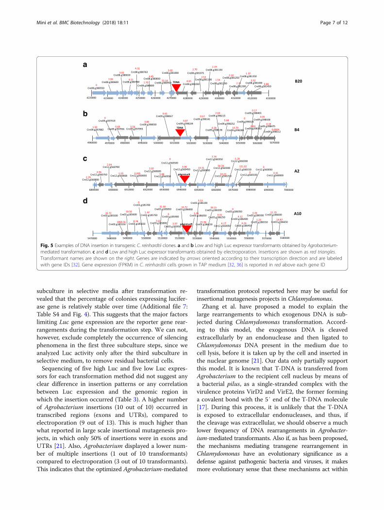
coverage of about 20×. The reads were aligned on theChlamydomonas genome and the pAgroLucR vector se-quences and the insertion points where determined usingpaired ends mapping to both the algal genome and the vec-tor. The results are summarized in Table 3. One of theAgrobacterium transformants did not carry a detectable in-sertion, presumably because the insertion occurred in agenomic region not present in the available genome assem-bly. The remainder of the insertions occurred, for the mostpart, in genes (5′ and 3′ UTR and exons) with only a mi-nority occurring in intergenic regions. One Agrobacteriumand three electroporation transformants carried insertionsin multiple chromosomal regions. Examples of DNA inte-gration are given in Fig. 5 for one low and one high Lucexpressor obtained by both Agrobacterium-mediated trans-formation and electroporation.
DiscussionAlthough Agrobacterium mediated transformation inChlamydomonas was first reported in 2004 [6], up tonow it has not been widely adopted. Previous publica-tions [6–8], demonstrate the successful expression of ex-ogenous genes and, in one case, stable integration in thenucleus. It has been proposed that, similar to plants,transformation of Chlamydomonas by Agrobacteriumcould provide stable integration and lower copy num-bers, potentially leading to fewer problems with trans-gene co-suppression and instability. However, no dataon the rearrangements/insertion points and copy numberare available for Agrobacterium-mediated transformantsand, most importantly, no comparative data are availablewith widely used methods, such as electroporation.The experiments described here indicate that the cw15
strain is more susceptible to Agrobacterium transform-ation than the CC125 strain. This may be due to the factthat cw15 is a cell wall deficient mutant and may bemore susceptible to Agrobacterium infection. Analysis ofa large set of transformants indicated that, regardless ofthe method of transformation used, just a small percent-age (10% to 20%) of the transformants show significantLuc expression (Table 2). In the case of electroporation,all the transformants analyzed contain the Paro select-able gene, while about 1/3 also contain a non-rearrangedLuc gene. Transformation with Agrobacterium results in ahigher number of false positive colonies (Table 2), lackingboth genes. This may be due to low level expression of theParo gene in Agrobacterium, leading to inactivation ofparomomycin during the co-cultivation step, or to thelong incubation of plates from Agrobacterium-mediatedtransformation, leading to light-mediated degradation ofparomomycin, or to interference by the antibiotics used toremove Agrobacterium cells.
a
b
c
Fig. 4 Temporal stability of Luc activity in Chlamydomonastransformants obtained through electroporation or Agrobacterium-mediated transformation. Twenty Chlamydomonas transformantsshowing the highest level of luciferase expression were collected fromtwo experiments (experiments A and B: ten transformants chosen foreach experiment, see Additional file 7: Table S4) and analyzed forluciferase expression at the 3rd (black bars) and the 20th (gray bars)cycles of subculture after the initial transformation event. a Electroporationwith pAgroLucR b Agrobacterium-mediated trasformation withpAgroLucR c Agrobacterium-mediated trasformation with pAgroLucL.Luminescence is expressed as CPS normalized for 105 cells
Mini et al. BMC Biotechnology (2018) 18:11 Page 5 of 12

In cells transformed with pAgroLucR a gradient of de-letions is observed, with very high deletion frequenciesnear the RB, i.e. away from the selectable marker gene(Fig. 2, Additional file 3: Figure S3 and Additional file 4:
Figure S4). A slight effect of the LB and RB on deletionfrequencies was observed, with the RB being more proneto deletion than LB, as observed in plants [20]. Lumino-metric assays performed at the 3th and 20th cycles of
Table 3 Information on the insertion sites and Luc expression of the different transformants. Five low and five high Luc expressorswere collected and analyzed for the characterization of the insertion site both for Agrobacterium transformation and electroporation.The first column reports the name of the transformant selected (see Additional file 7: Table S4). Luc activity of the transformants atthe 3rd and 20th subculture in selective medium is expressed as average values of CPS (counts per second) normalized for 105 cells± standard deviation of three biological replicates. Chr #: chromosome number in which DNA integration occurred. Insertion pointswere determined from the Chlamydomonas reinhardtii structural annotation v5.5 [32]. Gene annotation was taken from the Chlamydomonasreinhardtii functional annotation v 5.5 [32]. Gene expression refers to RNA-Seq experiments conducted on wild type cells grown inTAP medium [32, 36] and are reported in FPKM (fragments per kilobase per million mapped fragments)
Transformant Luc activity 3rdsubculture
Luc activity 20thsubculture
Chr#
Insertionpoint
Gene name Gene annotation Gene expression(FPKM)
CPS ± sd CPS ± sd
Transformation method: Agrobacterium
B 20 9 ± 2 8 ± 1 8 Gene (3′ UTR) Cre08.g381050 Senescence-associated gene 12 4.65
A 14 38 ± 11 18 ± 2 1 Gene (exon) Cre01.g051900 Ubiquinol-cytochrome Creductase iron-sulfur subunit
166.65
B 9 5 ± 1 64 ± 12 12 Gene (5′UTR) Cre12.g486000 n.a. 1.49
B 12 7 ± 1 69 ± 13 17 Gene (exon) Cre17.g736700 n.a. 0
A 18 48 ± 10 16 ± 2 1 Gene (exon) Cre01.g015250 DNA binding; DNA-directedDNA polymerases
4.13
A 6 13,306 ± 1218 6979 ± 926 3 Gene (exon) Cre03.g204200 n.a. 0
A 7 19,240 ± 1945 21,210 ± 1694 n.d. n.d. n.d. n.d. n.d.
B 4 15,202 ± 2795 8006 ± 1259 9 Gene (5′UTR) Cre09.g398067 Rotamase FKBP 1 9.45
B 6 12,460 ± 1239 7751 ± 643 17 Gene (exon) Cre17.g699600Cre08.g377150
Sedoheptulose-bisphosphatase 14.62
8 Gene (exon) n.a. 18.19
B 11 16,458 ± 892 8762 ± 831 8 Gene (3′ UTR) Cre08.g379400 n.a. 2.95
Transformation method: Electroporation
A 2 7 ± 3 49 ± 6 12 Gene (3′ UTR) Cre12.g560450 n.a. 3.08
A 3 2 ± 0 68 ± 9 1 Gene (exon) Cre01.g014150 MATE efflux family protein 5.20
4 Gene (exon) Cre04.g223550 n.a. 0.42
A 4 4 ± 1 24 ± 5 13 Gene: 3′ UTR Cre13.g569850 Ammonium transporter 1;2 0.89
A 5 37 ± 11 61 ± 7 12 Gene (exon) Cre12.g559450 Calcium-dependent lipid-bindingfamily protein
37.98
A 7 1 ± 1 21 ± 5 11 Gene (exon) Cre11.g467556 n.a. 2.73
5 Gene (5′UTR) Cre05.g232100 n.a. 6.87
A 10 17,849 ± 1022 16,393 ± 3650 3 Gene (3′ UTR) Cre03.g186050 n.a. 1.26
A 11 11,347 ± 645 3932 ± 771 2 2 Genes, both3′UTR
Cre02.g098700; ABC-2 type transporter familyprotein
0.63
Intergenicregion
Cre02.g098750 STELAR K+ outward rectifier 2.77
8 / /
B 11 17,253 ± 1316 20,099 ± 2464 3 Gene (intron) Cre03.g165700 Pyruvate decarboxylase-2 87.52
A 18 13,348 ± 753 15,958 ± 1918 3 Intergenicregion
/ / /
B 18 12,482 ± 499 13,977 ± 1959 16 Intergenicregion
/ / /
n.d. not determined, n.a. not available
Mini et al. BMC Biotechnology (2018) 18:11 Page 6 of 12
subculture in selective media after transformation re-vealed that the percentage of colonies expressing lucifer-ase gene is relatively stable over time (Additional file 7:Table S4 and Fig. 4). This suggests that the major factorslimiting Luc gene expression are the reporter gene rear-rangements during the transformation step. We can not,however, exclude completely the occurrence of silencingphenomena in the first three subculture steps, since weanalyzed Luc activity only after the third subculture inselective medium, to remove residual bacterial cells.Sequencing of five high Luc and five low Luc expres-
sors for each transformation method did not suggest anyclear difference in insertion patterns or any correlationbetween Luc expression and the genomic region inwhich the insertion occurred (Table 3). A higher numberof Agrobacterium insertions (10 out of 10) occurred intranscribed regions (exons and UTRs), compared toelectroporation (9 out of 13). This is much higher thanwhat reported in large scale insertional mutagenesis pro-jects, in which only 50% of insertions were in exons andUTRs [21]. Also, Agrobacterium displayed a lower num-ber of multiple insertions (1 out of 10 transformants)compared to electroporation (3 out of 10 transformants).This indicates that the optimized Agrobacterium-mediated
transformation protocol reported here may be useful forinsertional mutagenesis projects in Chlamydomonas.Zhang et al. have proposed a model to explain the
large rearrangements to which exogenous DNA is sub-jected during Chlamydomonas transformation. Accord-ing to this model, the exogenous DNA is cleavedextracellularly by an endonuclease and then ligated toChlamydomonas DNA present in the medium due tocell lysis, before it is taken up by the cell and inserted inthe nuclear genome [21]. Our data only partially supportthis model. It is known that T-DNA is transferred fromAgrobacterium to the recipient cell nucleus by means ofa bacterial pilus, as a single-stranded complex with thevirulence proteins VirD2 and VirE2, the former forminga covalent bond with the 5′ end of the T-DNA molecule[17]. During this process, it is unlikely that the T-DNAis exposed to extracellular endonucleases, and thus, ifthe cleavage was extracellular, we should observe a muchlower frequency of DNA rearrangements in Agrobacter-ium-mediated transformants. Also if, as has been proposed,the mechanisms mediating transgene rearrangement inChlamydomonas have an evolutionary significance as adefense against pathogenic bacteria and viruses, it makesmore evolutionary sense that these mechanisms act within
c
d
b
a
Fig. 5 Examples of DNA insertion in transgenic C. reinhardtii clones. a and b Low and high Luc expressor transformants obtained by Agrobacterium-mediated transformation. c and d Low and high Luc expressor transformants obtained by electroporation. Insertions are shown as red triangles.Transformant names are shown on the right. Genes are indicated by arrows oriented according to their transcription direction and are labeledwith gene IDs [32]. Gene expression (FPKM) in C. reinhardtii cells grown in TAP medium [32, 36] is reported in red above each gene ID
Mini et al. BMC Biotechnology (2018) 18:11 Page 7 of 12

the Chlamydomonas cell, where the bacterial or viral DNAis naked, rather than in the extracellular space, where it iscontained within a bacterial cell or a viral capsid. We thuspropose that the introduction of exogenous DNA in theChlamydomonas cell triggers an endonucleolytic mechan-ism aimed at its degradation, and that this mechanism pref-erably acts within the nucleus, when the nucleoprotein T-DNA complex is disassembled and the T-DNA is naked,before insertion into the nuclear DNA.
ConclusionsChlamydomonas has been proposed as a system forheterologous protein expression [22]. However, lowlevels of expression and rearrangements of the trans-forming DNA prevent it from being a competitive pro-duction platform with respect to bacterial, fungal oranimal cells. In the present work we compared Agrobac-terium-mediated transformation with electroporation ofChlamydomonas in order to verify if the former can alle-viate these problems. Regardless of the method used totransform Chlamydomonas cells, extensive rearrange-ments occur at the Luc gene, which is not under selectivepressure. This drawback was only partially compensatedby the reduced Luc silencing in Agrobacterium transfor-mants. Thus, differently from what observed in plants,Agrobacterium-mediated transformation does not presentsignificant advantages in terms of higher or more stableexpression. In order to increase transgene stability and ex-pression, mutants affected in transgene silencing [23] orin exogenous DNA cleavage can be of great help. Theknowledge gained from Agrobacterium-mediated trans-formation may be instrumental in the latter endeavour.
MethodsChlamydomonas strains and culture conditionsThe Chlamydomonas reinhardtii cell wall-deficient cw15and cell wall-proficient CC125 (+) strains [24], wereused for all experiments. Cells were grown photomixo-trophically in Tris Acetate Phosphate (TAP) medium[24] at 25 °C under continuous irradiation with fluores-cent white light (60 μE m− 2 s− 1). In the case of cw15strain the growth medium was supplemented with 1%(w/v) D-sorbitol.For luciferase assay, the transformants were grown in
1.5 ml of TAP medium at 160 rpm at 80 μE m− 2 s−1on arotary shaker until they reach the stationary phase(about 48 hs) in 24-well blocks (Qiagen, cat. n. 19,583).The cultures were then diluted 1:20 in 4 ml of TAPmedium and grown for 48 h. The plates were coveredwith Breathe-Easy membrane (Sigma-Aldrich, cat. n.Z763624-100EA) to prevent evaporation without limit-ing gaseous and light exchange. Frozen cell pellets rela-tive to 200 μl of each culture were re-suspended in 40 μlof lysis buffer (Renilla Luciferase Assay System,
Promega, cat. E2820), lysed at room temperature for15 min on a rotary shaker (750 rpm) and then incubatedon ice until assayed activity. Data were normalized usingoptical absorbance of at least two biological replicatesfor each transformant.
Plasmid constructionIn order to obtain the pAgroR (Fig. 1, panel a) andpAgroL plasmids (map not shown), pSL18 [25] wasdigested with NotI and KpnI. The digestion producestwo fragments, 3255 and 2867 bp long, that were bluntended using the Klenow Fragment (Roche, cat. n.11,008,404,001). The 3255 bp fragment containing theparomomycin expression cassette and the PSAD pro-moter and terminator regulatory elements was gel puri-fied and used for the next cloning step. The plasmidpKCRTI [26] (derived from pCAMBIA 1390) wasdigested with BglII and XhoI releasing four fragments of13, 865, 3391 and 6906 bp long. The digestion was sub-jected to filling in by Klenow and the 6906 bp fragmentcontaining all the genetic elements required for Agrobac-terium propagation and transfer into the host nucleus(including the LB and RB) was gel purified. The twofragments of 3255 bp, originating from pSL18, and6906 bp, originating from pKCRTI, were blunt clonedand PCR was performed using specific oligonucleotidesto discriminate the two different orientations of the frag-ments. The suffix R and L in pAgro vectors refers to theposition of the empty PSAD expression cassette respectto the borders (R: the PSAD cassette is close to the RB;L: the PSAD cassette is close to the LB). To buildpAgroLucR and pAgroLucL plasmids, Renilla reniformisluciferase coding sequence was isolated by PCR fromplasmid PSAD:cRLuc [27] with the following oligos:crLuc-EcoRI for CAGCGAATTCATGGCCAGCAAGGTGTACGAC and cRLuc-XbaI rev CAGTCTAGATTACGTATCGTTCTTCAGC (EcoRI and XbaI restrictionsites are reported in bold, gene specific sequence initalics is underlined). The PCR product was then clonedEcoRI-XbaI into pAgroR and pAgroL generating respect-ively pAgroLucR and pAgroLucL (Fig. 1, panel b and c).The bacterial strain used for cloning was XL1Blue fromStatagene. Bacterial transformation was carried out byelectroporation.
DNA extraction from Chlamydomonas cells and PCRanalysisSingle colonies of Chlamydomonas cells were picked andresuspended in 200 μl of 5% (w/v) chelex solution (Sigma-Aldrich, cat. n. C7901-25G) in a 96-well plate. The platewas incubated 10 min at 100 °C, 10 min on ice and centri-fuged at 6000 g for 3 min. One microliter of the super-natant was used to perform PCR analysis. The sequencesof oligonucleotides used to screen Chlamydomonas
Mini et al. BMC Biotechnology (2018) 18:11 Page 8 of 12

transformants are reported in Additional file 7: Table S1.β-tubulin was used as positive control for DNA extraction,paromomycin oligonucleotides were used to screen thepositivity to the antibiotic, kanamycin oligonucleotideswere used to evaluate residual Agrobacterium contamin-ation while cRLuc for 710 and PSAD Ter 276 were usedto check the presence of luciferase gene.Chlamydomonas colonies were analysed by PCR after
the three transfers in 1,5% TAP agar plate containing10 μg/μl paramomycin. In the case of Chlamydomonastransformants obtained through co-cultivation method500 μg/μl cefotaxime and 500 μg/μl carbenicillin wereadded in order to remove residual Agrobacterium cells.Ninety-six colonies were analyzed by PCR for eachtransformation experiment.
Chlamydomonas electroporationChlamydomonas cells were cultured in TAP medium to3–6 × 10⁶ cells/ml and then centrifuged at 3000 g for5 min at 4 °C. The pellet was washed once with coldTAP containing 60 mM D-sorbitol and resuspended atfinal concentration of 2 × 10⁸ cells/ml. 250 μl (5 × 10⁷
cells) of this culture was mixed with 200 ng of linearizedvector in a 0,4 cm cuvette (BIORAD, cat. n. 165–2081)and electroporated at 25 μF, 2000 V/cm. The time con-stant ranged between 13 and 16 ms. The electroporatedcells were recovered overnight in TAP medium at lowlight (30 μE m− 2 s− 1) and then plated on 1% TAP agarplate containing 10 μg/μl paramomycin in the case ofvectors described in Fig. 1. For electroporation, thepAgroLucR vector (Fig. 1b) was linearized with AgeI.Two independent electroporation experiments were per-formed, for each electroporation ten transformations werecarried out. Ninety-six transformants for each transform-ation were analysed by PCR.
Agrobacterium-mediated transformation with publishedmethodsIn the first set of experiments, Chlamydomonas cellswere transformed according to [28]. Briefly, LBA4404(Takara cat. n° 9115) and C58C1 Agrobacterium cells,kindly donated by Prof. Edgardo Filippone, were trans-formed through electroporation with the pAgroR vector(Fig. 1, Panel a) and plated on LB containing 100 μg/mlkanamycin and 100 μg/ml rifampicin (LBA4404 strain)or 200 μg/ml kanamycin and 100 μg/ml rifampicin(C58C1 strain) and grown for 48 h at 28 °C. Five singlecolonies from each transformation plate were inoculatedin 1.5 ml of LB medium containing the appropriate anti-biotics and analysed by PCR in order to check the pres-ence of the right and left borders with the followingpairs of oligonucleotides: PSAD Ter for GATTTCGCTGATTGATACGG and R border rev TAAACGCTCTTTTCTCTTAGG, producing an amplicon of 894 bp, L
border for TGGCAGGATATATTGTGGTG and Parobox rev CTGGACTGGGAGCGGTGT, producing anamplicon of 1039 bp. All the five picked colonies showedthe right amplicons and one of them was inoculated inLB medium plus antibiotics to prepare glycerol stocks.For each transformation experiment 100 μl of glycerolstock were inoculated in 15 ml liquid YEB medium -containing 30 μg/ml rifampicin and 100 μg/ml kanamy-cin - and grown for 24 h at 28 °C. The cells where thencentrifuged at 2700 g for 30 min and resuspended inliquid TAP medium containing 100 μM acetosyringoneto an A595 of 0.5. 107 Chlamydomonas cells were platedon a 90 mm petri dish and grown for two days at 80 μEm− 2 s− 1 in continuous light to allow a lawn of cells tobe formed. 200 μl of the bacterial suspension obtainedas described above was spread on the lawn of Chlamy-domonas cells for co-cultivation. After co-cultivation for48 h at 30 μE m− 2 s− 1 cells were harvested at 1500 g for5 min washed twice with liquid TAP medium containing500 μg/μl cefotaxime by centrifugation at 1500 g for5 min. Finally the cells were resuspended in TAP at 5 ×10⁷ cells/ml concentration and 1 ml was then platedonto 1% agar TAP plates containing 10 μg/μl paramo-mycin, 500 μg/μl cefotaxime and 500 μg/μl carbenicillin.Paromomycin is the selective antibiotic for transform-ation while cefotaxime and carbenicillin were used to killAgrobacterium cells. Single colonies appeared after 7 daysof growth at 30 μE m− 2 s− 1. These colonies were pickedand transferred on new TAP agar plates containing allthe three antibiotics. Even if we could observe severalcolonies on the primary transformation plate, after twoor three transfers on selective medium they died, prob-ably because they were false positive or because the vec-tor did not integrate stably into the nucleus. In a secondround of experiments we followed the protocol de-scribed in [7], but in this case we could not observe anycolony on the primary transformation plates.
Development of a modified protocol for Agrobacterium-mediated transformationIn order to set up a protocol for Chlamydomonas trans-formation through Agrobacterium in our laboratory, wefollowed the indications by [19]. Glycerol stocks of theAgrobacterium strains LBA4404 and C58C1 transformedwith pAgroR vector obtained as described in the previ-ous paragraph were inoculated in 15 ml of AB medium(pH 7) [29] containing 0.5% (w/v) glucose, 30 μg/ml ri-fampicin and 100 μg/ml kanamycin. The flasks weregrown at 28 °C for 24 h, then centrifuged at 2700 g for30 min and finally resuspended in 40 ml of AB inductionmedium (AB medium containing 0.5% glucose, 20 μMMES pH 5,6 and 100 μM acetosyringone) and grownovernight at 28 °C. Before co-cultivation the cell densitywas adjusted to A595 = 0,5 with AB induction medium.
Mini et al. BMC Biotechnology (2018) 18:11 Page 9 of 12
Fifty ml of Chlamydomonas culture grown for 2 days inTAP medium at approximately 107 cells/ml were centri-fuged at 1500 g for 5 min and resuspended in ABmedium at a final cell density of 108 cells/ml. For co-cultivation, 100 μl of microalgae corresponding to 107
cells and 40 μl of Agrobacterium were spotted on 1%agar AB induction medium plates. In total 14 spots, cor-responding to 108 Chlamydomonas cells, were plated oneach 1% agar AB induction medium plate and the plateswere grown at 30 μE m− 2 s− 1 for 48 h. The cells werethen harvested from each plate, washed twice with liquidTAP medium containing 500 μg/μl cefotaxime, centri-fuged at 1500 g for 5 min, resuspended in liquid TAPmedium at a density of 5 × 10⁷ cells/ml and plated ontwo TAP agar plates containing 10 μg/μl paramomycin,500 μg/μl cefotaxime and 500 μg/μl carbenicillin. After1 week of growth, colonies started to appear. Transform-ation efficiency is expressed as an average of the numberof paromomycin resistant colonies obtained per 108 ofChlamydomonas cells.Paromomycin resistant colonies were transferred for
three subculture rounds on 1.5% TAP agar plates con-taining the selective antibiotics and then analyzed byPCR. Two independent transformation experiments foreach vector were performed and 96 colonies for each ex-periment were analysed by PCR.
Luciferase assayLuciferase assay was performed using the Renilla Lucifer-ase Assay System (Promega, cat. n. E2820) as described in[27]. The luminescence of the wild type (background lu-minescence) was subtracted from the luminescence of thetransformants. All the transformants showing a level ofLuc activity > 3 fold than the background were consideredLuc-positive. The luminescence values reported are rela-tive to 105 cells. At least two biological and two technicalreplicates were analyzed for each transformant.
Whole genome sequencing and bioinformatic analysesTotal DNA was extracted using the DNeasy Plant MiniKit (Qiagen, Cat No. 69104) from pellets obtained from8 ml of mid-log phase cultures (concentration of 5 × 10⁶
cells/ml), grown in TAP medium as described above.DNA quality and concentration was checked by running
samples on 0.5% agarose gel and by fluorometry (Qubit,ThermoFisher Scientific), respectively. For each sample,one microgram of DNA was used to generate paired-endIllumina libraries, which were then sequenced on a HiSeq2500 Illumina sequencer generating about 8 millionspaired-end reads on average. Reads were cleaned ofadapters and quality trimmed with Cutadapt v. 1.8.1 [30]and Trimmomatic v. 0.33 [31]. A composite reference wasprepared by combining Chlamydomonas reinhardtii gen-ome v. 5.5 [32] and the pAgroLucR vector sequence. All
libraries were aligned to the reference using Bowtie2 v.2.2.7 [33] as single ends, to avoid any bias due to pairingin the alignment results (Additional file 7: Table S5). Inorder to isolate the insertion signals, only pairs with a hitboth on the alga genome and the vector were retained.Finally, for each transformed sample, all signals were com-pared to the wild type and insertion points were calledwith MACS v. 2.1.1 [34].
Additional files
Additional file 1: Figure S1. PCR confirmation of the presence of theParo selectable marker. Fourteen Paro resistant colonies (1–14) obtainedby co-cultivation of the cw15 strain with Agrobacterium C58C1 cellsharbouring the pAgroR plasmid were analyzed by PCR for the presenceof the endogenous β-tub gene (used as positive control for DNA extraction),Paro gene (selectable marker) and Kan gene (diagnostic of residualAgrobacterium contamination). Transformants 1–12 and 14 have theParo gene, while the results on transformant 13 are inconclusivesince the colony is still contaminated by Agrobacterium. Wt: cw15strain; P: pAgroR plasmid; C+: positive cw15 transformant; C-: no DNA. M: 1Kb Plus ladder (Life Technologies). Oligonucleotide sequences are shown inAdditional file 7: Table S1. (PPTX 380 kb)
Additional file 2: Figure S2. Retention of the Luc transgene in a set ofChlamydomonas colonies transformed with the pAgroLucR plasmid. cw15cells were co-cultivated with the Agrobacterium C58C1 strain harboringthe pAgroLucR plasmid. The DNA extracted was analyzed for the presenceof the genes shown in Additional file 1: Figure S1 plus the Luc gene. Thevast majority of the transformants, although positive for the presence of theParo gene, do not contain an intact Luc gene. A and B are two independentcontrol transformants, positive for the presence of the Paro and Lucgenes. P: pAgroLucR plasmid. C-: negative control. M: 1 Kb Plus DNALadder (Life Technologies). Oligonucleotide sequences are shown inAdditional file 7: Table S1. (PPTX 1875 kb)
Additional file 3: Figure S3. PCR analysis on a set of nine independenttransformants obtained through Agrobacterium to study the deletionpattern long the T-DNA. Chlamydomonas cw15 cells were co-cultivatedwith Agrobacterium C58C1 strain carrying the pAgroLucR plasmid. SixPCR reactions were performed on extracted DNA with nested pairs ofoligonucleotides annealing in the T-DNA from the LB to the RB (PanelA). The results (Panel B) show that there is a gradient of deletions fromthe LB to the RB. β-tubulin was used as positive control for DNA extraction.M: 1 Kb Plus DNA Ladder (Life Technologies); wt: cw15 strain; P: pAgroLucR;−: negative control. Oligonucleotide sequences are reported in Additionalfile 7: Table S2. (PPTX 204 kb)
Additional file 4: Figure S4. PCR analysis on a set of nine independenttransformants obtained through electroporation to study the deletionpattern along the T-DNA. Chlamydomonas cw15 cells were electroporatedwith the pAgroLucR plasmid. Six PCR reactions were performed on extractedDNA with nested pairs of oligonucleotides annealing in the T-DNA from theLB to the RB (Panel A). The results (Panel B) show that there is a gradient ofdeletions from the LB to the RB. β-tubulin was used as positive controlfor DNA extraction. M: 1 Kb Plus DNA Ladder (Life Technologies); wt:cw15 strain; P: pAgroLucR; −: negative control. Oligonucleotide sequencesare reported in Additional file 7: Table S2. (PPTX 128 kb)
Additional file 5: Figure S5. Deletion pattern on the T-DNA in thepAgroLucR transformants obtained though co-cultivation of Chlamydomonaswith Agrobacterium cells transformed with the pAgroLucR plasmid. Thefigure shows respectively a PCR analysis of a set of 29 independent cw15transformants obtained with C58C1 Agrobacterium cells carrying thepAgroLucR vector. Wt: cw15, P: pAgroLucR plasmid; C-: negative control.Oligonucleotide sequences are reported in Additional file 7: Table S3.(PPTX 1196 kb)
Additional file 6: Figure S6. Deletion pattern on the T-DNA in thepAgroLucL transformats obtained though co-cultivation of
Mini et al. BMC Biotechnology (2018) 18:11 Page 10 of 12

Chlamydomonas with Agrobacterium cells transformed with thepAgroLucL plasmid. The Figure shows a PCR analysis of a set of 29independent transformants obtained co-cultivating cw15 cells with C58C1Agrobacterium cells carrying the pAgroLucL vector. Wt: cw15, P: pAgroLucLplasmid; C-: negative control. Oligonucleotide sequences are reported inAdditional file 7: Table S3. (PPTX 2320 kb)
Additional file 7: Table S1. Oligonucleotides used to screenChlamydomonas transformants. Table S2. Oligonucleotides used tostudy the T-DNA deletion pattern. Table S3. Oligonucleotides used to studythe influence of R and L borders on T-DNA rearrangements. Table S4.Luciferase activity data at the 3rd and 20th subcultures. Table S5. NGSlibrary mapping statistics. (DOCX 75 kb)
AbbreviationsChr: Chromosome; CPS: Counts per second; FPKM: Fragments per kilobaseper million mapped fragments; HSP70/RBCS2: 70 kDa heat shock protein/ribulose bisphosphate carboxylase/oxygenase small subunit; LB: Left border;Luc: Luciferase; nt: Nucleotides; Paro: Paromomycin; PSAD: PhotosystemI reaction center subunit II; RB: Right border; TAP: Tris Acetate Phosphate;UTR: Untraslated region; β-tub: β-tubulin
AcknowledgementsPart of the computing resources and the related technical support used forthis work have been kindly provided by CRESCO/ENEAGRID High PerformanceComputing infrastructure and its staff [35]. This work was supported by theItalian Ministry of Agriculture, Hydrobio project.
FundingThis work was financially supported by the Italian Ministry of Agriculture,Hydrobio project. The funding body had no role in study design, in thecollection, analysis and interpretation of data, in the writing the Ms. and inthe decision to submit the article for publication.
Availability of data and materialsThe raw data relative to Tables 1 and 2 are available upon request. TheIllumina sequence data have been submitted as Bioproject [PRJNA395035] toNCBI sequence read archive under accession number [SRP113153].
Authors’ contributionsPM, OCD, SV, PP, GA and PF produced data. PM, OCD, GA, PF and GG analyzeddata. PF and GG coordinated the study and wrote the manuscript. All authorsreviewed the results and approved the final version of the manuscript.
Ethics approval and consent to participateNot applicable
Consent for publicationNot applicable
Competing interestsThe authors declare that they have no competing interests.
Publisher’s NoteSpringer Nature remains neutral with regard to jurisdictional claims inpublished maps and institutional affiliations.
Author details1ENEA, Italian National Agency for New Technologies, Energy andSustainable Economic Development, Casaccia Research Center, 00123 Rome,Italy. 2University of Rome “La Sapienza”, Piazzale Aldo Moro, 5, 00185 Rome,Italy.
Received: 8 June 2017 Accepted: 15 January 2018
References1. Kindle KL, Schnell RA, Fernandez E, Lefebvre PA. Stable nuclear transformation
of Chlamydomonas using the Chlamydomonas gene for nitrate reductase.J Cell Biol. 1989;109(6 Pt 1):2589–601.
2. Debuchy R, Purton S, Rochaix JD. The argininosuccinate lyase gene ofChlamydomonas reinhardtii: an important tool for nuclear transformationand for correlating the genetic and molecular maps of the ARG7 locus.EMBO J. 1989;8(10):2803–9.
3. Kindle KL. High-frequency nuclear transformation of Chlamydomonasreinhardtii. Proc Natl Acad Sci U S A. 1990;87(3):1228–32.
4. Dunahay TG. Transformation of Chlamydomonas reinhardtii with siliconcarbide whiskers. BioTechniques. 1993;15(3):452-455, 457-458, 460.
5. Shimogawara K, Fujiwara S, Grossman A, Usuda H. High-efficiency transformationof Chlamydomonas reinhardtii by electroporation. Genetics. 1998;148(4):1821–8.
6. Kumar SV, Misquitta RW, Reddy VS, Rao BJ, Rajam MV. Genetic transformation ofthe green alga—Chlamydomonas reinhardtii by Agrobacterium tumefaciens.Plant Sci. 2004;166(3):731–8.
7. Pratheesh PT, Vineetha M, Kurup GM. An efficient protocol for theAgrobacterium-mediated genetic transformation of microalgaChlamydomonas reinhardtii. Mol Biotechnol. 2014;56(6):507–15.
8. Barahimipour R, Neupert J, Bock R. Efficient expression of nuclear transgenesin the green alga Chlamydomonas: synthesis of an HIV antigen anddevelopment of a new selectable marker. Plant Mol Biol. 2016;90(4–5):403–18.
9. Specht E, Miyake-Stoner S, Mayfield S. Micro-algae come of age as aplatform for recombinant protein production. Biotechnol Lett.2010;32(10):1373–83.
10. Neupert J, Shao N, Lu Y, Bock R. Genetic transformation of the model greenalga Chlamydomonas reinhardtii. Methods Mol Biol. 2012;847:35–47.
11. Valvekens D, Montagu MV, Van Lijsebettens M. Agrobacterium tumefaciens-mediated transformation of Arabidopsis thaliana root explants by usingkanamycin selection. Proc Natl Acad Sci U S A. 1988;85(15):5536–40.
12. Dai S, Zheng P, Marmey P, Zhang S, Tian W, Chen S, Beachy RN, Fauquet C.Comparative analysis of transgenic rice plants obtained by Agrobacterium-mediated transformation and particle bombardment. Mol Breed.2001;7(1):25–33.
13. Sizova I, Fuhrmann M, Hegemann P. A Streptomyces rimosus aphVIII genecoding for a new type phosphotransferase provides stable antibioticresistance to Chlamydomonas reinhardtii. Gene. 2001;277(1–2):221–9.
14. Fuhrmann M, Hausherr A, Ferbitz L, Schodl T, Heitzer M, Hegemann P.Monitoring dynamic expression of nuclear genes in Chlamydomonasreinhardtii by using a synthetic luciferase reporter gene. Plant Mol Biol.2004;55(6):869–81.
15. Schroda M, Blocker D, Beck CF. The HSP70A promoter as a tool for theimproved expression of transgenes in Chlamydomonas. Plant J.2000;21(2):121–31.
16. Fischer N, Rochaix JD. The flanking regions of PsaD drive efficient geneexpression in the nucleus of the green alga Chlamydomonas reinhardtii.Mol Gen Genomics. 2001;265(5):888–94.
17. Gelvin SB. Agrobacterium-mediated plant transformation: the biology behindthe “gene-jockeying” tool. Microbiol Mol Biol Rev. 2003;67(1):16–37.
18. Vernade D, Herrera-Estrella A, Wang K, Van Montagu M. Glycine Betaineallows enhanced induction of the Agrobacterium tumefaciens vir genes byAcetosyringone at low pH. J Bacteriol. 1988;170(12):5822–9.
19. Gelvin SB. Agrobacterium virulence gene induction. In: Wang K, editor.Agrobacterium protocols, vol. 1. Totowa: Humana Press; 2006.
20. Tzfira T, Li J, Lacroix B, Citovsky V. Agrobacterium T-DNA integration:molecules and models. Trends Genet. 2004;20(8):375–83.
21. Zhang R, Patena W, Armbruster U, Gang SS, Blum SR, Jonikas MC. High-throughput genotyping of green algal mutants reveals random distributionof mutagenic insertion sites and Endonucleolytic cleavage of transformingDNA. Plant Cell. 2014;26(4):1398–409.
22. Rasala BA, Muto M, Lee PA, Jager M, Cardoso RM, Behnke CA, Kirk P,Hokanson CA, Crea R, Mendez M, et al. Production of therapeutic proteinsin algae, analysis of expression of seven human proteins in the chloroplastof Chlamydomonas reinhardtii. Plant Biotechnol J. 2010;8(6):719–33.
23. Neupert J, Karcher D, Bock R. Generation of Chlamydomonas strains thatefficiently express nuclear transgenes. Plant J. 2009;57(6):1140–50.
24. Harris EH: The Chlamydomonas sourcebook. A comprehensive guide tobiology and laboratory use.; 1989.
25. Depège N, Bellafiore S, Rochaix JD. Role of chloroplast protein kinase Stt7 inLHCII phosphorylation and state transition in Chlamydomonas. Science.2003;299(5612):1572–5.
26. Diretto G, Al-Babili S, Tavazza R, Papacchioli V, Beyer P, Giuliano G. Metabolicengineering of potato carotenoid content through tuber-specificoverexpression of a bacterial mini-pathway. PLoS One. 2007;2:e350.
Mini et al. BMC Biotechnology (2018) 18:11 Page 11 of 12

27. Ferrante P, Catalanotti C, Bonente G, Giuliano G. An optimized, chemicallyregulated gene expression system for Chlamydomonas. PLoS One.2008;3(9):e3200.
28. Rajam MV, Kumar SV. Green Alga (Chlamydomonas reinhardtii). MethodsMol Biol. 2006;344:421–33.
29. Miller JH. Experiments in molecular genetics. Cold Spring Harbor: ColdSpring Harbor Laboratory; 1972.
30. Martin M. Cutadapt removes adapter sequences from high-throughputsequencing reads. EMBnet J. 2011;17(1):10–2.
31. Bolger AM, Lohse M, Usadel B. Trimmomatic: a flexible trimmer for Illuminasequence data. Bioinformatics. 2014;30(15):2114–20. https://doi.org/10.1093/bioinformatics/btu170.
32. Merchant SS, Prochnik SE, Vallon O, Harris EH, Karpowicz SJ, Witman GB,Terry A, Salamov A, Fritz-Laylin LK, Marechal-Drouard L, et al. TheChlamydomonas genome reveals the evolution of key animal and plantfunctions. Science. 2007;318(5848):245–50.
33. Langmead B, Salzberg SL. Fast gapped-read alignment with Bowtie 2. NatMethods. 2012;9(4):357.
34. Zhang Y, Liu T, Meyer CA, Eeckhoute J, Johnson DS, Bernstein BE, NusbaumC, Myers RM, Brown M, Li W. Model-based analysis of ChIP-Seq (MACS).Genome Biol. 2008;9(9):R137.
35. Ponti G, Palombi F, Abate D, Ambrosino F, Aprea G, Bastianelli T, Beone F,Bertini R, Bracco G, Caporicci M. The role of medium size facilities in theHPC ecosystem: the case of the new CRESCO4 cluster integrated in theENEAGRID infrastructure. In: High Performance Computing & Simulation(HPCS), 2014 International Conference on, IEEE; 2014. p. 1030–3.
36. Castruita M, Casero D, Karpowicz SJ, Kropat J, Vieler A, Hsieh SI, Yan W, Cokus S,Loo JA, Benning C, et al. Systems biology approach in Chlamydomonas revealsconnections between copper nutrition and multiple metabolic steps. PlantCell. 2011;23(4):1273–92.
• We accept pre-submission inquiries
• Our selector tool helps you to find the most relevant journal
• We provide round the clock customer support
• Convenient online submission
• Thorough peer review
• Inclusion in PubMed and all major indexing services
• Maximum visibility for your research
Submit your manuscript atwww.biomedcentral.com/submit
Submit your next manuscript to BioMed Central and we will help you at every step:
Mini et al. BMC Biotechnology (2018) 18:11 Page 12 of 12