biolistic and agrobacterium-mediated genetic
TRANSCRIPT

BIOLISTIC AND AGROBACTERIUM-MEDIATED GENETIC
TRANSFORMATION OF IMMATURE AND MATURE EMBRYOS OF SPRING
WHEAT CULTIVAR SARATOVSKAYA-29
A Thesis
by
ARMAN A. KOPBAYEV
Submitted to the Office of Graduate Studies of Texas A&M University
in partial fulfillment of the requirements for the degree of
MASTER OF SCIENCE
May 2004
Major Subject: Molecular and Environmental Plant Sciences
brought to you by COREView metadata, citation and similar papers at core.ac.uk
provided by Texas A&M Repository

BIOLISTIC AND AGROBACTERIUM-MEDIATED GENETIC
TRANSFORMATION OF IMMATURE AND MATURE EMBRYOS OF SPRING
WHEAT CULTIVAR SARATOVSKAYA-29
A Thesis
by
ARMAN A. KOPBAYEV
Submitted to Texas A&M University in partial fulfillment of the requirements for the degree of
MASTER OF SCIENCE
Approved as to style and content by: __________________________ ________________________ J. H. Gould K. Rathore (Chair of Committee) (Member) __________________________ _________________________ Z. J. Chen M. Binzel (Member) (Chair of Molecular and Environmental Plant Sciences Faculty) __________________________ C. T. Smith (Head of Department)
May 2004
Major Subject: Molecular and Environmental Plant Sciences

iii
ABSTRACT
Biolistic and Agrobacterium – Mediated Genetic Transformation of
Immature and Mature Embryos of Spring Wheat Cultivar Saratovskaya-29.
(May 2004)
Arman A. Kopbayev, B.S., Zhezkazgan University, Kazakhstan
Chair of Advisory Committee: Dr. J. H. Gould
Plant transformation provides a promising methodology of introducing new genes that encode desirable traits to a wide range of crop plants. Success in genetic transformation has been achieved in many of the important crop species, such as soybean, cotton, rice, corn. However, wheat, one of the major crops of the world, has been considered to be difficult to transform via either Agrobacterium or biolistic bombardment (Rakszegi et al., 2001). There have been limited studies on A. tumefaciens-mediated transformation of cereals, including wheat, because of the overall refractory character of host-pathogen interactions between Agrobacterium and the cereal plants (Gould et al., 1991; Hiei et al., 1994; Cheng et al., 1997). While the genetic transformation of rice using Agrobacterium has become routine, only a few successful studies of Agrobacterium- mediated transformation of wheat have been reported, and these involved a model spring wheat, Triticum aestivum cultivar Bobwhite (Cheng et al., 1997). Model genotypes are developed for ease of plant regeneration in tissue culture and both Agrobacterium and biolistic mediated transformation methods require regeneration of plants in tissue

iv
culture. More success has been achieved in obtaining fertile transgenic wheat plants by particle bombardment, or biolistics method (Vasil et al., 1992; Weeks et al., 1993; Becker et al., 1994; Zhou et al., 1995; Altpeter et al., 1996). Wheat plants of the model system cultivar Bobwhite were used in most of these studies as well. The primary objective of this study was to use the callus-based transformation
procedures mentioned above with a non-model cultivar of hexaploid spring wheat
Saratovskaya-29, widely grown in Kazakhstan, to test the genotype dependence of the
previously developed transformation protocols with respect to stable transfer of DNA
and regeneration of transgenic plants. The spring wheat cultivar Saratovskaya-29
(Albidum-24/ Lutescens-55-11) was chosen for the study as being one of the most
widely grown wheat cultivars both in Russia and Kazakhstan. It was bred in early 50’s in
the Research Institute of the South-East, Saratov. Because of its drought resistance and
good baking quality traits, Saratovskaya-29 reached a peak of nearly 21.2 mln ha in the
former USSR in 1996 (Martynov and Dobrotvorskaya, 1996). Economical importance
of this cultivar makes it an appropriate candidate for further improvement of
economically significant traits. Another objective of the study described was to compare
the transformation efficiencies and inheritance in the transgenic plants produced.

v
This thesis is dedicated to my family.
To my Dear Father, the most influential person in my life, who showed me what the
attitude towards life’s responsibilities should be.
To my Beloved Mother, who brought me into this world and made me who I am with
constant dedication and unconditional support.
To my Brother, who was many times a teacher for me, being in fact my junior, and
provided absolute help and support during my time here and all the times before.

vi
ACKNOWLEDGEMENTS
It would have been simply impossible to accomplish the tremendous work implied by
the thesis goals without the constant support and friendly attitude of many wonderful
people I was honored to know. I would like to offer my sincere gratitude to them.
There is a person whose guidance, incredible scientific expertise and great human
qualities were absolutely critical for this work’s successful accomplishment. I would like
to thank Dr. Jean Gould with all my heart for being an example of accomplished
scientist and exceptional personality. You will always be the very definition of dedicated
scientist to me.
I am also very thankful to my committee members, Dr. Keerti Rathore and Dr.
Jeffrey Chen. I had the pleasure of working in Dr. Rathor’s lab and taking a class from
Dr. Chen, and I’ve benefited a lot from their various backgrounds and amazing ability to
share their vast knowledge in a most approachable and friendly manner. Special thanks
go to Dr. Chandrakarnath Emani, who was a tremendous help with whatever obstacles
I’ve faced during my research, and from whom I learned the basics of gene gun
operating, among many other things. Sincere thanks go to Dr. John Hemphill, Ms.
Maryanne Arnold and Ms. Michelle Raisor, who were also there for me under all
circumstances, and to whom I owe memories of my laboratory work experience that
were very pleasant and graced with a truly friendly environment.

vii
I should express my sincere gratitude to my fellow graduate students; Ms. Hui Mei,
Mr.Jaewong Moon, Mr. Karim Traore, Mr.Tesfamichael Kebrom, for their constant
assistance and wonderful friendship. I should also thank my dear friends Mr.Sam
Patton, Ms. Irina Nasadiuk, Ms. Dinara Khalmanova, Mr. Ilyas Jumambaev and Mr.
Nurbol Mameshev for friendship and good memories.
I would also like to thank Dr. PH Quail for the kind provision of plasmid pAHC 25
used in the biolistics experiments.

viii
TABLE OF CONTENTS
Page
ABSTRACT …………………………………………………………............ iii
DEDICATION …………………………………………………………........ v
ACKNOWLEDGEMENTS ………………………………………………….. vi
TABLE OF CONTENTS ……………………………………………….......... viii
LIST OF FIGURES ………………………………………………………..... x
LIST OF TABLES ……………………………………………………………. xii
CHAPTER
I INTRODUCTION ……………………………………………………... 1
Wheat and Methods of Plant Transformation ……………………...... 1
II BIOLISTIC AND AGROBACTERIUM-MEDIATED GENETIC
TRANSFORMATION OF IMMATURE AND MATURE
EMBRYOS OF SPRING WHEAT CULTIVAR
SARATOVSKAYA-29…………………………………………....... 16
Introduction ………………………………………………………… 16
Materials and Methods……………………………………………… 18
Results……………………………………………………………..... 30

ix
CHAPTER Page
Discussion …………………………………………………………. 75
III CONCLUSIONS …………………………………………………………. 79
LITERATURE CITED……………………………………………………….. 82
VITA …………………………………………………………………………. 94

x
LIST OF FIGURES
Page
Fig. 2-1 T-DNA region of the plasmid pTOK233 ………………………………. 22
Fig. 2-2 Schematic diagram of the vector pAHC25 …………………………….. 22
Fig. 2-3 Culture schedule for wheat cultivar Saratovskaya-29 ………………….. 34 Fig. 2-4 Transient GUS expression in calli inoculated with Agrobacterium and assayed with X-Gluc …………………………………………………….. 38
Fig. 2-5 Transient GUS expression in calli subjected to particle bombardment and assayed with X-Gluc ………………………………………………… 39
Fig. 2-6 An example of PCR amplified bands of UidA gene in wheat plants (R0) regenerated following Agrobacterium inoculation ……………………… 41 Fig. 2-7 An example of PCR amplified bands of UidA gene in wheat plants (R0) regenerated following Agrobacterium inoculation with subsequent probing for the UidA gene ………………………………………………. 42 Fig. 2-8 An example of PCR amplified bands of NptII gene in wheat plants (R0) regenerated following Agrobacterium inoculation ……………………… 43 Fig. 2-9 An example of PCR amplified bands of NptII gene in wheat plants (R0) regenerated following Agrobacterium inoculation with subsequent probing for the NptII gene ………………………………………………. 44 Fig. 2-10 An example of PCR amplified bands of Hpt gene in wheat plants (R0) regenerated following Agrobacterium inoculation ……………………… 45 Fig. 2-11 An example of PCR amplified bands of Hpt gene in wheat plants (R0) regenerated following Agrobacterium inoculation with subsequent probing for the Hpt gene ………………………………………………… 46 Fig. 2-12 An example of PCR amplified bands of UidA gene in regenerated (R0) wheat plants from callus submitted to microprojectile bombardment ….. 47 Fig. 2-13 PCR based Southern blot analysis of regenerated wheat plants (R0) probed for the Hpt gene …………………………………………………. 51

xi
Page Fig. 2-14 PCR-based Southern blot analysis of regenerated wheat plants (R0) probed for the NptII gene ……………………………………………….. 54 Fig. 2-15 PCR-based Southern blot analysis of regenerated wheat plants (R0) probed for the UidA gene ………………………………………………. 55 Fig. 2-16 PCR-based Southern blot analysis of regenerated wheat plants (R0) transformed via particle bombardment and probed for UidA gene …….. 56 Fig. 2-17 PCR amplified bands of UidA gene in R1 generation progeny of the wheat plant S9 …………………………………………………………… 59 Fig. 2-18 PCR amplified bands of NptII gene in R1 generation progeny of the wheat plant S9 …………………………………………………………… 60 Fig. 2-19 PCR amplified bands of Hpt gene in R1 generation progeny of the wheat plant S9 …………………………………………………………… 61 Fig. 2-20 PCR-based Southern blot analysis of R1 generation progeny of the wheat plant S9 probed for the UidA gene ………………………………. 62 Fig. 2-21 PCR-based Southern blot analysis of R1 generation progeny of the wheat plant S9 probed for the NptII gene ………………………………. 63 Fig. 2-22 PCR-based Southern blot analysis of R1 generation progeny of the wheat plant S9 probed for the Hpt gene ………………………………… 64 Fig. 2-23 PCR amplified bands of UidA gene in R1 generation progeny of the wheat plant Em2 ………………………………………………………… 65 Fig. 2-24 PCR amplified bands of NptII gene in R1 generation progeny of the wheat plant Em2 ………………………………………………………… 66 Fig. 2-25 PCR amplified bands of Hpt gene in R1 generation progeny of the wheat plant Em2 ………………………………………………………… 67 Fig. 2-26 PCR amplified bands of UidA gene in R1 generation progeny of the wheat plant Gi2 …………………………………………………………. 68 Fig. 2-27 PCR amplified bands of Bar gene in R1 generation progeny of the wheat plant Gi2 ………………………………………………………….. 69

xii
LIST OF TABLES
Page
Table 2-1 Effects of exogenous benzyladenine and zeatin on regeneration frequency of immature embryos of wheat cv Saratovskaya- 29 ……….. 32 Table 2-2 Total number of plants recovered from Agrobacterium and biolistic transformation procedures ………………………………………………. 33 Table 2-3 Effect of two pre-inoculation procedures on transient expression of the transferred GUS gene in wheat tissues after cocultivation with A.tumefaciens LBA4404(pTOK233) ………………………………….. 36 Table 2-4 Transient GUS expression in calli transformed either by Agrobacterium or microprojectile bombardment ……………………………………….. 37 Table 2-5 PCR amplification and PCR-based Southern blot of primary regenerated (R0) plants derived from callus inoculated with Agrobacterium ………… 48 Table 2-6 PCR amplification and PCR-based Southern blot of primary regenerated (R0) plants transformed via particle bombardment ……………………… 49 Table 2-7 PCR amplification of R1 progeny of plants inoculated with Agrobacterium ………………………………………………………….. 70 Table 2-8 PCR amplification of R1 progeny of plants transformed via particle bombardment ……………………………………………………………. 72 Table 2-9 Total number of PCR positive R0 plants with PCR positive R1 progeny …………………………………………………………………... 74
1
CHAPTER I
INTRODUCTION
WHEAT AND METHODS OF PLANT TRANSFORMATION
World Wheat Production and Importance
Total world wheat production for 2003/04 was forecast to be 2.292 billion bushels
(USDA, August 2003). Although this global wheat production was approximately 32
million tons lower than in the previous year, wheat remains the world’s most cultivated
grain crop. Historically, increase of world wheat production between 1943 and 1978
averaged 3.3% per year. The production increase at the beginning of that period was due
to both expansion of production area and increased per acre yields. In the 1960’s yield
increase was due to improved varieties and extensive use of irrigation, pesticides and
fertilizers. The great impact of these new varieties was called the “green revolution”.
Between 1982 and1991, the rate of wheat production slowed down to 1.5% per year. The
present decreasing tendency in the world’s production of wheat could be characterized
by the fall in wheat production in the so-called traditional exporting countries, such as
US, Australia, Canada, Europe and Argentina. For example, US wheat production
decreased 30% in the 3 years to 2003, while wheat consumption in the world has
increased from 549,272 tons in 1999/2000 to 565,483 in 2002/2003 and is forecast to
increase further (USDA, August 2003).
_______________
This thesis follows the format and style of the journal Plant Physiology.

2
On the other hand, countries such as India, Russia and Ukraine are becoming more
significant wheat exporters; however, the grain exported by these countries is of low
quality characterized by inadequate flour strength (P value, hard wheat cultivars have
higher P values in comparison with extensibility L values) and extensibility (L value,
high L values characterize soft wheat cultivars), which make them unsuitable for use in
traditional items as French style bread, pastry, cookies and crackers (US wheat
associates/Wheat letter 17 January 03). Both problems of decreased wheat production
and low cultivar quality produced by developing countries might be partly resolved
through the introduction of new genetically modified varieties with desirable traits
introduced by means of plant transformation methods.
Genetics of Wheat
Wheat represents the Poaceae (alternative name-Gramineae ) family and the genus
Triticum. According to different classifications, number of species in the genus varies
from 5 to 27 (Merezhko, 1998). In some classifications, this genus includes species of
wheat and Aegilops (Morris and Sears, 1967). This genus includes diploid (n=14),
tetraploid (n=28) and hexaploid (n=42) species. Bread wheat (Triticum aestivum) is
hexaploid (AABBDD). Other economically significant species are tetraploid T. durum
and T. compactum. Three groups of polyploids are recognized among wheat species
(Zohary and Feldman, 1962); species in each group have one genome in common and
differ in other genomes. T. aestivum (AABBDD) belongs to the group A along with the
tetraploids T. turgidum (AABB) and T. timopheevi (AAGG). Triticum polyploids behave

3
as genomic amphidiploids; their chromosomes pair in a diploid-like fashion and the
mode of inheritance is disomic. The diploid-like behavior of T.aestivum is due to
suppression of pairing of homeologous chromosomes by the Ph1 locus on the long arm
of the chromosome 5B (Riley and Chapman, 1958). T. aestivum contains 2 genomes
homologous with the A and B genomes of T. turgidum. Aegilops tauschii is the most
likely donor of the D genome (Morris and Sears, 1967). The B genome donor is not
identified conclusively. T. speltoides (genome S) appears to be the most likely candidate;
screen of the speltoides-specific sequence against the genomes of tetraploid and
hexaploid wheat allowed to suggest existence of related, but modified B genome in
hexaploid wheat compared to modern T.speltoides (Daud and Gustafson, 1996). It was
hypothesized that B genome could have differentiated from the G genome of T.
timopheevi, or that both B and G genomes are modified S genomes, coming from an
initial amphidiploid (AASS), which may have undergone exchange of the chromosome
segments with other amphidiploids or diploids, such as T. longissima (genome S1) or
T.bicornis (genome Sb) (Feldman et al., 1995). Hybridization of T. monococcum (var.
boeoticum) and T. speltoides is believed to give origin to the tetraploid wheat group.
Hexaploid wheat is believed to have arisen about 5000 years ago, when genomes of
tetraploid wheat (T. turgidum, 2n=28, AABB) and Asian goatgrass (A.tauschii, 2n=14,
DD) were combined via amphidiploidisation. The tremendous variability of hexaploid
wheats suggests that numerous hybridizations involving different genotypes of
A.tauschii may have taken place. Dvorak et al. (1998) investigated polymorphism of the
restriction fragments at 53 single-copy loci, the rRNA locus Nor3 and high-molecular

4
glutenin locus Glu1 in the D genome of T.aestivum and A. tauschii. It was found that D
genomes of investigated forms of T. aestivum are closely related to the gene pool of
A.tauschii ssp. strangulata, from Transcaucasia and southwestern Caspian Iran, and all
investigated T.aestivum forms appear to share a single D genome gene pool, which is
contrary to the hypothesis that several A.tauschii parents were involved in the evolution
of T.aestivum (Dvorak et al., 1998).
Many wild perennial species closely related to wheat; such genera as Aegilops,
Agropyron, Eremopyron and Haynalidia might be mentioned. Along with wheat and rye
(Secale) they form subtribe Triticinae of the tribe Triticeae of the grass family
(Simmonds, 1976). Wild species are important sources of many traits which may aid
genetic diversity of wheat. Numerous attempts of wide interspecific hybridization
between wheat and the wild relatives have been made; however, this approach has not
been successful due to low affinity between homologous chromosomes of crossed
species that leads to poor chromosome pairing and sterility of the progeny. Measures
such as embryo rescue, ovule culture, protoplast fusion and grafting along with use of
bridging species allowed production of fertile hybrids containing new introgressed
alleles. Hybrids of wheat and Aegilops, Agropyron, Thinopirum, Elymus, Leymus have
been obtained; production of hybrids between wheat and more than 50 wild perennial
species was confirmed (Sharma, 1995). Many resistances and morphologically
beneficial traits have been transferred to wheat by means of wide hybridization:
examples include transfer of disease resistance to Puccinia recondita from Aegilops
umbellulata to wheat by backcrossing nulli 5B amphidiploid hybrids of T. aestivum/A.

5
umbellulata to wheat (Riley, Kimber, Law, 1967) and resistance to Cephalosporum
stripe disease, obtained by the T. aestivum/Thinopyrum ponticum hybrid AT3425
(2n=56) (Manthre et al., 1985). Recent wide hybridization studies have considered
transfer of the various resistance genes from wheat to its wild relatives (Zemetra et al.,
1998; Gressel, 2000). With the advancement of plant transformation methods,
development of systematic approach for preventing possibility of such transfer is
important.
Transgenic Technology in the Wheat Research
Several methods developed by traditional breeding, such as direct hybridization of
plants with adapted germplasm, marker-assisted introgression or induced mutagenesis
have been used in the past for the generation of genetic variability in the wheat. Gene
transfer approaches, developed on the basis of latest achievements in molecular biology,
provide new opportunities to increase wheat genetic diversity through the transfer of
beneficial genes from virtually any organism. Transgenic technology overcomes the
limits of traditional breeding both in terms of the spectrum of potential gene donors and
the possibility of introducing only the desired novel genes. The introduction of genes to
produce genetically modified crops (GM) may lead to improvement of the yield quality
and reduction of yield losses through weed, pests, and pathogen factors. Improvement in
the quality of yield may be achieved through modification of dough quality, dietetic
traits of proteins and increase in the yield of micronutrients, such as zink, iron, and
vitamins. Reduction of yield losses may be achieved by introduction of various genes

6
conferring resistance to diseases, pests, or abiotic stresses. It should be noted that
production of transgenic plants is a complex procedure, including introduction of foreign
DNA into host cells, integration of foreign nucleotide sequences into the host genomic
DNA, expression of new genes in a controlled way, and stable inheritance of the new
trait (Rakszegi et al., 2001).Transgenic methodology promises increase in the genetic
variability of wheat, in a ways impossible through traditional breeding.
Methods of Gene Transfer to the Cereals
All currently used cereal transformation techniques are divided into direct gene
transfer methods (protoplast-based method, particle bombardment), and Agrobacterium-
mediated gene transfer, standing alone as a method based on the naturally evolved
ability of pathogenic A. tumefaciens to transform a plant host during infection (Rakszegi
et al., 2001). For successful transformation to occur, the transferred gene must be
incorporated into a chromosome of the target plant cell and faithfully copied through
successive mitoses. It is important that the transformation event(s) also be heritable, that
is, incorporated into the plant’s germline and inherited by the plant’s progeny. To
achieve a successful transformation, a set of criteria must be met, including: 1)
competence of target tissues for propagation or regeneration; 2) availability of agents for
selection of transgenic tissues; 3) ability to recover transgenic plants at a reasonable rate;
4) a simple, efficient, genotype-independent and cost effective transformation process;
5) tight timeframe in culture to avoid somaclonal variation (tissue culture derived
mutations, often producing sterile plants) (Hansen and Wright, 2001). Currently, three

7
methods appear to fulfill these criteria: protoplast-based transformation, biolistics or
microprojectile bombardment, and Agrobacterium-mediated transformation. Whichever
transformation technique is used, a so-called ‘transformation model’ system, a plant
species or cultivar that is amenable to in vitro culture is used first, and method is later
extended to elite genotypes. For wheat, the cultivar “Bobwhite’ represents such a model
system, and it was successfully used in transformation experiments.
Protoplast-Based Method of Gene Transfer
Plant-protoplast based gene transfer was the first method to be developed for
introducing foreign genetic material into plant germplasm. Initial step of this technique
is isolation of protoplasts of plant cells by mechanical or enzymatic removal of the cell
wall. The DNA of interest is subsequently added to the protoplast suspension that is then
treated to encourage uptake. Some foreign DNA may be taken up by the cells and some
is incorporated into the plant’s genome. Protoplasts can be transformed by
Agrobacterium or by direct transfer methods, facilitated by polyethylene glycol
treatment, electroporation or liposomes (Shillito, 1999). Protoplasts can be obtained
from an established suspension cell line of callus initiated from immature embryos,
immature inflorescences, mesocotyls, immature leaf bases and anthers. The major
drawback of the method is low regeneration ability of protoplasts along with the extreme
genetic specificity. Cereal suspension cultures were shown to lose their embryogenic
potential (DiMaio and Shillito, 1989) and accumulate genetic abnormalities (Karp, 1991)
when in culture for a long period of time. Among major crops, this method was shown to

8
be reproducible in rice and maize (Birch, 1997; Golovkin et al., 1993). In several studies
sterile transgenic rice plants were obtained; observed abnormal ploidy was suggested as
a source of sterility (Chair, Legavre, Guiderdoni, 1996). For wheat, several stably
transformed suspension cell cultures were obtained (Vasil et al., 1991), but attempts to
regenerate plants from those cultures were unsuccessful.
Genetic transformation by electroporation might be considered as a derivative of
protoplast-based transformation method. High-voltage electrical pulses allow uptake of
the foreign DNA through cell membranes from a surrounding buffer solution. Recently,
optimum conditions for DNA transfer into mature embryos of barley via electroporation
were developed (Gurel, Gozukirmisi, 2000). Electroporation was used successfully
along with PEG treatment for the protoplast-based transformation of maize (Fromm et
al., 1986). However, this method also has disadvantages, particularly critical importance
of target tissue preparation and lower amounts of foreign DNA delivered into the target
cells in comparison with microprojectile bombardment; similar problem of the
integration of multiple copies of the foreign genes may occur (Rakszegi et al., 2001).
Biolistic (Microprojectile Bombardment) Method
Biolistics, or microprojectile bombardment method of plant transformation was
introduced back in the late 1980’s (Sanford, 1988). This method is based on the delivery
of gold or tungsten particles coated with DNA of interest into the target cells by
acceleration. The acceleration can be provided by gun powder, by gases, such as helium
or CO2, or by an electric discharge. Any kind of plant tissue could be used as an explant

9
for microprojectile bombardment, which is an advantage over other methods; however,
regeneration of bombarded tissues into fertile plants can be problematic. Therefore, even
though the choice of the target tissue is unrestrained, one with the higher regeneration
ability in vitro is preferable. Another advantage of this method is that there are no major
biological barriers such as those present in case of Agrobacterium or protoplast-based
transformation (Rakszegi et al., 2001). Biolistics is currently the most widely used
method for direct gene transfer and by far the most reliable for the production of the
fertile transgenic wheat plants (Rakszegi et al., 2001). Numerous research groups have
obtained transgenic wheat plants of T0 generation and confirmed T1 progeny using
microprojectile bombardment (Vasil et al., 1992; Weeks et al., 1993; Nehra et al., 1994;
Becker et al., 1994; Altpeter et al., 1996; Zhang et al., 2000; Wright et al., 2001). Several
genes of agronomic importance have been incorporated into wheat, such as rice chitinase
gene (Chen et al., 1998), a barley-seed class-II chitinase (Bliffeld et al., 1999), the
stylbene synthase gene (Leckband and Lorz , 1998) , the barley yellow mosaic virus coat
protein gene (Karunaratne et al., 1996), high-molecular weight (HMW) glutenin subunit
genes (Altpeter et al., 1996a; Blechl and Anderson, 1996), and a barley trypsin inhibitor
gene (Altpeter et al., 1999). Alterations in the standard transformation protocol, such as
preculture of the explant material, use of smaller size microprojectile particles and
osmotic pretreatment of the target tissue have yielded improvement in transformation
efficiencies (Finer et al., 1999). Effect of the different DNA/gold precipitation processes,
types and sizes of particles and tissue culture variables on the transformation efficiency
was extensively studied (Rasco-Gaunt et al., 1999).

10
Infertility and transgene silencing might be named as the major drawbacks of the
biolistics technique. General use of tissue-culture responsive, but agronomically less
desirable ‘model’ genotypes also limits applicability of the method, along with
somaclonal variation mutations that are often induced in tissue culture. Mechanisms of
transgene silencing are not clearly understood; it is often observed when multiple copies
of a transgene are integrated, or when inserted genes contain sequence homology to an
endogenous gene (Muller et al., 1996). Study on the inheritance and stability of an
Act1D-uidA::nptII expression cassette of the spring wheat cultivar ‘Fielder’ has shown
relation between high methylation and loss of transgene activity (Demeke et al., 1999).
Authors speculated that multiple integrations of the transgene may have triggered
transgene methylation followed by transgene silencing and distortion of segregation
ratios. Anti-sense RNA production and heterochromatization of the transgenic locus
have also been suggested as mechanisms possibly involved in transgene silencing. Chen
et al., (1998) observed loss of the rice chitinase chi 11 gene expression in T1 progeny of
wheat transgenic plants, while bar gene expression was unaffected. Since chi11 gene
was driven by CaMV35S promoter and bar gene by ubiquitin promoter, authors
suggested that selective inactivation of the CaMV35 S promoter may have taken place.
Interestingly, the same promoter was effective in T1 progeny of transformed rice (Hiei et
al., 1994), indicating that the CaMV35S promoter is more prone to silencing in wheat,
than in rice. Transgene silencing may be minimized by careful choice of the promoter
and reporter gene constructs, use of matrix-associated regions (MARs) or scaffold
attachment regions (SARs) to insulate transgenes from surrounding chromatin, and use

11
of Agrobacterium-mediated transformation method, which tends to result in low copy
transformation and simple integration patterns (Demeke et al., 1999).
Agrobacterium-Mediated Gene Transformation Method
Agrobacterium tumefaciens is a soil bacterium which belongs to the genus
Agrobacterium of the family Rhizobiaceae, which includes both saprophytic and
pathogenic bacterial species. Interestingly, recent findings of the 16sDNA sequence
similarities suggest that Agrobacterium and Rhizobium to be combined into one
monophyletic clade (Young et al., 2001), although this suggestion is disputed (Farrand et
al., 2003). Agrobacterium tumefaciens is a pathogenic bacterium that has a naturally
evolved mechanism to transfer genes into the chromosomes of host plant cells. To date
Agrobacterium appears to be the only known organism capable of performing permanent
gene transfer to members of other taxonomic kingdoms. Crown gall is a disease
naturally occurring among perennial plants caused by Agrobacterium. Specific segment
of the Ti-plasmid, T-DNA, might be engineered by initial disarming (removal of the
bacterial tumorigenic genes contained in the T-DNA) and insertion of a selectable
marker and genes of interest. Other important factors required for successful
Agrobacterium-mediated transformation are the bacterial virulence (vir) genes,
regulating mechanism of host inoculation, and phenolic inducers of virulence, such as
acetosyringone, that are released by wounded plant cells, triggering expression of the vir
genes and initiating transformation process. Agrobacterium-mediated transformation is
advantageous in terms of the relative simlplicity of the protocol and minimal equipment

12
cost. The yields of transgenics obtained are usually 10-30%, the transferred genes are
often present in single or low copy, incorporated in a stable manner, and inherited in the
plant’s progeny (Gould, 1997). It was thought that Agrobacterium was incapable of
transferring large (>10 kb) tracts of DNA, until it was demonstrated that the BAC
fragments of 120-150 kb could be transferred (Hamilton et al., 1996).
Until recently it was believed that Agrobacterium-mediated plant transformation could
only be used successfully with dicotyledonous plants, being natural hosts of
A.tumefaciens; monocotyledonous plants including cereals were excluded from the range
of A.tumefaciens hosts partly due to demonstrated inability to produce signaling
compounds for activation of vir genes (Usami et al., 1987) and lack of a typical wound
response. But the idea of principal possibility of DNA transfer to monocot plants via
A.tumefaciens has found its enthusiastic investigators. Maize was the first of the cereal
species shown to be susceptible to Agrobacterium infection (Graves and Goldman, 1986;
Grimsley et al., 1987). Later, it was shown that use of dividing meristematic cells as
target cells for transformation (Gould et al., 1991) along with addition of vir inducing
compounds, such as acetosyringone and nopaline in maize (Gould et al., 1991) and
potato wound exudate for yam (Schafer et al., 1987) and rice (Chan et al., 1993) leads to
successful T-DNA transformation and expression in monocotyledons along with
inheritance of transferred genes. Choice of meristematic cells as target cells might be
explained by their critical importance (Gould et al., 1991; Chan et al., 1993; Delbreil et
al., 1993). Another advantage of meristematic cells is general ability of other types of
monocotyledonous cells to lose dedifferentiation ability at early developmental stages,

13
which might lead to decrease in response to bacterial infections (Graves et al., 1992);
that might serve as an explanation for past unsuccessful attempts to transform
monocotyledons via stem and leaf tissue inoculations. Finally, meristematic cells might
secrete compounds that induce the vir genes. Particular advantage of shoot apical
meristem-based Agrobacterium transformation was that of genotype-independent plant
regeneration and avoidance of passage through callus intermediate and somaclonal
variation mutations, which impact fertility, yield and qualitative traits (Gould, 1997).
The 1990’s and current decade might be considered as a breakthrough period in the
transformation of cereal plants. Success in Agrobacterium-mediated transformation of
maize (Gould et al., 1991; Ishida et al., 1996; Zhao et al., 2002), rice (Rainieri et al.,
1990; Chan et al., 1993; Hiei et al., 1994; Aldemita and Hodges, 1996; Kant et al., 2001;
Ming et al., 2001), and barley (Tingay et al., 1997) was achieved. Hiei et al. (1994), used
superbinary vectors pLG121Hm and pTOK233, having additional copies of vir genes
outside the T-DNA region on the binary vector, and immature embryos as explant
source. The authors proved stable transformation of the rice plants of cv japonica by use
of the sequence analysis of the plant/T-DNA border junction regions. Cheng et al.
(1997), Khanna and Daggard (2003), Hu et al. (2003) achieved successful
transformation of wheat with obtainment of T0 transgenic plants and T1 transgenic
progeny. Work of Khanna and Daggard is of particular interest; using superbinary
vector pHK21 with an extra set of vir genes outside the T-DNA region and polyamine
spermidine as a principal supplement of regeneration media, they were able to obtain 17
stably transformed plants of spring wheat cv Veery 5 from 587 immature embryo-

14
derived calli, when the same procedure with ordinary binary vector recovered no
transformants. Authors suggest that use of superbinary vectors containing additional
copies of inductive vir genes might be a critical factor in the development of high-
efficiency transformation protocols.
Various factors influence bacterial and plant cells involved in a process of
Agrobacterium infection. These factors need to be analyzed and optimized for each
species. Wu et al. (2003) have analyzed set of factors influencing Agrobacterium-
mediated transformation of wheat, and discovered that such factors as embryo size,
duration of preculture, inoculation and cocultivation, and the presence of acetosyringone
and Silwet L-77 in the media induced significant differences in T-DNA delivery and
regeneration. Authors demonstrated that conditions favoring T-DNA delivery are not
necessarily the same as those favoring the recovery of the stable transformation events.
For example, T-DNA delivery was shown to increase with shorter pre-culture times,
longer inoculation and higher Silwet L-77 concentrations, whereas the ability of the
explants (immature embryos) to survive cocultivation increased with exactly the
opposite condition changes applied. Applications of 0.01% Silwet L-77 and 200µM
acetosyringone were shown to increase T-DNA delivery without losing the regeneration
potential of the immature embryos in the four wheat varieties tested (spring wheat:
Bobwhite, Canon; winter wheat: Florida, Cadenza). Methodology of Agrobacterium-
mediated wheat transformation is still under development; immature embryo-derived
calli appear to be the preferred tissue source. Attempts to use other types of explants for
Agrobacterium-mediated transformation, as inflorescence tissue, weren’t successful

15
(Amoah et al., 2001). The most efficient application of Agrobacterium-mediated
transformation was achieved with limited range of model genotypes, of which BobWhite
shows higher amenability to Agrobacterium infection. Use of superbinary vectors for
inoculation, careful formulation of regeneration media to overcome decrease in
regeneration response from inoculated tissue culture explants, and optimization of
factors influencing Agrobacterium-mediated transformation, such as timing of
inoculation and cocultivation, and concentration of acetosyringone and surfactant
applied, appear to be main means of increasing effectiveness of Agrobacterium-mediated
transformation of wheat. Of interest are attempts to combine Agrobacterium-mediated
and particle bombardment procedures; for example, bombardment of apical meristemes
of tobacco cv. Xanthi and sunflower was used for wounding before applying
Agrobacterium solution, and this procedure was more effective in inducing transient
GUS expression then standard gene gun bombardment protocol (Bidney et al.,1992).
Similar approach was successfully used for the Agrobacterium-mediated transformation
of recalcitrant rice cultivar Zhonghua 8 (ssp. japonica) (Ming et al., 2001).
16
CHAPTER II
BIOLISTICS AND AGROBACTERIUM-MEDIATED GENETIC
TRANSFORMATION OF IMMATURE AND MATURE EMBRYOS OF SPRING
WHEAT CULTIVAR SARATOVSKAYA-29
INTRODUCTION
Agrobacterium-Mediated Wheat Transformation
As it was noted earlier, monocotyledonous plants were considered recalcitrant for
Agrobacterium-mediated transformation. However, this notion was questioned when
reports indicating Agrobacterium-mediated gene transfers in non-cereal monocot plants
asparagus (Hernalsteens et al., 1984) and dioscorea (Schafer et al., 1987) appeared.
Further reports demonstrated that the cereals, maize (Graves and Goldman, 1986; Gould
and Smith, 1989; Gould et al., 1991) and rice (Raineri et al., 1990; Chan et al., 1993)
could also be transformed via Agrobacterium. Gould et al. (1991) reported the first
production of transgenic cereal plants (maize) and the production of transgenic progeny,
demonstrating at the molecular level that Agrobacterium-mediated transformations of a
cereal were stable and inherited. Chan et al. (1993) first reported successful production
of transgenic rice plants by inoculating immature embryos, and performed molecular and
genetic analysis of the progeny to prove the stable, heritable transformation. Further
demonstration of successful transformation of cereals mediated by Agrobacterium was
marked in the papers on the rice transformation (Hiei et al., 1994) and maize (Ishida et
al., 1996). These papers provided the data that allowed accepting the concept and helped

17
to establish routine Agrobacterium-mediated transformation protocols. However,
studies on Agrobacterium-mediated transformation of wheat (Triticum aestivum L.) were
limited and less reproducible. Hess et al. (1990) attempted pipetting of Agrobacterium
solution into spikelets of wheat, but the protocol used was not readily reproducible, and
inheritance of the new construct was not proved. Deng et al. (1990) were among the first
to demonstrate formation of opine-synthesizing tumors on infected bases of leaves and
spike stems of wheat. But achieving efficient stable transformation was problematic and
demanded a set of criteria to be met.
According to a popular review (Potrykus, 1991), criteria on which success of
transformation process is strongly dependent are genotype, explant source, and medium
composition. The choice of the spring wheat cultivar Saratovskaya-29 for this study was
based on its wide distribution and economical importance both in Kazakhstan and
Russia. In addition, Saratovskaya-29 was one of the most common parents of spring
wheat cultivars released in the former U.S.S.R. between 1980 and 1996, with ancestral
contribution for Kazakhstani cultivars reaching 26.5% (Martynov and Dobrotvorskaya,
1996). Progeny spring cultivars of Saratovskaya-29 would be expected to respond
similarly to transformation and tissue culture procedures developed for this cultivar.
Choice of the immature embryo-derived calli as an explant source for this study was
based on previous successes in Agrobacterium-mediated transformation using this type
of explant (Cheng et al., 1997; Khanna and Daggard, 2003). Mature embryo-derived
calli, that developed directly from the precultured mature seed, were also used in this
study. Callus-based methods for plant regeneration were considered appropriate for the
18
use in the study, because of the responsiveness of the Saratovskaya-29 cultivar during
the development in vitro for callus growth and organogenesis from unfertilized ovaries
(Kopbayev et al., 2000). A. tumefaciens strain carrying binary vector pTOK233 (Hiei et
al., 1994) was used in Agrobacterium-mediated transformation, and the pAHC25
plasmid (Christensen, Quail, 1996) was used in and gene gun bombardment
transformation procedure. Both constructs were used successfully for the transformation
of the cereal crop plants (Hiei et al., 1994; Ratnayaka, 1999, Atpeter et al., 1996). The
plant growth regulator 2, 4-dichlorophenoxyacetic acid (2, 4-D) was chosen for the
callus induction medium and zeatin for the regeneration medium. These growth
regulators were used previously for the transformation of wheat (Altpeter et al., 1996;
Cheng et al., 1997), and 2, 4-D was already shown to induce callus growth for
Saratovskaya-29 (Kopbayev et al., 2000).
The objectives of the study were: 1) transform and regenerate plants of elite non-
model spring wheat cultivar Saratovskaya-29 using Agrobacterium-mediated and
biolistic methods of transformation; study 2) transient expression of transferred marker
gene (UidA in both cases); 3) transformation efficiency and 4) inheritance of the foreign
genes.
MATERIALS AND METHODS
Preparation of Plant Material and Culture
Seeds of the spring wheat cultivar Saratovkaya-29 were obtained courtesy of Dr.
Mukhambetzhanov (Almaty State University, Kazakhstan). Procedures used for both
19
Agrobacterium and biolistic gene transfer operations are summarized in the Figure on p.
34. Calli initiated from mature seeds and immature embryos were used in this study as
an explant source. Particle bombardment transformation was performed on the same
group of calli that were used for Agrobacterium-mediated transformation (Dai et al.,
2001).
Plant Growth Conditions, Isolation and Callus Induction Culture
Wheat plants (Triticum aestivum L., cv. Saratovskaya-29) were grown in a growth
chamber at 20◦C/15◦C day/night temperature and 16 h photoperiod at 1000 µE-m2-s-1.
Immature embryos were collected from flower spikes 10 to14 days after anthesis, and
mature embryos were collected in 7 to 10 days after the grain filling. Spikes were used
fresh both for the isolation of immature or mature embryos. For isolation of immature
embryos, immature seeds were surface sterilized with 70% ethanol for two min. and
25% Chlorox with 0.1% Tween 20 for 25 min, followed by a rinse of four changes of
sterile distilled water. For isolation of mature embryos, seeds were surface sterilized
with 70% ethanol for two min., subjected to 70% Chlorox with 0.1% Tween 20 for 1 h,
then to 30% hydrogen peroxide for 1h, followed by four rinses of sterile distilled water.
Immature embryos (0.5-1.5 mm in size) were aseptically removed with forceps under
sterile conditions in the laminar air- flow hood and placed scutellum side up on MS
medium (Murashige and Skoog, 1962) supplemented with 2 µg/mL-1 2,4-D, 20 µg/mL-1
sucrose, 500 µg/mL-1 glutamine and 100 µg/mL-1 casein hydrolysate (MS+ medium)
(Altpeter et al., 1996). The same isolation procedure was applied to mature embryos.

20
Callus induction medium, described above, was used both for Agrobacterium and
particle bombardment procedures. Both immature and mature embryos were precultured
in the dark on the callus induction medium for 3 to 5 days prior to the transformation.
Agrobacterium Strain and Culture Condition
The Agrobacterium strain LBA 4404 (TOK233) was grown using two pre-inoculation
procedures developed for Agrobacterium, by Ratnayaka (1999) and by Lichtenstein and
Draper (Lichtenstein and Draper, 1985). In the first of pre-inoculation procedures
mentioned (Ratnayaka, 1999), Agrobacterium strain LBA 4404 (pTOK233) was grown
10-24 hours on LB media (Sigma, St. Louis) solidified with 15 g of Bacto Agar at pH
7.0 (GIBCO BRL, Gaithersburg, MD), containing hygromycin (50 µg mL-1) ( Sigma, St.
Louis, MO). Bacteria were then removed from the plate using the glass spatula and
suspended in Agrobacterium activator medium (AAM), (Hiei et al., 1994), virulence
activator acetosyringone was added at 100 µM immediately before inoculation. The
optical density (OD) of the bacterial suspension was measured using a
spectrophotometer and OD value of 0.6 was taken as the optimal concentration for
inoculation. In the second procedure, Agrobacterium strain LBA 4404 (pTOK233) was
grown overnight on the solidified LB medium containing 50 µg mL-1 hygromycin. A
single bacterial colony was removed using a toothpick and suspended in a liquid LB
medium containing 50 µg mL-1 hygromycin, at volume of 2 ml per tube. Culture tubes
were incubated at room temperature, and placed on an orbital shaker for 12-16 hours at
1600 rpm. The resulting bacterial suspension was added into 10 ml of pre-induction

21
medium (PIM), acetosyringone added at 2 µl/mL-1, and incubated on a shaker for an
additional 10-12 hours (Lichtenstein and Draper, 1985) before inoculation.
Plasmids
The hybrid binary vector pTOK233 is the cointegrated form of the two plasmids
pTOK162 and pGL2-IG generated by homologous recombination (Komari, 1990 b). T-
DNA region of binary vector pTOK233 is shown in Fig.2-1 and vector pAHC25 is
shown in figure 2-2.
The T-DNA region of pTOK 233 plasmid contains genes Npt, Hpt and UidA genes
(Hiei et al., 1994). The beta-glucuronidase gene (UidA) encoding beta-glucuronidase
GUS enzyme (Jefferson, 1987) was modified to contain an eukariotic intron in the N-
terminal region of the coding sequence; this intron was fused with the 35S promoter of
cauliflower mosaic virus (Vacancy et al., 1990). The role of this intron is to enhance
expression of the UidA gene by eukaryotic plant cells and inhibit expression of UidA by
Agrobacterium by occupying the prokaryotic binding site in the promoter. The
Agrobacterium strain LBA4404(pTOK233) was supplied courtesy of Japan Tobacco
Inc., Japan.
The plasmid pAHC25 (Christensen and Quail, 1996) was used in particle
bombardment transformation procedure, courtesy of Dr. PH Quail (USDA, Albany CA).
This plasmid contains the selectable bar gene, encoding the enzyme phosphinothricin
acetyltransferase (PAT) and the GUS gene (UidA) encoding ß-glucuronidase, both

22
Figure 2-1. T-DNA region of plasmid pTOK233 (redrawn from Hiei et al., 1994).
Abbreviations: BR, right border; BL, left border; NptII, neomycin phosphotransferase II;
GUS, ß-glucuronidase (UidA); Hpt, hygromycin phosphotransferase; NOS, nopaline
synthase promoter; 35S, Cauliflower Mosaic Virus promoter (CaMV35S); TNOS, 3’
eukaryotic terminator signal of nopaline synthase (NOS); T35S, 3’ eukaryotic terminator
signal of 35S RNA.
Figure 2-2. Schematic diagram of the vector pAHC25 (redrawn from Christensen and
Quail, 1996). Bold straight line, Ubi-1 promoter sequences; filled box, Ubi-1 exon;
angled line, Ubi-1 intron; blank open box, nopaline synthase 3’untranslated sequence;
thin straight line, pUC8 sequence. Arrow at the Ubi-1 exon signifies transcription start
site. GUS, ß-glucuronidase; BAR, phosphinothricin acetyltransferase.

23
driven by separate maize ubiquitin (Ubi-1) promoter (Christensen and Quail, 1996). The
plasmid was constructed by excising the Ubi-GUS-NOS containing HindIII fragment
from pAHC27 and subcloning it into HindIII-digested UBI-BAR containing pAHC-20
plasmid (Christensen and Quail, 1996).
Agrobacterium Co-cultivation Procedure
Calli developed from both immature and mature embryos were collected into 50 ml
sterile test tubes (Cell Star, Greiner Bio-one), and the Agrobacterium suspension in
AAM medium (Hiei et al., 1994) or in PIM medium (Lichtenstein and Draper, 1985)
was added. The tubes were placed under vacuum for 5 min (ROC-R Vacuum-MFG
Corp., Benton Harbor, MI). Calli were then plated on filter paper on co-cultivation
medium (half-strength MS salts plus 3% sucrose) in darkness for 2-3 days at 22◦C.
Particle Bombardment Procedure
Preparation and delivery of DNA-coated gold particles was performed according to
Vasil and Vasil (Vasil V and Vasil IK, 1999). The Biolistictm Particle Delivery System
PDS-100 (E. I. Dupont deNemours & Co. Biotech. System Division, Wilmington, DE)
was used in the study. Standard 1100 psi microcarrier disks were used for all
experiments. Calli were precultured on high osmotic medium (MS+ medium
supplemented with 0.2 M mannitol and 0.2 M sorbitol) for 4 hours prior and 18 hours
after bombardment (Altpeter et al., 1996).
24
Selection and Plant Regeneration
Hygromycin was used for selection of both immature and mature embryo-
developed calli in Agrobacterium-mediated transformation procedure because the
CaMV35S promoter used with the Hpt gene is known to be active in callus tissue (Hiei
et al., 1994), while the Nos promoter of NptII gene is not as active in callus. After co-
cultivation, calli were collected into sterile test tubes and washed in an antibiotic
solution to kill Agrobacterium (Clavamox, Smith Kline-Beecham; Gould, Magallanes,
1999) at concentration of 250 µg mL-1 for 15 minutes. Calli were then transferred to MS
media supplemented with 2 µg mL-1 2, 4-D and 250 µg/mL-1 Clavamox (amoxicillin
trihydrate/ clavulanate potassium, Veterinary Research Triangle Park, NC) and cultured
for 1 week. After 1 week, calli were transferred to selective callus induction media
supplemented with 2 µg/mL-1 2,4 –D, 250 µg/mL-1 Clavamox and 50 µg/mL-1
hygromycin. After 3-4 weeks, viable embryogenic calli were selected and transferred to
the same media for another 3-4 weeks. Calli that survived this treatment, were
transferred to regeneration media (MS media supplemented with 0.5 µg/mL-1 zeatin and
250 µg/mL-1 Clavamox), which did not contain the selective antibiotic, hygromycin.
Culture plates were kept at a growth chamber with a 16 h light and 8 h dark photoperiod
at 26◦ C. Developing shoots were transferred to MS media with half-strength salts and
vitamins, supplemented with 15 µg/mL-1 sucrose and 50 µg/mL-1 hygromycin but
without any hormones (MS/2) for 1-2 cycles of 14 days each (Altpeter et al., 1996).
When rooted shoots were about 15-20 mm long, they were transferred to ‘Jiffy’ peat

25
pellets and cultured under the ‘Ziploc’ bags for 5-7 days, then transferred to soil and
grown to maturity in a growth chamber under the conditions described above.
In the particle bombardment transformation procedure, selection and regeneration
protocol of Altpeter et al. (1996) was followed. After post-bombardment osmotic
treatment calli were transferred to callus induction media MS+ (same as in
Agrobacterium procedure) without the selective agent (bialaphos) (Product No.: B 131,
PhytoTechnology Laboratories, Shawnee Mission, KS) for 7 to 10 days. Calli were then
transferred to selective callus induction medium (MS+ supplemented with 2 µg/mL-1
2,4-D and 4µg/mL-1 bialaphos) for two subsequent culture cycles ~ 1 month each.
Surviving calli have been transferred to regeneration media described above for
Agrobacterium procedure, without selection. Developing shoots were transferred to
MS/2 media supplemented with 5 µg/mL-1 bialaphos. Once shoots were approximately
15-20 mm long, they were transferred into ‘Jiffi’ peat pellets for 5-7 days and then
transferred to soil. As Agrobacterium transformed plants, they were grown to maturity in
growth chamber.
Analysis
GUS Histochemical Assay
The GUS (beta-glucuronidase) histochemical assay procedure developed by Jefferson
(Jefferson, 1987) was used to observe the expression of the transferred UidA (GUS, beta-
glucuronidase) gene. Two to three days following bombardment 3 to 4 calli from each
plate were collected and treated with X-Gluc (5 Bromo-4-Chloro-3-Indoxyl-beta-D-

26
glucuronic acid, Cat. 28056/2, Biosynth Ag) in 50 mM Sodium Phosphate buffer
(Jefferson, 1987). After adding calli to solution, samples were incubated at 37◦ C
overnight. Tissues were then examined under the microscope, and cells exhibiting active
expression of the UidA gene were observed as blue colored areas. The GUS
histochemical assay was also applied to leaf pieces of the regenerated wheat plants 1 to 2
weeks after the transfer to soil. Leaf samples were surface sterilized (25% commercial
bleach for 15 min.) and then treated with X-Gluc with subsequent 37◦C incubation
overnight. Leaf samples were then placed in 95% ethyl alcohol for 3-4 h and examined
under microscope for UidA expression.
DNA Isolation
Leaf tissues from young seedlings were extracted for DNA using a urea-phenol
extraction method (Shure et al., 1983) and Potassium ethyl xanthogenate (PEX)
extraction method (Williams and Ronald, 1994). DNA concentration was measured
using a fluorimeter (model TKO 100, Hoefer Scientific Instruments, San Francisco); calf
thymus DNA (Sigma, St. Louis) was used as a standard for the estimation.
PCR
UidA Gene
PCR to amplify UidA sequences was performed in a volume of 25 µl using a PCR
kit (PCR Master kit, Roche Diagnostics Corporation, Indianapolis, IN). Amount of plant
DNA samples was adjusted to ~ 100ng/25 µL-1 volume reaction. Amplifications were

27
carried for 30 cycles (denaturation: 94◦C, 30 sec.; annealing: 48◦C, 1 min.; extension:
72◦C, 3 min.). Sequences of the UidA primers used for PCR amplification were: primer1:
5’TTCGGTGATGATAATCGGCTGTTCGGTG 3’ and primer 2: 5’GGTTATCAGCG
CGGAAGTC 3’. Expected fragment size was ~ 1.6 kb for both pTOK233 and pAHC25
plasmids, since primers were taken from the exon sequence of the UidA gene in both
plasmids. PCR products were analyzed by gel electrophoresis in a 0.6% agarose (Sigma,
St.Louis, MO). The size of the amplified fragments were determined with reference to
molecular weight marker Lambda DNA/EcoRI+HindIII Markers (Cat.No G173A,
Promega, Madison, WI) used as a size standard for fragments larger than 1 kb. Gel was
stained with Ethidium Bromide (GIBCO BRL, Gaithersburg, MD) to view fragments.
NptII Gene
PCR amplification of the NptII gene was performed in a volume of 25 µl using PCR
kit (PCR Master kit, Roche Diagnostics Corporation, Indianapolis, IN). Amount of plant
DNA samples was adjusted to ~100 ng/25 µL-1 volume reaction. Amplifications were
carried for 30 cycles (denaturation: 94◦C, 30 sec.; annealing: 48◦C, 1 min.; extension:
72◦C, 3 min.) Sequences of the NptII primers used for PCR amplifications were: primer
1: 5’ AGACAATCGGCTGCTCTGAT 3’ and primer 2: 5’ AGCCAACGCTATGTCC
TGAT 3’. Expected fragment size was ~ 593 bp. PCR products were analyzed by
electrophoresis in a 0.6% agarose. Size of the fragment was determined with reference to
PCR molecular weight markers (Cat.No G316A, Promega, Madison, WI) used as a 1 kb
size standard. Gel was stained with Ethidium Bromide (Gibco BRL, Gaithersburg, MD).

28
Hpt Gene
PCR amplification of the Hpt gene was performed in a volume of 25 µl using PCR
kit (PCR Master kit, Roche Diagnostics Corporation, Indianapolis, IN). Amount of plant
DNA samples was adjusted to ~ 100 ng/25 µL-1volume reaction. Amplifications were
carried for 38 cycles (denaturation: 94◦C, 30 sec.; annealing: 55◦C, 30 sec.; extension:
72◦C, 1 min.). Sequences of the Hpt primers for PCR amplifications were: primer 1:
5’GAAAAAGCCTGAACTCACCG 3’ and primer 2: 5’ ACATTGTTGGAGCCGAAA
TC 3’. Expected fragment size was ~ 570 bp. PCR products were analyzed by
electrophoresis in a 0.6% agarose gel. Size of the fragment was determined with
reference to PCR molecular weight markers (Cat. No G316A, Promega, Madison, WI)
used as a 1 kb size standard. Gel was stained with Ethidium Bromide (Gibco BRL,
Gaithersburg, MD).
Bar Gene
PCR was performed in a volume of 25 µl using PCR kit (PCR Master kit, Roche
Diagnostics Corporation, Indianapolis, IN). Amount of plant DNA samples was adjusted
to ~ 100 ng/25 µL-1 volume reaction. Amplifications were carried for 40 cycles
(denaturation: 95◦C, 30 sec.; annealing: 52◦C, 30 sec.; extension: 72◦C, 1 min.).
Sequences of the bar primers used for PCR amplifications were: primer 1:
5’AGGACAGAGCCACAAACACC 3’ and primer 2: 5’ AAATCCCTTTGCCAAAAC
C 3’. Expected fragment size was ~ 390 bp. PCR products were analyzed by
electrophoresis in a 0.6% agarose gel. Size of the fragment was determined with
reference to PCR molecular weight markers (Cat.No G316A, Promega, Madison, WI)
29
used as a 1 kb size standard. Gel was stained with Ethidium Bromide (Gibco BRL,
Gaithersburg, MD).
PCR Blotting
DNA samples from putative transgenic plants were PCR amplified using programs
described above and run out on agarose gel. After visualization of bands, gel was blotted
using alkaline transfer method (Sambrook et al., 1989). DNA was transferred to
Hybond-N membrane (Amersham, Arlington Heights, IL) and Southern hybridization
(Southern, 1976) was carried essentially according to Sambrook et al. (1989). Pre-
hybridization was carried out according to the manufacturer’s protocol (Amersham Life
Science). Pre-hybridization solution of 10 ml (2.5 ml 20×SSC, 0.5 ml Denhardt solution,
0.5 ml 10% (w/v) SDS, purified H2O added up to 10 ml) was prepared fresh for each
procedure. Herring sperm DNA (Cat.No D-6898, Lot 051K7071, Sigma, St.Louis, MO)
was denatured for 5min. at 95◦C and added to the prehybridization solution at a final
concentration of 100µg/mL-1. The blotted membrane-bound DNA was hybridized with
random primed 32P probe using Random Prime Labeling Kit (Random Primers DNA
Labeling System, Invitrogen Life Technologies, Carlsbad, CA). The same solution was
used for hybridization. Prehybridization was performed at 65◦C for 4 hours and
hybridization was performed at 65◦C for 16-18 hours. Membranes were washed in
rinsing solutions as follows: 50ml of 2×SSC, 0.1% (w/v) SDS at 65ºC for 25 min., and
0.1×SSC, 0.1% (w/v) SDS at 65ºC for 25 min. Then membranes were placed under X-
ray sheets (Kodak X-Omat-AR film, Lot72200902, Eastman Kodak Company,

30
Rochester, NY), covered with protective sheets and placed in a -70ºC refrigerator. Film
was developed 3 to 7 days later, depending on the strength of the hybridization signal.
RESULTS
Hygromycin B Selection of Callus
Calli initiated from mature seeds were plated on MS media supplemented with 2,4-
D (2µg/mL-1) and different levels of selection agent hygromycin B (5, 10, 25, 30 and 50
µg/mL-1) after seeds were cultured on MS basic medium (5 calli were placed in each
Petri plate). Calli began to show necrotic regions after 3 days in media containing 25, 30
and 50 µg/mL-1 hygromycin, and all calli were dead in 9 days on 50 µg/mL-1g
hygromycin. Hygromycin B at a concentration of 50µg/mL-1 was chosen as an optimum
for further selection in Agrobacterium mediated transformation experiments.
The herbicide Bialaphos used in the selection of gene gun bombarded plants was
applied in concentration of 4µg/mL-1and 5 µg/mL-1, similar to those described by
Altpeter et al. (1996).
Regeneration of Plants
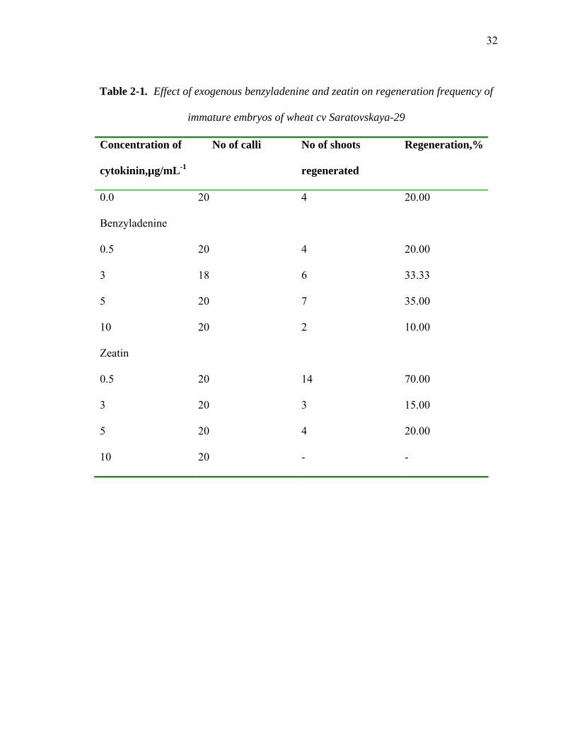
Five levels of the cytokinins benzyladenine (BA) and zeatin were tested for
effectiveness in shoot initiation, and the concentrations of: 0 µg/mL-1, 0.5µg/mL-1, 3
µg/mL-1, 5 µg/mL-1 and 10 µg/mL-1 were tested. Immature embryos were precultured on
MS medium containing 2, 4-D (2 µg/mL-1) for 15-17 days to obtain viable morphogenic

31
callus, and then transferred to MS media with given concentrations of cytokinin.
Results of the experiment are shown in Table 2-1.
Test showed that MS medium supplemented with 0.5 µg/mL-1 zeatin was optimal for
shoot initiation. In addition, MS medium containing 0.5 µg/mL-1 zeatin was chosen as a
medium for shoot initiation in all transformation experiments. MS with half strength
salts and vitamins without any selection (MS/2) was used for shoot elongation for about
a week, followed by MS/2 medium containing 5 µg/mL-1bialaphos in biolistic
bombardment procedure, and by MS/2 containing 50 µg/mL-1 hygromycin in
Agrobacterium mediated transformation procedure. Data on total recovery of plants from
immature and mature embryos is summarized in Table 2-2. Seven plants in total were
recovered in particle bombardment procedure, four of these originated from immature
embryos, and three-from mature embryos (Table 2-2). In Agrobacterium-mediated
transformation procedure, recovery was achieved with 17 plants, 6 of which originated
from immature embryos and 11-from mature embryos (Table 2-2). Protocol for the
obtainment of plants of cv Saratovskaya-29 following Agrobacterium-mediated and
particle bombardment procedures is shown in Figure 2-3.
32
Table 2-1. Effect of exogenous benzyladenine and zeatin on regeneration frequency of
immature embryos of wheat cv Saratovskaya-29
Concentration of
cytokinin,µg/mL-1
No of calli No of shoots
regenerated
Regeneration,%
0.0 20 4 20.00
Benzyladenine
0.5 20 4 20.00
3 18 6 33.33
5 20 7 35.00
10 20 2 10.00
Zeatin
0.5 20 14 70.00
3 20 3 15.00
5 20 4 20.00
10 20 - -

33
Table 2-2. Total number of plants recovered from Agrobacterium and biolistic
transformation procedures
Procedure Total No. of embryos
No. of plants recovered
Rate: plant recovery
Agrobacterium- mediated
transformation
Mature embryos
452
11
2.43 %
Immature embryos
622
6
0.96 %
Total Agrobacterium
1074
17
1.58 %
Particle
Bombardment
Mature embryos
460
3
0.65 %
Immature embryos
526
4
0.76 %
Total particle bombardment
986
7
0.70 %

34
Figure 2-3. Culture schedule for wheat cultivar Saratovskaya-29.
Agrobacterium-mediated procedure Microprojectile bombardment procedure
Procedure Time Medium Procedure Time Medium Callus
induction 3 to 5
days in the dark
MS+2µg/mL-1 2,4-D, 500µg/mL-1 glutamine,
100 µg/mL-1casein hydrolysate (MS+
medium)
Callus induction
3 to 5 days in the dark
MS+2µg/mL-1 2,4-D,
500µg/mL-1 glutamine, 100 µg/mL-1casein
hydrolysate (MS+ medium)
Inoculation
2-3 days in the dark
Pick a single colony from bacterial plate. Suspend in 2 ml of
liquid LB+50µg/mL hygromycin. Incubate on a shaker for 16 h.
Add resulting bacterial suspension into PIM
medium+acetosyringone at 2µl/mL-1. Incubate on a shaker for 10-12h. Add acetosyringone at
2µl/mL-1before inoculation.
MS with half strength
salts and vitamins (MS/2)+3% sucrose
Bombardment 4 hours before
bombardment and 18 hours
after
MS high osmotic medium
(MS+0.2M mannitol, 0.2M
sorbitol)
Standard 1100 psi microcarrier
discs used
Reculture 7 days MS+2µg/mL-12,4D, 250 µg/mL-1Clavamox
Reculture 7 days MS+2µg/mL-
12,4-D Reculture 2 cycles
3-4 weeks each
MS+2µg/mL-12,4-D, 250µg/mL-1Clavamox, 50µg/mL-1hygromycin
Reculture 2 cycles 3-4 weeks
each
MS+2µg/mL-
12,4-D, 4µg/mL-1
bialaphos
Regeneration 1-2 weeks
MS+0.5µg/mL-1zeatin, 250µg/mL-1Clavamox
Regeneration 1-2 weeks MS+0.5µg/mL-
1zeatin Transfer of regenerated
plants to Jiffy peat pellets
5-7 days Transfer of regenerated
plants to Jiffy peat pellets
5-7 days
Transfer of regenerated
(R0) plants to soil
6-8 weeks
Transfer of regenerated
(R0) plants to soil
6-8 weeks

35
GUS Assays
Comparative efficiency of two Agrobacterium preinoculation procedures of
Ratnayaka (Ratnayaka, 1999) and Lichtenstein and Draper (1985) was tested and
evaluated in terms of GUS expression. Efficiency was tested by exposing transformed
calli pieces to X-gluc treatment 48 h after cocultivation (Table 2-3). All inoculated calli
were collected and exposed to X-gluc treatment. The experiment was repeated two times
(2X) for both immature and mature embryo-derived calli. Total number of calli
containing blue spots with X-gluc was highest for the Lichtenstein and Draper
preinoculation procedure for both immature and mature embryo-derived calli. Procedure
of Lichtenstein and Draper (1985) was used for growing LBA4404 (pTOK233) culture
in all subsequent Agrobacterium-mediated transformation experiments.
Overall results of GUS histochemical assay for immature and mature embryo-
derived calli are summed up in Table 2-4. Transient GUS expression was evaluated in
both Agrobacterium-mediated and biolistic transformation procedures 2 to 3 days after
transfer to callus induction media. A significant difference in rate of transient GUS
expression between immature and mature embryo-derived calli transformed with particle
bombardment was observed, with former being superior. In case of Agrobacterium-
mediated transformation, rate of transient GUS expression was similar, with immature
embryo-derived calli demonstrating higher rates of uniform expression of GUS (data not
shown). Examples of transient GUS expression in calli transformed by Agrobacterium
and particle bombardment are shown in Figures 2-4 and 2-5 respectively.

36
Table 2-3. Effect of two pre-inoculation procedures on transient expression of the
transferred GUS gene in wheat tissues after cocultivation with A.tumefaciens
LBA4404(pTOK233)
Experimenta Preinoculation procedure of
Ratnayaka (1999)
Preinoculation procedure of
Lichtenstein and Draper (1985)
Total GUS+ % Total GUS+ %
1 25 3 12 24 9 37.6
2 16 12 46.1 23 18 78.2
3 15 6 40 21 9 42.8
4 22 8 36.3 23 11 47.8
a Experiments No 1,2 were performed with mature embryo-derived calli
Experiments No 3,4 were performed with immature embryo derived calli
GUS+ =GUS-positive

37
Table 2-4. Transient GUS expression in calli transformed by either Agrobacterium or
microprojectile bombardment
Experime
nt No.a
Microprojectile bombardment Agrobacterium-mediated
transformation
Total GUS+ % Total GUS+ %
1
114
25
21.9
135
62
45.9
2
158
80
50.6
136
61
44.8
a experiment No 1 was performed with mature embryo-derived calli
experiment No 2 was performed with immature embryo-derived calli

38
A
Figure 2-4. Transient GUS expression in calli inoculated with Agrobacterium a
assayed with X-Gluc. A, An example of a pattern of GUS spots. B, An example
uniform GUS expression. GUS spots, as well as uniform GUS expression, were
localized on the scutellum surface of the inoculated calli.
B
nd
of a
mostly

39
A B
Figure 2-5. Transient GUS expression in calli transformed via particle bombardment
and assayed with X-Gluc. A, An example of a pattern of GUS spots. B, An example of a
uniform GUS expression.

40
PCR Screening
All regenerated plants were screened for the presence of GUS (UidA), Npt, Hpt or
Bar genes using PCR amplification of fragments in the transferred genes. Seventeen
plants were regenerated from Agrobacterium inoculation. Seven of the 17 plants were
PCR positive for the one or more of the transformed genes. Seven plants were
regenerated from the gene gun bombardment procedure. Four of the 7 plants were PCR
positive for the transferred UidA gene. Examples of the PCR amplification reactions are
shown in Figures 2-6, 2-7 (UidA), Figures 2-8, 2-9 (NptII), Figures 2-10, 2-11 (Hpt),
Figure 2-12 (UidA for gene gun bombarded plants). Results of PCR amplifications and
PCR-based Southern blots for Agrobacterium and particle bombardment procedures are
shown in Tables 2-5 and 2-6 respectively.
Plant Identification System
For Agrobacterium procedure, inoculated plants derived from mature embryos were
labeled “S” (seeds), and plants derived from immature embryos were labeled “Em”
(embryos). In PCR reactions, an additional number appears in parentheses to identify
individual shoots from multiple shoots derived from the callus (e.g. S6 (2) is a DNA
sample extracted from the second shoot of the mature embryo-derived plant No. 6). For
plants subjected to biolistic bombardment, those derived from mature embryos were
labeled Gm (gun mature), and plants derived from immature embryos were labeled Gi
(gun immature).

41
* * * *
M + S1 S2 S3 S4 S5 S6
← 1.6 kb + S6 S7 S7 S8 S8 - (2) (2) * * * * * Figure 2-6. An example of PCR amplified bands of UidA gene in wheat plants (R0)
regenerated following Agrobacterium inoculation. A positive (+) control (transforming
pTOK233 plasmid), a negative (-) control (non-transgenic wild type plant DNA of cv
Saratovskaya-29), molecular weight markers (M) (Promega Lambda DNA markers, 21.2
kb ladder), and the expected UidA PCR product (1.6 kb) are indicated. DNA samples
used for the PCR reaction were taken from plants regenerated from mature embryos (S1-
8). * positive plants exhibiting bands of expected size.

42
M + - Em2Em3 S9 S8 S3
~ 1.6 kb →
Figure 2-7. An example of PCR amplified bands of UidA gene in wheat plants (R0)
regenerated following Agrobacterium inoculation with subsequent probing for the UidA
gene. A positive (+) control (transforming pTOK233 plasmid), a negative (-) control
(non-transgenic wild type plant DNA of cv Saratovskaya-29), molecular weight markers
(M) (Promega Lambda DNA markers, 21.2 kb ladder), and the expected UidA PCR
product (~1.6 kb) are indicated.

43
* * *
M + - S4 S1 S3 S8
← ~ 593 bp
+ - S9 Em2 Em3
* * Figure 2-8. An example of PCR amplified bands of NptII gene in wheat plants (R0) regenerated following Agrobacterium inoculation. A positive (+) control (pART27), a negative (-) control (non-transgenic plant DNA of cv Saratovskaya-29), molecular weight markers (M) (Promega PCR markers 1-kb ladder), and the expected PCR NptII product (~593 bp) are indicated. DNA samples used for PCR reaction were taken from plants regenerated from mature embryos (S1, S3, S4, S8 and S9) and immature embryos (Em2, Em3). * positive plants exhibiting bands of expected size.

44
+ - Em2Em3S9 S8 S3 M ~ 593 bp → Figure 2-9. An example of PCR amplified bands of NptII gene in wheat plants (R0) regenerated following Agrobacterium inoculation with subsequent probing for the NptII gene. A positive control (+) (transforming pTOK233 plasmid), a negative control (-) (non-transgenic DNA of cv Saratovskaya-29), molecular weight markers (Promega PCR markers, 1 kb ladder), and the expected PCR NptII product (~593 bp) are indicated.

45
* * * *
M + S1 S2 S3 S8 S9 - ~ 576 bp → M + Em1/ 2 / 3 / 4 / 5 / - Figure 2-10. An example of PCR amplified bands of Hpt gene in wheat plants (R0) regenerated following Agrobacterium inoculation. A positive (+) control (transforming
pTOK233 plasmid), a negative control (non-transgenic wild type DNA of cv
Saratovskaya-29), molecular weight markers (M) (Promega PCR markers, 1-kb ladder)
and the expected PCR Hpt product (~ 576 bp) are indicated. DNA samples used for PCR
reaction were taken from plants regenerated from mature embryos (S1, S2, S3, S8 and
S9) and immature embryos (Em1, Em2, Em3, Em4 and Em5). * positive plants
exhibiting bands of expected size.
46
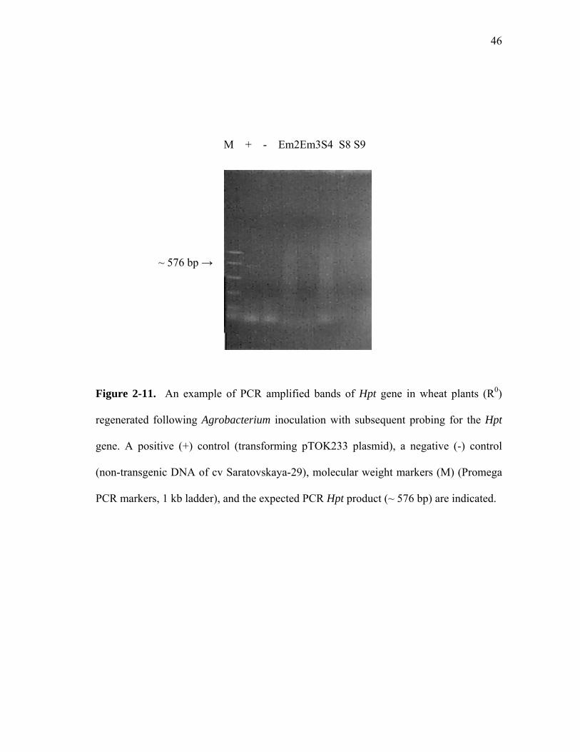
M + - Em2Em3S4 S8 S9
~ 576 bp →
Figure 2-11. An example of PCR amplified bands of Hpt gene in wheat plants (R0)
regenerated following Agrobacterium inoculation with subsequent probing for the Hpt
gene. A positive (+) control (transforming pTOK233 plasmid), a negative (-) control
(non-transgenic DNA of cv Saratovskaya-29), molecular weight markers (M) (Promega
PCR markers, 1 kb ladder), and the expected PCR Hpt product (~ 576 bp) are indicated.

47
* * * * + Gi - GmGmGiGi M 1 1 2 2 3 ~ 1.6 kb → Figure 2-12. An example of PCR amplified bands of UidA gene in regenerated (R0) wheat plants from callus submitted to microprojectile bombardment. A positive (+)
control (transforming pAHC25 plasmid), a negative (-) control (non-transformed wild
type DNA of the cv Saratovskaya-29), molecular weight markers (M) (Promega Lambda
DNA markers, 21.1 kb ladder) and the expected PCR UidA product (~ 1.6 kb) are
indicated. DNA samples for PCR reaction were taken from plants regenerated from
immature embryos (Gi 1-3) and mature embryos (Gm1,Gm2). * positive plants
exhibiting bands of expected size.

48
Table 2-5. PCR amplification and PCR-based Southern blot of primary
regenerated (Ro) plants derived from callus inoculated with Agrobacterium
Plant
Number Hpt gene PCR
Hpt gene Hybridization
NptII gene PCR
NptII gene Hybridization
UidA gene PCR
UidA gene Hybridization
S1 + + + + S2 - - - S3 - + + S4 - - - - - S5 - - - S6 - - + S7 - - + S8 + + + + + + S9 - + + + + + S10 - - - S11 - - - Em1 - - - Em2 - - - - + + Em3 - - + + - - Em4 - - - Em5 - - - + = PCR and/or Southern blot positive plants - = plants that did not produce bands in PCR amplification reaction and/or PCR-based Southern blot

49
Table 2-6. PCR amplification and PCR-based Southern blot of primary regenerated
(R0) plants transformed via particle bombardment Plant number UidA PCR UidA
Hybridization Bar PCR Bar
Hybridization Gi1 + + - - Gi2 + + - - Gi3 + + - - Gi4 - - - Gm1 - - - - Gm2 + + - - Gm3 - - -
+ = PCR positive plants - = plants that did not produce bands in PCR amplifications and/or PCR-based Southern blot

50
Molecular Analysis of Regenerated Plants Five plants regenerated following Agrobacterium inoculation and five plants
regenerated following microprojectile bombardment were subjected to the PCR
amplification of the transferred genes and Southern blotting of PCR amplified
fragments. PCR amplified UidA gene fragment from plasmid pTOK233 was used as a
probe for the UidA gene in PCR amplified DNA of Agrobacterium-transformed plants,
and the PCR amplified UidA fragment from plasmid pAHC25 was used as a probe for
the UidA gene in PCR amplified DNA of microprojectile bombarded plants. DNA from
plasmid pG35BarB (Rathore et al., 1993), PCR amplified with Bar gene primers, was
used as a probe for the Bar gene in PCR amplified DNA of microprojectile bombarded
plants. DNA from plasmid pCAM1300, PCR amplified with Hpt primers, and DNA
from plasmid pART27 (Gleave, 1992), PCR amplified with NptII primers, was used as
probes for the Hpt and NptII genes respectively in Agrobacterium-transformed R0 plants.
Hpt Gene
Results of PCR-based Southern blot analysis of R0 regenerated plants for the
presence of hpt gene are shown in Figure 2-13. As it was noted, the hpt probe was PCR
amplified DNA from Hpt gene of plasmid pCAM1300. Non-transformed DNA from
wheat plants of cv Saratovskaya-29 was used as a negative control, and PCR amplified
DNA from plasmid pCAM1300 was used as a positive control. Plants S8 (lane 5) and S9
(lane 7), both mature embryo derived, showed fragments that hybridized to the probe.

51
* *
+ - Em2 Em3 S4 S8 S9
~ 576 bp → Figure 2-13. PCR-based Southern blot analysis of regenerated wheat plants (R0) probed for the Hpt gene. Positive control (+) (PCR amplified DNA of plasmid pCAM 1300),
negative control (-) (non-transgenic DNA of cv Saratovskaya-29), and the amplified Hpt
fragment of the expected size (~576 bp)
performed on Hpt gene PCR gel shown i
presence of the Hpt gene.
are indicated. Southern blot analysis was
n Fig. 2-11. * positive plants exhibiting

52
NptII Gene
Results of PCR-based Southern blot analysis of R0 regenerated wheat plants for
the presence of the NptII gene are shown in figure 2-14. The probe for the NptII gene
was prepared using PCR amplified product from the plasmid pART27, which was
purified using MinElute PCR Purification Kit Protocol (MinElute Handbook, QIAGEN,
2001) and then used for the preparation of the radioactive probe. Non-transformed DNA
of cv Saratovskaya-29 was used as a negative control, and PCR amplified DNA of the
plasmid pART27 was used as a positive control. Fragments of the DNA from plants
Em2 (lane 3, immature embryo derived plant) and S9 (lane 5, mature embryo derived
plant) hybridized to the probe.
UidA Gene
Figure 2-15 shows the results of the PCR-based blot analysis of Agrobacterium
transformed plants for the presence of UidA gene. The probe for the UidA gene was
prepared using PCR amplified product from the plasmid pTOK233 which was further
purified using MinElute PCR Purification Kit Protocol. Non-transformed DNA of cv
Saratovskaya-29 was used as a negative control, and PCR amplified DNA of the plasmid
pTOK233 was used as a positive control. Fragments of the DNA from plants Em2 (lane
4, immature embryo derived plant), S9 (lane 6, mature embryo derived plant), and S8
(lane 7, mature embryo derived plant) hybridized to the probe. Plant Em2 shows faint
band in comparison with other UidA positive plants and positive control, suggesting
insertion of fewer copies of the gene during the transformation process.

53
UidA Gene ( Microprojectile Bombardment Procedure)
Fig. 2-16 shows results of the PCR-based Southern blot of the regenerated (R0)
plants transformed via microprojectile bombardment and probed for the UidA gene. The
probe for the UidA gene was prepared using PCR amplified product from the plasmid
pAHC25 further purified using MinElute PCR Purification Kit Protocol. Non-
transformed DNA of cv Saratovskaya-29 was used as a negative control, and PCR
amplified DNA of the plasmid pAHC 25 was used as a positive control. Fragments of
the DNA from the plants Gi1 (lane 2, immature embryo derived plant), Gm2 (lane 5,
mature embryo derived plant), Gi2 (lane 6, immature embryo derived plant), and Gi3
(lane 7, immature embryo derived plant) hybridized to the probe.
It should be noted that no hybridization to the Bar probe was obtained during
the PCR-based Southern blot of the regenerated (R0) plants transformed via particle
bombardment, except a single case. The probe for the Bar gene was prepared using PCR
amplified product from pG35barB plasmid (Rathore et al., 1993), supplied courtesy of
Dr. Rathore.

54
* *
+ -Em2Em3S8S9S3
~ 576 bp →
Figure 2-14. PCR-based Southern blot analysis of regenerated wheat plants (R0)
probed for the NptII gene. Positive control (+) (PCR amplified DNA of plasmid
pART27), negative control (non-transgenic DNA of cv Saratovskaya-29), and the NptII
amplified fragment of expected size (~ 576 bp) are indicated. Southern blot analysis was
performed on NptII gene PCR gel shown in Fig. 2-9. * positive plants exhibiting
presence of the NptII gene.

55
* * *
+ - Em2 Em3 S9 S8 S3
~ 1.6 kb →
Figure 2-15. PCR-based Southern blot analysis of regenerated wheat plants (R0) probed for the UidA gene. Positive control (+) ( PCR amplified DNA of plasmid pTOK233), negative control (non-transgenic DNA of cv Saratovskaya-29), and UidA amplified fragment of expected size (~1.6 kb) are indicated. Southern blot analysis was performed on UidA gene PCR gel shown in Fig. 2-7. * positive plants exhibiting presence of the UidA gene.

56
* * * * + Gi1 -Gm1Gm2Gi2Gi3 ~ 1.6 kb → Figure 2-16. PCR-based Southern blot analysis of regenerated wheat plants (R0) transformed via particle bombardment and probed for UidA gene. Positive control (+) (PCR amplified DNA of plasmid pAHC25), negative control (-) (non-transgenic DNA of cv Saratovskaya-29), and the UidA amplified fragment of expected size (~ 1.6 kb) are indicated. Southern blot analysis was performed on UidA gene PCR gel shown in Fig. 2-12. * positive plants exhibiting presence of the UidA gene.

57
R1 Generation To verify inheritance of the transferred genes, DNA from the developing shoots
and/or embryos of the R1 progeny of the 9 R0 plants was extracted, using urea-phenol
extraction method (Shure et al., 1983) and PCR amplified for the presence of the
transferred genes. Progeny of 5 mature embryo-derived R0 plants transformed with
Agrobacterium (S1, S2, S3, S6 and S9) 2 immature-embryo-derived R0 plants
transformed with Agrobacterium (Em2, Em3), and 2 R0 plants transformed with
microprojectile bombardment (Gi2, Gm2) was evaluated. Results of the PCR
amplification of the UidA, NptII and Hpt genes in the progeny of the plant S9 are shown
in Figures 2-17, 2-18, and 2-19 respectively. With 4 progeny tested, 3 were PCR positive
for the amplified UidA fragment of expected size (~ 1.6 kb), 4 were PCR positive for the
amplified NptII fragment, and 3 were PCR positive for the amplified Hpt fragment.
Results of the PCR-based Southern blot analysis of the progeny of plant S9 for the
presence of the UidA, NptII and Hpt genes are shown in Figures 2-20, 2-21, and 2-22
respectively. PCR-based Southern blot analysis of DNA of the 4 progeny of the S9 plant
showed progeny plants S9-1, S9-3 and S9-4 hybridizing to UidA probe, plants S9-1, S9-
2, S9-3, and S9-4 hybridizing to NptII probe, and plants S9-2, S9-3, and S9-4
hybridizing to Hpt probe. Results of these PCR-based Southern blots might suggest
possible inheritance of the transferred genes in R1 progeny of the S9 plant. Results of the
PCR amplification of the UidA, NptII and Hpt genes in the progeny of the plant Em2 are
shown in Figures 2-23, 2-24, and 2-25 respectively. With 10 progeny tested, 4 were

58
PCR positive for the amplified UidA gene fragment, 9 were PCR positive for the
amplified NptII gene fragment, and 8 were PCR positive for the amplified Hpt fragment.
Results of PCR amplification of the UidA and Bar genes in the progeny of the plant Gi2
are shown in Figures 2-26 and 2-27 respectively. With 8 progeny tested, 1 was PCR
positive for the amplified Bar gene fragment, and 7 were PCR positive for the amplified
UidA fragment.
Overall results of the PCR amplification of the transferred genes in the progeny of
the tested R0 plants transformed with Agrobacterium inoculation or microprojectile
bombardment are summed up in Tables 2-7 and 2-8 respectively. Although use of PCR-
based Southern blot procedure in the study does not allow suggesting number of gene
copies inserted as a result of transformation procedure applied, progeny of all 9 R0 plants
tested were PCR positive for one or more genes transferred, thus suggesting a possibility
that both genetic constructs were transferred and inherited. Total number of PCR
positive R0 plants with PCR positive R1 progeny is shown in Table 2-9.
Young shoots and roots of R1 progeny of R0 PCR positive plants were subjected to
X-gluc assay. No GUS expression was observed with material of all 7 plants tested,
whereas PCR amplification of the UidA gene was observed in R1 progeny of all these
plants. Absence of GUS expression in the R1 progeny might be explained by possible
silencing of the UidA gene. Absence of the GUS expression in R1 progeny of primary
transformed plants was observed before and several hypotheses explaining this event
were suggested (Demeke et al., 1999).

59
* * *
M + - S9-1S9-2S9-3S9-4
~ 1.6 kb →
Figure 2-17. PCR amplified bands of UidA gene in the R1 generation progeny of the wheat plant S9. Positive (+) control (transforming pTOK233 plasmid), negative (-) control (no DNA template), molecular weight markers (M) (Promega Lambda DNA markers, 21.1 kb-ladder) and the UidA amplified fragment of expected size (~1.6 kb) are indicated. * positive plants exhibiting bands of expected size.

60
* * * M + - S9-1S9-2S9-3S9-4 ←~ 593 bp Figure 2-18. PCR amplified bands of NptII gene in the R1 generation progeny of the wheat plant S9. Positive control (+) (transforming pTOK233 plasmid), negative control (-) (no DNA template), molecular weight markers (M)(Promega PCR markers, 1kb- ladder), and the amplified NptII band of expected size are indicated. * positive plants exhibiting bands of expected size.

61
* * * M + - S9-1S9-2S9-3S9-4 ← ~576 bp Figure 2-19. PCR amplified bands of Hpt gene in the R1 generation progeny of the wheat plant S9. Positive control (+) (transforming pTOK233 plasmid), negative control (-) (no DNA template), molecular weight markers (Promega PCR markers, 1-kb ladder),
and the amplified Hpt fragment of the expected size (~ 576 bp) are indicated. * positive
plants exhibiting bands of expected size.

62
* * * + - S9-1S9-2 S9-3S9-4
~ 1.6 kb →
Figure 2-20. PCR-based Southern blot analysis of R1 generation progeny of the wheat
plant S9 probed for the UidA gene. Positive control (+) (transforming plasmid
pTOK233), negative control (-) (no DNA template), and UidA amplified fragment of
expected size (~ 1.6 kb) are indicated. Southern blot analysis was performed on UidA
gene PCR gel shown in Fig. 2-17. * positive plants exhibiting presence of the UidA
gene.
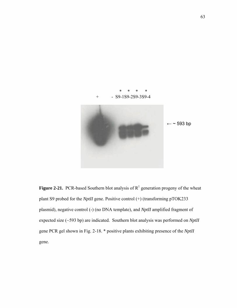
63
* * * * + - S9-1S9-2S9-3S9-4 ← ~ 593 bp
Figure 2-21. PCR-based Southern blot analysis of R1 generation progeny of the wheat
plant S9 probed for the NptII gene. Positive control (+) (transforming pTOK233
plasmid), negative control (-) (no DNA template), and NptII amplified fragment of
expected size (~593 bp) are indicated. Southern blot analysis was performed on NptII
gene PCR gel shown in Fig. 2-18. * positive plants exhibiting presence of the NptII
gene.

64
* * * + - S9-1 S9-2 S9-3 S9-4
← ~ 576 bp
Figure 2-22. PCR-based Southern blot analysis of R1 generation progeny of the wheat
plant S9 probed for the Hpt gene. Positive control (+) (transforming pTOK233 plasmid),
negative control (-) (no DNA template), and amplified Hpt fragment of expected size (~
576 bp) are indicated. Southern blot analysis was performed on Hpt gene PCR gel shown
in Fig. 2-19. * positive plants exhibiting presence of the Hpt gene.

65
* * * *
M + - Em2-1/- 2 /- 3 /- 4 / - 5 /– 6 / -7 / - 8 / -9 / -10
1.6kb→
Figure 2-23. PCR amplified bands of UidA gene in the R1 generation progeny of the
wheat plant Em2. Positive control (+) (transforming plasmid pTOK233), negative
control (-) (no DNA template), molecular weight markers (M) (Promega Lambda DNA
Markers, 21.1-kb ladder), and the UidA amplified band of expected size (~1.6 kb) are
indicated. * positive plants exhibiting bands of expected size.

66
* * * * * * * * *
M + - Em2-1/ -2 / -3 / -4 / -5 / -6 / -7 / -8 / - 9 / - 10 ~ 593 bp → Figure 2-24. PCR amplified bands of NptII gene in the R1 generation progeny of the
wheat plant Em2. Positive control (+) (transforming pTOK233 plasmid), negative
control (-) (no DNA template), molecular weight markers (M) (Promega PCR markers,
1-kb ladder), and NptII amplified fragment of the expected size (~ 593 bp) are indicated.
* positive plants exhibiting bands of expected size.

67
* * * * * * * *
M + - Em2-1/ -2 / -3 /-4 / -5/ - 6 / - 7 / - 8/ -9 / - 10
~ 576 bp →
Figure 2-25. PCR amplified bands of Hpt gene in R1 generation progeny of the wheat
plant Em2. Positive control (+) (transforming plasmid pTOK233), negative control (-)
(no DNA template), molecular weight markers (M) (Promega PCR Markers, 1-kb
ladder), and the Hpt amplified band of expected size (~ 576 bp) are indicated. * positive
plants exhibiting bands of expected size.

68
* * * *
M + - Gi2-1Gi2-2Gi2-3Gi2-4
~ 1.6 kb → M + - Gi2-5Gi2-6Gi2-7Gi2-8 * * * Figure 2-26. PCR amplified bands of UidA gene in R1 generation progeny of wheat
plant Gi2. Positive control (+) (transforming plasmid pAHC25), negative control (-) (no
DNA template), molecular weight markers (M) (Promega Lambda DNA Markers, 21.1
kb- ladder) and UidA amplified band of expected size (~ 1.6 kb) are indicated. * positive
plants exhibiting bands of expected size.

69
* M + - Gi2-1 Gi2-2 Gi2-3 Gi2-4
~ 393 bp → M + - Gi2-5 Gi2-6 Gi2-7 Gi2-8 Figure 2-27. PCR amplified bands of Bar gene in R1 generation progeny of the wheat
plant Gi2. Positive control (+) (PG35BarB plasmid), negative control (-) (no DNA
template), molecular weight markers (M) (Promega PCR markers, 1-kb ladder), and Bar
amplified band of the expected size are indicated. * positive plants exhibiting bands of
expected size.

70
Table 2-7. PCR amplification of R1 progeny of plants inoculated with Agrobacterium
No of the plant
progeny
UidA gene PCR NptII gene PCR Hpt gene PCR
S1-1 + + +
S1-2 - - -
S1-3 - - -
S1-4 + + -
S2-1 - + -
S2-2 - - -
S2-3 - - -
S2-4 - - +
S2-5 + + +
S3-1 + + -
S3-2 - + -
S3-3 + + +
S4-3 + + +
S6-1 + + -
S6-2 - - -
S6-3 - - +
S6-4 - - -
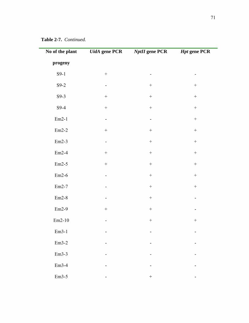
71
Table 2-7. Continued.
No of the plant
progeny
UidA gene PCR NptII gene PCR Hpt gene PCR
S9-1 + - -
S9-2 - + +
S9-3 + + +
S9-4 + + +
Em2-1 - - +
Em2-2 + + +
Em2-3 - + +
Em2-4 + + +
Em2-5 + + +
Em2-6 - + +
Em2-7 - + +
Em2-8 - + -
Em2-9 + + -
Em2-10 - + +
Em3-1 - - -
Em3-2 - - -
Em3-3 - - -
Em3-4 - - -
Em3-5 - + -

72
Table 2-7. Continued.
No of plant
progeny
UidA gene PCR NptII gene PCR Hpt gene PCR
Em3-6 - + -
Em3-7 - - -
Em3-8 + + -
Table 2-8. PCR amplification of R1 progeny of plants transformed with particle
bombardment
No of the plant progeny UidA gene PCR Bar gene PCR
Gi2-1 + +
Gi2-2 + -
Gi2-3 + -
Gi2-4 + -
Gi2-5 + -
Gi2-6 + -
Gi2-7 - -

73
Table 2-8. Continued.
No of the plant progeny UidA gene PCR Bar gene PCR
Gi2-8 + -
Gm2-1 - -
Gm2-2 - -
Gm2-3 - -
Gm2-4 + -
Gm2-5 - -
Gm2-6 + -
Gm2-7 - -
Gm2-8 - -
Gm2-9 - -
Gm2-10 - -

74
Table 2-9. Total number of PCR positive R0 plants with PCR positive R1 progeny
Procedure
Number of
plantsa
R0
PCR+b
R0
PCR BlotbNumber of R0 with R1
PCR+b,c
Rate: PCR+ R0 with R1
PCR+a
Agrobacterium
Mature embryos Immature embryos
11
6
6
1
5
1
4 1
4/452=0.88%
1/622=0.16%
Particle Bombardment Mature embryos Immature embryos
3
4
1
3
1
3
1
3
1/460=0.22%
3/526=0.57%
a values from Table 2-2 b values from Table 2-5 and 2-6 c values from Table 2-7 and 2-8

75
DISCUSSION The principal goal of this study was to determine the applicability of two most
widely used transformation techniques to transfer foreign genes into the genome of an
elite Russian spring wheat cultivar Saratovskaya-29. As it was noted, this cultivar was
one of the most common parents of modern Kazakhstani spring wheat cultivars
(Martynov and Dobrotvorskaya, 1996). Results of the PCR amplification analysis of the
R0 original transformed plants and R1 progeny along with PCR-based Southern blot
analysis suggest that genomic integration and inheritance of the transferred genes might
have taken place with both Agrobacterium-mediated and particle bombardment
transformation procedures. Two sources of tissue explants were used in the study-
immature and mature embryo-derived calli. Putative transgenic R0 plants with probable
inheritance of one or more transferred genes by R1 generation were obtained from both
types of explants used in the study. Successful transformation of wheat using A.
tumefaciens was first reported by Cheng et al. (1997). Model cultivar “Bobwhite” was
used in this study, as well as in several other works (Weeks et al., 1993; Becker et al.,
1994; Hu et al., 2003). Problem of adjustment of well-established transformation
techniques for routine use with elite cultivars is of importance, in author’s opinion.
Therefore, in the study described, attempt was made to develop routine transformation
and regeneration protocol for elite cultivar allowing for transferred genes to be inherited
stably and in predictable manner. Our study produced first transgenic progeny for the
cultivar Saratovskaya-29. Comparison of two pre-inoculation procedures performed in
the study allowed to discover that procedure by Lichtenstein and Draper (1985) gives
76
higher rates of UidA gene transient expression, which might have been a result of
increased virulence of the A.tumefaciens strain LBA4404, used in the study.
Regeneration protocol that allowed producing fertile plants from transformed tissue
explants was developed, with plant recovery reaching 1.58% for Agrobacterium-
mediated procedure, and 0.7% for particle bombardment procedure. All obtained plants
were fertile and produced viable seeds. It should be noted that length of culture time was
increased in comparison with the field growth, which might be explained by the use of
callus-based method, having additional dedifferentiation developmental step, and post-
transformational stress.
Molecular analysis of the R0 and R1 plants revealed no PCR amplification or
hybridization of the Bar gene, except a single case in R1 progeny of plant Gi2, whereas
PCR results for the presence of UidA gene were positive for both R0 and R1 generation
plants. But R0 plants, obtained in the particle bombardment procedure, were developing
on bialaphos-containing media, suggesting that Bar gene was transferred and was being
expressed. Most likely explanation for this observation might be the PCR artifact, caused
by non-optimal conditions of the PCR reaction that were used for the amplification of
the Bar gene, preventing efficient amplification of the Bar sequence. Also, additional
bands were observed in several cases along with the expected band of the transferred
gene. Suboptimal PCR conditions, such as improper annealing temperature, unsuitable
primers or excessive number of amplification cycles used might have caused appearance
of these additional bands as well. PCR reactions were performed for the detection of the

77
presence of transferred genes. Genomic Southern blot is required for answering the
question about the origin of these additional bands.
Kohil et al. (1999) discovered 19 bp palindromic sequence inside CaMV35S
promoter that might act as a recombination hotspot. In the study of Ratnayaka (1999),
loss of UidA gene was observed with plasmid pTOK233 used for Agrobacterium-
mediated transformation. Alignment of the amplified fragment of one of the R1 plants
with sequence of 35S promoter allowed to discover loss of the part of the 35S sequence
in the plant genomic DNA. Recombination that occurred between 35S promoter sites
was suggested as cause of the deletion of the part of 35S promoter sequence and
fragment containing UidA gene. Author suggests that the design of the vector
(pTOK233) with two duplicated 35S sites might have caused the loss of UidA gene. The
same reason might have caused loss of UidA gene in several progeny plants of Em2
plant (particularly, Em-3, Em-6, Em-7, Em-8 and Em-10, where NptII and Hpt genes
were amplified by PCR procedure). However, PCR amplification of the DNA of R1
progeny of several regenerated plants (particularly, S1, S2 and S6 progeny was used)
with CaMV35S forward and reverse primers revealed amplification of ~1.8 kb expected
fragment approximately including 35S promoter sites. This fragment was observed both
in pTOK233 plasmid and in R1 progeny plant DNA, thus suggesting that the 35S
promoter passed into R1 generation of Agrobacterium-transformed plants intact (data not
shown). Thus, deletion of a part of 35S sequence did not occur in the mature embryo-
derived plants tested.

78
Agrobacterium-mediated transformation had several advantages in the study
described. Regeneration of Agrobacterium-inoculated plants required less time than for
those transformed via particle bombardment, and grain yield was also higher in most
cases. Results of PCR amplification of DNA of R1 progeny of the original transgenic
plants, obtained via Agrobacterium-mediated transformation, for the presence of the
transferred genes suggests inheritance of these genes for all plants tested. Data obtained
suggest further use of Agrobacterium-mediated method for the transformation of the
cultivar Saratovskaya-29 as a main transformation method.

79
CHAPTER III
CONCLUSIONS
To our knowledge, this is the first demonstration of putative transformation of
cultivar Saratovskaya-29. One of the most widely used explant sources for crop plant
transformations is callus derived from somatic tissues. Our research suggests the
applicability of callus and regeneration of plants via organogenesis in the establishment
of a transformation method for the elite wheat cultivar Saratovskaya-29, supporting
previous reports on successful transformation of other wheat varieties using callus as an
explant source (Vasil et al., 1992; Altpeter et al., 1996; Cheng et al., 1997; Weir et al.,
2001; Khanna and Daggard, 2003). Use of the pTOK233 plasmid for the
Agrobacterium-mediated transformation of wheat demonstrated that this genetic
construct, successfully used earlier for the Agrobacterium-mediated transformation of
rice (Hiei et al., 1994; Ratnayaka et al., 1999), could be stably transformed into wheat
genome as well. Amplification of the UidA gene was especially evident in both R0 and
R1 generations of transformants, though several cases of absence of amplification of
UidA gene were observed. Earlier cases of loss of UidA gene in R1 and R2 progeny of
rice transformed with pTOK233 plasmid due to homologous recombination between two
CaMV35S promoter sites (Ratnayaka, 1999) weren’t observed in our study, and
presence of 35S promoter fragments was observed in amplification reactions of R1
progeny of original transformants.
80
Sterility is a frequently observed problem in plants regenerated from callus; however
in this study, all plants recovered from both transformations procedures were fertile and
produced viable progeny, although grain yield was lower in comparison with wild type
plants (data not shown). Phenotypic abnormalities observed with the original
transformants suggest that somaclonal variations might have taken place during the
prolonged tissue culture stage of the development of transferred explants. It might be
important to develop regeneration protocol allowing decrease in the longevity of tissue
culture step with a view to avoid increase in somaclonal variation.
Our study has shown a slight superiority of an Agrobacterium-mediated
transformation method in terms of overall plant recovery ratio and the number of PCR-
positive plants obtained. Genomic blot analyses of this progeny generation are needed
to determine if the transferred genes, detected in the DNA by PCR, have actually been
incorporated into chromosomal DNA.
This approach to transformation might be suggested for further use with the cultivar
studied, to transfer beneficial genes to this cultivar. Specifically, the Saratovskaya-29
cultivar suffers from susceptibility to fungal diseases, such as leaf rust (Puccinia
recondita f. ssp. triticii) and mildew. Introduction of fungal resistance gene(s) might aid
this cultivar in terms of grain yield and resistance. In a recent study, cDNAs encoding
antifungal protein Ag-AFP (barley class II chitinase) from Aspergillus giganteus were
shown to be introduced and expressed in wheat, causing significant reduction in
formation of leaf rust and powdery mildew (Oldach et al., 2001).

81
Both immature and mature embryo-derived calli might be used for transformation,
since in our study putative transgenic plants were regenerated from both types of explant
source. Also, use of shoot apices as an explant source might be suggested, since in this
case time span for the tissue culture step would be significantly decreased, and the
inoculation process might be more effective. Unlimited dedifferentiation ability of
meristematic cells makes them an appropriate target for the plant transformation;
meristematic cells were already used successfully for the transformation of such major
crop plants as maize (Gould et al., 1991) and rice (Chan et al., 1993). Transformation of
the cultivar Saratovskaya-29 using calli and other explants such as the shoot apex (Smith
et al., 1992) or floral dip (Dong et al., 2001) might be an important additional step in
discovering most efficient method of transformation for the described cultivar.

82
LITERATURE CITED
Aldemita RR, Hodges TK (1996) Agrobacterium tumefaciens-mediated transformation
of japonica and indica rice varieties. Planta 199: 612-617
Altpeter F, Vasil V, Srivastava V, Stoger E, Vasil IK (1996) Accelerated production
of transgenic wheat (Triticum aestivum L.) plants. Plant Cell Rep 16: 12-17
Altpeter F, Diaz I, McAuslane H, Gaddour K, Carbonero P, Vasil IK (1999)
Increased insect resistance in transgenic wheat stably expressing trypsin inhibitor
CMe. Mol Breeding 5: 53-63
Amoah BK, Wu H, Sparks C, Jones HD (2001) Factors influencing Agrobacterium-
mediated transient expression of uidA in wheat inflorescence tissue. J Exp Bot 52:
1135-1142
Becker D, Brettshneider R, Lorz H (1994) Fertile transgenic wheat from
microprojectile bombardment of the scutellum tissue. Plant J 5: 299-301
Bidney D, Scelonge C, Martich J, Burrus M, Sims L, Huffman G (1992)
Microprojectile bombardment of plant tissues increases transformation frequency by
Agrobacterium tumefaciens. Plant Mol Biol 18: 301-313
Birch RG (1997) Plant transformation: problems and strategies for practical application.
Annual Rev of Pl Physiol and Pl Mol Biol 48: 297-326
Blechl AE, Anderson OD (1996) Expression of a novel high-molecular-weight glutenin
subunit in transgenic wheat. Nature Biotech 14: 875-879

83
Bliffeld M, Mundy J, Potrykus I, Futterer J (1999) Genetic engineering of wheat or
increased resistance to powdery mildew disease. Theor and Appl Genet 98: 1079-
1086
Chair H, Legavre T, Guiderdoni E (1996) Transformation of haploid, microspore-
derived cell suspension protoplasts of rice (Oryza sativa L.) Plant Cell Rep 10: 77-
770
Chan MT, Chang HH, Ho SL, Tong WF, Yu SM (1993) Agrobacterium mediated
production of transgenic rice plants expressing a chimeric ß-amylase promoter/-
glucuronidase gene. Plant Mol Biol 22: 491-506
Chen WP, Gu X, Liang GH, Muthukrishnan S, Chen PD, Liu DJ, Gill BS (1998)
Introduction and constitutive expression of a rice chitinase gene in bread wheat using
biolistic bombardment and the bar gene as a selectable marker. Theor and Appl
Genet 97: 1296-1306
Cheng M, Fry JE, Pang S, Zhou H, Hironaka CM, Duncan D, Conner TW, Wan Y
(1997) Genetic transformation of wheat mediated by Agrobacterium tumefaciens.
Plant Physiol 115: 971-980
Christensen AH, Quail PH (1996) Ubiquitin promoter-based vectors for high-level
expression of selectable and/or screenable marker genes in monocotyledonous
plants. Transgenic Res 5: 213-218
Dai S, Zheng P, Marmey P, Zhang S, Tian W, Chen S, Beachy R, Fauquet C (2001)
Comparative analysis of transgenic rice plants obtained by Agrobacterium-mediated
transformation and particle bombardment. Mol Breeding 7: 25-33

84
Daud HM, Gustafson JP (1996) Molecular evidence for Triticum speltoides as a B-
genome progenitor of wheat (Triticum aestivum). Genome 39: 543-548
Delbreil BP, Guerche P, Jullien M (1993) Agrobacterium-mediated transformation of
Asparagus officinalis L. long-term embryogenic callus and regeneration of
transgenic plants. Plant Cell Rep 12: 129-132
Demeke T, Hucl P, Baga M, Caswell K, Leung N, Chibbar RN (1999) Transgenic
inheritance and silencing in hexaploid wheat. Theor Appl Genet 99: 947-953
Deng W-Y, Lin X-Y, Shao Q-Q (1990) Agrobacterium tumefaciens can transform
Triticum aestivum and Hordeum vulgare of gramineae. Sci in China (Series B) 33:
27-33
DiMaio JJ, Shillito RD (1989) Cryopreservation technology for plant cell cultures.
Journal of Tissue Culture Methods 12: 163-169
Dong J, Kharb P, Teng W, Hall TC (2001) Characterization of rice transformation via
an Agrobacterium-mediated inflorescence approach. Mol Breeding 7: 187-194
Dvorak J, Luo MC, Yang ZL, Zhang HB (1998) The structure of Aegilops tauschii
gene pool and the evolution of hexaploid wheat. Theor Appl Genet 97: 654-670
Farrand SK, van Berkum PB, Oger P (2003) Agrobacterium is definable genus of the
family Rhizobiaceae. Int J Syst Evol Microbiol 53: 1681-1689
Feldman M, Lupton FGH, Miller TE (1995) Wheats. In J Simmonds, NW Smartt, eds,
Evolution of Crop Plants. Longman Scientific & Technical, New York, pp 184-192
Finer JJ, Finer KR, Ponappa T (1999) Particle bombardment mediated transformation.
Plant Biotech 240: 59-80

85
Fromm ME, Taylor LP, Walbot V (1986) Stable transformation of maize after gene
transfer by electroporation. Nature 319: 791-793
Gleave AP (1992) A versatile binary vector system with a T-DNA organizational
structure conductive to efficient integration of cloned DNA into the plant genome.
Plant Mol Biol 20: 1203-1207
Golovkin MV, Abraham M, Morosz S, Bottka S, Feher A, Dudits P (1993)
Production of transgenic maize plants by direct DNA uptake into embryogenic
protoplasts. Plant Sci Limerick 90: 41-52
Gould JH, Smith RH (1989) Transformation systems for corn. 44th Corn & Sorghum
Res Conf, Am Seed Trade Assoc, New York, ppl-10
Gould JH, Devey ME, Hasegawa O, Ulian EC, Peterson G, Smith RH (1991)
Transformation of Zea mays L. using Agrobacterium tumefaciens and the shoot apex.
Plant Physiol 95: 426-434
Gould JH (1997) Transformation of the cereals using Agrobacterium. In Molecular
Biology, Vol 62. Recombinant Gene Expression Protocols. R. Tuan Humana Inc.,
Totowa, New Jersey, pp 491-501
Gould JH, Magallanes-Cedeno ME (1999) Adaptation of cotton shoot apex culture to
Agrobacterium-mediated transformation. Plant Mol Bio Rep 16: 283
Graves ACF, Goldman S (1986) The transformation of Zea mays seedlings with
Agrobacterium tumefaciens. Plant Mol Biol 43: 50-56

86
Graves AE, Goldman S, Banks SW, Graves ACF (1988) Scanning electron
microscope studies of Agrobacterium tumefaciens attachment to Zea mays,
Gladiolus sp., and Triticum aestivum. J Bacteriol 169: 1745-1746
Gressel J (2000) Molecular biology of weed control. Transgenic Res 9: 355-382
Grimsley N, Hohn T, Davis J, Hohn B (1987) Agrobacterium mediated delivery of
infectious maize streak virus into maize plants. Nature 325: 177-179
Gurel F, Gozukirmisi N (2000) Optimization of gene transfer into barley (Hordeum
vulgare L.) mature embryos by tissue electroporation. Plant Cell Rep 19: 787-791
Hamilton CM, Frary A, Lewis C, Tanksley SD (1996) Stable transfer of intact high
molecular weight DNA into plant chromosomes. Proc Natl Acad Sci 93: 9975-9979
Hansen G, Wright MS (1999) Recent advances in the transformation of plants. Trends
in Pl Sci 4: 226-231
Hernalsteens J-P, Thia-Toong L, Schell J, van Montagu M (1984) An
Agrobacterium-transformed cell culture from the monocot Asparagus officinalis.
EMBO J 13: 3039-3041
Hess D, Dressler K, Nimmrichter R (1990) Transformation experiments by pipetting
Agrobacterium into the spikelets of wheat (Triticum aestivum L.) Plant Sci 72: 233-
244
Hiei Y, Ohta S, Komari T, Kumashiro T (1994) Efficient transformation of rice
(Oryza sativa L.) mediated by Agrobacterium and sequence analysis of the
boundaries of the T-DNA. The Plant J 6: 271-282

87
Hu T, Metz S, Chay C, Zhou HP, Biest N, Chen G, Cheng M, Feng X, Radionenko
M, Lu F (2003) Agrobacterium-mediated large-scale transformation of wheat
(Triticum aestivum L.) using glyphosate selection. Plant Cell Rep 21: 1010-1019
Ishida Y, Saito H, Ohta S, Hiei Y, Komari T, Kumashiro T (1996) High-efficiency
transformation of maize (Zea mays L.) mediated by Agrobacterium tumefaciens. Nat
Biotech 14: 745-750
Jefferson RA (1987) Assaying chimeric genes in plants: the gusA gene fusion system.
Plant Mol Biol Rep 5: 387-405
Kant T, Kothari SL, Kononowicz-Hodges H, Hodges TK (2001) Agrobacterium
tumefaciens-mediated transformation of rice using coleoptile and mature seed-
derived callus. J of Pl Biochem and Biotech 10: 121-126
Karp A (1991) On the current understanding of somaclonal variation. Oxford Surveys
of Pl Mol and Cell Biol 7: 1-58
Karunaratne S, Sohn A, Mouradov A, Scott J, Steinbiss H-H, Scott KJ (1996)
Transformation of wheat with the gene encoding the coat protein of barley yellow
mosaic virus. Australian J of Pl Physiol 23: 429-435
Khanna HK, Daggard GE (2003) Agrobacterium tumefaciens-mediated transformation
of wheat using a super binary vector and a polyamine-supplemented regeneration
medium. Plant Cell Rep 21: 429-436
Kohil A, Griffiths S, Palacios N, Twyman RM, Vain P, Laurie DA, Christou P
(1999) Molecular characterization of transforming plasmid rearrangements in
rice reveals a recombination hotspot in the CaMV35S promoter and confirms the

88
predominance of microhomology mediated recombination. The Plant J 17: 591-
601
Komari T (1990b) Transformation of cultured cells of Chenopodium quinoa by binary
vectors that carry a fragment of DNA from the virulence region of pTiBo542. Plant
Cell Rep 9: 303-306
Kopbayev AA, Mukhambetzhanov KK, Mukhambetzhanov SK (2000) Regeneration
of wheat plants by unfertilized ovary culture. Biotech Theory and Practice 1-2: 160-
161
Leckband G, Lorz H (1998) Transformation and expression of a stilbene synthase gene
of Vitis vinifera L. in barley and wheat for increased fungal resistance. Theor Appl
Genet 96: 1004-1012
Lichtenstein CP, Draper J (1985) Genetic engineering of plants. In DM Glover, ed,
DNA Cloning, Vol 2. IRL Press, London, UK, pp 108-110
Manthre DE, Johnston RH, Martin JM (1985) Sources of resistance to
Cephalosporum gramineum in Triticum and Agropyron species. Euphitica 34: 419-
424
Martynov SP, Dobrotvorskaya TV (1996) The most common parents of Russian
spring wheat released from 1980 to 1996. The Russian Acad of Agric Sci. AWN Vol
43, http://wheat.pw.usda.gov/ggpages/awn/43/awn43c2.html
Merezhko AF (1998) Impact of plant genetic resources on wheat breeding. Euphitica
100: 295-303

89
Ming XT, Yuan HY, Wang LJ, Chen ZL (2001) Agrobacterium-mediated
transformation of rice with help of bombardment. Acta Bot Sin 43: 72-76
Morris R, Sears ER (1967) The cytogenetics of wheat and its relatives. In KS
Quisenberry, LP Reitz, eds, Wheat and Wheat Improvement, No13. Amer Soc of
Agronomy Inc Publisher, Madison, Wisconsin, pp 19-87
Muller E, Lorz H, Lutticke S (1996) Variability of transgene expression in clonal cell
lines of wheat. Plant Sci 114: 71-82
Murashige T, Skoog F (1962) A revised medium for rapid growth and bioassays with
tobacco tissue cultures. Plant Physiol 15: 473-497
Nehra NS, Chibbar RN, Leung N, Caswell K, Mallard C, Steinhauer L, Baga M,
Kartha KK (1994) Self-fertile transgenic wheat plants regenerated from isolated
scutellar tissues following microprojectile bombardment with two distinct gene
constructs. Plant J 5: 285-297
Oldach KH, Becker D, Lorz H (2001) Heterologous expression of genes mediating
enhanced fungal resistance in transgenic wheat. Mol Plant Microbe Interact 14: 832-
838
Potrykus I (1991) Gene transfer to plants: assessment of published approaches and
results. Annual Rev of Plant Physiol and Plant Mol Biol 42: 205-225
Raineri DM, Bottino P, Gordon MP, Nester EW (1990) Agrobacterium
transformation of rice (Oryza sativa L.) Bio/Technology 8: 33-38
Rakszegi M, Tamas S, Szucs P, Tamas L, Bedo Z (2001) Current status of wheat
transformation. Journal of Plant Biotechnology 3: 67-81

90
Rasco-Gaunt S, Riley A, Barcelo P, Lazzeri PA (1999) Analysis of particle
bombardment parameters to optimize DNA delivery into wheat tissues. Plant Cell
Rep 19: 118-127
Rathore KS, Chowdhury VK, Hodges TK (1993) Use of bar as a selectable marker
and for the production of herbicide-resistant rice plants from protoplasts. Plant Mol
Biol 21: 871-884
Ratnayaka IJ (1999) Agrobacterium-mediated gene transformation of rice: comparison
of callus and shoot apex derived plants. PhD dissertation. Texas A&M University,
College Station
Riley R, Chapman V (1958) Genetic control of the cytologically diploid behaviour of
hexaploid wheat. Nature 182: 713-715
Riley RG, Kimber G, Law CN (1967) Cytogenetics (recognition of intergenic
chromosomal homologies by interference with 5b system). In Annual Rep Plant
Breeding Institute, 1966-67. Plant Breeding Institute, Cambridge, UK, pp 105-116
Sambrook J, Fritch EF, Maniatis T (1989) Molecular Cloning: a Laboratory Manual.
Cold Spring Harbor Laboratory Press, Cold Spring Harbor, New York
Sanford JC (1988) A biolistic process-a new concept in gene transfer and biological
delivery. Trends Biotechnology 6: 229-302
Schafer W, Corz A, Kahl G (1987) T-DNA integration and expression in a monocot
crop plant after induction of Agrobacterium. Nature 327: 529-532
Sharma HC (1995) How wide can a wide cross be? Euphitica 82: 43-64

91
Shillito R (1999) Methods of genetic transformations: electroporation and polyethylene
glycol treatment. In IK Vasil, ed, Molecular Improvement of Cereal Crop. Kluwer
Academic Publishers, Dordrecht, Netherlands, pp 9-20
Shure MS, Wessler N, Fedoroff N (1983) Molecular identification of the waxy locus in
maize. Cell 35: 225-233
Simmonds NW (1976) Evolution of Crop Plants. Longman, London.
Smith RH, Gould JH, Uliah EC (1992) Transformation of plants via the shoot apex.
US patent No. 5,164,310.
Southern EM (1975) Detection of specific sequences among DNA fragments separated
by gel electrophoresis. J Mol Biol 98: 503-517
Tingay S, McElroy D, Kalla R, Fieg S, Wang M, Thornton S, Brettell R (1997)
Agrobacterium tumefaciens-mediated barley transformation. The Plant J 11: 1369-
1376
Usami S, Morikawa S, Takebe I, Machida Y (1987) Absence in monocotyledonous
plants of the diffusible plant factors inducing T-DNA circularization and vir gene
expression in Agrobacterium. Mol Gen Genet 209: 221-226
U.S. Department of Agriculture, National Agricultural Statistics Service (2003)
Small Grains 2003 Summary. http://usda.mannlib.cornell/edu/reports
Vacancy G, Schmidt R, Sanchez OA, Willmitzer L, Roca-Soza M (1990)
Construction of an intron containing marker gene: splicing of the intron in transgenic
plants and its use in monitoring early events in Agrobacterium-mediated
transformation. Mol Gen Genet 220: 245-250

92
Vasil IK, Vasil V (1999) Transformation of wheat via particle bombardment. In RD
Hall, ed, Plant Cell Culture Protocols, Vol 111. Humana Press, Totowa, New Jersey,
pp 349-358
Vasil V, Castillo AM, Fromm ME, Vasil IK (1992) Herbicide resistant fertile
transgenic plants obtained by microprojectile bombardment of regenerable
embryogenic callus. Bio/Technology 10: 1553-1558
Weeks JT, Anderson OD, Blechl A (1993) Rapid production of multiple independent
lines of fertile transgenic wheat (Triticum aestivum L.) Plant Physiol 102: 1077-1084
Weir B, Gu X, Wang MB, Upadhyaya N, Elliott AR, Brettell RIS (2001)
Agrobacterium tumefaciens-mediated transformation of wheat using suspension cells
as a model system and green fluorescent protein as a visual marker. Australian J of
Pl Physiol 28: 807-818
Wheat Letter (2003) Changing world wheat production patterns drive dramatic shifts in
trade. US Wheat Associates, http://www.uswheat.org/marketnews.ncf
Williams C, Ronald PC (1994) Rapid, homogenization-free isolation of rice DNA for
PCR. Nucl Acids Res 22: 1917-1918
Wright M, Dawson J, Dunder E, Suttie J, Reed J, Kramer C, Chang Y, Novitzky R,
Wang H, Artim-Moore L (2001) Efficient biolistic transformation of maize ( Zea
mays L.) and wheat (Triticum aestivum L.) using the phosphomannose isomerase
gene, pmi, as a selectable marker. Plant Cell Rep 20: 429-436
Wu H, Sparks C, Amoah B, Jones HD (2003) Factors influencing successful
Agobacterium-mediated genetic transformation of wheat. Plant Cell Rep 21: 659-668

93
Young JM, Kuykendall LD, Martinez-Romero E, Kerr A, Sawada H (2001) A
revision of Rhizobium Frank 1889, with an emended description of the genus, and
the inclusion of all species of Agrobacterium Conn 1942 and Allorhizobium undicola
de Lajudie et al.1998 as new combinations: Rhizobium radiobacter, R. rhizogenes, R.
tubi, R. undicola and R. vitis. Int J Syst Evol Microbiol 51: 89-103
Zemetra RS (1998) Potential for gene transfer between wheat ( Triticum aestivum) and
jointed goatgrass (Aegilops cylindrical). Weed Sci 46: 313-317
Zhang S, Rybszynski JJ, Landenberg WG, Mitra A, French R (2000) An efficient
wheat transformation procedure: transformed calli with long-term morphogenic
potential for plant regeneration. Plant Cell Rep 18: 959-966
Zhao ZY, Gu WN, Cai TS, Tagliani L, Hondred D, Bond D, Schroeder S, Rudert
M, Pierce D (2002) High throughput genetic transformation mediated by
Agrobacterium tumefaciens in maize. Mol Breeding 8: 323-333
Zhou H, Arrowsmith JW, Fromm ME, Hironaka CM, Taylor ML, Pajeau ME,
Brown SM, Santino CG, Fry JE (1995) Glyphosate-tolerant CP4 and GOX gene as
a selectable marker in wheat transformation. Plant Cell Rep 15: 159-163
Zohary D, Feldman M (1962) Hybridization between amphidiploids and the evolution
of polyploids in the wheat (Aegilops-Triticum) group. Evolution 16: 44-61

94
VITA
Name: Arman A. Kopbayev
Education: B.S., 1997, Biology and Geography
Zhezkazgan University, Zhezkazgan, Kazakhstan
Winner of Governmental “Bolashak” scholarship for M.S. studies
M.S., 2004, Molecular and Environmental Plant Sciences
Texas A&M University, College Station, TX 77843, USA
Permanent address: 5 Esenberlin Str., Apt.9, 477000 Zhezkazgan, Kazakhstan.