research article effect of nanoclay dispersion on the...
TRANSCRIPT

Research ArticleEffect of Nanoclay Dispersion on the Properties ofa Commercial Glass Ionomer Cement
Muhammad A. Fareed1,2 and Artemis Stamboulis1
1 School of Metallurgy and Materials, University of Birmingham, Edgbaston, Birmingham B15 2TT, UK2 FMH College of Medicine and Dentistry, University of Health Sciences Lahore, Lahore 54000, Pakistan
Correspondence should be addressed to Muhammad A. Fareed; [email protected]
Received 26 January 2014; Revised 5 June 2014; Accepted 24 July 2014; Published 26 August 2014
Academic Editor: Feng Huei Lin
Copyright © 2014 M. A. Fareed and A. Stamboulis. This is an open access article distributed under the Creative CommonsAttribution License, which permits unrestricted use, distribution, and reproduction in any medium, provided the original work isproperly cited.
Objective.The reinforcement effect of polymer-grade montmorillonite (PGV and PGN nanoclay) on Fuji-IX glass ionomer cementwas investigated.Materials andMethod.PGVand PGVnanoclays (2.0 wt%)were dispersed in the liquid portion of Fuji-IX. Fourier-transform infrared (FTIR) spectroscopy and gel permeation chromatography (GPC) were used to quantify acid-base reaction andthe liquid portion of GIC. The mechanical properties (CS, DTS, FS, and 𝐸𝑓) of cements (n = 20) were measured at 1 hour, 1 day,and 1 month. The microstructure was examined by cryo-SEM and TEM. Results. FTIR shows that the setting reaction involves theneutralisation of PAA by the glass powder which was linked with the formation of calcium and aluminium salt-complexes. Theexperimental GICs (C-V and C-N) exhibited mechanical properties in compliance to ISO standard requirement have higher valuesthan Fuji-IX cement. There was no significant correlation of mechanical properties was found between C-V and C-N. The averageMw of Fuji-IX was 15,700 and the refractive index chromatogram peak area was 33,800. TEMobservation confirmed that nanoclayswere mostly exfoliated and dispersed in the matrix of GIC. Conclusion. The reinforcement of nanoclays in GICs may potentiallyproduce cements with better mechanical properties without compromising the nature of polyacid neutralisation.
1. Introduction
Since the discovery of glass ionomer cement (GIC) byWilsonand Kent [1] the family of GICs have evolved into a diversegroup of dental materials that include direct restoratives,luting cements, liners, bases, atraumatic restoratives, and pitand fissure sealants [2].They are available as conventional andresin-modified products. The conventional GIC consists offluoroaluminosilicate glass powder and an aqueous solutionof poly(acrylic acid) (PAA) copolymer. The conventionalGICs undergo a chemical acid-base reaction by mixingthe powder and the liquid portion. The GICs are versatiledental restorative materials due to the several beneficialproperties such as chemical adhesion to the tooth structure,biocompatibility, and release of fluoride [2, 3]. However, thelow mechanical strength and early water sensitivity makethem unsuitable for use in load-bearing areas of molar teeth[4]. The properties of a conventional GIC are influenced bythe glass-powder and chemical composition of the polymer
liquid. Various modifications and the developments of glass-powder and polymer liquid have been introduced to improvethe mechanical properties of GICs by altering the chemistry[5].
The incorporation of small amount of montmorillonite(nanoclay) in the polymer matrix leads to a remarkableimprovement of the mechanical, physical, and chemicalproperties of the resulting composite as compared to conven-tional materials [6]. Polymer-nanoclay composites involvethe interaction of polymer matrix with the nanoplates of clayand are formed by the dispersion of lowweight percentages ofnanoclay into polymers. Montmorillonite (MMT) nanoclayshave attracted much research interests over the past decadeand arewidely used for dispersion in polymers due to the highaspect ratio and the large interface of the polymer-nanoclayinteraction [7]. The mechanical performance (stiffness andstrength) was enhanced significantly with addition of smallamount (0.5–5.0 wt%) of nanoclay in polymers [8].The appli-cation of different types of nanoclays in dental restorative
Hindawi Publishing CorporationInternational Journal of BiomaterialsVolume 2014, Article ID 685389, 10 pageshttp://dx.doi.org/10.1155/2014/685389

2 International Journal of Biomaterials
materials resulted in improved physical, mechanical, andadhesion properties [9, 10]. The exfoliation of nanoclaysin the liquid portion of GIC is the foremost towards thedevelopment of nanoclay reinforced glass ionomer cements.In this study, the reinforcement effect of polymer grademontmorillonite was explored by dispersion in the liquidportion of a GIC and subsequent properties of a commercialconventional GIC (Fuji-IX, GC Co.).
2. Materials and Methods
2.1. Experimental Materials. Conventional glass ionomercement Fuji-IX GP (GC Corporation, Tokyo Japan, batchnumber 0704281, shade A2) was used as a control group. Fuji-IX glass powder is mainly composed of fluoroaluminosilicateglass containing Si, Al, Sr, and Na. Fuji-IX liquid is composedof poly(acrylic acid), copolymers of carboxylic acid, andtartaric acid. Purified polymer-grade (PG) montmorillonite(PGV and PGN nanoclay) was obtained from Nanocor Inc.(Chicago, IL, USA). The PGV and PGN nanoclay consistedof silicates nanoplates of 1 nm thickness (lengths were upto 1000 nm) and were purified to a level greater than 98%montmorillonite by the manufacture. Nanoclays were dis-persed in Fuji-IX liquid (FL) by the exfoliation-adsorptionmethod as reported previously [11]. Briefly, PGV and PGNnanoclay (0.16 grams = 2.0 wt%) were weighed on a balance(TS4000, Ohaus, Pine Brook, NJ, USA) and were mixedin FL on a hot plate (Stable Temp Cole-Parmer IL, USA)using a magnetic-stirrer at 100 rpm for 24 hours at 75∘C.The schematic presentation of polymer solutions prepared isshown in Table 1.
2.2. Cements Specimen Preparation. Cylindrical specimens(6 × 4mm) for compressive strength (CS) test, disk-shapedspecimens (2 × 4mm) for diametral-tensile strength (DTS)test, and rectangular bar-shaped specimens (25 × 2 × 2mm)for three-point bend flexural strength (FS) test were prepared.Cement specimens were fabricated at room temperatureusing split brass moulds. A PTFE dry-film spray (PR Mouldrelease RS-7 Rocol Leeds, UK) was used to prevent cementadhesion to the mould. Fuji-IX glass powder and the corre-sponding liquid were mixed using a stainless-steel spatula ona paper mixing-pad with a powder to liquid ratio of 3.6 : 1 asrecommended by the manufacturer (Table 1).Themould wasslightly overfilled and gently pressed with an acetate-sheetand glass-slides within 60 seconds of the end of mixing. Themould was tightened using a C-shaped screw clamp and wasstored in a desiccatormaintained at 37.5∘C temperature and at95% humidity for one hour.The set specimens were removedfrom the mould after one hour and defective specimenswere discarded. The cement specimens were conditioned indistilled water at 37.5∘C for 1 hour, 1 day, and 1 month beforemechanical testing.
2.3. Measurement of Mechanical Properties. Mechanical test-ing was performed on a screw-driven Instron machine(Model 5566, Instron Corporation, HighWycombe, UK) at across-head speed of 1.0mm/min. Twenty specimens (𝑛 = 20)
Table 1: Compositions and abbreviations of different experimentalgroups.
Group Liquid composition Powder composition
FL∗ Polyacrylic acidcopolymer Control
FL-V FL 2wt% PGV nanoclayFL-N FL 2wt% PGN nanoclayGIC GIC liquid GIC powder
F-IX FL Fluoroaluminosilicateglass
C-V FL-V F-IX powderC-N FL-N F-IX powder∗Fuji-IX liquid (FL) is from Fuji-IX GP (F-IX), GC Cooperation, Japan.
Figure 1: The custom made Wilson’s oscillating rheometer.
were prepared for each of the three glass ionomer cementsfor each storage time (1 hour, 1 day, and 1 month) to measureCS, DTS, and FS (Table 1). The CS was calculated from theequation CS = 4𝑃/𝜋𝑑2, where 𝑃 is the maximum forceapplied at fracture and 𝑑 is the diameter of the specimen[12]. The DTS was determined from the relationship; DTS =2𝑃/𝜋DT,where𝑃 is load at fracture and𝐷 is the diameter and𝑇 is the thickness of the specimen [13]. The FS in three-pointbending was obtained from the formula FS = 3FL/2𝑏ℎ2,where L (20.0mm) is the span between the two supports,b is the breadth, and ℎ is the height of the specimen [14].The flexural modulus (𝐸𝑓) was calculated from the data ofthree-point bend test at one-month storage time by drawinga tangent to the steepest initial straight-line portion of theload-deflection curve. 𝐸𝑓 was calculated from the equation𝐸𝑓 = 𝑙
3𝑚/4𝑏𝑑
3, where 𝐸𝑓 is the modulus of elasticity inbending, 𝑙 is the distance between the two supports, m is theslope of the tangent to the initial straight-line portion of theload-deflection curve N/mm of deflection, 𝑏 is the width, and𝑑 is the depth (height) of the bar shaped specimen [15].
2.4. Working Time and Setting Time. Theworking time (WT)and the setting times (ST) of the cements were determinedat an ambient temperature (21–25∘C) by a modified Wilsonoscillating rheometer shown in Figure 1. The oscillatingrheometer consisted of two aluminium platens of 0.50mm
International Journal of Biomaterials 3
deep groves to hold the plastic cement mass and the distancebetween the two platens was 1.0mm. The WT and STwere calculated by the changes in the oscillatory motion ofthe lower platen recorded on a software programme (RDPElectronics Limited Wolverhampton, UK) using an excelchart and were determined by calculating the time takento reach 95% and 5% of the initial (maximum) amplitudeof oscillation, respectively [16]. The values reported are theaverage of three traces of each cement group.
2.5. Statistical Analysis. Statistical analysis was performedby using the Minitab 15.0 (Minitab Limited Coventry, UK).Analysis of variance (ANOVA) was conducted with post hocTukey’s test comparisons (𝑃 < 0.05) at the associated 95%confidence interval.
2.6. Fourier Transform Infrared (FTIR) Spectroscopy. FTIRspectra of the polymer liquids (FL, FL-V, and FL-N) andglass ionomer cement (F-IX and C-V) were obtained on aNicolet FTIR spectrometer (FT-Roman Module, MGNA-IR860) equipped with a mid-infrared source using a DTGSdetector having a XT-KBr beam-splitter with a Golden GateSingle ReflectionDiamondATR attachment. For each sample100 scans were recorded with a 4 cm−1 resolution in therange of 2500–700 cm−1. The setting reaction of F-IX andC-V cements was studied by the series spectra for one hourby placing the plastic mass of cements on Diamond ATRattachment of spectrometer at one minute after the start ofmixing.The cementmasswas surrounded by awet tissue afterthreeminutes of the start of themixing to prevent dislocationby dehydration.
2.7. Molecular Weight Measurement. The number-averagemolecular weight (𝑀𝑛), the weight-average molecular weight(𝑀𝑤), and polydispersity (𝑀𝑤/𝑀𝑛) of Fuji-IX liquid (FL)were measured at Rapra Technology Limited (Shrewsbury,UK) employing gel permeation chromatography (GPC) usinga Vicotek TDA 301 (Column Oven and Detector System withassociated Pump and Auto-sampler) at 30∘C and at a flowrate of 1.0mL/min. The GPC (size exclusion chromatogra-phy) system was calibrated with sodium polyacrylates cali-brants. Solution for GPC analysis was prepared by dissolving50mg sample in 10mL of eluent (0.2M NaNO3, 0.01MNaH2PO4 pH ∼ 7) and was left overnight to dissolve andthen was filtered through 0.45𝜇m PVDF membrane priorto chromatography. The data was analysed using PolymerLaboratories Cirrus software.
2.8. Electron Microscopy. A cryoscanning electron micro-scope (cryo-SEM) Philips XL30 ESEM-FEG (Philips Co.,Japan) at 15 keV at a humidity level of 95% was used tostudy the fractured surface of the cement specimens fromthree-point bend test. The GIC samples were cryofixed byplunging it into subcooled nitrogen close to the freezing pointof nitrogen at −210∘C and then the samples were transferredto the cold-stage of the SEM cryopreparation chamber.After sputter coating with gold, the sample was transferred
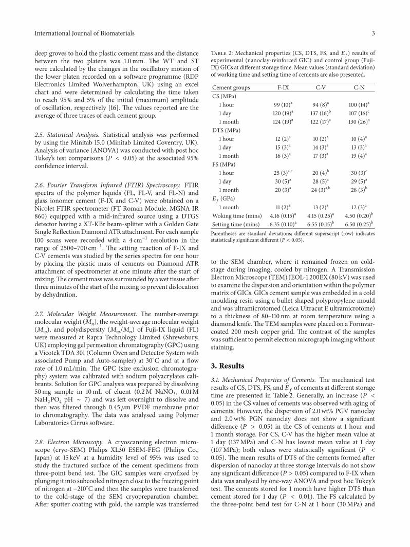
Table 2: Mechanical properties (CS, DTS, FS, and 𝐸𝑓) results ofexperimental (nanoclay-reinforced GIC) and control group (Fuji-IX) GICs at different storage time.Mean values (standard deviation)of working time and setting time of cements are also presented.
Cement groups F-IX C-V C-NCS (MPa)
1 hour 99 (10)a 94 (8)a 100 (14)a
1 day 120 (19)a 137 (16)b 107 (16)c
1 month 124 (19)a 122 (17)a 130 (26)a
DTS (MPa)1 hour 12 (2)a 10 (2)a 10 (4)a
1 day 15 (3)a 14 (3)a 13 (3)a
1 month 16 (3)a 17 (3)a 19 (4)a
FS (MPa)1 hour 25 (3)a,c 20 (4)b 30 (3)c
1 day 30 (5)a 28 (5)a 29 (5)a
1 month 20 (3)a 24 (3)a,b 28 (3)b
𝐸𝑓 (GPa)1 month 11 (2)a 13 (2)a 12 (3)a
Woking time (mins) 4.16 (0.15)a 4.15 (0.25)a 4.50 (0.20)b
Setting time (mins) 6.35 (0.10)a 6.55 (0.15)b 6.50 (0.25)b
Parentheses are standard deviations; different superscript (row) indicatesstatistically significant different (𝑃 < 0.05).
to the SEM chamber, where it remained frozen on cold-stage during imaging, cooled by nitrogen. A TransmissionElectronMicroscope (TEM) JEOL-1 200EX (80 kV) was usedto examine the dispersion and orientationwithin the polymermatrix of GICs. GICs cement sample was embedded in a coldmoulding resin using a bullet shaped polypropylene mouldand was ultramicrotomed (Leica Ultracut E ultramicrotome)to a thickness of 80–110 nm at room temperature using adiamond knife.The TEM samples were placed on a Formvar-coated 200 mesh copper grid. The contrast of the sampleswas sufficient to permit electronmicrograph imagingwithoutstaining.
3. Results
3.1. Mechanical Properties of Cements. The mechanical testresults of CS, DTS, FS, and 𝐸𝑓 of cements at different storagetime are presented in Table 2. Generally, an increase (𝑃 <0.05) in the CS values of cements was observed with aging ofcements. However, the dispersion of 2.0 wt% PGV nanoclayand 2.0wt% PGN nanoclay does not show a significantdifference (𝑃 > 0.05) in the CS of cements at 1 hour and1 month storage. For CS, C-V has the higher mean value at1 day (137MPa) and C-N has lowest mean value at 1 day(107MPa); both values were statistically significant (𝑃 <0.05). The mean results of DTS of the cements formed afterdispersion of nanoclay at three storage intervals do not showany significant difference (𝑃 > 0.05) compared to F-IX whendata was analysed by one-way ANOVA and post hoc Tukey’stest. The cements stored for 1 month have higher DTS thancement stored for 1 day (𝑃 < 0.01). The FS calculated bythe three-point bend test for C-N at 1 hour (30MPa) and

4 International Journal of Biomaterials
3500 3000 2500 2000 1500 1000
1040
33611088
1134
FL-N
FL-V
FL
1237
1447
1630
1706Ab
sorb
ance
(a.u
.)
Wavenumbers (cm−1)
Figure 2: FTIR spectra of Fuji-IX liquid (FL) and solutions formedafter dispersion of 2 wt% PGV and PGN nanoclays in FL.
1 month (28MPa) storage time was statistically significant(𝑃 < 0.05) compared to F-IX (25MPa and 20MPa). Theaverage FS values of F-IX cementwere better at 1 day (30MPa)and do not show any significant difference (𝑃 > 0.05)compared to C-V and C-N. Similarly, 𝐸𝑓 of cement formedafter the nanoclays dispersion (C-V and C-N) at 1 monthstorage showed no significant difference (𝑃 > 0.05) comparedto the F-IX control group.
3.2. Working Time and Setting Time. The results of theworking time measurements demonstrated increase (𝑃 <0.05) in working time for C-N and similar working time forC-V cements compared to F-IX. The addition of nanoclayresulted in increased (𝑃 < 0.05) setting time for C-N andC-V, 6.55 (0.10) minute and 6.50 (0.25) minute, respectively(Table 2).
3.3. Fourier Transform Infrared (FTIR) Spectroscopy. FTIRspectra of Fuji-IX liquid (FL) and the solutions preparedafter the dispersion of 2 wt% of PGV and PGN nanoclay(FL-V and FL-N) are shown in Figure 2. Table 3 gives themeasured wavenumbers and their correlation with knownvibration frequencies. No obvious peak shift was observedin FL after dispersion of nanoclay. The peak at 1706 cm−1attributed to the C=O stretching vibrations in the carboxylicgroup which is lost when it neutralised, whereas the peak at1630 cm−1 was associated with –OH bending vibrations ofcarboxyl group [17]. The peaks at 1088 cm−1 and 1134 cm−1suggested the presence of tartaric acid in FL [17]. In thespectrum of FL-V and FL-N, the presence of a weak peakat 1040 cm−1 was attributed to the Si–O stretching mode inPGV and PGN nanoclay. The series spectra of glass ionomercement (F-IX and C-V) at different time interval for 1 hourare shown in Figure 3 and peak assignments are given inTable 3. The weak peak at 1160 cm−1 can be assigned to theC–OH stretching vibration of tartaric acid in F-IX. Thisband decreased in intensity andmoved to lower wavenumberat 1055 cm−1. The strong band at 1405 to 1451 cm−1 was
Table 3: Description of FTIR peaks assignment present in thespectra of Fuji-IX liquid and Fuji-IX cements shown in Figures 2and 3.
Wavenumber cm−1 Assignment Reference
GIC1705 C=O stretching vibration [17]
1635 O–H stretching vibration ofmonomer [17]
1640 C=C stretching of monomer [17]
1625 Asymmetric C=O stretching ofAl-polycarboxylate [17–19]
1554 Asymmetric C=O stretching ofCa-polycarboxylate [17, 19]
1450 Symmetric C=O stretching ofAl-polycarboxylate [17–19]
1405 Symmetric C=O stretching ofCa-polycarboxylate [17, 19]
1644 Water sorption band in cement [18]
1640–1590 COO− asymmetric stretchingvibration [17–19]
1450–1405 COO− symmetric stretchingvibration [17–19]
1160, 1050 C–OH stretching vibration [17, 20]948 Si–OH stretching vibration [20]940–1200 Si–O stretching vibration [17, 20]
GIC Liquid
3354 H bonded O–H stretchingvibration [29]
1706 C=O stretching vibration incarbonyl group [17]
1630 –OH bending vibration incarboxylic group [17]
1134 Tartaric acid in F-IX [17, 19]1080 Tartaric acid in F-IX [17, 19]1040 Si–O stretching vibration [20, 30]
due to the formation of calcium and aluminium polysaltswhich increased in intensity and became prominent afterone hour [17–19]. At one hour after mixing, the symmetricand asymmetric stretching vibrations of aluminium poly-acrylate were present at approximately 1450 and 1625 cm−1,respectively (Figure 3(a)). The peak around 1644 cm−1 wasassigned to water sorption but an increase in this absorbanceband was small in comparison to changes due to acid-base reaction in the similar band [18]. A strong absorbanceband at 948 cm−1 due to the stretching vibrations of Si–OH was present continuously over the time periods studied.The asymmetric stretching vibrations in Si–O that usuallyappear between 940 and 1200 cm−1 are an indication forsilica gel formation by acid degradation of glass powder [20].Figure 3(b) shows the setting reaction of cements (C-V) and astrong peak at 940 cm−1 was associated with the asymmetricstretching vibrations in Si–OH. The bending vibration at1625 cm−1 moved to higher wavenumbers and attributed to

International Journal of Biomaterials 5Ab
sorb
ance
(a.u
.)
Wavenumber (cm−1)
Time
1min1981
1704
1644
1625
1594
15541498
1451
1405
948
11601055
2000 1800 1600 1400 1200 1000 800
(a)
Abso
rban
ce (a
.u.)
Wavenumber (cm−1)
Time
1min
2000 1800 1600 1400 1200 1000 800
1980
1704
164216251498
14511405
11631045
940
(b)
Figure 3: Real time FTIR analysis of the setting reaction of F-IXcement (a) and C-V cement (b) at different time intervals for onehour.
the formation of polyacrylate salts whereas, unlike F-IX, thepeak at 1594 cm−1 was not present. A gradual increase in thepeak intensity at 1642 cm−1 was observed with cements agingas expected.
3.4. Molecular Weight Determination. Figure 4 shows anoverlay of the computed molecular weight distribution ofduplicate run. The polymer solution of Fuji-IX liquid (FL)has a peak in the range of 15,600. The polymer contentwas calculated from the refractive index detector response.The refractive index chromatogram peak area for the FLwas 33,800 indicating the amount of polymer concentrationpresent in the solution. The average𝑀𝑤 was 15,700,𝑀𝑛 was3,970, and𝑀𝑤/𝑀𝑛 was 39.
3.5. Electron Microscopy. The representative cryo-SEMmicrographs of the fractured surface of bar-shapedspecimens demonstrated few or no microcracks on thesurface of glass ionomer cements and the size of the glassparticles was measured to be in 5 𝜇m range (Figure 5). Dueto the small wt% of nanoclays used, it was difficult to studythe dispersion of nanoclays in GIC by scanning electronmicroscopy. Therefore, transmission electron microscopy(TEM) was employed to study the structure and interaction
0.85
0.65
0.45
0.25
0.1
0
100 1000 10000 100000
Molecular weight
dw/d
log
Peak area
15,600
3,970
39
33,800
Mw
Mn
Mw/Mn
Figure 4: GPC plot showing the molecular weight distribution ofFL.
of nanoclays and GICs. The TEM micromorphologicalappearance of glass fillers, GIC matrix, and nanoclays ispresented in Figure 6. The different phases of glass ionomercement were readily observed from the TEM micrographsof C-V cement (Fuji-IX with 2wt% PGV nanoclays). Theporous structure of glass filler particles indicated the acidattack of PAA resulting in the formation of siliceous hydrogellayers all around the glass core. The porous nature of glassfiller particles was retained within the silica gel layers afterdepletion of ions from the surface of glass particles [20].The presence of nanoclays within the GIC matrix was alsoobserved which supported the interaction of PAA withnanoclays. TEM observation confirmed that the layers ofnanoclays were mostly exfoliated and dispersed in the matrixof GICs after mixing the PAA liquid constituent containingnanoclay with the aluminosilicate glass powder.
4. Discussion
The polymer-grade nanoclay was dispersed in the liquidportion of F-IX to study the effect on cement properties.The dispersion of nanoclays in poly(acrylic acid) solutionof F-IX increased the interlayer space of the nanoclay totrap polymer molecules. The possible reaction of PAA withPGV and PGN nanoclays and ion exchange with sodiumions on silicate plates occur during the process of nanoclaydispersion. Polymer chains of PAA can adsorb onto thesurface of clay galleries by forming hydrogen bonds or by ion-dipole interaction [11].The chemical and physical interactionof nanoclay resulted in an ionic bonding at the interfacebetween GIC polymer matrix and the nanoclay which canimprove mechanical properties [9]. However, if nanoclayswere not fully dispersed and penetrated by polymer chains,then agglomeration of nanoclays would be observed.
Brittle dental restoratives such as GIC have tensilestrength values lower than the compressive strength becausecrack propagation is favoured by tensile forces. The mechan-ical properties (CS, DTS, FS, and 𝐸𝑓) of cements beforeand after reinforcement with nanoclays showed that allthe cements became stronger as they matured at 1 monthof storage because it is expected that the setting reaction

6 International Journal of Biomaterials
(a) (b)
(c) (d)
Figure 5: Cryo-SEM micrographs of the fractured surface of GIC at various magnifications demonstrating (a) micrograph at lowestmagnification (×250) showing the absence of the cracks and ((b), (c), (d)) micrographs at high magnifications (×1998 and ×3997) indicatingthe presence of pores, glass particles, and the matrix phase of GIC.
will continue with time and that the mechanical propertiesshould improve with time. Xie et al. studied the mechanicalbehaviour of GICs and reported that conventional GICs hadhigher values of modulus than resin-modified GIC due tothe more flexible polymer matrix [21]. In the present work,the modulus of nanoclay clay reinforced cement (C-V and C-N) was higher than control cement, but statistically it wasnot significant. Therefore, it is suggested that the modulusof GICs after the dispersion of 2 wt% of nanoclays makesthesematerials suitable for use in load bearing applications aswell as for nonload bearing areas. The cements formed afterthe dispersion of nanoclay generally exhibited higher valuesor similar value to control group for CS and FS at differentstorage time but DTS was not statistically different than con-trol group. The significant improvement in the mechanicalbehaviour of mechanical properties may be restricted by theadjustment in the powder to liquid ratio of the GIC systemsafter dispersion and the processing technique of nanoclaysdispersion in PAA solution. Therefore, it is possible that thegraft polymerization of poly(acrylic acid) on the surface ofnanoclay would improve the strength of GIC system [9].The results of the WT and ST depicted a small increase in
both the WT and ST of glass ionomer cements preparedafter nanoclay dispersion compared to the control group.The dental cement should have a long working time afterwhich it should set rapidly to meet the clinical requirementin oral cavity without disturbing the inherited properties ofthe materials.The slight deflection in the results to the highervalues of WT and ST may be due to the room temperatureused during the experiments instead of 37∘C. However, suchvariations in the results may be accepted due to the natureof Wilson’s rheometers which is largely dependent upon theelastic tension and the physical properties of the differentspring coils used in oscillating rheometers. Previous studiesshowed the effect of temperature on the working and settingcharacteristics of cements and an increase in WT and ST atlower temperatures (8∘C) [16].
In the F-IX liquid the concentration of PAA in waterestimated from refractive index chromatogram peak area was33wt % and the 𝑀𝑤 of FL that was measured by GPC atRAPRA was in the range of 15.000. It is expected, however,that the molecular weight of PAA of a conventional GICwould have an effect on the nanoclays exfoliation and specif-ically on the interlayer spacing as the number of polymer

International Journal of Biomaterials 7
(a)
(b) (c)
M
M
M
M
NN
N
H
H
G
G
G
Figure 6: TEM micrographs of Fuji-IX cement after the dispersion of 2% nanoclay in liquid portion (C-N) showing the interaction ofnanoclays within GIC system, ((a) and (b)) at lowermagnification and (c) at highermagnification indicating remnant glass core (G), nanoclay(N), siliceous hydrogel layers around the periphery of glass filler core (H), cement matrix (M), and fully reacted hydrogel within the matrix(white bold arrow).
entanglements has an effect on the mobility of the chainswithin the interlayer spacing. This cannot be confirmed bythe current study as the effect of PAA molecular weight onthe dispersion of nanoclays was not investigated. Kirwan etal. reported that at low pH the attachment of polycarboxylatemolecules on hematite surface was not dependent on thechain length [22]. One of the reasons for this could be thedifference in the molecular weight as well as the concentra-tion of the polymer in the solution. In order to elucidate theeffect of the molecular weight and polymer concentration onthe dispersion and exfoliation of nanoclays, amore systematicstudy should be carried out.
The conventional GIC sets by means of an acid-basereaction between an aqueous PAA solution and a fluoroa-luminosilicate glass. It is, therefore, important to study thesetting reaction of GICs in order to understand what exactlycontrols the properties of GICs at themolecular level. Duringthe setting reaction of GICs, poly-acrylate-salt units andtartrate-salt units appear at different wavenumbers makingit possible to monitor the activity of various metal cations.The vibration bands from calcium acrylate and aluminiumacrylate appear due to the carboxylate stretching vibration[19]. However, FTIR spectroscopy is only suitable for semi-quantitative analysis, since the loss of the carbonyl group
absorption band during the neutralization overlaps withthe formation of the asymmetric COO− salt band and theabsorption bands of the polycarboxylic acid overlap with thestrong absorption bands of water at 1642 and 1705 cm−1 [19].The FTIR spectra of the FL-N and FL-V were not dissimilarfrom the spectrum of Fuji-IX liquid. The effects of acidtreatment on the structural modification of nanoclays werereported by Madejova et al. which suggested the successiverelease of the central atoms from the octahedral layer and therelease of Al from the Si tetrahedral sheets [23]. In the caseof PAA which is considered a weak acid, it can be suggestedthat the protons from the –COOH groups enter the nanoclaylayers and attack the structural –OH groups resulting indehydroxylation of nanoplates connected with the successiverelease of the central atoms. This can be demonstrated byobserving the changes in the characteristic absorption bandsattributed to the vibration of –OH groups and octahedralcations (1642 cm−1 and 940 cm−1), respectively. The settingreaction of GIC involves the neutralisation of PAA by theglass powder, which is associated with the formation of cal-cium and aluminium salt complexes. The acid neutralisationextent can be determined from the absorbance changes at1704 and 1555 cm−1 [17]. In comparison to F-IX cement, thereal time spectra of C-V show that the peak at 1625 cm−1

8 International Journal of Biomaterials
moved to higher wavenumbers and there was an absence ofpeak at 1594 cm−1 unlike F-IX spectra after one hour fromthe start of cement mixing. Additionally, the presence ofthe strong absorbance band at 948 cm−1 in both cementswas due to the stretching vibrations of Si–OH of the glasspowder and no significant changes were found in this bandover the duration of one hour. In the F-IX real time FTIRspectrum it is clear that Al- and Ca-tartrates form almost atthe same time. However, often it is difficult to differentiatebetween Ca-poly-acrylates and Al-poly-acrylates resulting inlack of strong evidence that Ca-poly-acrylates form first. Inaddition, MAS-NMR spectroscopy studies have shown thatoctahedral hydrated Al3+ (sixfold coordinated aluminiumAl(VI)) the type of Al3+ that forms Al-poly-acrylates formsalready at very early stage during the setting reaction [24,25]. The setting reaction of GICs based on three differentfluoroaluminosilicate glass compositions (LG125, ART10, andLG26Sr) using 27Al MAS-NMR spectroscopy was recentlystudied by Zainuddin et al. [26] and it was reported that thesetting reaction depended strongly on the glass compositionused to form the cement. Specifically, when the glass wasrich in phosphorus, the presence of Al–O–P species had aninfluence on the dissolution of the glass during the acid attackand consequently an effect on the ion release. In the case ofthe Sr substituted glass, the setting reaction was completedafter one day, whereas the setting reaction continued up to1 year in the case of glass having low phosphorus contents.Furthermore, they reported the presence of Al(VI) withobvious conversion of Al(IV) to Al(VI) at two minutesafter the start of the GIC setting reaction. Considering allthe above, it is clear that there is no strong evidence tosupport the idea that Ca-poly-acrylates form earlier than Al-poly-acrylates and Ca and Al compete equally to crosslinkthe polyacrylic acid chains during the cement formation.Nonetheless, it was expected that the FTIR analysis wouldgive more information on the role of nanoclays in the settingreaction; however, the spectra of cements that containednanoclays did not give a better insight.
The SEM observation of GICs revealed that the fracturedsurface of cement specimens consists of both large and smallglass particles which can readily be distinguished from thepolymer matrix. The cracks formations on GIC surface wereavoided by the use of cryo-SEM. Cryo-SEMmicrographs didnot show the cracks over the surface of GIC samples com-pared to conventional SEM. Such crack formations duringSEM observations are due to the dehydration of cementsunder high vacuum conditions. Figure 5 shows that glassparticles were embedded in the cement matrix and someof the glass particles are also observed on the surface ofcements dislodged during the three-point test. Although theaverage size of glass particles was measured approxi-mately5 𝜇m in the micrograph of the Fuji-IX cement, there is apossibility that much smaller glass particles may also bepresent. Transmission electron microscopy (TEM) showedthat the cement-forming reaction resulted in the dissolu-tion of the glass to form an amorphous gel around theglass particle. The mesoporous appearance of glass particleetched with PAA and its association with the nanoclay can
be observed in Figure 6. Tay et al. described the porousstructure of glass filler particles in ChemFlex (GIC) as “seed-like” inclusions and reported that seed-like inclusions wereretained within the silica gel layer after the depletion of ionsfrom the surfaces of glass particles in conventional glassionomer cements [27]. They observed a 150–300 nm thicksiliceous hydrogel phase around the glass core and suggestedthat smaller glass particles reacted completely with PAAforming siliceous-hydrogel rich phases within the cementmatrix. On the other hand, Barry et al. suggested that theporous glass filler inclusions present in conventional GICformulations represent segregated regions of a fluoride-richphase in certain reactive glass compositions [28]. Extendingthe explanations of Tay et al. to our work, the presenceof siliceous hydrogel phases surrounding the remnant glasscore was observed in the TEM micrographs of nanoclayreinforced GIC (C-N). The thickness of this phase seems tobe only a few hundred nanometers. The smaller particlesforming fully reacted hydrogel phases (core glass has fullyreacted) are also shown in Figure 6 (white bold arrow). TheTEM of GICs reported in the present study confirmed thepresence of porous glass filler particles and our findingsare in good agreement with the abovementioned studies.Moreover, the presence of electron-dense (dark gray) zonesin the majority of the polymer matrix (M) in Figure 5 wasprobably due to the formation of a polysalt matrix whenmetallic ions reacted with PAA. In the setting reaction ofGICs, bonds are formed between the polycarboxylic from thePAA matrix and aluminium and/or calcium ions from theglass particles resulting in salt bridges. The unreacted glassfiller particles and/or unreacted PAA matrix may constitutea microstructure with inferior mechanical properties. It istherefore assumed that nanoclay reinforcement may reducethe amount of unreacted PAA matrix but it is difficult tocomment on such interaction.
5. Conclusions
The optimum nanoclay dispersion in the polymer liquidcomponent of the cement is crucial. The dispersion ofnanoclays in the liquid portion of F-IX and the reinforcementeffect on GICs were determined. Although the dispersion ofnanoclays was successfully achieved, a small improvement inthe mechanical properties of the GIC systems was observed.The reinforcement of 2 wt% nanoclay generally resultedin improved mechanical behaviour. The incorporation ofnanoclay possibly does not compromise the acid-base neu-tralization reaction with a minimal effect on WT of GICbut provides the reinforcement at nanoscale. Various factorscan affect the improvement of the mechanical properties incements; for example, the adjustment in the P/L ratio of theGIC systems, the polymer concentration,molecular weight ofPAA, the size of nanoclays, the nanoclay surfacemodificationmethod, and the processing technique used are importantfactors for the stability of the PAA-nanoclay dispersion. Itis suggested that the dispersion of nanoclays in lower than2.0 wt% (1-2.0 wt%) inGICsmay potentially produce cementswith better mechanical properties.

International Journal of Biomaterials 9
Disclaimer
The authors have no relationship with the manufacturerslisted in this paper.
Conflict of Interests
The authors declare that there is no conflict of interestsregarding the publication of this paper.
Acknowledgments
The authors would like to thank Dr. M. Dadras at Universityof Neuchatel Switzerland for helping with cryo-SEM studiesandDr. Neigel Bubb at LeedsDental Institute for helpingwithrheological equipment.
References
[1] A. D.Wilson and B. E. Kent, “The glass-ionomer cement: a newtranslucent dental fillingmaterial,” Journal of Applied ChemistryBiotechnology, vol. 21, pp. 313–318, 1971.
[2] G. J. Mount, “Glass ionomers: a review of their current status,”Operative dentistry, vol. 24, no. 2, pp. 115–124, 1999.
[3] M. J. Tyas, “Clinical evaluation of glass-ionomer cement resto-rations,” Journal of Applied Oral Science, vol. 14, pp. 10–13, 2006.
[4] R. C. Randall and N. H. E.Wilson, “Glass-ionomer restoratives:a systematic review of a secondary caries treatment effect,”Journal of Dental Research, vol. 78, no. 2, pp. 628–637, 1999.
[5] R. Peez and S. Frank, “The physical-mechanical performanceof the new Ketac Molar Easymix compared to commerciallyavailable glass ionomer restoratives,” Journal of Dentistry, vol.34, no. 8, pp. 582–587, 2006.
[6] M. Yoonessi, H. Toghiani, W. L. Kingery, and C. U. PittmanJr., “Preparation, characterization, and properties of exfoli-ated/delaminated organically modified clay/dicyclopentadieneresin nanocomposites,”Macromolecules, vol. 37, no. 7, pp. 2511–2518, 2004.
[7] A. Okada and A. Usuki, “Twenty years of polymer-claynanocomposites,” Macromolecular Materials and Engineering,vol. 291, no. 12, pp. 1449–1476, 2006.
[8] J. H. Park and S. C. Jana, “The relationship between nano- andmicro-structures and mechanical properties in PMMA-epoxy-nanoclay composites,” Polymer, vol. 44, no. 7, pp. 2091–2100,2003.
[9] L. Solhi,M.Atai, A.Nodehi,M. Imani, A.Ghaemi, andK.Khos-ravi, “Poly(acrylic acid) grafted montmorillonite as novel fillersfor dental adhesives: synthesis, characterization and propertiesof the adhesive,” Dental Materials, vol. 28, no. 4, pp. 369–377,2012.
[10] A. H. Dowling, A. Stamboulis, and G. J. P. Fleming, “The influ-ence ofmontmorillonite clay reinforcement on the performanceof a glass ionomer restorative,” Journal of Dentistry, vol. 34, no.10, pp. 802–810, 2006.
[11] M. A. Fareed and A. Stamboulis, “Nanoclays reinforced glassionomer cements: dispersion and interaction of polymer grade(PG) montmorillonite with poly(acrylic acid),” Journal of Mate-rials Science:Materials inMedicine, vol. 25, no. 1, pp. 91–99, 2014.
[12] ISO, “Water-based cements-part 1: powder/liquid acid-basecements,” ISO 9917-1:2007, International Organization for Stan-dardisation, 2009.
[13] L. Wang, P. H. P. D’Alpino, L. G. Lopes, and J. C. Pereira,“Mechanical properties of dental restoratives materials: relativecontribution of laboratory tests,” Journal of Applied Oral Sci-ences, vol. 11, pp. 162–167, 2003.
[14] ISO 4049:2000, “Dentistry, Dentistry-Polymer-based filling,restorative and lutingmaterials,” International Organization forStandardisation, 2000.
[15] ASTM D790-07 Standard Test Methods for Flexural Propertiesof Unreinforced and Reinforced Plastics and Electrical InsulatingMaterials, ASTM, 2007.
[16] G. J. Pearson and A. S. Atkinson, “Effects of temperature changeon the working and setting characteristics of water-based dentalcements,” Dental Materials, vol. 3, no. 5, pp. 275–279, 1987.
[17] A. M. Young, S. A. Rafeeka, and J. A. Howlett, “FTIR investi-gation of monomer polymerisation and polyacid neutralisationkinetics and mechanisms in various aesthetic dental restorativematerials,” Biomaterials, vol. 25, no. 5, pp. 823–833, 2004.
[18] A. M. Young, “FTIR investigation of polymerisation and poly-acid neutralisation kinetics in resin-modified glass-ionomerdental cements,” Biomaterials, vol. 23, no. 15, pp. 3289–3295,2002.
[19] J. W. Nicholson, “Chemistry of glass-ionomer cements: areview,” Biomaterials, vol. 19, no. 6, pp. 485–494, 1998.
[20] E. A. P. de Maeyer, R. M. H. Verbeeck, and C. W. J. Vercruysse,“Infrared spectrometric study of acid-degradable glasses,” Jour-nal of Dental Research, vol. 81, no. 8, pp. 552–555, 2002.
[21] D. Xie, W. A. Brantley, B. M. Culbertson, and G. Wang,“Mechanical properties and microstructures of glass-ionomercements,” Dental Materials, vol. 16, no. 2, pp. 129–138, 2000.
[22] L. J. Kirwan, P. D. Fawell, and W. Van Bronswijk, “In situ FTIR-ATR examination of poly(acrylic acid) adsorbed onto hematiteat low pH,” Langmuir, vol. 19, no. 14, pp. 5802–5807, 2003.
[23] J. Madejova, J. Bujdak, M. Janek, and P. Komadel, “ComparativeFT-IR study of structural modifications during acid treatmentof dioctahedral smectites and hectorite,” Spectrochimica ActaA: Molecular and Biomolecular Spectroscopy, vol. 54, no. 10, pp.1397–1406, 1998.
[24] A. Stamboulis, R. V. Law, and R. G. Hill, “Characterisationof commercial ionomer glasses using magic angle nuclearmagnetic resonance (MAS-NMR),” Biomaterials, vol. 25, no. 17,pp. 3907–3913, 2004.
[25] A. Stamboulis, S. Matsuya, R. G. Hill et al., “MAS-NMRspectroscopy studies in the setting reaction of glass ionomercements,” Journal of Dentistry, vol. 34, no. 8, pp. 574–581, 2006.
[26] N. Zainuddin, N. Karpukhina, R. G.Hill, andR. V. Law, “A long-term study on the setting reaction of glass ionomer cements by27Al MAS-NMR spectroscopy,”Dental Materials, vol. 25, no. 3,pp. 290–295, 2009.
[27] F. R. Tay, E. L. Pashley, C.Huang et al., “Theglass-ionomer phasein resin-based restorative materials,” Journal of Dental Research,vol. 80, no. 9, pp. 1808–1812, 2001.
[28] T. I. Barry, D. J. Clinton, and A. D. Wilson, “The structureof a glass-ionomer cement and its relationship to the settingprocess,” Journal of Dental Research, vol. 58, no. 3, pp. 1072–1079,1979.
10 International Journal of Biomaterials
[29] D. Gao and R. B. Heimann, “Structure and mechanical proper-ties of superabsorbent poly(acrylamide)-montmorillonite com-posite hydrogels,” Polymer Gels and Networks, vol. 1, no. 4, pp.225–246, 1993.
[30] J. Billingham, C. Breen, and J. Yarwood, “Adsorption ofpolyamine, polyacrylic acid and polyethylene glycol on mont-morillonite: an in situ study using ATR-FTIR,” VibrationalSpectroscopy, vol. 14, no. 1, pp. 19–34, 1997.

Submit your manuscripts athttp://www.hindawi.com
ScientificaHindawi Publishing Corporationhttp://www.hindawi.com Volume 2014
CorrosionInternational Journal of
Hindawi Publishing Corporationhttp://www.hindawi.com Volume 2014
Polymer ScienceInternational Journal of
Hindawi Publishing Corporationhttp://www.hindawi.com Volume 2014
Hindawi Publishing Corporationhttp://www.hindawi.com Volume 2014
CeramicsJournal of
Hindawi Publishing Corporationhttp://www.hindawi.com Volume 2014
CompositesJournal of
NanoparticlesJournal of
Hindawi Publishing Corporationhttp://www.hindawi.com Volume 2014
Hindawi Publishing Corporationhttp://www.hindawi.com Volume 2014
International Journal of
Biomaterials
Hindawi Publishing Corporationhttp://www.hindawi.com Volume 2014
NanoscienceJournal of
TextilesHindawi Publishing Corporation http://www.hindawi.com Volume 2014
Journal of
NanotechnologyHindawi Publishing Corporationhttp://www.hindawi.com Volume 2014
Journal of
CrystallographyJournal of
Hindawi Publishing Corporationhttp://www.hindawi.com Volume 2014
The Scientific World JournalHindawi Publishing Corporation http://www.hindawi.com Volume 2014
Hindawi Publishing Corporationhttp://www.hindawi.com Volume 2014
CoatingsJournal of
Advances in
Materials Science and EngineeringHindawi Publishing Corporationhttp://www.hindawi.com Volume 2014
Smart Materials Research
Hindawi Publishing Corporationhttp://www.hindawi.com Volume 2014
Hindawi Publishing Corporationhttp://www.hindawi.com Volume 2014
MetallurgyJournal of
Hindawi Publishing Corporationhttp://www.hindawi.com Volume 2014
BioMed Research International
MaterialsJournal of
Hindawi Publishing Corporationhttp://www.hindawi.com Volume 2014
Nano
materials
Hindawi Publishing Corporationhttp://www.hindawi.com Volume 2014
Journal ofNanomaterials