remembering first impressions: effects of intentionality ...uleman, 2003; j. willis & todorov,...
TRANSCRIPT

Remembering first impressions: Effects of intentionalityand diagnosticity on subsequent memory
Roee Gilron & Angela H. Gutchess
Published online: 3 December 2011# Psychonomic Society, Inc. 2011
Abstract People rely on first impressions every day as animportant tool to interpret social behavior. While research isbeginning to reveal the neural underpinnings of firstimpressions, particularly through understanding the role ofdorsal medial prefrontal cortex (dmPFC), little is knownabout the way in which first impressions are encoded intomemory. This is surprising because first impressions arerelevant from a social perspective for future interactions,requiring that they be transferred to memory. The presentstudy used a subsequent-memory paradigm to test theconditions under which the dmPFC is implicated in theencoding of first impressions. We found that intentionallyforming impressions engages the dmPFC more than doesincidentally forming impressions, and that this engagementsupports the encoding of remembered impressions. Inaddition, we found that diagnostic information, which morereadily lends itself to forming trait impressions, engages thedmPFC more than does neutral information. These resultsindicate that the neural system subserving memory forimpressions is sensitive to consciously formed impressions.The results also suggest a distinction between a socialmemory system and other explicit memory systemsgoverned by the medial temporal lobes.
Keywords Episodic memory . Impression formation .
Prefrontal cortex .Medial temporal lobes
First impressions have a profound effect on our everydaylives. We use them to determine who we should approach
and who we should avoid. They can be a deciding factor inmate choice, trustworthiness judgments, and hiring deci-sions. Moreover, there is evidence that they may influencecourt decisions (Zebrowitz & McDonald, 1991; Zebrowitz& Montepare, 2008), election results (Olivola & Todorov,2010; Verhulst, Lodge, & Lavine, 2010), and professionalevaluations (Ambady & Rosenthal, 1993). A growingnumber of studies are examining the way in which wequickly and automatically make trait impressions of othersand use that knowledge (Cloutier, Kelley, & Heatherton,2010; Uleman, Saribay, & Gonzalez, 2008; Van Overwalle,2009; Van Overwalle & Labiouse, 2004), but few (Harvey,Fossati, & Lepage, 2007; Mitchell, Macrae, & Banaji,2004; Schiller, Freeman, Mitchell, Uleman, & Phelps,2009) have examined the conditions under which weremember these impressions. This is surprising, becausethe memory of these impressions has the capacity toinfluence our future actions. Though current researchsuggests that we are experts at forming quick, automaticimpressions, little is known about the processes that supportretaining these impressions in long-term memory.
Though much can be learned about first impressions frombehavioral measures, an investigation of the factors thatinfluence first-impression formation and the correspondingneural underpinnings would allow one to ask more nuancedquestions about forming impressions and their storage inmemory. Accumulating evidence in the memory literature hassuggested that the broad distinction between the neuralsubstrates supporting semantic, episodic, and proceduralmemory may also extend to distinct classes of elaborativesemantic encoding processes, perhaps including those in thesocial domain. From their review of the literature, Macrae andcolleagues suggested that largely disparate neural networksare activated during the successful formation of memories inresponse to verbal, visual, emotional, and self-referential
R. Gilron :A. H. Gutchess (*)Department of Psychology, Brandeis University,415 South Street, MS 062, P.O. Box 549110, Waltham,MA 02454-9110, USAe-mail: [email protected]
Cogn Affect Behav Neurosci (2012) 12:85–98DOI 10.3758/s13415-011-0074-6
processing, consistent with the idea that different processescontribute to the formation of distinct varieties of episodicmemories (Macrae, Moran, Heatherton, Banfield, & Kelley,2004). Recent investigations have upheld this division, inparticular as it relates to the processing of social andemotional information (Gutchess, Kensinger, & Schacter,2010; Haas & Canli, 2008; Harvey et al., 2007; Mitchell etal., 2004). Though the hippocampus plays a key role in theencoding network for memory for many classes of informa-tion, additional disparate brain regions support specificsubcategories. Thus, as in a comparable system that aids inencoding emotional information into memory (Schacter,Gutchess, & Kensinger, 2009), there may be a dedicatedsystem for encoding first impressions (and more broadly,social information) into memory. Given how important socialinteraction is to the human condition, we would expect tofind evidence for the contributions of a social cognitionnetwork to the encoding of first impressions into memory.
Applying a social neuroscience approach to understandhow people form impressions of others advances ourunderstanding of the component processes and the feedfor-ward and feedback loops that shape our perceptions ofothers (for reviews, see Ames, Fiske, & Todorov, 2011;Rule & Ambady, 2008). A number of neural regionsrespond to impression formation, reflecting the complexityof the processes involved and the interconnectedness of thenetwork that allows impressions to be invoked so instan-taneously. These regions include the amygdala, whichresponds to emotional and evaluative conditions (Schilleret al., 2009) and to appearance-based cues, such astrustworthiness (Said, Baron, & Todorov, 2009; Winston,Strange, O’Doherty, & Dolan, 2002); the caudate nucleus,which responds to reward and feedback (Delgado,Nystrom, Fissell, Noll, & Fiez, 2000); the superior temporalsulcus, which responds to others’ intentions (Saxe, Xiao,Kovacs, Perrett, & Kanwisher, 2004); and the fusiformgyrus, which is invoked by face processing (Winston,Henson, Fine-Goulden, & Dolan, 2004). The dorsal medialprefrontal cortex (dmPFC), a region of the frontal cortex,has been particularly implicated in impression formation, aswell as in a wide array of social processes (Amodio &Frith, 2006; D’Argembeau et al., 2007; Harvey et al., 2007;Macrae et al., 2004; Mitchell, Cloutier, Banaji, & Macrae,2006; Mitchell, Macrae, & Banaji, 2004, 2005, 2006).Furthermore, virtually all studies that have investigated firstimpressions in their manifestation as trait judgments haveimplicated the dmPFC (Mason, Dyer, & Norton, 2009;Mitchell, 2008; Mitchell, Ames, Jenkins, & Banaji, 2009;Mitchell, Cloutier, et al., 2006; Mitchell, Macrae, & Banaji,2004, 2005, 2006; Todorov, Gobbini, Evans, & Haxby,2007; the exception is Heberlein & Saxe, 2005, whosecontrol was an emotional task, which might also engagedmPFC). Not only does the dmPFC respond to social
information, but it also mediates the encoding of firstimpressions into memory (Mitchell et al., 2004). InMitchell et al.’s (2004) study, participants read sentencesdepicting actions that were paired with a picture of a face.Participants were asked either to form an impression of theface–sentence pair or to remember the sequence of thepresented actions. While forming an impression, dmPFCactivity was higher when the face–statement pair was laterremembered rather than later forgotten. However, activityin this region did not predict successful encoding whenparticipants were oriented to the sequence of statements. Incontrast, the right hippocampus predicted successful encod-ing for the sequencing task, but not for the impression task.Mitchell et al.’s (2004) study suggests that successfulencoding of social information, but not of nonsocialinformation, may be mediated by the dmPFC.
DmPFC not only contributes to the encoding ofimpressions, but also is more engaged under specificconditions of impression formation. Mitchell, Cloutier, etal. (2006) found that the dmPFC was sensitive to the natureof the information on which an impression was based.Participants viewed face–sentence pairs with sentencesdepicting actions that were either diagnostic for a specifictrait (i.e., behaviors that conveyed information that allowedone to form an impression, such as generous, boring, lazy,or friendly) or nondiagnostic (i.e., behaviors that did notstrongly indicate a specific personality trait, such as “hephotocopied the article”). When participants were told toform an impression, dmPFC was engaged, but the activitydid not differ for diagnostic versus nondiagnostic informa-tion, suggesting that the region is highly engaged whenforming impressions, regardless of the content of theinformation. However, when impression formation wasunintentional (i.e., participants focused on the sequencesin which statements were presented), dmPFC responsedifferentiated diagnostic from nondiagnostic trials. Thissuggests that when incidentally forming impressions, theregion selectively and spontaneously responds to informa-tion that supports impression formation, and that it can beengaged when attempting to form an impression consciously,even when the available information does not indicate aparticular trait.
Taken together, these studies highlight the important rolethat the dmPFC plays in encoding first impressions.However, the conditions under which the region contributesto impression formation are little understood. Mitchell,Cloutier, et al.’s (2006) surprising finding that the dmPFCdoes not differentiate between diagnostic and neutralinformation when people explicitly attempt to form impres-sions (although this is not the case for implicit impressionformation) suggests that an individual’s goals and the typeof information with which he is presented have implicationsfor how impressions are stored in memory for future use.
86 Cogn Affect Behav Neurosci (2012) 12:85–98

Importantly, this study did not test memory, so it isunknown whether these differential effects also influencethe encoding of trait impressions into memory.
In addition to the dmPFC, one may expect to find thatstructures implicated in memory formation, such as thehippocampus and medial temporal lobes, would alsosupport memory for impressions. However, the hippo-campus did not emerge in a study investigating theencoding of impressions, and in fact was only implicatedin forming memories for the nonsocial-sequencingcomparison condition (Mitchell et al., 2004). This isconsistent with the patient literature, in which patientswith damaged hippocampi are able to learn trait associa-tions to faces, but not when the amygdalae and temporallobes are damaged (Todorov & Olson, 2008), andcircumstantially from patient H.M., who exhibited somesemantic knowledge of famous people postoperatively(O’Kane, Kensinger, & Corkin, 2004). In the neuro-imaging literature, however, it is unclear whether Mitchellet al.’s (2004) use of a sequencing task is the bestnonsocial comparison task with which to assess thecontribution of the hippocampus to the encoding ofimpressions, because the hippocampus is implicated insequencing, regardless of memory demands (Eichenbaum,2004; Lehn et al., 2009). A comparison task that is notknown to be hippocampally dependent may be moreappropriate to test the involvement of the dmPFC and thehippocampus in the successful formulation of memories.
Thus, we can make a number of predictions regardingthe function of a dedicated social memory system. Wewould expect this system to respond differentially on thebasis of intentionality, such that intentionally formedimpressions would contribute to encoding success differ-ently than would incidentally formed impressions. Thoughsome research has suggested that intentionality does notaffect the processing of trait impressions (Todorov &Uleman, 2003; J. Willis & Todorov, 2006), other researchhas suggested that additional neural regions are recruitedduring intentional impression formation, perhaps reflectingbroader consideration of the available information in orderto confirm initial impressions (Ma, Vandekerckhove, VanOverwalle, Seurinck, & Fias, 2011). Explicit effort supportsthe ability to recognize emotional facial expressions,although implicit conditions reveal impairments in patientswith lesions to the orbitofrontal cortex (M. L. Willis,Palermo, Burke, McGrillen, & Miller, 2010). Given theimportance of the orbitofrontal cortex to emotional pro-cessing, this finding suggests that explicit judgments recruitand utilize additional resources as compared to implicitimpression formation. Mitchell, Cloutier, et al. (2006) alsofound differences in dmPFC activation in implicit but notexplicit person perception, suggesting that additionalresources may be recruited when explicitly forming
impressions even for nondiagnostic events. Thus, we expectthat information that easily lends itself to forming traitimpressions (e.g., diagnostic information) would engagedmPFC structures, as well as contributing to encodingsuccess more than neutral information does.
Thus, our study aimed to investigate the neuralmechanisms supporting the encoding of trait impressionsinto memory. Through it, we hoped to assess theimportance of one’s state of mind (intention to form animpression) and of the type of information that one ispresented with (whether diagnostic of a particular trait ornot) when making trait impressions, as well as the wayin which these factors influence the social cognitionnetwork in the brain.
Method
Participants
A group of 22 participants (11 male, 11 female) completedthe study (mean age = 21.8, SD = 3.28) in exchange forpayment of $25/h. Two of the participants were excludedfrom the analysis (1 male, 1 female), one for excessivemovement in the scanner (>10 mm), and the other for near-chance performance (52.3% across all conditions) on thetask. All participants signed informed consent forms andwere screened for fMRI eligibility, including right-handedness; English learned before the age of 8; goodneurological, psychological, and physical health; and theabsence of medications that affect the central nervoussystem and devices or implants contraindicated for mag-netic resonance scanning. The protocol was approved bythe Brandeis University and Massachusetts General HospitalInstitutional Review Boards.
Materials and procedure
The encoding stimuli consisted of 240 face–sentence pairs,with half of the faces (120) female and half male. The faceshad neutral expressions and were evenly distributed acrossfour different age groups, ranging from 18 to 89, and eachface was displayed once during encoding. The faces werecolor photographs selected from the Center for VitalLongevity Face Database: https://pal.utdallas.edu/facedb/request/index
The first independent variable was the type of encodingtask performed by the participants: impression based orsemantic based. During the encoding phase, participantswere asked to make one of two judgments regarding eachface–sentence pair. One task emphasized the semanticnature of the behavior, in which we asked participants toindicate whether the action the person performed took place
Cogn Affect Behav Neurosci (2012) 12:85–98 87

at home or away from home (SEM); this served as acomparison task that was not highly social or evaluative. Inthe other task, participants were asked to think of a trait thatdescribed the person depicted in the face–sentence pair andto decide whether this trait was positive or negative (IMP),a highly social task.
The content of the sentences served as the secondindependent variable. Half of the sentences (120) werediagnostic, in that they implied one of 24 traits (e.g.,boring); 12 of these traits were positive, and 12negative.1 The remaining 120 sentences were “neutral”and described an action that did not readily lend itself to atrait judgment, such as “He made a peanut butter and jellysandwich.” All of the diagnostic sentences had previouslybeen used in studies by Mitchell, Macrae, and Banaji(2004, 2006), and the neutral ones were created by theexperimenters. Each sentence appeared below a faceduring the encoding phase (see Fig. 1). Participants werenot asked to intentionally memorize the face–sentencepairs.
Participants performed the task during six functionalruns, each lasting 6 min and consisting of 40 face–sentencepairs, with 20 male and 20 female targets. The faces wereequally distributed across each age group. Face–sentencepairs were pseudorandomly assigned to either the SEM(nonsocial) or IMP (social) condition, for a total of fourexperimental conditions: SEM–diagnostic, SEM–neutral,IMP–diagnostic, and IMP–neutral. Each face–sentence pairwas presented for 6 s, during which time the participantsmade a response with a buttonpress to indicate “home” or“not home,” for SEM judgments, or “positive” or “nega-tive,” for IMP judgments (see Fig. 1). The 6-s trial lengthwas necessary because the reaction times to longersentences were approximately 5 s, due to the amount ofinformation to be processed (e.g., condition label, face,sentence). Baseline trials in which participants saw afixation cross were intermixed throughout the run. Trialswere presented in a jittered design, with intertrial intervalsranging from 0 to 12 s, and were ordered using optseq2(Dale, 1999).
Stimuli were assigned to conditions in a within-subjectsdesign. Four counterbalanced versions were used in orderto account for stimulus-specific effects. Across participants,the face–sentence pairs originally coupled with the SEM(nonsocial) task were recoupled with the IMP (social) task,and vice versa, and new face–sentence pairs were formed inan attempt to reduce face-specific effects. Both pairs werecounterbalanced for trait (i.e., faces previously paired withdiagnostic statements were now paired with neutral state-ments, and vice versa) and gender (i.e., sentences paired
with a female face were switched so that they were pairedwith a male face).2
Between the encoding and retrieval phases was anapproximately 12-min retention interval, during which timestructural images of the brain were acquired while theparticipants did not perform any task. During the retrievalphase, participants saw two faces and a target sentence thathad previously been paired in the encoding phase with one ofthe faces (see Fig. 1 bottom). Participants chose the face thatthey remembered as having been paired with the targetsentence during encoding and indicated their response with abuttonpress during the 6-s trial interval. As in the encodingphase, participants completed 40 trials during each of the sixruns; each run lasted 6 min. Each face was presented twiceduring recognition, once as the correct answer and once as alure, and whether each particular face appeared first as acorrect answer or a lure was counterbalanced acrossparticipants. The first three retrieval runs contained novelface pairs, whereas in the remaining runs the same pairingsof faces were repeated with the remaining sentences. Facepairs were always from the same age group and werematched on gender. In addition, faces were matched acrossconditions (e.g., a face originally encoded in the IMPcondition was paired with a lure also encoded in the IMPcondition) but were not matched in terms of sentencediagnosticity. Runs were also balanced with regard toencoding conditions, such that face–sentence pairs from allconditions present at encoding were included in eachencoding run. The retrieval data will be analyzed in aseparate study.
Prior to the encoding and retrieval stages of the experiment,participants practiced the task with additional face–sentencepairs. Participants also completed measures to assess cogni-tive ability, including the Vocabulary subscale of the ShipleyInstitute of Living Scale (Shipley, 1986) and the digitcomparison task (Hedden et al., 2002; modeled afterSalthouse & Babcock’s, 1991, letter comparison task) tomeasure speed of processing. The experimental task waspresented using E-Prime software (Psychology SoftwareTools, Pittsburgh, PA), which also recorded participants’yes/no responses and reaction times from a button box.
Image acquisition and data analysis
The data were acquired with a Siemens Avanto 1.5 T scanner,using an echo-planar imaging sequence (TR = 2,000 ms, TE =
1 Some examples of negative and positive traits are motivated, honest,confident (positive) versus lazy, dishonest, inconsiderate (negative).
2 In the process of counterbalancing, an experimental error occurred intwo of the counterbalancings, in which 10 face–sentence pairs wereassigned to the SEM–neutral condition instead of the IMP–neutralcondition. As a result, some participants in these counterbalancegroups (n = 8) viewed 60 face–sentence pairs in each of the twodiagnostic conditions, but received 50 pairs in the IMP–neutralcondition and 70 pairs in the SEM–neutral condition.
88 Cogn Affect Behav Neurosci (2012) 12:85–98

40 ms, FOV = 200 mm, flip angle = 90º) to acquire26 AC/PC-oriented slices 3.2 mm thick with a 10% skip.Slices covered most of the cortex, with the exception of thedorsal portion of the parietal lobes and the ventral portion ofthe temporal lobes. Stimuli were back-projected onto ascreen behind the scanner and viewed by the participantsusing a mirror attached to the head coil. High-resolutionanatomical images were acquired using a multiplanar rapidlyacquired gradient echo (MP-RAGE) sequence.
The preprocessing and data analysis were conductedwith SPM5 (Wellcome Trust Centre for Neuroimaging,London, UK). Functional images were slice-time corrected,realigned to the first image in order to correct for motion,normalized to the Montreal Neurological Institute template,resampled to 3-mm cubic voxels, and spatially smoothedusing a 6-mm full-width half maximum isotropic Gaussiankernel (Dale, 1999).
We used a subsequent-memory (Dm) paradigm in orderto sort our imaging results according to the success of
memory formation (Wagner et al., 1998). In a Dmparadigm, one separates encoding trials on the basis of thesuccess of memory formation (i.e., whether the trials werelater remembered or forgotten at the time of retrieval).Thus, we used the recognition data to distinguish all face–sentence pairs that were incorrectly identified at retrievaland binned these as “forgotten.” We then binned all of theface–sentence pairs that were correctly identified at retriev-al as “remembered.” This analysis approach, applied to theencoding data, allows one to identify brain regions that aremore engaged during the successful encoding of informa-tion (i.e., remembered > forgotten), in what will be referredto as a Dm effect.
To model each participant’s data, events were convolvedwith a canonical hemodynamic response function in anevent-related design. A total of eight regressors werecreated for the combination of conditions: orientation atencoding (IMP/SEM), sentence diagnosticity (diagnostic/neutral), subsequent memory (remembered/forgotten), and
Fig. 1 Experimental displays atencoding and retrieval. (i) Fourstimuli are displayed, represent-ing the four encoding condi-tions, either impression (a and c)or semantic (b and d), denotedby the question posed at the topof the screen. Each face waspaired with either a diagnostic(a and b) or a neutral (c and d)sentence, determined by thecontent of the sentence at thebottom of the screen. Faces werematched across all conditionsfor gender and age. Stimuli werepresented without the condition-identifying boxes shown here inthe top right of panels A–D. (II)Participants indicated with abuttonpress (left or right) whichface they had previously seenpaired with the sentence
Cogn Affect Behav Neurosci (2012) 12:85–98 89

a ninth regressor was included for participants who hadtrials with no response. Regressors were also created tomodel the six separate runs. Contrasts of interest weredefined using these regressors of interest and then estimatedfor each participant’s fMRI data.
In addition to the regressors stemming from the studydesign, we introduced a parametric regressor when model-ing the data to control for the effects of face attractiveness.Face attractiveness has the capacity to influence firstimpressions, most notably by adding to a general positiveimpression (Langlois et al., 2000) as well as activating thesocial network (such as dmPFC; Ishai, 2007; Liang,Zebrowitz, & Zhang, 2010). To control for these effects,we added an attractiveness rating for each face as aparametric regressor. Ratings were based on the averageattractiveness rating provided by a separate sample of 15participants (9 male, 6 female) in the laboratory. Partic-ipants responded to the prompt of “how attractive is thisperson?” on a 1–7 scale.
We then pooled the relevant contrasts across participantsin a series of random-effects whole-brain group analyses.These were thresholded at p < .001 at the voxel level andwith a spatial extent threshold of 10 voxels. Note that thisthreshold surpasses an overall correction level of p < .05,which can be achieved through the combination of a voxel-level correction of p < .001 and a cluster-level threshold of7 voxels. To estimate the cluster-level threshold, we used ascript that determined the number of contiguous voxelsrequired to achieve an overall correction of p < .05, on thebasis of the parameters of the data (e.g., slice thickness) andthe selected voxel-level threshold (as in Slotnick, Moo,Segal, & Hart, 2003). We highlight regions in medialprefrontal cortex (particularly BA 10/32/8) because priorstudies have identified these regions as relevant for theencoding of social information (Amodio & Frith, 2006;Harvey et al., 2007; Mitchell, Cloutier, et al., 2006;Mitchell et al., 2004; Schiller et al., 2009). We also focusedon regions of the medial temporal lobes (MTL) that havebeen broadly implicated in explicit encoding processes(Macrae et al., 2004; Squire, 2004), although, to date, littleevidence has associated MTL with the encoding of socialinformation. By comparing two different types of tasks(IMP/SEM), we have the potential to distinguish theinvolvement of MTL regions in the encoding of socialstimuli during conditions conducive to impression forma-tion, relative to more semantic or knowledge-based con-ditions. This may be a sensitive comparison with which toreveal a role for this region in the encoding of social stimulior to further support the separation of a “social memorysystem” from an MTL-based explicit memory system.
To explore memory effects in regions that responded toinformation relevant for impression formation, we firstidentified regions responding to the social relevance of the
information as an effect of the orientation of the participant(IMP vs. SEM) or the content of the information (diagnos-tic vs. neutral) in whole-brain contrasts. We identifiedmedial prefrontal and MTL regions emerging from theseanalyses, and in a second step, probed these regions fororthogonal effects of memory by focusing on interactionsor main effects of memory (remembered/forgotten). Thisallowed us to assess the extent to which regions involved inthe processing of social information also respond tomemory formation. To characterize the activity in eachindividual condition, we used MarsBaR (Brett, Anton,Valabregue, & Poline, 2002) to extract percent signalchange from each region of interest (ROI) based on thethree factors of interest (intentionality, diagnosticity, mem-ory), each with two levels: Participants formed firstimpressions with an intentional focus on social information(IMP) or incidentally with a more semantic, nonsocial focus(SEM); the sentences were either diagnostic or neutral; andeach pair was either remembered or forgotten.
As an additional test of the involvement of MTL in theencoding of social information, we conducted an a priori ROIanalysis on MTL regions. To do so, we created anatomicalmasks of MTL regions, including hippocampal, parahippo-campal, and amygdala regions, using PickAtlas software(Maldjian, Laurienti, Kraft, & Burdette, 2003). We thenapplied these masks to the SPM analyses. While this ROIanalysis overlapped with the whole-brain approach describedearlier, it offered a more lenient test to detect the contributionof MTL regions to the encoding of social information,regions that have not emerged in the literature to date.
Results
Behavioral results
We conducted a 2 × 2 repeated measures ANOVA to probethe effects of sentence diagnosticity (diagnostic/neutral) andtask orientation (IMP/SEM) on memory performance. Wefound a main effect for sentence diagnosticity, such thatdiagnostic (M = 69.7%, SD = 8.2%) face–sentence pairswere better remembered than neutral (M = 64.4%, SD =8.4%) face–sentence pairs, F(1, 19) = 21.4, p < .001, ηp
2 =.53. Surprisingly, memory performance was not significantlybetter in the IMP condition (M = 67.9%, SD = 8.9%) than inthe SEM condition (M = 66.2%, SD = 8.5%), F(1, 19) =0.67, p = .42, ηp
2 = .03. The interaction was also notsignificant (F<1). See the data in Fig. 2.
fMRI results
As expected, our neuroimaging analysis contrasting taskorientation (IMP > SEM) identified neural regions previ-
90 Cogn Affect Behav Neurosci (2012) 12:85–98

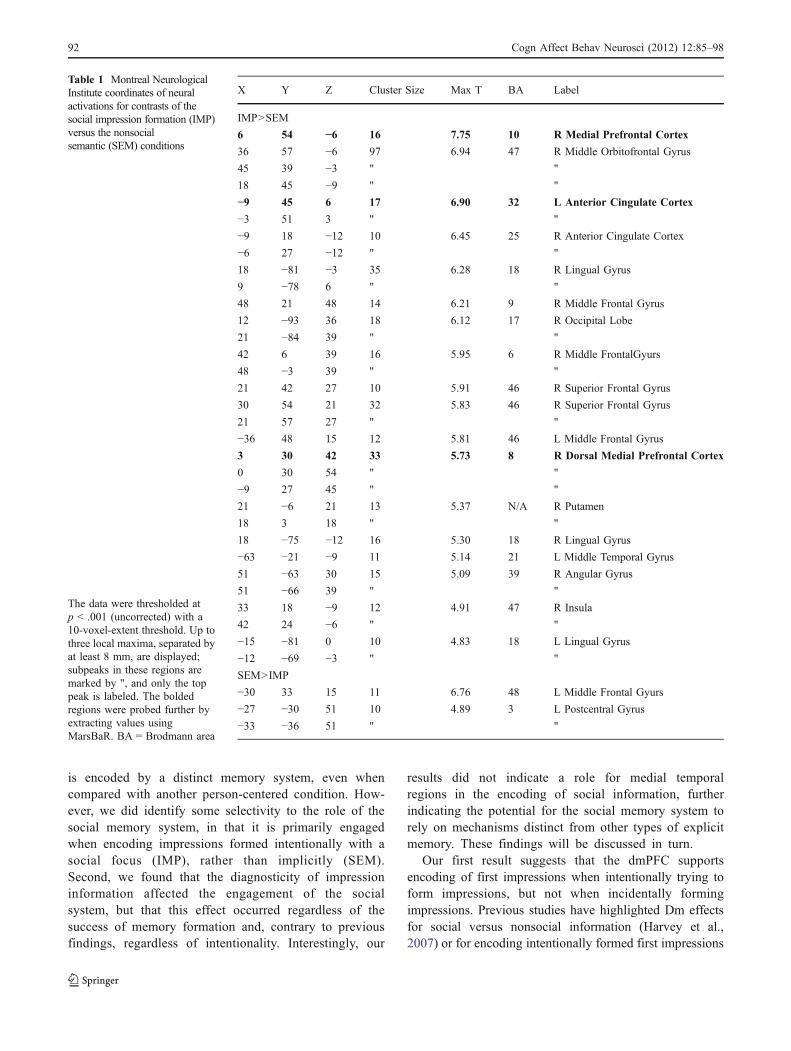
ously implicated in person evaluation, particularly medialprefrontal cortex. This contrast yielded brain regions thatresponded more to intentionally formed impressions with asocial focus (IMP) than to incidentally formed impressions(SEM) and suggested that intentional impression formationevokes more social (e.g., medial prefrontal), emotional(e.g., orbitofrontal and insula), and semantic (temporallobes) processing than does incidental impression formation(Table 1). Of the regions that emerged in the IMP > SEMcontrast, we selected two ROIs in medial prefrontal regions,along with anterior cingulate cortex, for further analysis ofmemory effects (bolded in Table 1), because in previousresearch these regions had shown robust activation inresponse to person evaluation, particularly during success-ful encoding of information into memory (Harvey et al.,2007; Mitchell, 2008; Schiller et al., 2009). No mPFC orMTL regions emerged from the contrast of SEM > IMP.
A region in dmPFC (3, 30, 42; see Fig. 3) near thosepreviously implicated in person evaluation was the onlyregion to exhibit effects of encoding success. The region wasinitially selected from the random-effects analysis because itrevealed a main effect of intentionality (verified in the ROIanalysis: F(1, 19) = 22.06, p < .001, ηp
2 = .53), with greaterdeactivation for the SEM (nonsocial) condition than forthe IMP (social) condition. In addition, there was anorthogonal main effect of diagnosticity, F(1, 19) = 331,p < .001, ηp
2 = .94, with greater deactivation for diagnostic(M = −0.88%, SD = 0.39) than for neutral sentences(M = −0.36%, SD = 0.35). In addition, we found aninteraction between intentionality and memory, F(1, 19) =4.55, p < .05, ηp
2 = .19, such that a Dm effect emerged forthe IMP condition but not for the SEM condition. Tocharacterize the nature of the memory effects acrossconditions, we conducted two-way ANOVAs separately forthe IMP (social) and SEM (nonsocial) conditions with thefactors Diagnosticity (diagnostic/neutral) and Memory (re-membered/forgotten). For intentionally formed impressions(IMP), a main effect of memory emerged, F(1, 19) = 6.34,p < .03, ηp
2 = .25, such that the dmPFC deactivated more forremembered than for forgotten sentences. For incidentally
formed impressions (SEM), we did not find a main effect ofmemory (p = .47). None of the other ROIs selected in thecontrast of IMP > SEM reached significance in ANOVAswith the Diagnosticity or Memory factors.
The contrast of diagnostic and neutral revealed a numberof regions suggesting classic memory networks (e.g.,hippocampus) and social processes (e.g., mPFC) in additionto the lingual and fusiform gyri, likely consistent withincreased processing time for diagnostic sentences (asdiscussed in Mitchell, Cloutier, et al., 2006). As in Mitchell,Cloutier, et al. (2006), we found activity in the dmPFCassociated with person perception. We performed ANOVAson medial prefrontal and MTL regions (bolded in Table 2),but none of the ROIs returned significant main effects orinteractions involving memory.
In the a priori ROI analyses of MTL regions, based onanatomical masks, no significant effects involving memoryemerged in the contrast of IMP > SEM or diagnostic >neutral in the restricted volumes. To further probe thisregion, we contrasted remembered versus forgotten col-lapsed across all conditions, and also separately estimatedthe contrast for the IMP (social) and SEM (nonsocial)conditions. These analyses also failed to reveal significanteffects.
Although a direct contrast of memory (remembered/forgotten), diagnosticity (diagnostic/neutral), and inten-tionality (semantic/intentional) pooled across participantswould have allowed for a whole-brain test of anyinteractions between all three factors, this exploratoryanalysis did not return any regions that achievedsignificance.
Discussion
Our neuroimaging results reveal two primary findings aboutthe memory system engaged in the encoding of socialinformation, one of which is related to encoding, the otherto processing social information. First, our findings con-verge with previous data indicating that social information
Fig. 2 Memory effects. Thebehavioral results indicated thatdiagnostic face–sentence pairs(leftmost bars) were betterremembered than neutral ones(rightmost bars). **p < .001
Cogn Affect Behav Neurosci (2012) 12:85–98 91
is encoded by a distinct memory system, even whencompared with another person-centered condition. How-ever, we did identify some selectivity to the role of thesocial memory system, in that it is primarily engagedwhen encoding impressions formed intentionally with asocial focus (IMP), rather than implicitly (SEM).Second, we found that the diagnosticity of impressioninformation affected the engagement of the socialsystem, but that this effect occurred regardless of thesuccess of memory formation and, contrary to previousfindings, regardless of intentionality. Interestingly, our
results did not indicate a role for medial temporalregions in the encoding of social information, furtherindicating the potential for the social memory system torely on mechanisms distinct from other types of explicitmemory. These findings will be discussed in turn.
Our first result suggests that the dmPFC supportsencoding of first impressions when intentionally trying toform impressions, but not when incidentally formingimpressions. Previous studies have highlighted Dm effectsfor social versus nonsocial information (Harvey et al.,2007) or for encoding intentionally formed first impressions
Table 1 Montreal NeurologicalInstitute coordinates of neuralactivations for contrasts of thesocial impression formation (IMP)versus the nonsocialsemantic (SEM) conditions
The data were thresholded atp < .001 (uncorrected) with a10-voxel-extent threshold. Up tothree local maxima, separated byat least 8 mm, are displayed;subpeaks in these regions aremarked by ", and only the toppeak is labeled. The boldedregions were probed further byextracting values usingMarsBaR. BA = Brodmann area
X Y Z Cluster Size Max T BA Label
IMP>SEM
6 54 −6 16 7.75 10 R Medial Prefrontal Cortex
36 57 −6 97 6.94 47 R Middle Orbitofrontal Gyrus
45 39 −3 " "
18 45 −9 " "
−9 45 6 17 6.90 32 L Anterior Cingulate Cortex
−3 51 3 " "
−9 18 −12 10 6.45 25 R Anterior Cingulate Cortex
−6 27 −12 " "
18 −81 −3 35 6.28 18 R Lingual Gyrus
9 −78 6 " "
48 21 48 14 6.21 9 R Middle Frontal Gyrus
12 −93 36 18 6.12 17 R Occipital Lobe
21 −84 39 " "
42 6 39 16 5.95 6 R Middle FrontalGyurs
48 −3 39 " "
21 42 27 10 5.91 46 R Superior Frontal Gyrus
30 54 21 32 5.83 46 R Superior Frontal Gyrus
21 57 27 " "
−36 48 15 12 5.81 46 L Middle Frontal Gyrus
3 30 42 33 5.73 8 R Dorsal Medial Prefrontal Cortex
0 30 54 " "
−9 27 45 " "
21 −6 21 13 5.37 N/A R Putamen
18 3 18 " "
18 −75 −12 16 5.30 18 R Lingual Gyrus
−63 −21 −9 11 5.14 21 L Middle Temporal Gyrus
51 −63 30 15 5.09 39 R Angular Gyrus
51 −66 39 " "
33 18 −9 12 4.91 47 R Insula
42 24 −6 " "
−15 −81 0 10 4.83 18 L Lingual Gyrus
−12 −69 −3 " "
SEM>IMP
−30 33 15 11 6.76 48 L Middle Frontal Gyurs
−27 −30 51 10 4.89 3 L Postcentral Gyrus
−33 −36 51 " "
92 Cogn Affect Behav Neurosci (2012) 12:85–98

versus memorizing order (Mitchell et al., 2004). However,both of these studies contrasted against control conditionsthat did not require participants to make evaluative, person-centered judgments. Therefore, the previous results mayreflect the act of evaluation rather than a dedicated socialprocess per se, or may reflect differences in the attentiondevoted to evaluating a single individual. By contrasting asocial task (form impression, IMP) with a person-centeredand evaluative task emphasizing semantic rather than socialcomponents (judging the location where a behavioroccurred, SEM), we showed in the present study that whenforming and encoding first impressions intentionally, thedmPFC is recruited. We believe that this result advancesour current knowledge because it better characterizes theinvolvement of the dmPFC in social evaluation, as well ashighlighting its role in encoding first impressions intomemory.
Somewhat surprisingly, this result is based on differ-ences in deactivations, as opposed to previous studies inwhich activations have been found (Mitchell et al., 2004).Closer scrutiny of our neural data in the remembered tasksacross both intentional- and incidental-encoding tasksreveals that the differences across conditions seem to bedriven by the forgotten rather than by the rememberedtrials. We believe this pattern of deactivation is in line withthe activity of the “task-negative” network of the brain, or
the “default mode” network (Buckner, Andrews-Hanna, &Schacter, 2008; Raichle et al., 2001). Previous studies havefound that activity of the default mode network hampersencoding, such that deactivating it would support bettermemory performance (Daselaar et al., 2009). Whileposterior regions, such as posterior cingulate, precuneus,and bilateral ventral posterior parietal cortices, haveemerged more consistently in the literature, some workhas implicated anterior regions as well, such as anteriorcingulate and medial prefrontal cortices, such that deacti-vation supports successful encoding (Kim, Daselaar, &Cabeza, 2010).
Though the previous studies did not implicate dmPFC aspart of the task-negative network, it is possible that thesocial nature of our stimuli and the unique neural regionsengaged to encode this type of information account forthese differences. Previous studies (Daselaar et al., 2009;Kim et al., 2010) focused on the encoding of words, scenes,and faces, but did not incorporate stimuli relevant tosocioemotional goals. Under such conditions, dmPFC couldbe associated with the network of regions deactivatedduring encoding and activated during retrieval. Deactivat-ing this network to support encoding processes implies thatfocusing on internal processes detracts from the ability tofocus on external stimuli and successfully encode them. Inour task, this could mean that focusing too much on internal
Fig. 3 Subsequent memoryeffects in dmPFC. A region inthe dmPFC (3, 30, 42), whichemerged from the contrast ofimpression formation (IMP, so-cial) > semantic (SEM, nonso-cial), showed an interaction oforientation with memory. Rstands for “remembered,” and Ffor “forgotten.” The region con-tributed to memory formationfor the IMP condition, but notthe SEM condition
Cogn Affect Behav Neurosci (2012) 12:85–98 93

cognition, perhaps retrieving autobiographical memories offamiliar individuals with appearances similar to the targetstimulus or creating associations based on facial featuresalone, impairs one’s ability to form a memory trace of theface–behavior association presented in the study (seeShrager, Kirwan, & Squire, 2008, for a discussion ofsimilar effects in the hippocampus). This is consistent withour pattern of results showing less deactivation (i.e., greateractivity) in the forgotten trials of the impression condition,
perhaps reflecting a failure to inhibit distracting internalassociations that hamper successful memory formation.Although one might expect to see a similar pattern forremembered versus forgotten trials in the semantic condition(incidentally forming impressions), it may be that internalinformation is most interfering when one is focused onimpression formation. Thus, when trying to intentionally formimpressions, inhibiting interference is important in order forone to later remember externally presented information
Table 2 Montreal NeurologicalInstitute coordinates of neuralactivations for the contrasts ofdiagnostic (DIAG) versusneutral (NEU)
The data were thresholded at p <.001 (uncorrected) with a 10-voxel-extent threshold. Up tothree local maxima, separated byat least 8 mm, are displayed;subpeaks in these regions aremarked by ", and only the toppeak is labeled. The boldedregions were probed further byextracting values using Mars-BaR. BA = Brodmann area
X Y Z Cluster Size Max T BA Label
DIAG > NEU
−12 −81 −6 14,627 100.03 18 L Lingual Gyrus
−9 −75 3 " "
51 −18 −18 " "
3 48 48 145 35.91 9 R Medial Prefrontal Cortex
−9 54 45 " "
−15 54 30 " "
−27 51 −33 12 31.09 N/A N/A (out of brain)
−18 −51 75 14 22.50 5 R Postcentral Gyrus
−24 51 −42 " "
−33 42 39 23 20.69 40 L Inferior Parietal
−3 21 −6 21 20.24 25 L Anterior Cingulate Cortex
−48 −15 0 22 19.99 48 L Superior Temporal Gyrus
−15 −66 −30 17 17.64 N/A L Cerebellum
−6 −78 −30 " "
12 −66 −33 14 16.32 N/A R Cerebellum
−27 −45 24 10 14.62 N/A White Matter
9 72 9 10 13.75 10 R Medial Prefrontal Cortex
9 69 21 " "
0 57 15 27 13.66 10 Dorsal Medial Prefrontal Cortex
12 −18 −39 10 11.65 N/A N/A (out of brain)
0 −57 −18 23 11.47 N/A Cerebellum
30 81 −18 16 11.28 19 R Fusiform Gyrus
21 12 57 11 11.20 6 R Superior Frontal Gyrus
−21 −3 −18 16 11.17 N/A L Hippocampus
51 −24 21 12 10.21 40 R Postcentral Gyrus
36 −27 −3 13 10.15 N/A R Hippocampus
−6 −39 69 14 8.78 4 L Paracentral
−48 −69 −15 10 8.75 19 L Fusiform Gyrus
NEU > DIAG
45 −45 48 30,757 66.22 40 R Inferior Parietal Lobe
−39 −21 63 " "
42 −42 39 " "
−33 51 −48 107 63.75 N/A N/A (out of brain)
−39 60 −36 " "
−30 48 −39 " "
33 54 −36 119 46.61 N/A N/A (out of brain)
27 54 −42 " "
36 51 −48 " "
94 Cogn Affect Behav Neurosci (2012) 12:85–98

leading to an impression. Future research explicitly testing thebehavioral and neural effects of potential interference, such asfrom facial characteristics (e.g., Zebrowitz & Montepare,2008), during the encoding of face–behavior pairings wouldhelp to resolve this question, particularly if the effects varywith the goals of the task (e.g., incidental vs. intentionalimpression formation).
Our second major finding is that diagnostic informationthat easily lends itself to forming an impression deactivatesthe dmPFC more than does neutral information. We wouldexpect that diagnostic information would engage regionsimplicated in forming impressions more strongly thanwould neutral information, regardless of orientation. Thisis because diagnostic information lends itself more easilytoward forming trait inferences (as shown by Uleman et al.,2008). However, Mitchell, Cloutier, et al. (2006) found thatdiagnostic information fails to engage the dmPFC morethan does neutral information when one intentionally formsimpressions, and that diagnosticity differentially affectsneural engagement only when one incidentally (uncon-sciously) forms impressions. Mitchell, Cloutier, et al.’sinterpretation was that when one is intentionally formingimpressions, everything is “diagnostic” (even neutralinformation), but when unconsciously forming impressions,only diagnostic information activates frontal regions asso-ciated with forming impressions.
In contrast to their findings, we find that both intentionaland incidental impression formation engage the dmPFC,such that it deactivates more for diagnostic than for neutralinformation. In conjunction with our finding of bettermemory for diagnostic than for neutral face–sentence pairs,our data lend support to the notion that increased depth ofprocessing may be associated with dmPFC activity forsocial tasks. This idea is consistent with previous behav-ioral studies showing that depth of processing contributes tobetter memory (Craik & Lockhart, 1972; Craik & Tulving,1975) and, in the social domain, that making complexjudgments about peoples’ traits leads to better memory thando simple judgments of their sex (Bower & Karlin, 1974;Wenger & Ingvalson, 2002). Again, our pattern of greaterdeactivation, rather than activation, for diagnostic informa-tion is surprising, but perhaps reflects the mismatchbetween behavioral information and facial appearance,which is more salient for diagnostic than for neutral trials.Relative to prior studies that did not find this pattern, theneed to integrate face and behavior could be more salientfor one-shot impression formation tasks, as were employedin the present study.
Another possible explanation for differences from theprior studies may lay in the different designs employedacross studies. Though both we and Mitchell et al. (2004)used similar statements in our studies, our procedure wasvery different, in that we presented each face once, paired
with a single unique sentence, whereas Mitchell et al.(2004) presented each face paired with 10 differentsentences. Pooling impression information across multipletrials may decrease the importance of diagnostic informa-tion on any single trial when intentionally forming animpression of an individual. Our use of a single actor–single behavior design may be more consistent withresearch on spontaneous trait inferences (STI), accordingto Uleman et al.’s (2008) claim that in order to generate themost robust STI, one must be presented with a single orvery few related behaviors and integrate these with an actorrepresentation. In contrast, “integrating meanings and/orevaluations of one target’s many behaviors is less likely tooccur spontaneously and requires high levels of relevantchronic goals” (p. 333). This argument indicates that a morenaturalistic setting, in which we form impressions based ona range of behaviors, is not ideal for forming a lasting,distinct first impression. With a single defined behavior,there is evidence that first impressions occur spontaneously,even in the absence of conscious effort to create animpression (Ambady, Krabbenhoft, & Hogan, 2006;Todorov & Uleman, 2002, 2003; Uleman et al., 2008).
Because our study presented only a single sentence foreach person, this might have increased the perceiveddiagnosticity of each sentence, in contrast to studies withmultiple sentences converging on a single trait. Distinctimpression formation processes may be recruited when oneis more concerned with updating and comparing impres-sions to current knowledge (e.g., Schiller et al., 2009), asopposed to forming initial impressions (as in our study).Although more research will be needed to elucidate theunderlying processes that contribute to encoding firstimpressions, we believe that our finding highlights thesensitivity of dmPFC to diagnostic information, regardlessof the state of mind one adopts. One would expect that asystem that operates as seemingly automatic, such asimpression formation (Todorov et al., 2007; J. Willis &Todorov, 2006), would be sensitive to the type ofinformation at all times, whether impressions are incidentalor intentional.
Surprisingly, diagnosticity did not influence the role thatthe dmPFC played in encoding impressions. This isparticularly unexpected because the behavioral memorymeasures indicated that diagnostic information was betterremembered than neutral information. While this suggeststhat we are better at encoding impressions that are based onmeaningful behavior than impressions that do not containtrait diagnostic information, one might also expect inten-tionality to impact memory (e.g., Mitchell et al., 2004).This was not the case for our data: Intentionally andincidentally formed impressions were encoded equally well.However, orientation did affect the engagement of dmPFCduring successful versus unsuccessful encoding trials. This
Cogn Affect Behav Neurosci (2012) 12:85–98 95

apparent inconsistency between the behavioral and neuralmeasures of memory may reflect the greater sensitivity ofneural measures in some instances, allowing us to reveal acontribution of intentionality using neural measures. However,this potential for greater sensitivity may rely heavily on theselectivity of particular regions for specific processes.
Our results are consistent with those of Mitchell et al.(2004) in indicating the lack of an MTL contribution to theencoding of social information. This is surprising given thepervasive nature of MTL contributions to explicit memory(Shrager et al., 2008; Squire, 2009; Tulving & Markowitsch,1998), although a small body of literature has suggested thatamnesics may be able to form new memories of impressionsof others under some circumstances, despite MTL impair-ment (Johnson, Kim, & Risse, 1985; Todorov & Olson,2008). Importantly, MTL regions that did emerge in ourdiagnostic > neutral contrast did not show a Dm effect. Thiswas further tested using an anatomical MTL mask in order tohave greater sensitivity to detect effects, and these resultsalso indicated that MTL regions do not respond significantlyor differentiate between social and nonsocial tasks orbetween information that is diagnostic or neutral. Althoughwe are limited in the inferences that we can make based on anull finding, our data add to the growing evidence that socialmemory formation may not be MTL dependent.
While our focus was on mPFC and MTL contributionsto the encoding of impressions, our analyses also probedeffects of the orientation and content of information morebroadly. While some of the regions implicated in intentionalover incidental impression formation are consistent withprior studies (Schiller et al., 2009) that have suggested arole of emotion (e.g., insula) and face processing (e.g.,fusiform) in forming impression of others, our results alsoidentify some novel regions, such as the anterior cingulatecortex (ACC). The ACC is known to be involved indecision making and conflict monitoring (Pochon, Riis,Sanfey, Nystrom, & Cohen, 2008), which might suggestthat intentional impression formation involves deeperprocessing and may allow one to be better able to accountfor ambiguity or inconsistency (e.g., a pretty face engagedin an ugly behavior). Notably, the reverse contrast (SEM >IMP) did not produce any MTL regions, suggesting that ourcontrast was successful in contrasting social with nonsocialevaluation while avoiding activation of classic memorynetworks.
While we have achieved some success in differentiatingconditions loading more heavily on social information, suchas diagnosticity and intentional trait impressions, fromthose that invoke these processes less, it would have beenhelpful to have a true control condition that did not involvesocial information. This would have allowed us to furtherdifferentiate social from nonsocial information processing,potentially allowing us to more directly investigate the role
of the MTL in encoding nonsocial and social information.In addition, the relatively lengthy trials might have resultedin some blurring of the intentional trait impressioncondition and the unintentional semantic condition. Theamount of time available in which to deeply processinformation, as well as the interspersing of trial typeswithin a participant, might have encouraged participants toform impressions even on the semantic trials. Such apossibility may account for the lack of memory differencesacross these conditions.
In conclusion, our finding that encoding first impres-sions relies on the dmPFC only when intentionally trying toform impressions highlights the importance of orientationand the unique role played by this region when intention-ally forming first impressions. It adds to our currentknowledge in that it shows that this is true not only incomparison to a nonsocial sequencing task, but also whencompared to a more nuanced, person-centered evaluativetask. In addition, we found a role for the dmPFC inprocessing diagnostic information that easily lends itself toimpression formation, as compared to neutral information.This highlights the role of the region as being dedicated toforming first impressions, and it may indicate the impor-tance of the task in engaging this region. Compared withprevious findings, our results suggest that the processesdiffer when impressions are based on single versus multiplebehaviors. Further investigations will likely highlight thetypes of diagnostic information that most impact impressionformation, the effects of multiple versus singular impres-sion formation, and the roles of these regions duringretrieval of first impressions from memory.
Author note The authors thank Leslie Zebrowitz, Don Katz, andJason Mitchell for valuable feedback in the initial stages of theresearch; Nancy Dennis for helpful suggestions; and Jennifer Colemanfor assistance with data collection and analysis. The project waspartially supported by the Brandeis University Theodore and JaneNorman Fund and the National Institute on Aging (Grant R21AG032382), and portions of the research were conducted while A.H.G. was a fellow of the American Federation for Aging Research.The Athinoula A. Martinos Center for Biomedical Imaging issupported by the National Center for Research Resources (Grant P41RR14075) and by the MIND Institute. R.G. is now located at Tel AvivUniversity.
References
Ambady, N., Krabbenhoft, M. A., & Hogan, D. (2006). The 30-secsale: Using thin-slice judgments to evaluate sales effectiveness.Journal of Consumer Psychology, 16, 4–13.
Ambady, N., & Rosenthal, R. (1993). Half a minute: Predictingteacher evaluations from thin slices of nonverbal behavior andphysical attractiveness. Journal of Personality and SocialPsychology, 64, 431–441. doi:10.1037/0022-3514.64.3.431
96 Cogn Affect Behav Neurosci (2012) 12:85–98

Ames, D. L., Fiske, S. T., & Todorov, A. (2011). Impressionformation: A focus on others’ intents. In J. Decety & J. Cacioppo(Eds.), Oxford handbook of social neuroscience (pp. 419–433).New York, NY: Oxford University Press.
Amodio, D. M., & Frith, C. D. (2006). Meeting of minds: The medialfrontal cortex and social cognition. Nature Reviews Neurosci-ence, 7, 268–277. doi:10.1038/nrn1884
Bower, G. H., & Karlin, M. B. (1974). Depth of processing pictures offaces and recognition memory. Journal of Experimental Psychol-ogy, 103, 751–757. doi:10.1037/h0037190
Brett, M., Anton, J.-L., Valabregue, R., & Poline, J.-B. (2002). Regionof interest analysis using an SPM toolbox. Sendai, Japan: Paperpresented at the 8th International Conference on FunctionalMapping of the Human Brain.
Buckner, R. L., Andrews-Hanna, J. R., & Schacter, D. L. (2008). Thebrain’s default network: Anatomy, function, and relevance todisease. In A. Kingstone & M. B. Miller (Eds.), The year incognitive neuroscience 2008 (Annals of the New York Academyof Sciences, Vol. 1124, pp. 1–38). Malden, MA: Blackwell.doi:10.1196/annals.1440.011
Cloutier, J., Kelley, W. M., & Heatherton, T. F. (2010). The influenceof perceptual and knowledge-based familiarity on the neuralsubstrates of face perception. Social Neuroscience, 6, 63–75.doi:10.1080/17470911003693622
Craik, F. I. M., & Lockhart, R. S. (1972). Levels of processing: Aframework for memory research. Journal of Verbal Learning andVerbal Behavior, 11, 671–684. doi:10.1016/S0022-5371(72)80001-X
Craik, F. I. M., & Tulving, E. (1975). Depth of processing and retention ofwords in episodic memory. Journal of Experimental Psychology.General, 104, 268–294. doi:10.1037/0096-3445.104.3.268
D’Argembeau, A., Ruby, P., Collette, F., Degueldre, C., Balteau, E.,Luxen, A., & Salmon, E. (2007). Distinct regions of the medialprefrontal cortex are associated with self-referential processingand perspective taking. Journal of Cognitive Neuroscience, 19,935–944. doi:10.1162/jocn.2007.19.6.935
Dale, A. M. (1999). Optimal experimental design for event-relatedfMRI. Human Brain Mapping, 8, 109–114.
Daselaar, S. M., Prince, S. E., Dennis, N. A., Hayes, S. M., Kim, H.,& Cabeza, R. (2009). Posterior midline and ventral parietalactivity is associated with retrieval success and encoding failure.Frontiers in Human Neuroscience, 3, 13. doi:10.3389/neuro.09.013.2009
Delgado, M. R., Nystrom, L. E., Fissell, C., Noll, D. C., & Fiez, J. A.(2000). Tracking the hemodynamic responses to reward andpunishment in the striatum. Journal of Neurophysiology, 84,3072–3077.
Eichenbaum, H. (2004). Hippocampus: Cognitive processes andneural representations that underlie declarative memory. Neuron,44, 109–120. doi:10.1016/j.neuron.2004.08.028
Gutchess, A. H., Kensinger, E. A., & Schacter, D. L. (2010). Functionalneuroimaging of self-referential encoding with age. Neuropsycho-logia, 48, 211–219. doi:10.1016/j.neuropsychologia.2009.09.006
Haas, B. W., & Canli, T. (2008). Emotional memory function, personalitystructure and psychopathology: A neural system approach to theidentification of vulnerability markers. Brain Research Reviews, 58,71–84. doi:10.1016/j.brainresrev.2007.10.014
Harvey, P.-O., Fossati, P., & Lepage, M. (2007). Modulation ofmemory formation by stimulus content: Specific role of themedial prefrontal cortex in the successful encoding of socialpictures. Journal of Cognitive Neuroscience, 19, 351–362.doi:10.1162/jocn.2007.19.2.351
Heberlein, A. S., & Saxe, R. R. (2005). Dissociation betweenemotion and personality judgments: Convergent evidence fromfunctional neuroimaging. NeuroImage, 28, 770–777.doi:10.1016/j.neuroimage.2005.06.064
Hedden, T., Park, D. C., Nisbett, R., Ji, L.-J., Jing, Q., & Jiao, S.(2002). Cultural variation in verbal versus spatial neuropsycho-logical function across the life span. Neuropsychology, 16, 65–73. doi:10.1037/0894-4105.16.1.65
Ishai, A. (2007). Sex, beauty and the orbitofrontal cortex. Interna-tional Journal of Psychophysiology, 63, 181–185. doi:10.1016/j.ijpsycho.2006.03.010
Johnson, M. K., Kim, J. K., & Risse, G. (1985). Do alcoholicKorsakoff’s syndrome patients acquire affective reactions?Journal of Experimental Psychology: Learning, Memory, andCognition, 11, 22–36. doi:10.1037/0278-7393.11.1.22
Kim, H., Daselaar, S. M., & Cabeza, R. (2010). Overlapping brainactivity between episodic memory encoding and retrieval: rolesof the task-positive and task-negative networks. NeuroImage, 49,1045–1054. doi:10.1016/j.neuroimage.2009.07.058
Langlois, J. H., Kalakanis, L., Rubenstein, A. J., Larson, A., Hallam,M., & Smoot, M. (2000). Maxims or myths of beauty? A meta-analytic and theoretical review. Psychological Bulletin, 126, 390–423. doi:10.1037/0033-2909.126.3.390
Lehn, H., Steffenach, H. A., van Strien, N. M., Veltman, D. J., Witter, M.P., & Haberg, A. K. (2009). A Specific role of the humanhippocampus in recall of temporal sequences. Journal of Neurosci-ence, 29, 3475–3484. doi:10.1523/jneurosci.5370-08.2009
Liang, X. Y., Zebrowitz, L. A., & Zhang, Y. (2010). Neural activationin the oreward circuito shows a nonlinear response to facialattractiveness. Social Neuroscience, 5, 320–334. doi:10.1080/17470911003619916
Ma, N., Vandekerckhove, M., Van Overwalle, F., Seurinck, R., & Fias,W. (2011). Spontaneous and intentional trait inferences recruit acommon mentalizing network to a different degree: Spontaneousinferences activate only its core areas. Social Neuroscience, 6,123–138. doi:10.1080/17470919.2010.485884
Macrae, C. N., Moran, J. M., Heatherton, T. F., Banfield, J. F., & Kelley,W. M. (2004). Medial prefrontal activity predicts memory for self.Cerebral Cortex, 14, 647–654. doi:10.1093/cercor/bhh025
Maldjian, J. A., Laurienti, P. J., Kraft, R. A., & Burdette, J. H. (2003).An automated method for neuroanatomic and cytoarchitectonicatlas-based interrogation of fMRI data sets. NeuroImage, 19,1233–1239. doi:10.1016/s1053-811900169-1
Mason, M. F., Dyer, R., & Norton, M. I. (2009). Neural mechanisms ofsocial influence. Organizational Behavior and Human DecisionProcesses, 110, 152–159. doi:10.1016/j.obhdp.2009.04.001
Mitchell, J. P. (2008). Contributions of functional neuroimaging to thestudy of social cognition. Current Directions in PsychologicalScience, 17, 142–146. doi:10.1111/j.1467-8721.2008.00564.x
Mitchell, J. P., Ames, D. L., Jenkins, A. C., & Banaji, M. R. (2009).Neural correlates of stereotype application. Journal of CognitiveNeuroscience, 21, 594–604. doi:10.1162/jocn.2009.21033
Mitchell, J. P., Cloutier, J., Banaji, M. R., & Macrae, C. N. (2006a).Medial prefrontal dissociations during processing of trait diag-nostic and nondiagnostic person information. Social Cognitiveand Affective Neuroscience, 1, 49–55. doi:10.1093/scan/nsl007
Mitchell, J. P., Macrae, C. N., & Banaji, M. R. (2004). Encoding-specific effects of social cognition on the neural correlates ofsubsequent memory. Journal of Neuroscience, 24, 4912–4917.doi:10.1523/JNEUROSCI.0481-04.2004
Mitchell, J. P., Macrae, C. N., & Banaji, M. R. (2005). Formingimpressions of people versus inanimate objects: Social–cognitiveprocessing in the medial prefrontal cortex. NeuroImage, 26, 251–257. doi:10.1016/j.neuroimage.2005.01.031
O’Kane, G., Kensinger, E. A., & Corkin, S. (2004). Evidence forsemantic learning in profound amnesia: An investigation withpatient HM. Hippocampus, 14, 417–425. doi:10.1002/hipo.20005
Olivola, C. Y., & Todorov, A. (2010). Elected in 100 milliseconds:Appearance-based trait inferences and voting. Journal of Non-verbal Behavior, 34, 83–110. doi:10.1007/s10919-009-0082-1
Cogn Affect Behav Neurosci (2012) 12:85–98 97

Pochon, J. B., Riis, J., Sanfey, A. G., Nystrom, L. E., & Cohen, J. D.(2008). Functional imaging of decision conflict Journal of Neuro-science, 28, 3468–3473. doi:10.1523/JNEUROSCI.4195-07.2008
Raichle, M. E., MacLeod, A. M., Snyder, A. Z., Powers, W. J.,Gusnard, D. A., & Shulman, G. L. (2001). A default mode ofbrain function. Proceedings of the National Academy of Sciences,98, 676–682. doi:10.1073/pnas.98.2.676
Rule, N. O., & Ambady, N. (2008). First impressions: Peeking at theneural underpinnings. In N. Ambady & J. J. Skowronski (Eds.),First impressions (pp. 35–56). New York, NY: Guilford Press.
Said, C. P., Baron, S. G., & Todorov, A. (2009). Nonlinear amygdalaresponse to face trustworthiness: Contributions of high and lowspatial frequency information. Journal of Cognitive Neurosci-ence, 21, 519–528. doi:10.1162/jocn.2009.21041
Salthouse, T. A., & Babcock, R. L. (1991). Decomposing adult agedifferences in working memory. Developmental Psychology, 27,763–776. doi:10.1037/0012-1649.27.5.763
Saxe, R., Xiao, D. K., Kovacs, G., Perrett, D. I., & Kanwisher, N.(2004). A region of right posterior superior temporal sulcusresponds to observed intentional actions. Neuropsychologia, 42,1435–1446. doi:10.1016/j.neuropsychologia.2004.04.015
Schacter, D. L., Gutchess, A. H., & Kensinger, E. A. (2009). Specificityof memory: Implications for individual and collective remembering.In P. Boyer & J. V.Wertsch (Eds.),Memory in mind and culture (pp.83–111). New York, NY: Cambridge University Press.
Schiller, D., Freeman, J. B., Mitchell, J. P., Uleman, J. S., & Phelps, E.A. (2009). A neural mechanism of first impressions. NatureNeuroscience, 12, 508–514. doi:10.1038/nn.2278
Shipley, W. C. (1986). Shipley Institute of Living Scale. Los Angeles:Western Psychological Services.
Shrager, Y., Kirwan, C. B., & Squire, L. R. (2008). Activity in bothhippocampus and perirhinal cortex predicts the memory strengthof subsequently remembered information. Neuron, 59, 547–553.doi:10.1016/j.neuron.2008.07.022
Slotnick, S. D., Moo, L. R., Segal, J. B., & Hart, J., Jr. (2003). Distinctprefrontal cortex activity associated with item memory andsource memory for visual shapes. Cognitive Brain Research,17, 75–82. doi:10.1016/S0926-6410(03)00082-X
Squire, L. R. (2004). Memory systems of the brain: A brief historyand current perspective. Neurobiology of Learning and Memory,82, 171–177. doi:10.1016/j.nlm.2004.06.005
Squire, L. R. (2009). The legacy of patient HM for neuroscience.Neuron, 61, 6–9. doi:10.1016/j.neuron.2008.12.023
Todorov, A., Gobbini, M. I., Evans, K. K., & Haxby, J. V. (2007).Spontaneous retrieval of affective person knowledge in faceperception. Neuropsychologia, 45, 163–173. doi:10.1016/j.neuropsychologia.2006.04.018
Todorov, A., & Olson, I. R. (2008). Robust learning of affective traitassociations with faces when the hippocampus is damaged, but notwhen the amygdala and temporal pole are damaged. Social Cognitiveand Affective Neuroscience, 3, 195–203. doi:10.1093/scan/nsn013
Todorov, A., & Uleman, J. S. (2002). Spontaneous trait inferences arebound to actors’ faces: Evidence from a false recognition
paradigm. Journal of Personality and Social Psychology, 83,1051–1065. doi:10.1037/0022-3514.83.5.1051
Todorov, A., & Uleman, J. S. (2003). The efficiency of bindingspontaneous trait inferences to actors’ faces. Journal of Exper-imental Social Psychology, 39, 549–562. doi:10.1016/s0022-103100059-3
Tulving, E., & Markowitsch, H. J. (1998). Episodic and declarativememory: Role of the hippocampus. Hippocampus, 8, 198–204.
Uleman, J. S., Saribay, S. A., & Gonzalez, C. M. (2008). Spontaneousinferences, implicit impressions, and implicit theories. AnnualReview of Psychology, 59, 329–360. doi:10.1146/annurev.psych.59.103006.093707
Van Overwalle, F. (2009). Social cognition and the brain: A meta-analysis. Human Brain Mapping, 30, 829–858. doi:10.1002/hbm.20547
VanOverwalle, F., & Labiouse, C. (2004). A recurrent connectionist modelof person impression formation. Personality and Social PsychologyReview, 8, 28–61. doi:10.1207/S15327957PSPR0801_2
Verhulst, B., Lodge, M., & Lavine, H. (2010). The attractiveness halo:Why some candidates are perceived more favorably than others.Journal of Nonverbal Behavior, 34, 111–117. doi:10.1007/s10919-009-0084-z
Wagner, A. D., Schacter, D. L., Rotte, M., Koutstaal, W., Maril, A.,Dale, A. M., & Buckner, R. L. (1998). Building memories:Remembering and forgetting of verbal experiences as predictedby brain activity. Science, 281, 1188–1191. doi:10.1126/science.281.5380.1188
Wenger, M. J., & Ingvalson, E. M. (2002). A decisional component ofholistic encoding. Journal of Experimental Psychology: Learn-ing, Memory, and Cognition, 28, 872–892. doi:10.1037/0278-7393.28.5.872
Willis, J., & Todorov, A. (2006). First impressions: Making up yourmind after a 100-ms exposure to a face. Psychological Science,17, 592–598. doi:10.1111/j.1467-9280.2006.01750.x
Willis, M. L., Palermo, R., Burke, D., McGrillen, K., & Miller, L.(2010). Orbitofrontal cortex lesions result in abnormal socialjudgements to emotional faces. Neuropsychologia, 48, 2182–2187. doi:10.1016/j.neuropsychologia.2010.04.010
Winston, J. S., Henson, R. N. A., Fine-Goulden, M. R., & Dolan, R. J.(2004). fMRI-adaptation reveals dissociable neural representa-tions of identity and expression in face perception. Journal ofNeurophysiology, 92, 1830–1839. doi:10.1152/jn.00155.2004
Winston, J. S., Strange, B. A., O’Doherty, J., & Dolan, R. J. (2002).Automatic and intentional brain responses during evaluation oftrustworthiness of faces. Nature Neuroscience, 5, 277–283.doi:10.1038/nn816
Zebrowitz, L. A., & McDonald, S. M. (1991). The impact of litigants’baby-facedness and attractiveness on adjudications in smallclaims courts. Law and Human Behavior, 15, 603–623.doi:10.1007/BF01065855
Zebrowitz, L. A., & Montepare, J. M. (2008). Social psychologicalface perception: Why appearance matters. Social and PersonalityPsychology Compass, 2, 1497–1517.
98 Cogn Affect Behav Neurosci (2012) 12:85–98