regulatory t cells preventing detrimental autoimmune reactions jurnal sar aun qa/2013/5.pdf · tis,...
TRANSCRIPT
Send Orders for Reprints to [email protected]
The Open Autoimmunity Journal, 2013, 5, 1-5 1
1876-8946/13 2013 Bentham Open
Open Access
CD4+CD25+ Regulatory T Cells Preventing Detrimental Autoimmune Reactions
Muhaimin Rifa’i*
Department of Biology, Faculty of Sciences, Brawijaya University, Jl. Veteran Malang 65145, East Java, Indonesia
Abstract: Objective: Autoimmune diseases will occur when the immune response inflicts damage to tissues in the body. Attempts to overcome this disease have a lot to do but they have not yielded satisfying results. In this study, the function of CD4+CD25+ regulatory T cells was evaluated for therapeutic potential to prevent the development of autoreactive T cells.
Methods: The isolation of CD4+CD25+ regulatory T cells was performed by using FACS vantage. It was conducted by taking a spleen of 6-week-old C57BL/6 mice. Cells having high purity (2x10⁶) were intravenously injected into 3-week-old CD122-/- mice. In this experiment we used congeneic mice as donors and transferred hosts. The result of adoptive transfer was examined after 7 weeks.
Result: Here we showed that the transfer of CD4+CD25+ regulatory T cells into CD122-/- mice significantly prevented the development of abnormal T cells. The injection of CD4+CD25+ regulatory T cells derived from normal young mice into CD122-/- mice could prevent the development of activated memory T cells and these cells were essential for maintaining normal homeostasis (P <0.01).
Conclusion: In this study we obtained evidence that regulatory T cells (2x10⁶) derived from normal young mice could overcome diseases when transferred to the mouse models of autoimmune disorder.
Keywords: Regulatory T cells, autoimmune disease, autoreactive, CD4+CD25+.
INTRODUCTION
An autoimmune disorder is a condition occuring when the immune system mistakenly attacks and destroys healthy body tissue. These diseases are increased because of life style and genetic problem. One of the most important strate-gies to overcome the diseases is the use of regulatory T cells as therapy. Currently scientists classify regulatory T cells into two major groups, i.e., professional regulatory T cells and induced regulatory T cells [1-5]. Professional regulatory T cells emerge from thymus, whereas induced regulatory T cells emerges due to micro-environmental influence. Many types of regulatory T cells have been proposed, but CD4+CD25+ population is the most potent regulator which most researchers believe. In this study we show the importance of CD4+CD25+ regulatory as the component of immune system which can “cease-fire” between autoreactive cells in order not to damage tissue in the body. The immune system has evolved several mechanisms to control the ex-pansion and differentiation of activated T cell, including anergy, death, and regulation. Normally, immune system reactions are required to prevent a body against pathogen and tumor cells, but they can be harmful for an individual. Therefore, the immune system must be tightly regulated in order not to attack self-antigen [6, 7]. Currently it is known
*Address correspondence to this author at the Department of Biology, Faculty of Sciences, Brawijaya University, Jl. Veteran Malang 65145, East Java, Indonesia; Tel: 62-341-554403; Fax: 62-341-554403; E-mail: [email protected]
that regulatory T cells play an important role in such regulation. CD4+CD25+ T cells are the most important part of T cells known and well-documented as a potent regulatory T cell [8-10]. For better understanding of autoimmune disor-der, the mouse models of this disease have been engineered, namely CD122 knockout genes (CD122-/-). CD122 (IL-2/IL-15 receptor chain)-deficient mice are representative models of autoimmune disease with many symptoms such as gastri-tis, oophritis, orchitis, thyroiditis, pancreatitis, and colitis. CD122 (IL-2/IL-15 receptor chain)-deficient mice exhibit severe hyper-immunity [11], with augmented granulopoiesis and suppressed erythropoiesis. Severe anemia progresses and the mice die within 3-4 months after birth. The generation mechanism of activated memory-type T cells in CD122-/- mice is controversial. According to Rifa’i et al. [14], un-healthy condition in CD122-/- is due to autoreactivity of T cells. In these mice most T cells become activated memory and very few exist in naïve phenotype. Abbas and others [12, 13], state that the programmed cell death (apoptosis) defect in IL-2-/- or CD25-/- T cells can be applied to the survival mechanism of activated IL-2Rβ-deficient T cells, and such possibility has not been excluded. Until now there has been no direct evidence for which population among CD4 and CD8 T cells responsible to regulate normal homeostasis. It is important to look for an explanation either the loss of the regulatory system may underlie the occurrence of reactivity T cells, or another necessary factor such defective expression of CD122 molecules is required. In this study, we show that injection of CD4+CD25+ regulatory cells derived from

2 The Open Autoimmunity Journal, 2013, Volume 5 Muhaimin Rifa’i
normal mice into 3-week-old CD122-/- mice makes these hosts healthy and causes the phenotype to develop normally. This result indicates that CD4+CD25+ T cell population con-tains novel regulatory T cells that effectively control CD122-deficient T cells. The most important result is the fact that CD4+CD25+ regulatory T cells can ameliorate the condition of the mice that have been already sick for three weeks. This finding opens up opportunities to use regulatory T cells as therapeutic strategies for diseases involving cell activation. Since Rifa’i et al. [14] report that the usage of CD8+CD122+ regulatory T cells has a potential to inhibit autoimmune reaction in CD122-deficient mice when applied in neonate, it is important to combine these two regulatory T cells together.
MATERIALS AND METHODS
Animal
In this experiment we used C57BL/6-CD45.1/CD45.1 and CD122-/-- CD45.2/CD45.2 mice. They were housed in pathogen free facility. Regulatory T cells were isolated from C57BL/6-CD45.1/CD45.1 normal mice, while the hosts were CD122-/-- CD45.2/CD45.2 mice.
Antibodies
Phycoerythrin (PE) or allophycocyanin (APCn)-conjugated anti-mouse CD4 (clone GK1.5), FITC- or biotin-conjugated anti-mouse CD45.1 (clone A20), Fluorescein isothiocyanate (FITC)-conjugated anti-mouse CD44 (clone IM7), (FITC)-conjugated anti-mouse CD8 (clone 53-6.7), PE-conjugated anti-mouse CD62L (clone MEL-14), TER-119 (clone TER-119) antibodies were purchased from eBio-science Inc. (San Diego, CA). For FITC-conjugated anti-mouse CD69 (clone H1.2F3), biotin-conjugated anti-mouse CD122 (clone 5H4), FITC-conjugated anti-mouse CD25 (clone PC61.5), and FITC-conjugated anti-mouse Gr-1 (clone RB6-8C5), when we used biotin-conjugated antibod-ies, we visualized the antibodies by streptavidin-PE-Cy5 (eBioscience, San Diego, CA).
Flow Cytometry and Cell Sorting
To obtain highly purified CD4+CD25+ regulatory T cells we performed cell sorting by using FACS Vantage cell sorter (BD-Biosciences, San Jose, CA). Analytical flow cytometry was performed by using FACS CaliburTM flow cytometer (BD-Biosciences, San Jose, CA).
Hematocrit
Hematocrit was measured by taking peripheral blood from mice’s tails, and heparin-coated capillary tubes were used to inhibit coagulation. The capillary tubes were sealed with clay, and centrifuged (3500 rpm) for 20 minutes. Then, the length of packed RBC per total blood was measured.
Statistical Analysis
One-way ANOVA was used to analyze the data. The differ-ences between groups were considered significant at P<0.01. All results were presented as the mean of SD values of 5 mice in each group.
RESULTS
CD4+CD25+ regulatory T cells prevented the expansion of autoreactive and activated T cells. As shown in (Figs. 1-3), activated memory-type T cells in CD122-/- mice would increase in the age of 7 -weeks with markers of CD44+, CD62L-, and CD69+. In contrast, the same age CD122-/- mice which received 2x10⁶ CD4+CD25+ cells did not show such increase of activated memory T cells. The transfer of 2x10⁶ CD4+CD25- T cells to CD122-/- mice did not prevent the accumulation of activated memory type T cells. We have data that CD4+CD25+ cells increase in the 2-year-old mice compared to those in 6-week-old ones. However, in this experiment we did not check whether CD4+CD25+ cells in old mice contained potent regulatory cell.
IL-2 receptor chain (CD122) was essential for CD4+CD25+ development because host CD122-deficient mice which had been cured after receiving the transfer of CD4+CD25+ regulatory T cells had only donor type CD4+CD25+ regulatory T cells. It was very interesting that
Fig. (1). CD122-/- mice receiving CD4+CD25+ regulatory T cells at the age of 3 –weeks did not develop autoreactive or activated mem-ory type T cells. Spleen cells of CD122-/- mice were analyzed 7 weeks after receiving 2x10⁶ regulatory T cells. Spleen cells were stained with anti-CD45.1, anti-CD8, anti-CD4 and anti-CD44 antibodies, and analyzed by flow cytometry. Host cells, both CD4 and CD8 T were presented with dot-plots as population gated to CD8+CD45- or CD4+CD45.1-. Percentages of autoreactive or memory type T cells were shown in each panel with CD8+CD44+ or CD4+CD44+. Controls were CD122-/- mice (KO) without manipulation. Data were mean of SD values of five mice per group.

CD4+CD25+ Regulatory T Cells Preventing Detrimental Autoimmune Reactions The Open Autoimmunity Journal, 2013, Volume 5 3
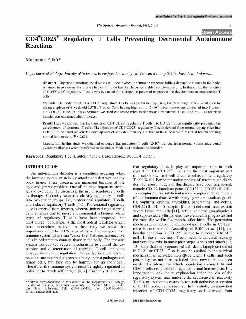
CD25 molecules could not be expressed by host even though the gene of the molecule was not deleted. It meant, genetical defect in CD122 gene affected CD25 gene product. The analysis using flow cytometer showed percentage of granu-locytic Gr-1+ cells in CD122-/- mice increased up to 82% in the bone marrow compared to about 55% in normal mice (data not shown). On the other hand, cells in erythroid line-age expressing TER-119 decreased in CD122-/- mice. Most TER-119+ cells in the bone marrow were VLA4+ erythro-blasts. These bone marrow changes affected the total condi-tion of mice. Changes of TER-119 expression influenced the number of both white and red blood cells in peripheral blood. In CD122-/- mice both CD4 and CD8 T cells increased and became activated, B cells dramatically decreased and almost missing from lymphoid tissue and circulatory (Fig. 4). The number of leukocytes markedly increased and hematocrit significantly decreased in CD122-/- mice. These symptoms were signs of anemia progression in these mice. CD122-/-
mice (3 –weeks -old) injected with 2x10⁶ CD4+CD25+ cells had normal TER-119+/Gr-1+ cell ratio in the bone marrow, leukocyte number in peripheral blood, and hematocrit. On the contrary, CD122-/- mice receiving 2x10⁶ CD4+CD25- cells had TER-119+/Gr-1+ cell ratio skewed toward Gr-1+ cells, low hematocrit, and increased the number of activated T cells in peripheral blood (data not shown). In CD122-/-
mice, CD4+CD25+ regulatory T cells could not be induced from CD4+CD25- cells, and the generation from bone mar-row was also impaired. It meant CD4+CD25+ regulatory T
cells had specific lineage different from another clone. In this case IL-2 signaling was suggested to play pivotal role for CD4+CD25+ regulatory T cell development, and the ab-sence of IL-2 receptor or this cytokine would affect the de-velopment of CD4+CD25+ regulatory cell.
The experiment results described that CD4+CD25+ regu-latory T cells controlled CD122-/- T cells, so that normal homeostasis could be maintained. We proposed that auto-immune disorders in CD122-/- mice were site effects of cell activation. In normal conditions, vertebrate activates immune system to overcome foreign antigen or invader. The best examples of foreign antigen or invader are viruses, bacteria and their product, cancer cell, and tissue or blood from donor. Antibodies will be produced by B cells to eliminate these foreign antigens which are notably harmful substances. In these mouse models of autoimmune disorder, B cell could not develop (Fig. 4). Consequently, the immunity related to the B cells also became impaired, that was why these mice had a short life in about 10-12 weeks.
CD4+CD25+ ameliorate erythropoiesis and B cell gen-eration. To know the existent of erythropoiesis we examined the expression of TER-119 antigen as a specific marker of erythroid-lineage [15-17]. In this experiment we showed that the number of TER-119+ cells in the bone marrow of CD122-
/- decreased before the manipulation with regulatory T cells. This erythroid suppression could be rescued by the transfer
Fig. (2). CD122-/- mice receiving CD4+CD25+ regulatory T cells at the age of 3- weeks developed naïve type T cells. Spleen cells of CD122-/- mice were analyzed 7 weeks after receiving 2x10⁶
regulatory T cells. Spleen cells were stained with anti-CD45.1, anti-CD8, anti-CD4 and anti-CD62L antibodies, and analyzed by flow cytometry. Host cells, both CD4 and CD8 T were presented with dot-plots as population gated to CD8+CD45- or CD4+CD45.1-. Percentages of naïve T cells were shown in each panel with CD8+CD62L+ or CD4+CD62L+. Controls were CD122-/- mice (KO) without manipulation. Data were mean of SD values of five mice per group.
Fig. (3). CD122-/- mice receiving CD4+CD25+ regulatory T cells at the age of 3- weeks did not develop early activated memory T cells. Spleen cells of CD122-/- mice were analyzed 7 weeks after receiving 2x10⁶ regulatory T cells. Spleen cells were stained with anti-CD45.1, anti-CD8, anti-CD4 and anti-CD69 antibodies, and analyzed by flow cytometry. Host cells, both CD4 and CD8 T, were presented with dot-plots as population gated to CD8+CD45- or CD4+CD45.1-. Percentages of early activated memory type T cells were shown in each panel with CD8+CD69+ or CD4+CD69+. Controls were CD122-/- mice (KO) without manipulation. Data were mean of SD values of five mice per group.
4 The Open Autoimmunity Journal, 2013, Volume 5 Muhaimin Rifa’i
of 2x10⁶ CD4+CD25+ cells into 3-week-old mice, whereas the transfer of 2x10⁶ CD4+CD25- conventional cells was not effective, and still showed the same decrease of TER-119+ cells as that in non-manipulated CD122-/- mice. CD4+CD25+ regulatory T cells seemed to be the best candidate for auto-immune therapy since this cells had major vast of array in the context of immune system. In this experiment we clearly showed that the transfer with CD4+CD25+ T cells could prevent anemia when we examined hematocrit composition in peripheral blood. In contrast, an injection of conventional CD4+CD25- cells with the same number of (2x10⁶) did not ameliorate anemia condition in CD122-/- mice. In this ex-periment we observed the power of CD4+CD25+ regulatory T cells when these cells were adoptively transferred to CD122-/- mice. The transfer of CD4+CD25+ T cells (2x10⁶) could normalize B cells development reflected by the in-creasing number of B220+ cells, instead of 2x10⁶ CD4+CD25-. The general condition of CD122-/- mice receiv-ing 2x10⁶ CD4+CD25- did not change when compared to non-treated ones, and they all died before 12 weeks of age. It meant the regulatory T cells only occupied CD4+CD25+ population and could not be induced from CD4+CD25- popu-lation. Many researchers believe CD4+CD25+ regulatory T cell is thymus origin and needs IL-2 signal to develop as functionally full competent cells.
In CD122-/- mice, granulocytic Gr-1+ cells were accumu-lated in bone marrow compartment. This accumulation was normalized when 3-week-old mice were transferred with 2x10⁶ CD4+CD25+ cells, instead of CD4+CD25- cells, in the same number. The measurement of leukocyte numbers in peripheral blood indicated that the increasing number of granulocytic Gr-1+ cells in bone marrow was closely related to that of leukocytes in peripheral blood. The increase of granulocytic Gr-1+ cells in bone marrow compartment and peripheral blood of CD122-/- mice could be one of compen-sations when the number of red blood cell decreased. By transferring 2x10⁶ CD4+CD25+ regulatory T cells to 3-week-old mice, instead of transferring 2x10⁶ CD4+CD25- cells, the
leukocyte number in peripheral blood of CD122-/- mice could be normalized. Peculiar feature of CD122-/- mice could be one of thymic atrophy manifestations. The thymic atrophy had a very broad impact, including lack regulatory of T cells and abnormal development of activated memory type cells.
DISCUSSION
One of the most exclusive features of CD4+CD25+ cells is its capability to change the nature of already activated T cells, at least in the case of CD122-/- mouse-derived T cells. This fact could be seen in rescued 3-week-old CD122-/- mice normalized after receiving CD4+CD25+ regulatory T cells. It meant that CD4+CD25+ regulatory T cells had capacity to rescue autoimmune attack even though the component of individual immune system had been activated. The mecha-nism in which CD4+CD25+ regulatory T cells “ceased fire” among activated and aberrant cells was still illusive. In the age of 3- weeks, CD122-/- mice usually are developed with activated memory T cells and autoimmune symptom. How-ever, transferred CD4+CD25+ regulatory T cells isolated from normal mice could reverse activated memory to naïve type. In this case CD44 and CD69 molecule were suppressed whereas CD62L was increased. After transferring with CD4+CD25+ regulatory T cells, CD44 and CD69 decrease significantly, it means that the status of the cells change from activated to naïve type. On the other hand, CD62L molecule expression increased after transferring with CD4+CD25+ regulatory T cells. CD62L was expressed in high level on naïve type and this molecule was lost in activated or memory type T cells. When T cells in the body were dominated by naïve type, they were likely healthy individuals. The capabil-ity of CD4+CD25+ regulatory T cells to reverse autoreactive cells could be related with production of IL-10 and TGF-β by these cells. There is a report that interposition of APC is required by CD4+CD25+ regulatory T cells in order for the cell to function effectively [18-21]. In this experiment we did not discriminate whether APC was involved in this system, but most probably APC did an intervention within in vivo system.
In studies by Malek et al. [22, 23], the defect of CD4+CD25+ regulatory T cells is proposed to be responsible for the increase of deregulated T cells in CD122-/- mice. It is based on the fact showing very few presence of CD4+CD25+ T cell in CD122-/- mice. The result of Malek et al. is consis-tent with our observation that CD122-/- mice develop autore-active cells causing death for mice in about 3 months old. CD4+CD25+ regulatory T cells play important role to control autoreactive cells expressing high level of CD69 and CD44 molecules and loss of CD62L molecule on their cell surface. The absent of CD4+CD25+ cells initiated autoimmune reac-tion due to the loss of regulatory function. Although the precise mechanism of CD4+CD25+ cell action remains ob-scure at present, but the functionally important of this cells should be a concern for researchers. Interestingly, most CD4+CD25+ regulatory T cells in young mice expressed FOXP3 molecule, that was why purified CD4+CD25+ regula-tory T cells from young mice were actually the same cell as CD4+CD25+FOXP3+ T cells (data not shown).
CONCLUSION
Fig. (4). B cells and erythrocytes normalization in CD122-/- mice that had received CD4+CD25+ regulatory T cells. Bone marrow cells of CD122-/- mice were analyzed 7 weeks after receiving 2x10⁶
regulatory T cells. Bone marrow cells were stained with anti-CD45.1, anti-TER-119 and anti-B220 antibodies, and analyzed by flow cytometry. B lineage cells (B220+) and erythroid lineage cells (TER-119+) were presented with dot-plots as population gated to B220+CD45- or TER119+CD45.1-. Controls were CD122-/- mice (KO) without manipulation. Data were mean of SD values of five mice per group.
CD4+CD25+ Regulatory T Cells Preventing Detrimental Autoimmune Reactions The Open Autoimmunity Journal, 2013, Volume 5 5
This study concludes that there are regulatory T cells in normal individual. If these cells are lost from the body, autoimmune reactions will occur. These autoimmune reactions can be relieved by transferring CD4+CD25+ regula-tory T cells derived from normal individuals, instead of transferring conventional CD4+CD25- cells. CD4+CD25+ regulatory T cells work as a potent regulator by suppressing activated and autoreactive T cell and these cells can also normalize the general condition including the number of B cells. Also, they can increase red blood cells lineage (TER119), as in CD122-/- which is deeply decreased. As the conclusive result, CD122-deficient mice receiving CD4+CD25+ regulatory T cells at the age of 3- weeks grow normally and remain healthy. Together this study explains that CD4+CD25+ regulatory T cells play pivotal roles to normalize homeostasis.
CONFLICT OF INTEREST
The author(s) confirm that this article content has no conflict of interest.
ACKNOWLEDGEMENTS
We are indebted to all laboratory members at Immunol-ogy Department, Nagoya University, Japan for their help in analyzing the data by using flow cytometry. We are also grateful to Desi Nuryati for critical reading of this manu-script and helpful discussions. This work was supported by grants from the Ministry of Science, Education, Sports and Culture of Japan.
ABBREVIATIONS
APC = antigen presenting cells
FITC = fluorescent iso-thiocyanate
PE = phycoerythrin
REFERENCES
[1] Suciu-Foca N, Manavalan JS, Cortesini R. Generation and function of antigen-specific suppressor and regulatory T cells. Transpl Im-munol 2003; 11: 235-44.
[2] Chai JG, Xue SA, Coe D, et al. Regulatory T cells, derived from naive CD4+CD25− T cells by in vitro Foxp3 gene transfer, can in-duce transplantation tolerance. Transplantation 2005; 79: 1310-6.
[3] Rieger K, Loddenkemper C, Maul J, et al. Mucosal Foxp3+ regula-tory T-cells are numerically deficient in acute and chronic GvHD. Blood 2006; 107(4): 1717-23.
[4] Firestein GS. Mechanisms of inflammation and tissue repair. In: Goldman L, Schafer AI, Eds. Cecil Medicine. 24th ed. Philadel-phia, Pa: Saunders Elsevier 2011; chap 47.
[5] Goronzy JJ, Weyand CM. The innate and adaptive immune sys-tems. In: Goldman L, Schafer AI, Eds. Cecil Medicine. 24th ed. Philadelphia, Pa: Saunders Elsevier 2011; chap 44.
[6] Shevach EM. Regulatory T cells in autoimmunity. Annu Rev Immunol 2000; 18: 423-49.
[7] Chatenoud L, Salomon B, Bluestone JA. Suppressor T cells--they're back and critical for regulation of autoimmunity. Immunol Rev 2001; 182: 149-63.
[8] Samy ET, Parker LA, Sharp CP, Tung KS. Continuous control of autoimmune disease by antigen-dependent polyclonal CD4+CD25+ regulatory T cells in the regional lymph node, J Exp Med 2005; 202: 771-81.
[9] Sakaguchi S. Naturally arising CD4+ regulatory T cells for immu-nologic self-tolerance and negative control of immune responses. Annu Rev Immunol 2004; 22: 531-62.
[10] Albert MH, Liu Y, Anasetti C, Yu XZ. Antigen-dependent sup-pression of alloresponses by Foxp3-induced regulatory T cells in transplantation. Eur J Immunol 2005; 2598-607.
[11] Suzuki H, Kundig TM, Furlonger C, et al. Deregulated T cell activation and autoimmunity in mice lacking interleukin-2 receptor . Science 1995; 268: 1472-6.
[12] Refaeli Y, Van Parijs L, London CA, Tschopp J, Abbas AK. Bio-chemical mechanisms of IL-2-regulated Fas-mediated T cell apop-tosis. Immunity 1998; 8: 615-23.
[13] Chai JG, Xue SA, Coe D, et al. Regulatory T cells, derived from naive CD4+CD25− T cells by in vitro Foxp3 gene transfer, can in-duce transplantation tolerance. Transplantation 2005; 79:1310-16.
[14] Rifa’i M, Kawamoto Y, Nakashima I, Suzuki H. Essential Roles of CD8+CD122+ Regulatory T cells in the Maintenance of T Cell Ho-meostasis. J Exp Med 2004; 200(9): 1123-1124.
[15] Kina T, Ikuta K, Takayama E, et al. The monoclonal antibody TER-119 recognizes a molecule associated with glycophorin A and specifically marks the late stages of murine erythroid lineage. Br J Haematol 2000; 109: 280-7.
[16] Gondek DC, Lu LF, Quezada SA, Sakaguchi S, Noelle RJ. Cutting edge: contact-mediated suppression by CD4+CD25+ regulatory cells involves a granzyme B-dependent, perforin-independent mechanism. J Immunol 2005; 174: 1783-6.
[17] Koulnis M, Pop R, Porpiglia E, Shearstone JR, Hidalgo D, So-colovsky M. Identification and analysis of mouse erythroid pro-genitors using the CD71/TER119 Flow-cytometric Assay. J Vis Exp 2011; (54); e2809, doi:10.3791/2809.
[18] Takahashi T, Kuniyasu Y, Toda M, et al. Immunologic self-tolerance maintained by CD25+CD4+ naturally anergic and sup-pressive T cells: induction of autoimmune disease by breaking their anergic/suppressive state. Int Immunol 1998; 10: 1969-80.
[19] Thornton AM, Shevach EM. CD4+CD25+ immunoregulatory T cells suppress polyclonal T cell activation in vitro by inhibiting in-terleukin 2 production. J Exp Med 1998; 188: 287-96.
[20] Thornton AM, Shevach EM. Suppressor effector function of CD4+CD25+ immunoregulatory T cells is antigen nonspecific. J Immunol 2000; 164: 183-190.
[21] Setoguchi R, Hori S, Takahashi T, Sakaguchi S. Homeostatic maintenance of natural Foxp3+CD25+CD4+ regulatory T cells by interleukin (IL)-2 and induction of autoimmune disease by IL-2 neutralization, J Exp Med 2005; 201: 723-35.
[22] Malek TR, Yu A, Vincek V, Scibelli P, Kong L. CD4 regulatory T cells prevent lethal autoimmunity in IL-2R -deficient mice. Impli-cations for the nonredundant function of IL-2. Immunity 2002; 17: 167-78.
[23] Malek TR, Porter BO, Codias EK, Scibelli P, Yu A. Normal lym-phoid homeostasis and lack of lethal autoimmunity in mice con-taining mature T cells with severely impaired IL-2 receptors. J Im-munol 2000; 164: 2905-2914.
Received: May 21, 2013 Revised: August 03, 2013 Accepted: August 14, 2013
© Muhaimin Rifa’i; Licensee Bentham Open.
This is an open access article licensed under the terms of the Creative Commons Attribution Non-Commercial License (http://creativecommons.org/licen-ses/by-nc/3.0/) which permits unrestricted, non-commercial use, distribution and reproduction in any medium, provided the work is properly cited.