regulation of the structure and function of skeletal muscle and
TRANSCRIPT
Regulation of the Structure and Function of Skeletal Muscle and Tendon by
Myostatin
by
Christopher L. Mendias
A dissertation submitted in partial fulfillment of the requirements for the degree of
Doctor of Philosophy (Molecular and Integrative Physiology)
in The University of Michigan 2007
Doctoral Committee: Professor John A. Faulkner, Chair Associate Professor Susan V. Brooks Herzog Associate Professor Jeffrey F. Horowitz Assistant Professor Daniel E. Michele

ii
Acknowledgements
I would like to thank my committee for the help they have provided. My
advisor, John Faulkner, has taught me so much about the process and business
of science. His guidance, support, mentorship and colorful stories have been
very much appreciated. I would also like to thank the other members of my
committee, Susan Brooks, Jeffrey Horowitz and Daniel Michele, for their insight
and advice. I appreciate the time you have taken to help in my development as a
scientist.
I would also like to thank the many members of the Muscle Mechanics Lab
for their invaluable assistance. In particular, I would like to express my gratitude
for the assistance of two individuals, Cheryl Hassett and Dennis Claflin. Without
the technical assistance of these two individuals I would not have been able to
complete many of the studies in this dissertation.
I would like to thank my friends who have made my time here in Ann Arbor
a blast.
And finally, I would like to thank my family for their encouragement and
support from 2500 miles away, even they had no idea why I wanted to move to
Michigan from Arizona.
iii
Table of Contents Acknowledgements List of Figures List of Tables Chapters Chapter I. Introduction
Background and Hypotheses Skeletal Muscle Structure and Function Tendon Structure and Function Regulation of Muscle Growth and Atrophy Regulation of Muscle Growth and Atrophy by Myostatin
Methods Figures
References
Chapter II. Contractile Properties of Extensor Digitorum Longus and Soleus Muscles of Myostatin-Deficient Mice
Abstract Introduction Methods Results Discussion Acknowledgements Figures Tables References
Chapter III. Tendons of Myostatin-Deficient Mice are Small, Brittle and Hypocellular
Abstract Introduction Methods Results
ii
v
vi
1 3 8
11 16 19 24 32
38 39 42 49 52 56 57 62 65
70 71 74 80
iv
Discussion Acknowledgements Figures Tables References
Chapter IV. Summary and Conclusion Overview Discussion of Findings and Future Directions - Muscle Discussion of Findings and Future Directions - Tendon Summary References
84 88 89 93 96
100 100 104 108 111
v
List of Figures Figure 1.1 Schematic drawing of myostatin gene structure KO strategy and
genotype strategy
24
1.2 The myostatin deficient mouse model
25
1.3 Growth curves of male and female MSTN+/+, MSTN+/- and MSTN-/- mice.
26
1.4 Muscle fiber CSA distributions
27
1.5 Relative myofibrillar protein content
28
1.6 Myostatin signal transduction pathways
29
1.7 Ubiquitinated myosin heavy chain (Ub-MHC) protein and atrogin-1 expression from EDL and soleus muscles of MSTN+/+ and MSTN-/- mice
30
1.8 Atrogin-1 expression in primary myotubes treated with myostatin
31
2.1 Relative hydroxyproline content of EDL and soleus muscles from MSTN+/+, MSTN+/- and MSTN-/- mice
57
2.2 Myostatin increases col1α2 expression and procollagen I content of primary skeletal muscle myotubes
58
2.3 Force values for EDL and soleus muscles from MSTN+/+, MSTN+/- and MSTN-/- mice
59
2.4 Force deficits following contraction-induced injury to EDL muscles and soleus muscles
60
2.5 ActRIIB protein content of EDL and soleus muscles from MSTN+/+ mice
61
3.1 Tendon fibroblasts express the myostatin receptors and activate signal transduction cascades in the presence of myostatin
89
vi
3.2 Myostatin induces the proliferation of tendon fibroblasts
90
3.3 Myostatin induces the expression of scleraxis, tenomodulin and collagen Iα2 genes in tendon fibroblasts
91
3.4 Mechanical properties of tibialis anterior tendons of MSTN+/+ and MSTN-/- mice
92
vii
List of Tables Table 2.1 Anatomical properties of animals
62
2.2
Contractile properties of EDL and soleus muscles 63
2.3
Mechanical injury of EDL and soleus muscles 64
3.1
PCR primer sequences and expected amplicon sizes 93
3.2
Whole animal, muscle and tendon masses 94
3.3 Morphological and mechanical properties of tibialis anterior tendons
95
1
Chapter I
Introduction
Background and Hypotheses
The ability of skeletal muscle and tendon to adapt to environmental
changes, injury, illness and other physiological conditions is critical in
determining the overall health, mobility and athletic performance of an individual.
Determining the cellular and molecular mechanisms behind the adaptation of
skeletal muscle and tendon provides important insights into basic biological
processes and the design of therapies for the treatment of diseases and injuries.
Several cytokines have been identified as important regulators of skeletal muscle
mass. One of the most potent cytokines that regulate skeletal muscle mass is
myostatin (GDF-8). Myostatin is a member of the TGF-ß superfamily of
cytokines and the targeted inhibition of myostatin results in an up to two-fold
increase in skeletal muscle mass (54, 84). Due to the profound increase in
skeletal muscle mass that occurs as a result of the deficiency of myostatin, much
interest has focused on the targeted inhibition of myostatin in the treatment of
muscle injuries and wasting diseases (81).
While the inhibition of myostatin results in a clear muscle mass phenotype,
arguably with as great an impact on muscle mass as any other single cytokine,
the full range of mechanical consequences of myostatin deficiency are not
known. Using classical muscle mechanics experimental techniques in the
2
myostatin-deficient mouse model, along with contemporary molecular biology
experimental techniques, this doctoral dissertation determined the mechanisms
by which the deficiency of myostatin regulates the structure and function of
skeletal muscles and of tendons. This introductory chapter describes the
molecular biology of myostatin, the structure, function and adaptation of skeletal
muscle and tendon tissue, and preliminary experiments that were critical for the
research studies in Chapters II and III.
In Chapter II, the impact of the deficiency of myostatin on the contractile
properties and extracellular matrix composition of skeletal muscle was
determined. We hypothesized that the deficiency of myostatin increases the
maximum tetanic force (Po), but decreases the specific Po (sPo) of muscles and
increases the susceptibility of muscles to contraction-induced injury. To test
these hypotheses, we measured the in vitro contractile properties of EDL and
soleus muscles from MSTN+/+, MSTN+/- and MSTN-/- mice and subsequently
subjected the muscles to a lengthening contraction protocol. We also
determined the impact of myostatin on the connective tissue composition of
skeletal muscle. We hypothesized that the deficiency of myostatin decreases the
type I collagen content of muscle connective tissue. The type I collagen content
of EDL and soleus muscles from MSTN+/+, MSTN+/- and MSTN-/- mice was
determined, as well as the expression of type I collagen in cultured skeletal
muscle myotubes treated with myostatin. The results from Chapter II indicate
that the deficiency of myostatin increases the Po of muscles, increases the
3
susceptibility of muscles to injury and decreases the type I collagen content of
skeletal muscle connective tissue.
The experiments on skeletal muscles in Chapter II lead to the exploration
of the role of myostatin in the regulation of tendon structure and function in
Chapter III. We hypothesized that the deficiency of myostatin results in smaller,
stiffer, and more brittle tendons. To test this hypothesis, we measured the
stress-strain relationships of tendons from the tibialis anterior muscles of
MSTN+/+ and MSTN-/- mice. We also measured the expression of structural and
cell cycle regulatory genes in whole tendon tissue and in cultured tendon
fibroblasts treated with myostatin. Our studies of tendons lead to the novel
finding that, in addition to regulating skeletal muscle structure and function,
myostatin also regulates the structure and function of tendon tissue.
Skeletal Muscle Structure and Function
Skeletal muscles consist of hundreds to thousands, and sometimes
millions, of long, multinucleated fibers organized together by an extracellular
matrix. There are three general layers of extracellular matrix, or connective
tissue, in muscles – the outermost layer is the epimysium, the intermediate layer
is the perimysium and the inner most layer is the endomysium. Understanding
the structure and function of each of these three layers requires a hierarchical
approach. The structure and function of the epimysium and perimysium will be
discussed in a whole body and tissue level biomechanical context, whereas the
structure and function of the endomysium will be discussed in the context of
cellular and molecular biomechanics.
4
Epimysium and perimysium. The epimysium covers the surface of the
muscle and has important roles in force transmission and insulation of the
muscle (46). Processes from the epimysium extend into muscle tissue and form
the second layer of connective tissue, the perimysium. The perimysium contains
blood vessels, nerves and lymphatic ducts, and structurally divides muscle fibers
into functional groups called fascicles. The orientation of fascicles are important
determinants of the direction of the force a muscle can generate (55). The
variation in the number of fascicles allows muscles to adopt a complex geometry
and facilitate complicated movements at joints. There is a trade-off between the
angle at which the fascicles are oriented and the amount of force the fascicles
are able to generate. The angle at which the fascicles are oriented to the
insertion is known as the angle of pennation (θ). The force that a muscle fiber
can generate is proportional to the cosine of θ (50). As θ goes from 0º to 90º, the
cosine of θ goes from 1 to 0. Therefore, a fascicle can generate the greatest
amount of force when θ is 0º, and can generate no force when θ is 90º. An
interesting feature about the angle of pennation is that, as muscle fibers undergo
hypertrophy, there is an increase in θ (50). While a muscle that experiences
hypertrophy is generally able to generate a greater total force, due to the cosine
relationship between θ and net force development, the muscle must undergo
relatively greater increases in hypertrophy to generate greater net forces across
a joint.
In addition to providing a hierarchical structure to skeletal muscle fibers,
the epimysium and perimysium may also protect an injured muscle from further
5
damage. The epimysium and perimysium are composed mostly of the fibrillar
collagens, type I and type III (43). These collagens act as molecular springs and
exist in parallel with the muscle fibers (9). While some debate still exists on the
topic, the epimysium and perimysium are thought to protect muscle fibers against
stretch induced injury by taking up the load during stretch and protecting the
skeletal muscle fibers from further injury (4).
Endomysium. The innermost layer of connective tissue is the
endomysium. The endomysium is composed of two layers of mostly type I and
type III collagen that fuse to form a sheet-like structure that inserts into the
tendon (43). The endomysium is important in transmitting forces generated
within the muscle to the tendon as well as laterally to other muscle fibers (63).
The endomysium is connected to the basement membrane that directly
surrounds each muscle fiber. The basement membrane is composed mostly of
type IV collagen (43). Unlike the fibrillar type I and type III collagen, type IV
collagen forms a mesh-like network that surrounds the muscle fiber (9).
Embedded within the endomysium and basement membrane are two classes of
proteins that have important functions. The first class of proteins are responsible
for force transmission from the sarcomeres through the intramuscular connective
tissue and eventually to tendons and bone. These proteins, such as integrin and
fibronectin, provide a mechanical link between the structural proteins of muscle
fibers with the collagen network of the extracellular matrix (37). The second
class of proteins are released from the matrix after injury and are important in
initiating the recovery of muscle from injury (24, 30, 76). This second class of
6
proteins will be discussed in the section that focuses on contraction-induced
injury.
Molecular Mechanisms of Force Transmission in Skeletal Muscle. The
structure within muscle fibers that is responsible for the generation of force is the
sarcomere. The sarcomere is composed of three major components – the thick
filament that is surrounded by six thin filaments that are shared with the six
surrounding thick filaments. The thin filaments are imbedded in the Z-disks at
the end of each sarcomere (73). The thick filament contains the protein that is
the molecular motor that drives muscle contraction, myosin heavy chain. The
head of the myosin molecule interacts with the actin molecules on the thin
filaments. The myosin binding sites on the actin molecule are regulated by the
troponin protein complex. The thick filaments are anchored to the Z-disks via the
proteins titin and nebulin. The Z-disks are responsible for transmitting the force
generated within the sarcomere to the extracellular matrix (ECM) (11, 67). The
Z-disks accomplish this by linking to structures that transmit force both laterally
and longitudinally.
For the lateral transmission of force, the Z-disks link with a structure at the
plasma membrane called the costamere. The Z-disk interacts with the
costamere via the dystrophin class of proteins (11, 67). The dystrophin proteins
link the membrane bound dystroglycan complex, which is in turn linked to the
extracellular matrix via laminin and fibronectin. Longitudinal force transmission is
accomplished via a mechanical linkage between the Z-disk and the actin
cytoskeleton of the muscle cell (55). This force is transmitted to the ends of the
7
fibers at the location where the fibers insert into tendons. This linkage comes
about due to the association between the actin cytoskeleton with vinculin and
talin (12). Vinculin and talin transmit force across the plasma membrane using
the fibronectin receptor that is bound to fibronectin in the ECM (3, 12). The
contraction of a muscle is therefore dependent upon the effective transmission of
the forces generated by the interaction between actin and myosin molecules
through structural proteins, out to the endomysium and eventually to tendons that
link the muscles to bone.
Skeletal Muscle Contraction. There are three general types of skeletal
muscle contractions. The contractions are defined by the change in fiber length
that occurs subsequent to muscle activation (18). During a shortening
contraction, the force generated by the muscle is greater than the load placed on
the muscle. Consequently, the distance between the distal and proximal ends of
a muscle decrease and positive work is performed. During an isometric
contraction, the force generated by the muscle is equal to the load on the
muscle. Consequently, the length of the muscle fiber does not change
significantly and no net work is done. The amount of force a muscle can
generate is greatest during an isometric contraction and is referred to as the
maximum isometric tetanic force (Po). The specific maximum tetanic force (sPo)
is the value Po normalized to the CSA. During a lengthening contraction, the
load on the muscle is greater than the force generated by the muscle.
Consequently, the distance between the ends of the muscle increases and there
is net negative work done. Contraction-induced injury occurs during lengthening
8
contractions when the magnitude of the opposing force is sufficient to disrupt the
ultrastructure of individual or groups of sarcomeres (18). Clinically, contraction-
induced injuries are commonly referred to as muscle strains.
Tendon Structure and Function
Tendons are connective tissue structures responsible for the transmission
of the force developed by muscles to bones, and in doing so, enable the
contraction of muscles to lead to joint movements and locomotion. Force
transmission may also occur in the opposite direction, from bone through tendon
to muscle. The transmission of force in this direction allows for the storage of
elastic energy within the tendon and muscle, but may also lead to damage to the
muscle. During a lengthening contraction, tendons may protect muscle from
injury by limiting the strain placed on muscle fibers (23).
Tendon Structure. Tendon tissue is arranged in an hierarchical order,
similar to skeletal muscle. Fibroblasts are the major cellular component of
tendons and consequently are responsible for the maintenance, repair,
modification of the tendon ECM (37). The fundamental anatomical structure in
tendons is the tendon fiber (31, 82). The tendon fiber is composed of collagen
fibrils and other structural proteins and is wrapped in a layer of connective tissue
that carries nerve endings, capillaries and lymphatic ducts. Fibers coalesce to
form fascicles, that are surrounded by a second connective tissue layer that
contains arterioles, venules and axons of nerve cells. Tendon fascicles are
surrounded by a third connective tissue structure called the epitenon. The
synovial sheath surrounds the epitenon and secretes synovial fluid that helps to
9
cushion the tendon and reduce friction from adjacent tissues as the tendon is
stretched and relaxed. Early damage to tendon tissue can result in an
overproduction of tendon synovial fluid (42). Swelling in the synovium is referred
to as tenosynovitis and is an early clinical diagnostic sign that tendon tissue is
injured. Tendons are linked to muscle tissue by myotendinous junctions located
at the ends of the tendon (82). Many muscles have an aponeurosis or internal
tendon, that is an extension of the tendon into the muscle tissue. At the other
end, tendons are connected to bone by strong fibrous structures called entheses
(6).
Tendon Mechanical Properties. The proteins and molecules that make up
the tendon can be divided into two categories, based upon the mechanical
properties they impart to tendon – stiffness and viscoelasticity. The stiffness of a
tendon is an important determinant of the ability of the tendon to store elastic
energy. Type I and type III collagens are the major proteins that determine the
stiffness of a tendon (37). Both type I and type III collagens are triple helical
molecules (9). The amino acid residues that face the inner core of the triple helix
are able to form hydrogen bonds with each other, and it is this hydrogen bonding
that provides much of the molecular basis for the stiffness material property of
tendons (45). As the tendon is stretched the hydrogen bonds between amino
acid residues are broken and the breaking of these bonds gives off energy in the
form of heat (61). The tendon must be able to dissipate this heat energy,
because if the energy is not properly dissipated, the structural covalent bonds of
molecules in the tendon can be broken and this will lead to tendon rupture.
10
The second category of molecules impart the viscoelastic properties of
tendons. Viscoelasticity is important because this allows the tendon to resume
its original shape after the application of a strain (37). This second category of
molecules is comprised of elastin, proteoglycans and glycosaminoglycans.
These molecules are highly hydrophilic, and their ability to bind water molecules
allows for them to transfer the heat produced by the breaking of hydrogen bonds
within collagen to the water molecules, that are further able to dissipate heat
energy into surrounding tissues (61).
An additional factor that is an important determinant of the mechanical
properties of tendons is the covalent cross-linking between collagen molecules.
Cross-linking reduces the friction between collagen molecules as they are
stretched (61). The consequence of greater cross-linking between collagen
molecules is that the tendon becomes stiffer and can store mechanical energy
more efficiently without losing this energy in the form of heat. Cross-linking
between collagen molecules can occur via an enzymatic or a non-enzymatic
mechanism. The lysyl oxidase enzyme generates aldehyde groups on lysine
residues in collagen molecules (19). These highly reactive aldehyde groups can
form stable covalent bonds with other lysine residues. Non-enzymatic cross-
linking can occur when the amino group on the side-chains of amino acid
residues come into contact with a reducing sugar (65). In addition to cross-
linking amino acid residues between collagen molecules, this reaction generates
compounds called advanced glycation end products (AGEs) (65). These AGEs

11
accumulate in the tissue and can disrupt the hierarchical structure of tendons and
lead to tissue damage.
The structural and mechanical properties of tendons change with aging
and physical activity. Compared with younger tendons, aged tendon is stiffer
(60), has a lower rate of type I collagen synthesis (59) and is more prone to
rupture (33, 60). The density of fibroblasts in tendon is highest at birth and
steadily declines throughout the lifespan (56, 59). While conflicting reports have
been published (40, 79), tendon typically adapts to chronic physical activity by
increasing CSA, stiffness, peak stress, peak strain, type I collagen synthesis and
fibroblast activity (34, 47, 48, 72, 80, 86, 87).
Regulation of Muscle Growth and Atrophy
The regulation of muscle growth and atrophy involves a sensitive balance
between protein synthesis and degradation. Minor damage to muscle fibers
causes an adaptive response in which damaged proteins are removed and new
proteins are synthesized (20). With repetitive minor damage, such as the
damage that occurs during exercise sessions that involve repeated lengthening
contractions, there is a net increase in protein synthesis (17). This increase in
protein synthesis increases both the size and Po of individual fibers. However, if
the damage to muscle is of sufficient magnitude, such as the damage that occurs
following a muscle "strain", there is a robust activation of proteolytic enzymes
that cause the breakdown of both damaged proteins and healthy, functional
proteins (21, 30). The premature breakdown of functional proteins leads to
muscle atrophy and a decrease in both the size and Po of individual fibers.
12
Understanding the mechanical, cellular and molecular mechanisms that control
muscle growth and atrophy is critical in improving the current treatments
available for muscle injuries and diseases, as well as enhancing athletic
performance and maximizing strength gains in exercise programs.
Mechanical damage to muscle fibers can initiate a series of events that
lead to muscle hypertrophy and an increase in Po. Sarcomeres are usually
damaged only during lengthening contractions (10, 51, 52). The process of
mechanical damage to sarcomeres is best explained in terms of the length-
tension relationship of the sarcomere. The tension a muscle fiber can develop is
proportional to the amount of overlap between the thick and thin filaments (74).
The point of maximum tension occurs when the muscle is at a length at which the
maximum number of cross bridges can be formed. This is the point at which the
load opposing the muscle contraction is equal to the tension generated by the
muscle, an isometric contraction. As an actively contracting muscle is
lengthened, the available cross bridge binding sites steadily decreases. The
forces transmitted to the sarcomeres from the external load can damage the
ultrastructure of the sarcomere, and this disruption of sarcomere ultrastructure is
responsible for the immediate decrease in force production following injury (10,
16, 41). In addition to damaging the sarcomeres, lengthening contractions can
lead to disruption of the plasma membrane and endomysium due to shear forces
generated during the lengthening contraction (14, 44). This process of
sarcomere and membrane damage initiates a response that eventually leads to
repair of damaged structures and hypertrophy of the damaged muscle fiber.
13
Cellular Regulation of Muscle Growth and Atrophy. The nuclei within
skeletal muscle fibers are arrested at the G1 cell cycle checkpoint and are unable
to replicate (36). Following injury to a muscle fiber, the nuclei in the damaged
area undergo apoptosis (7, 70). Skeletal muscle stem cells, referred to as
satellite cells or myoblasts, reside in a space between the sarcolemma and the
basal lamina (58). Satellite cells, that normally exist in a quiescent state, become
activated, migrate to the site of injury, proliferate, and fuse with the damaged
fiber to repopulate the nuclei lost as a result of injury (24). Damage to the
endomysium releases inactive hepatocyte growth factor (HGF) (76). Reactive
oxygen species, produced by the nitric oxide synthase (NOS) enzyme, activate
HGF (77). Activated HGF binds to the c-met receptor on the plasma membrane
of satellite cells. HGF signaling activates the satellite cells from the quiescent
state and initiates the migration of satellite cells to the site of injury (76).
As satellite cells migrate to the site of injury, they also undergo
proliferation. The initiation of proliferation is brought on by an increase in the
expression of the basic helix-loop-helix (bHLH) transcription factor MyoD (24).
MyoD is one of four members of myogenic regulatory factor (MRF) family, that
include Myf-5, myogenin and MRF-4. The MRF family of transcription factors
initiate the "myogenic program" in these proliferating satellite cells (24). The
myogenic program changes the cellular morphology of satellite cells from a
fibroblastic type morphology to a muscle like morphology (75). Once in proximity
of the damaged muscle fiber, satellite cells fuse with each other to form
multinucleated structures called myotubes. These myotubes fuse with the
14
existing, damaged muscle fiber and can help bridge the gap between ruptured
ends of a muscle fiber. As the myotubes fuse with the muscle fiber, the nuclei
within the myotubes arrest in G1 (25, 36). A certain population of satellite cells
that underwent proliferation do not form myotubes, but instead resume a sub-
basal lamina position and return to quiescence (24). In addition to satellite cells,
fibroblasts and immune cells are attracted to the site of injury. The presence of
these cells helps to remove cellular debris and repair the ECM. If there is a
severe disruption of the ECM, fibroblasts can respond with an overproduction of
ECM (27). This overproduction of ECM results in the clinical condition of fibrosis,
or scar tissue accumulation. The mechanisms behind the formation of scar
tissue are of particular interest to clinicians, as this scar tissue is generally
disruptive to the proper function of muscle tissue and, once formed, is relatively
permanent (30).
Once the myotubes have fused with the damaged muscle fiber, the nuclei
from these myotubes occupy a centrally located position in the muscle fiber (24,
52). While nuclei in an uninjured fiber are typically located just beneath the
sarcolemma, taking up a central location presumably allows for greater surface
area contact of the nucleus with ribosomes and endoplasmic reticulum and thus
enhance the ability of muscle fibers to synthesize new proteins. During the repair
stages, the majority of proteins that are synthesized are responsible for repairing
the damaged fiber (26). While it is not clear exactly why the nuclei take up a
subsarcolemmal location after repair, the nuclei do not need as close a direct
association with endoplasmic reticulum, as mRNA that encodes sarcomeric
15
proteins is trafficked directly to the sarcomere, where translation and processing
occur (35). This strategy is likely in place as sarcomeric proteins are relatively
large, translation at the sarcomere minimizes the need for the muscle fiber to
have an elaborate protein trafficking system in place. The location of the nuclei
within the cytosol therefore provides a way to track the progress of repair in
skeletal muscle tissue.
Molecular Regulation of Muscle Growth and Atrophy. While the satellite
cells are undergoing migration, proliferation and fusion, the muscle fiber is
initiating the underlying processes that are responsible to begin the repair
process. Damage to the plasma membrane causes a persistent increase in
intracellular calcium levels (78). The influx of calcium initiates the persistent
activation of sarcomeres distal to the site of injury, and is likely the mechanism
behind the "muscle spasm" that is observed clinically after a muscle injury. This
influx of calcium also activates the proteolytic systems of muscle tissue (5, 29).
By three days following injury, the plasma membranes of the muscle fibers are
largely sealed off (64) and calcium homeostasis is restored (5).
The ubiquitin-proteasome proteolytic system is important in regulating
skeletal muscle mass and protein turn-over. Atrogin-1 (MAFbx) is a ubiquitin
ligase protein expressed in skeletal muscle (20, 32). Atrogin-1 directs the
polyubiquination of proteins (8). Once a protein is tagged with ubiquitin, that
protein is targeted to the proteasome organelle. The amount of ubiquitin tags on
a protein appears to be directly related to the speed at which the doomed protein
is degraded. Upon reaching the proteasome, the protein is broken down and its
16
constituent amino acids for recycled for use in the synthesis of new proteins.
The expression of atrogin-1 is regulated by the Forkhead box O (Foxo) family of
transcription factors (20, 32). The promoter region of the atrogin-1 gene contains
binding sites for the Foxo3 transcription factor that acts as a co-activator of
atrogin-1 transcription (68). Phosphorylation of Foxo3 by Akt inhibits the ability of
Foxo3 to enter the nucleus and thus blocks the ability of Foxo3 to act as a
transcription factor.
Regulation of Muscle Growth and Atrophy by Myostatin
Myostatin is a negative regulator of muscle mass (38, 39, 54, 84, 85, 90).
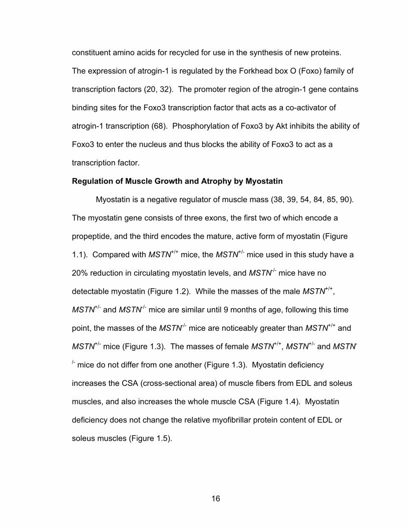
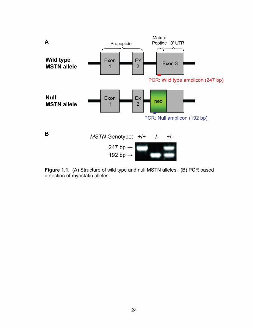
The myostatin gene consists of three exons, the first two of which encode a
propeptide, and the third encodes the mature, active form of myostatin (Figure
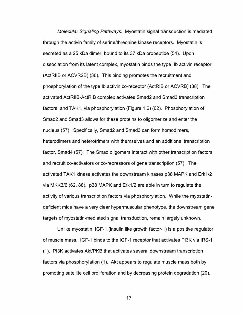
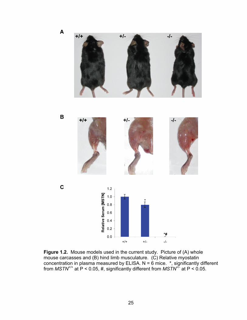
1.1). Compared with MSTN+/+ mice, the MSTN+/- mice used in this study have a
20% reduction in circulating myostatin levels, and MSTN-/- mice have no
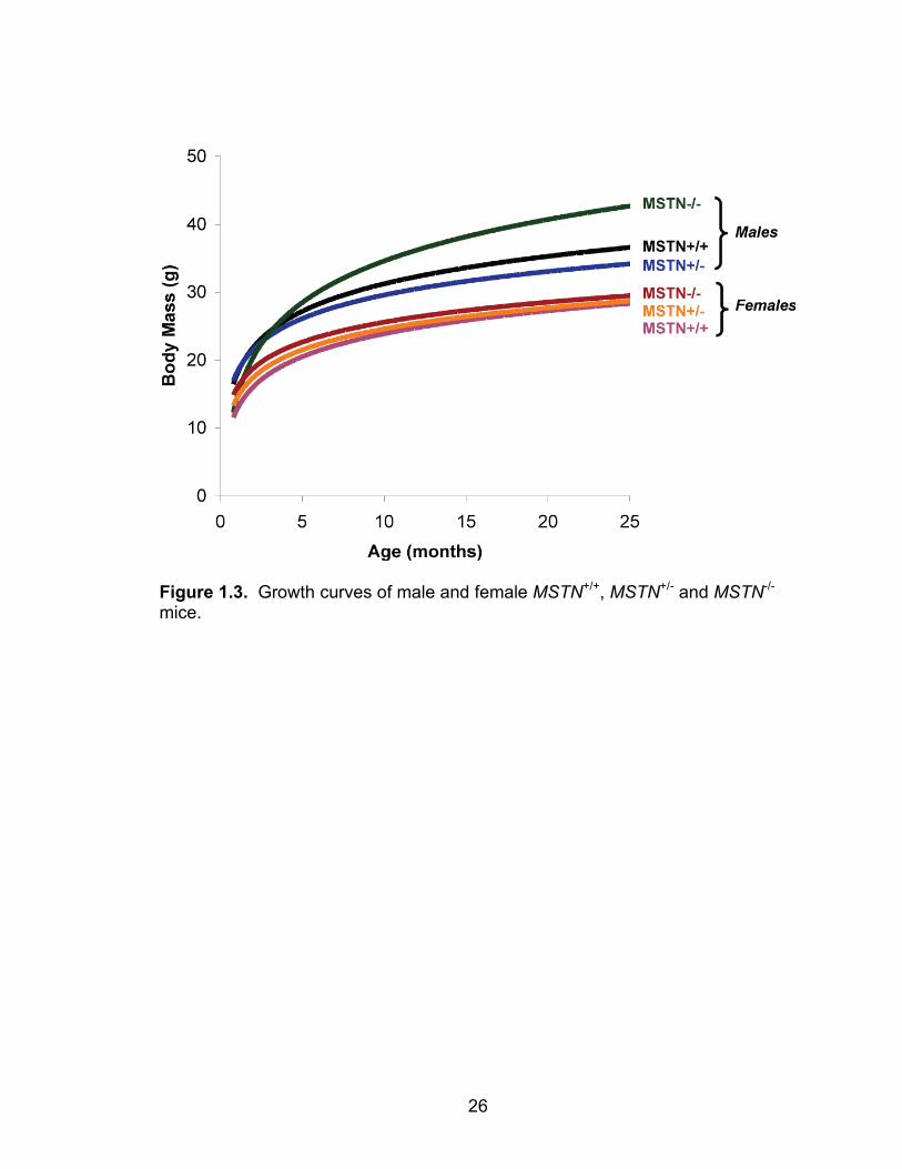
detectable myostatin (Figure 1.2). While the masses of the male MSTN+/+,
MSTN+/- and MSTN-/- mice are similar until 9 months of age, following this time
point, the masses of the MSTN-/- mice are noticeably greater than MSTN+/+ and
MSTN+/- mice (Figure 1.3). The masses of female MSTN+/+, MSTN+/- and MSTN-
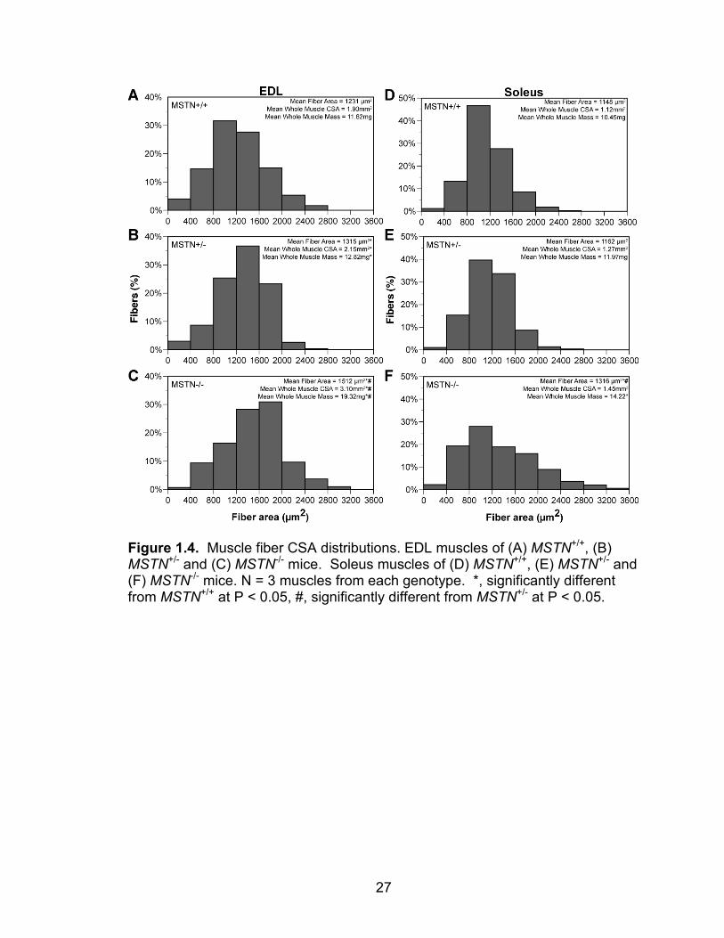
/- mice do not differ from one another (Figure 1.3). Myostatin deficiency
increases the CSA (cross-sectional area) of muscle fibers from EDL and soleus
muscles, and also increases the whole muscle CSA (Figure 1.4). Myostatin
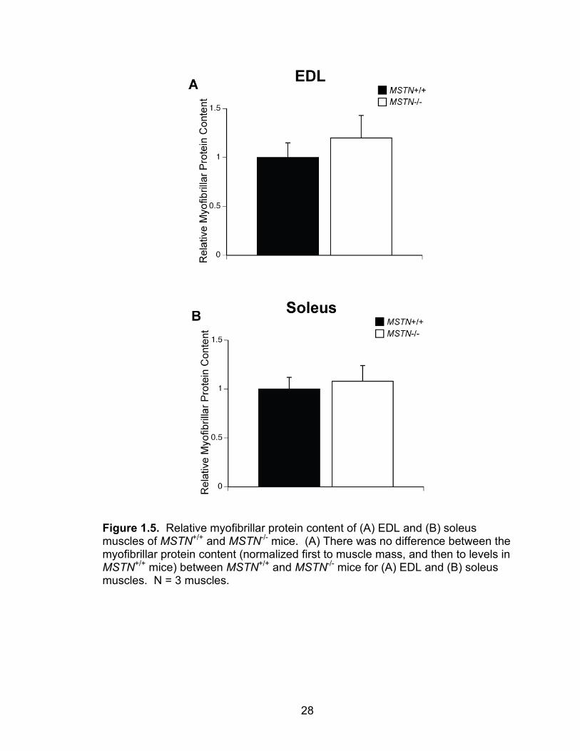
deficiency does not change the relative myofibrillar protein content of EDL or
soleus muscles (Figure 1.5).
17
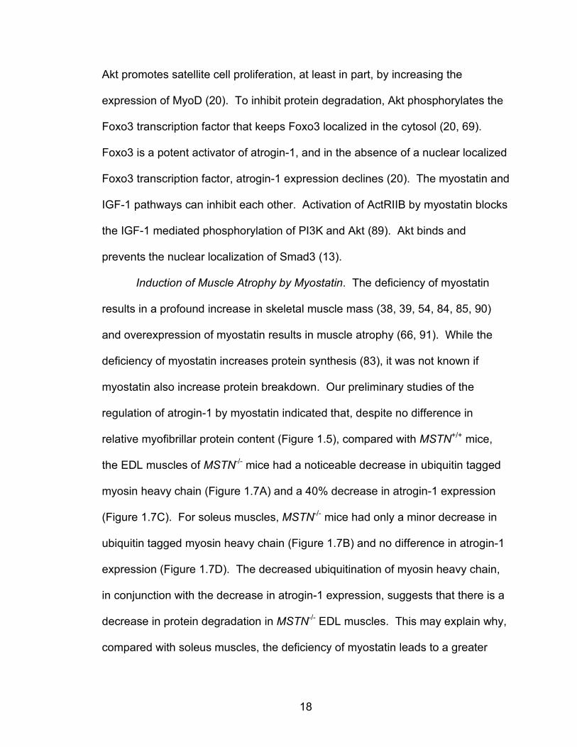
Molecular Signaling Pathways. Myostatin signal transduction is mediated
through the activin family of serine/threonine kinase receptors. Myostatin is
secreted as a 25 kDa dimer, bound to its 37 kDa propeptide (54). Upon
dissociation from its latent complex, myostatin binds the type IIb activin receptor
(ActRIIB or ACVR2B) (38). This binding promotes the recruitment and
phosphorylation of the type Ib activin co-receptor (ActRIB or ACVRB) (38). The
activated ActRIIB-ActRIB complex activates Smad2 and Smad3 transcription
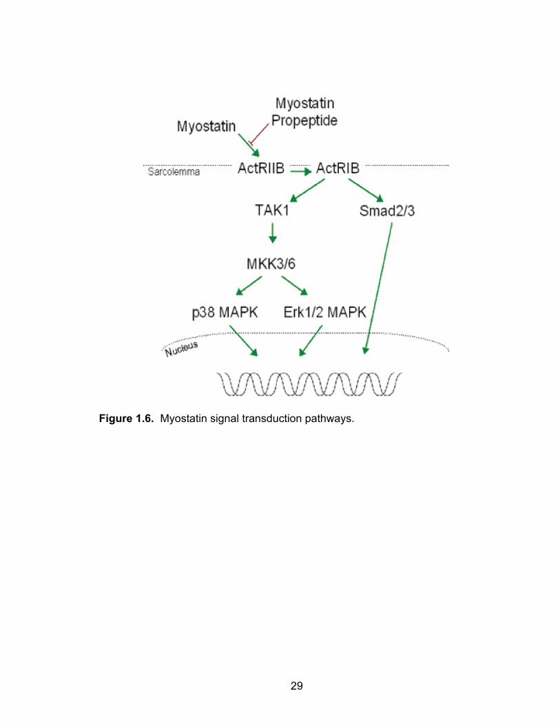
factors, and TAK1, via phosphorylation (Figure 1.6) (62). Phosphorylation of
Smad2 and Smad3 allows for these proteins to oligomerize and enter the
nucleus (57). Specifically, Smad2 and Smad3 can form homodimers,
heterodimers and heterotrimers with themselves and an additional transcription
factor, Smad4 (57). The Smad oligomers interact with other transcription factors
and recruit co-activators or co-repressors of gene transcription (57). The
activated TAK1 kinase activates the downstream kinases p38 MAPK and Erk1/2
via MKK3/6 (62, 88). p38 MAPK and Erk1/2 are able in turn to regulate the
activity of various transcription factors via phosphorylation. While the myostatin-
deficient mice have a very clear hypermuscular phenotype, the downstream gene
targets of myostatin-mediated signal transduction, remain largely unknown.
Unlike myostatin, IGF-1 (insulin like growth factor-1) is a positive regulator
of muscle mass. IGF-1 binds to the IGF-1 receptor that activates PI3K via IRS-1
(1). PI3K activates Akt/PKB that activates several downstream transcription
factors via phosphorylation (1). Akt appears to regulate muscle mass both by
promoting satellite cell proliferation and by decreasing protein degradation (20).
18
Akt promotes satellite cell proliferation, at least in part, by increasing the
expression of MyoD (20). To inhibit protein degradation, Akt phosphorylates the
Foxo3 transcription factor that keeps Foxo3 localized in the cytosol (20, 69).
Foxo3 is a potent activator of atrogin-1, and in the absence of a nuclear localized
Foxo3 transcription factor, atrogin-1 expression declines (20). The myostatin and
IGF-1 pathways can inhibit each other. Activation of ActRIIB by myostatin blocks
the IGF-1 mediated phosphorylation of PI3K and Akt (89). Akt binds and
prevents the nuclear localization of Smad3 (13).
Induction of Muscle Atrophy by Myostatin. The deficiency of myostatin
results in a profound increase in skeletal muscle mass (38, 39, 54, 84, 85, 90)
and overexpression of myostatin results in muscle atrophy (66, 91). While the
deficiency of myostatin increases protein synthesis (83), it was not known if
myostatin also increase protein breakdown. Our preliminary studies of the
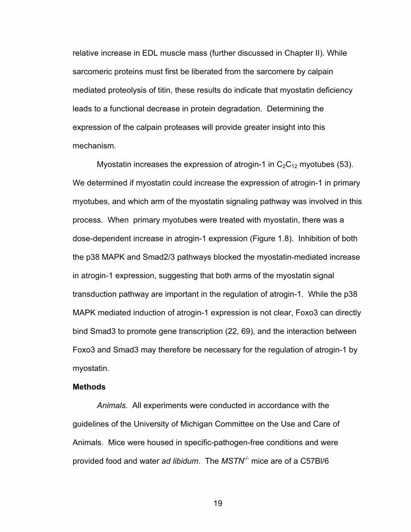
regulation of atrogin-1 by myostatin indicated that, despite no difference in
relative myofibrillar protein content (Figure 1.5), compared with MSTN+/+ mice,
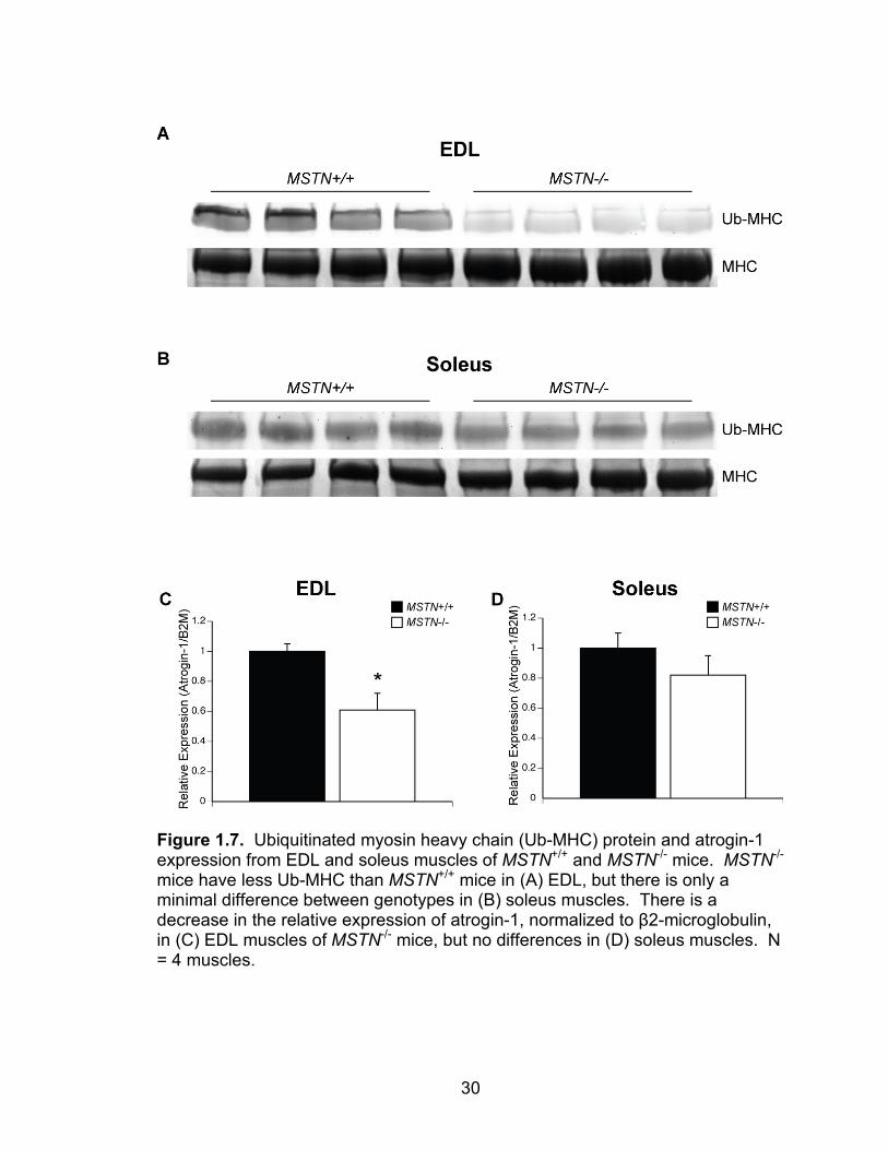
the EDL muscles of MSTN-/- mice had a noticeable decrease in ubiquitin tagged
myosin heavy chain (Figure 1.7A) and a 40% decrease in atrogin-1 expression
(Figure 1.7C). For soleus muscles, MSTN-/- mice had only a minor decrease in
ubiquitin tagged myosin heavy chain (Figure 1.7B) and no difference in atrogin-1
expression (Figure 1.7D). The decreased ubiquitination of myosin heavy chain,
in conjunction with the decrease in atrogin-1 expression, suggests that there is a
decrease in protein degradation in MSTN-/- EDL muscles. This may explain why,
compared with soleus muscles, the deficiency of myostatin leads to a greater
19
relative increase in EDL muscle mass (further discussed in Chapter II). While
sarcomeric proteins must first be liberated from the sarcomere by calpain
mediated proteolysis of titin, these results do indicate that myostatin deficiency
leads to a functional decrease in protein degradation. Determining the
expression of the calpain proteases will provide greater insight into this
mechanism.
Myostatin increases the expression of atrogin-1 in C2C12 myotubes (53).
We determined if myostatin could increase the expression of atrogin-1 in primary
myotubes, and which arm of the myostatin signaling pathway was involved in this
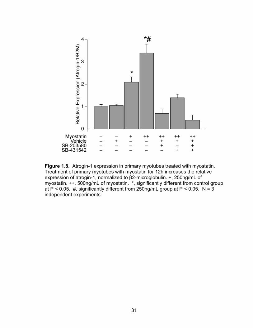
process. When primary myotubes were treated with myostatin, there was a
dose-dependent increase in atrogin-1 expression (Figure 1.8). Inhibition of both
the p38 MAPK and Smad2/3 pathways blocked the myostatin-mediated increase
in atrogin-1 expression, suggesting that both arms of the myostatin signal
transduction pathway are important in the regulation of atrogin-1. While the p38
MAPK mediated induction of atrogin-1 expression is not clear, Foxo3 can directly
bind Smad3 to promote gene transcription (22, 69), and the interaction between
Foxo3 and Smad3 may therefore be necessary for the regulation of atrogin-1 by
myostatin.
Methods
Animals. All experiments were conducted in accordance with the
guidelines of the University of Michigan Committee on the Use and Care of
Animals. Mice were housed in specific-pathogen-free conditions and were
provided food and water ad libidum. The MSTN-/- mice are of a C57Bl/6
20
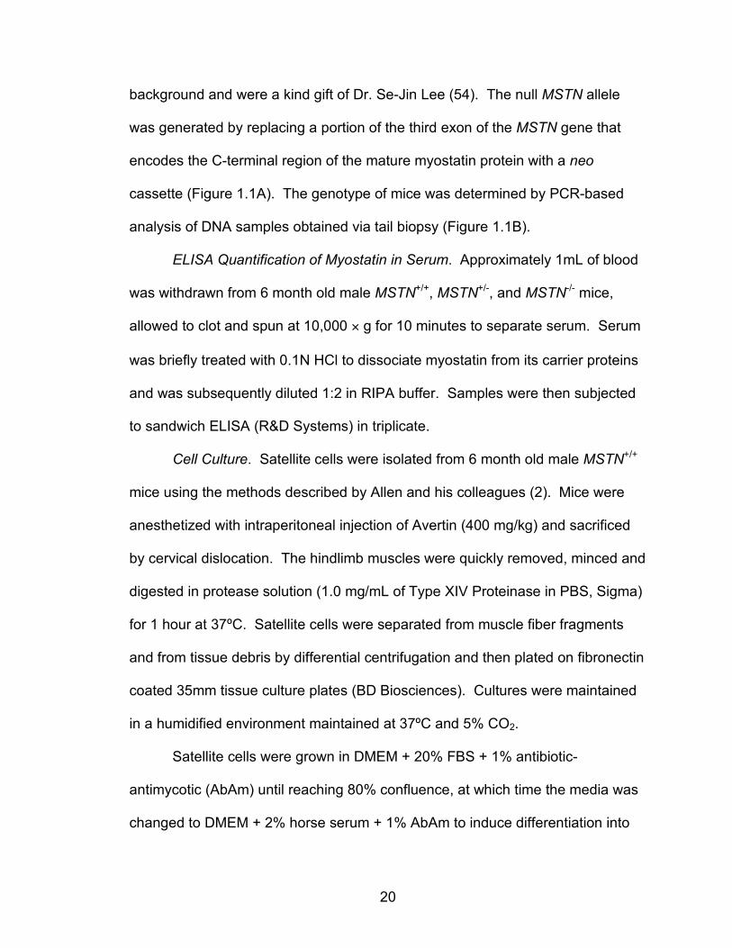
background and were a kind gift of Dr. Se-Jin Lee (54). The null MSTN allele
was generated by replacing a portion of the third exon of the MSTN gene that
encodes the C-terminal region of the mature myostatin protein with a neo
cassette (Figure 1.1A). The genotype of mice was determined by PCR-based
analysis of DNA samples obtained via tail biopsy (Figure 1.1B).
ELISA Quantification of Myostatin in Serum. Approximately 1mL of blood
was withdrawn from 6 month old male MSTN+/+, MSTN+/-, and MSTN-/- mice,
allowed to clot and spun at 10,000 × g for 10 minutes to separate serum. Serum
was briefly treated with 0.1N HCl to dissociate myostatin from its carrier proteins
and was subsequently diluted 1:2 in RIPA buffer. Samples were then subjected
to sandwich ELISA (R&D Systems) in triplicate.
Cell Culture. Satellite cells were isolated from 6 month old male MSTN+/+
mice using the methods described by Allen and his colleagues (2). Mice were
anesthetized with intraperitoneal injection of Avertin (400 mg/kg) and sacrificed
by cervical dislocation. The hindlimb muscles were quickly removed, minced and
digested in protease solution (1.0 mg/mL of Type XIV Proteinase in PBS, Sigma)
for 1 hour at 37ºC. Satellite cells were separated from muscle fiber fragments
and from tissue debris by differential centrifugation and then plated on fibronectin
coated 35mm tissue culture plates (BD Biosciences). Cultures were maintained
in a humidified environment maintained at 37ºC and 5% CO2.
Satellite cells were grown in DMEM + 20% FBS + 1% antibiotic-
antimycotic (AbAm) until reaching 80% confluence, at which time the media was
changed to DMEM + 2% horse serum + 1% AbAm to induce differentiation into
21
myotubes. Myotubes were maintained in culture for 5 days and then serum
starved for 24 hours by replacing the differentiation media with DMEM + Insulin-
Transferrin-Selenium Supplement (Invitrogen) + 1% AbAm. Recombinant murine
myostatin was produced in NS0 mouse myeloma cells (R & D Systems) and
dissolved into the starvation media at a final concentration of 250 or 500 ng/mL.
Stock solutions of the p38 MAPK inhibitor SB-203580 (15) and the Smad2/3
inhibitor SB-431542 (28) were prepared by dissolving these solid anhydrous
compounds in DMSO at a concentration of 10mM. SB-203580 and SB-431542
stock solutions were then added to starvation media containing myostatin and
0.5% DMSO at a final concentration of 10µM for SB-203580 and 5µM for SB-
431542. Cells were pretreated with SB-203580 or SB-431542 for 1 hour prior to
treatment with myostatin for 12 hours.
Myofibrillar Protein Content. Myofibrillar proteins were extracted from EDL
and soleus muscles using the methods of Solaro (71), modified to include the
addition of Leupeptin (Sigma). The protein content of the isolated myofibrils was
determined using a DC Protein Assay (Bio-Rad) and normalized to the wet mass
of the muscle.
SDS-PAGE and Immunoblot. Whole EDL and soleus muscles were
removed from anesthetized 6 month old MSTN+/+ and MSTN-/- mice, flash frozen
in liquid nitrogen and stored at -80ºC until use. Muscles and myotubes were
homogenized in Laemmli's sample buffer with 1:20 β-mercaptoethanol, 1:20
protease inhibitor cocktail (Sigma) and 1:40 phosphatase inhibitor cocktail
(Sigma) and then placed in boiling water for 5 minutes. Protein concentration of
22
the samples was determined using an RC DC Protein Assay (Bio-Rad). Equal
amounts of protein were loaded into 4% stacking, 7.5% resolving polyacrylamide
gels and subjected to electrophoresis. To detect total myosin heavy chain, gels
were stained with Coomassie Brilliant Blue (Bio-Rad). To detect ubiquitinated
myosin heavy chain, proteins were transferred from gels to a 0.45 µm
nitrocellulose membrane and stained with Ponceau S to verify equal protein
transfer. Membranes were blocked using casein (Vector Labs), incubated with
an HRPO tagged anti-ubiquitin antibody (Santa Cruz) and developed with
SuperSignal West Dura enhanced chemiluminescent reagents (Pierce
Biotechnology) and visualized using a FluorChem chemiluminescent
documentation system (Alpha Innotech).
RT-qPCR. RNA was isolated from samples using an RNeasy Fibrous
Tissue kit (Qiagen) and treated with DNase I. Poly-A mRNA was reverse
transcribed using an Omniscript RT system (Qiagen) and oligo(dT)15 primers.
cDNA was amplified using primers for atrogin-1 (forward: 5'-
ATTCTACACTGGCAGCAGCA-3'; reverse: 5'- TGTAAGCACACAGGCAGGTC-
3') and β2-microglobulin (forward: 5'-ATGGGAAGCCGAACATACTG-3'; reverse:
5'-CAGTCTCAGTGGGGGTGAAT-3') using a SYBR Green I PCR system
(Qiagen) with Uracil DNA Glycosylase (Invitrogen) in an Opticon 2 real-time
thermal cycler (Bio-Rad). qPCR reactions were conducted in quadruplicate for
each sample. C(t) values for atrogin-1 were normalized to β2-microglobulin
using the 2-ΔΔC(t) method. β2-microglobulin was selected as a normalizing gene
based on its stable expression in skeletal muscle tissue (49) and because its
23
expression did not differ between groups. The presence of single amplicons from
qPCR reactions were verified by melting curve analysis as well as
electrophoresis using a 2% agarose gel. Genomic DNA contamination was not
detected in qPCR reactions.
Statistical Analysis. Results are presented as means ± SE. KaleidaGraph
4.02 software was used to conduct statistical tests. For ELISA, histology and
gene expression data from cell culture experiments, differences between groups
were tested with a one-way ANOVA with α = 0.05. Fisher’s least significant post
hoc test was used to identify specific differences when significance was tested.
For all other data, differences between groups were tested with Student’s t-test
with α = 0.05.
24
Figure 1.1. (A) Structure of wild type and null MSTN alleles. (B) PCR based detection of myostatin alleles.
25
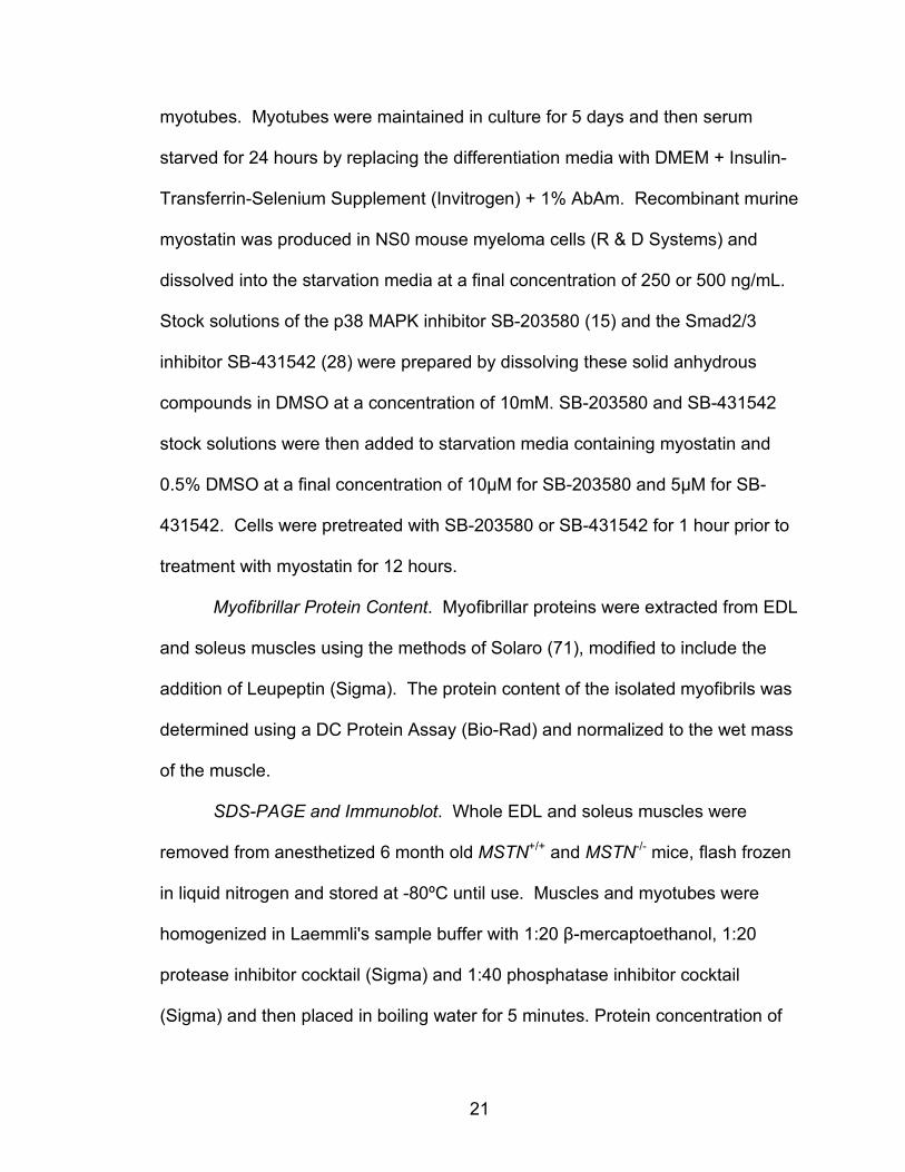
Figure 1.2. Mouse models used in the current study. Picture of (A) whole mouse carcasses and (B) hind limb musculature. (C) Relative myostatin concentration in plasma measured by ELISA. N = 6 mice. *, significantly different from MSTN+/+ at P < 0.05, #, significantly different from MSTN+/- at P < 0.05.
26
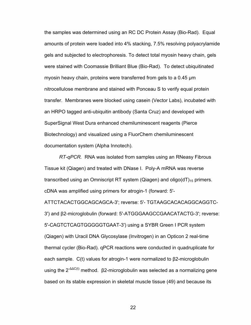
Figure 1.3. Growth curves of male and female MSTN+/+, MSTN+/- and MSTN-/- mice.
27
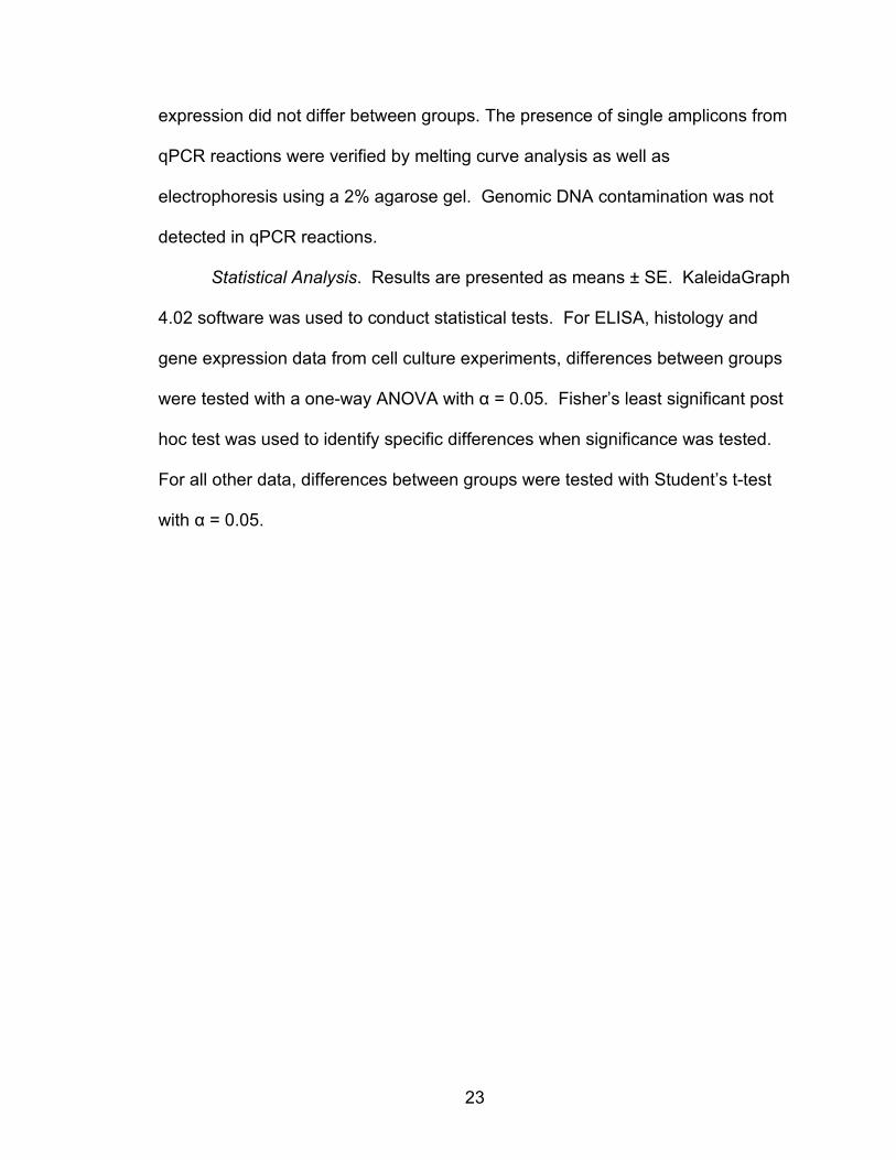
Figure 1.4. Muscle fiber CSA distributions. EDL muscles of (A) MSTN+/+, (B) MSTN+/- and (C) MSTN-/- mice. Soleus muscles of (D) MSTN+/+, (E) MSTN+/- and (F) MSTN-/- mice. N = 3 muscles from each genotype. *, significantly different from MSTN+/+ at P < 0.05, #, significantly different from MSTN+/- at P < 0.05.
28
Figure 1.5. Relative myofibrillar protein content of (A) EDL and (B) soleus muscles of MSTN+/+ and MSTN-/- mice. (A) There was no difference between the myofibrillar protein content (normalized first to muscle mass, and then to levels in MSTN+/+ mice) between MSTN+/+ and MSTN-/- mice for (A) EDL and (B) soleus muscles. N = 3 muscles.
29
Figure 1.6. Myostatin signal transduction pathways.
30
Figure 1.7. Ubiquitinated myosin heavy chain (Ub-MHC) protein and atrogin-1 expression from EDL and soleus muscles of MSTN+/+ and MSTN-/- mice. MSTN-/- mice have less Ub-MHC than MSTN+/+ mice in (A) EDL, but there is only a minimal difference between genotypes in (B) soleus muscles. There is a decrease in the relative expression of atrogin-1, normalized to β2-microglobulin, in (C) EDL muscles of MSTN-/- mice, but no differences in (D) soleus muscles. N = 4 muscles.
31
Figure 1.8. Atrogin-1 expression in primary myotubes treated with myostatin. Treatment of primary myotubes with myostatin for 12h increases the relative expression of atrogin-1, normalized to β2-microglobulin. +, 250ng/mL of myostatin. ++, 500ng/mL of myostatin. *, significantly different from control group at P < 0.05. #, significantly different from 250ng/mL group at P < 0.05. N = 3 independent experiments.
32
References 1. Adams G. Invited Review: Autocrine/paracrine IGF-I and skeletal muscle adaptation. J Appl Physiol 93: 1159-1167, 2002. 2. Allen RE, Temm-Grove CJ, Sheehan SM, and Rice G. Skeletal muscle satellite cell cultures. Methods Cell Biol 52: 155-176, 1997. 3. Anastasi G, Cutroneo G, Santoro G, and Trimarchi F. The non-junctional sarcolemmal cytoskeleton: the costameres. Ital J Anat Embryol 103: 1-11, 1998. 4. Arruda E, Mundy K, Calve S, and Baar K. Denervation does not change the ratio of collagen I and collagen III mRNA in the extracellular matrix of muscle. Am J Physiol Regul Integr Comp Physiol 292: R983-987, 2007. 5. Belcastro AN, Shewchuk LD, and Raj DA. Exercise-induced muscle injury: a calpain hypothesis. Mol Cell Biochem 179: 135-145, 1998. 6. Benjamin M, Kumai T, Milz S, Boszczyk B, Boszczyk A, and Ralphs J. The skeletal attachment of tendons--tendon "entheses". Comp Biochem Physiol, Part A Mol Integr Physiol 133: 931-945, 2002. 7. Biral D, Jakubiec-Puka A, Ciechomska I, Sandri M, Rossini K, Carraro U, and Betto R. Loss of dystrophin and some dystrophin-associated proteins with concomitant signs of apoptosis in rat leg muscle overworked in extension. Acta Neuropathol (Berl) 100: 618-626, 2000. 8. Bodine S, Latres E, Baumhueter S, Lai V, Nunez L, Clarke B, Poueymirou W, Panaro F, Na E, Dharmarajan K, Pan Z, Valenzuela D, DeChiara T, Stitt T, Yancopoulos G, and Glass D. Identification of ubiquitin ligases required for skeletal muscle atrophy. Science 294: 1704-1708, 2001. 9. Brinckmann J. Collagen - Primer in Structure, Processing and Assembly. Top Curr Chem 247: 1-229, 2005. 10. Brooks SV, Zerba E, and Faulkner JA. Injury to muscle fibres after single stretches of passive and maximally stimulated muscles in mice. J Physiol 488 ( Pt 2): 459-469, 1995. 11. Capetanaki Y, Bloch RJ, Kouloumenta A, Mavroidis M, and Psarras S. Muscle intermediate filaments and their links to membranes and membranous organelles. Exp Cell Res 313: 2063-2076, 2007. 12. Chopard A, Arrighi N, Carnino A, and Marini JF. Changes in dysferlin, proteins from dystrophin glycoprotein complex, costameres, and cytoskeleton in human soleus and vastus lateralis muscles after a long-term bedrest with or without exercise. Faseb J 19: 1722-1724, 2005. 13. Conery AR, Cao Y, Thompson EA, Townsend CM, Jr., Ko TC, and Luo K. Akt interacts directly with Smad3 to regulate the sensitivity to TGF-beta induced apoptosis. Nat Cell Biol 6: 366-372, 2004. 14. Consolino CM, and Brooks SV. Susceptibility to sarcomere injury induced by single stretches of maximally activated muscles of mdx mice. J Appl Physiol 96: 633-638, 2004. 15. Cuenda A, Rouse J, Doza Y, Meier R, Cohen P, Gallagher T, Young P, and Lee J. SB 203580 is a specific inhibitor of a MAP kinase homologue which is stimulated by cellular stresses and interleukin-1. FEBS Lett 364: 229-233, 1995.
33
16. Devor ST, and Faulkner JA. Regeneration of new fibers in muscles of old rats reduces contraction-induced injury. J Appl Physiol 87: 750-756, 1999. 17. Evans WJ. Protein nutrition and resistance exercise. Can J Appl Physiol 26 Suppl: S141-152, 2001. 18. Faulkner JA, Brooks SV, and Opiteck JA. Injury to skeletal muscle fibers during contractions: conditions of occurrence and prevention. Phys Ther 73: 911-921, 1993. 19. Gerriets J, Curwin S, and Last J. Tendon hypertrophy is associated with increased hydroxylation of nonhelical lysine residues at two specific cross-linking sites in type I collagen. J Biol Chem 268: 25553-25560, 1993. 20. Glass D. Molecular mechanisms modulating muscle mass. Trends in molecular medicine 9: 344-350, 2003. 21. Glass DJ. Skeletal muscle hypertrophy and atrophy signaling pathways. Int J Biochem Cell Biol 37: 1974-1984, 2005. 22. Gomis R, Alarcón C, He W, Wang Q, Seoane J, Lash A, and Massagué J. A FoxO-Smad synexpression group in human keratinocytes. Proc Natl Acad Sci USA 103: 12747-12752, 2006. 23. Griffiths RI. Shortening of muscle fibres during stretch of the active cat medial gastrocnemius muscle: the role of tendon compliance. J Physiol 436: 219-236, 1991. 24. Hawke T, and Garry D. Myogenic satellite cells: physiology to molecular biology. J Appl Physiol 91: 534-551, 2001. 25. Hawke TJ, Meeson AP, Jiang N, Graham S, Hutcheson K, DiMaio JM, and Garry DJ. p21 is essential for normal myogenic progenitor cell function in regenerating skeletal muscle. Am J Physiol Cell Physiol 285: C1019-1027, 2003. 26. Herndon DN, Dasu MR, Wolfe RR, and Barrow RE. Gene expression profiles and protein balance in skeletal muscle of burned children after beta-adrenergic blockade. Am J Physiol Endocrinol Metab 285: E783-789, 2003. 27. Huard J, Li Y, and Fu F. Muscle injuries and repair: current trends in research. The Journal of bone and joint surgery American volume 84-A: 822-832, 2002. 28. Inman G, Nicolás F, Callahan J, Harling J, Gaster L, Reith A, Laping N, and Hill C. SB-431542 is a potent and specific inhibitor of transforming growth factor-beta superfamily type I activin receptor-like kinase (ALK) receptors ALK4, ALK5, and ALK7. Mol Pharmacol 62: 65-74, 2002. 29. Jackman R, and Kandarian S. The molecular basis of skeletal muscle atrophy. Am J Physiol, Cell Physiol 287: C834-843, 2004. 30. Järvinen T, Järvinen T, Kääriäinen M, Kalimo H, and Järvinen M. Muscle injuries: biology and treatment. The American journal of sports medicine 33: 745-764, 2005. 31. Józsa LG, and Kannus P. Human tendons : anatomy, physiology, and pathology. Champaign, IL: Human Kinetics, 1997, p. ix, 574 p. 32. Kandarian S, and Jackman R. Intracellular signaling during skeletal muscle atrophy. Muscle Nerve 33: 155-165, 2006.
34
33. Kannus P, and Józsa L. Histopathological changes preceding spontaneous rupture of a tendon. A controlled study of 891 patients. The Journal of bone and joint surgery American volume 73: 1507-1525, 1991. 34. Kiiskinen A. Physical training and connective tissues in young mice--physical properties of Achilles tendons and long bones. Growth 41: 123-137, 1977. 35. Kiri A, and Goldspink G. RNA-protein interactions of the 3' untranslated regions of myosin heavy chain transcripts. J Muscle Res Cell Motil 23: 119-129, 2002. 36. Kitzmann M, and Fernandez A. Crosstalk between cell cycle regulators and the myogenic factor MyoD in skeletal myoblasts. Cell Mol Life Sci 58: 571-579, 2001. 37. Kjaer M. Role of extracellular matrix in adaptation of tendon and skeletal muscle to mechanical loading. Physiol Rev 84: 649-698, 2004. 38. Lee S, and McPherron A. Regulation of myostatin activity and muscle growth. Proc Natl Acad Sci USA 98: 9306-9311, 2001. 39. Lee S, Reed L, Davies M, Girgenrath S, Goad M, Tomkinson K, Wright J, Barker C, Ehrmantraut G, Holmstrom J, Trowell B, Gertz B, Jiang M, Sebald S, Matzuk M, Li E, Liang L, Quattlebaum E, Stotish R, and Wolfman N. Regulation of muscle growth by multiple ligands signaling through activin type II receptors. Proc Natl Acad Sci USA 102: 18117-18122, 2005. 40. Legerlotz K, Schjerling P, Langberg H, Brüggemann G, and Niehoff A. The effect of running, strength, and vibration strength training on the mechanical, morphological, and biochemical properties of the Achilles tendon in rats. J Appl Physiol 102: 564-572, 2007. 41. Lieber R, Woodburn T, and Fridén J. Muscle damage induced by eccentric contractions of 25% strain. J Appl Physiol 70: 2498-2507, 1991. 42. Lin T, Cardenas L, and Soslowsky L. Biomechanics of tendon injury and repair. Journal of biomechanics 37: 865-877, 2004. 43. Listrat A, Picard B, and Geay Y. Age-related changes and location of type I, III, IV, V and VI collagens during development of four foetal skeletal muscles of double-muscled and normal bovine animals. Tissue & cell 31: 17-27, 1999. 44. Lovering RM, and De Deyne PG. Contractile function, sarcolemma integrity, and the loss of dystrophin after skeletal muscle eccentric contraction-induced injury. Am J Physiol Cell Physiol 286: C230-238, 2004. 45. Lucero HA, and Kagan HM. Lysyl oxidase: an oxidative enzyme and effector of cell function. Cell Mol Life Sci 63: 2304-2316, 2006. 46. MacIntosh BR, Gardiner PF, and McComas AJ. Skeletal muscle : form and function. Champaign, IL: Human Kinetics, 2006, p. 423 p. 47. Maganaris C, Narici M, and Reeves N. In vivo human tendon mechanical properties: effect of resistance training in old age. Journal of musculoskeletal & neuronal interactions 4: 204-208, 2004. 48. Magnusson S, Hansen P, and Kjaer M. Tendon properties in relation to muscular activity and physical training. Scandinavian journal of medicine & science in sports 13: 211-223, 2003.
35
49. Mahoney D, Carey K, Fu M, Snow R, Cameron-Smith D, Parise G, and Tarnopolsky M. Real-time RT-PCR analysis of housekeeping genes in human skeletal muscle following acute exercise. Physiol Genomics 18: 226-231, 2004. 50. Maxwell L, Faulkner J, and Hyatt G. Estimation of number of fibers in guinea pig skeletal muscles. Journal of applied physiology 37: 259-264, 1974. 51. McCully KK, and Faulkner JA. Characteristics of lengthening contractions associated with injury to skeletal muscle fibers. J Appl Physiol 61: 293-299, 1986. 52. McCully KK, and Faulkner JA. Injury to skeletal muscle fibers of mice following lengthening contractions. J Appl Physiol 59: 119-126, 1985. 53. McFarlane C, Plummer E, Thomas M, Hennebry A, Ashby M, Ling N, Smith H, Sharma M, and Kambadur R. Myostatin induces cachexia by activating the ubiquitin proteolytic system through an NF-kappaB-independent, FoxO1-dependent mechanism. J Cell Physiol 209: 501-514, 2006. 54. McPherron A, Lawler A, and Lee S. Regulation of skeletal muscle mass in mice by a new TGF-beta superfamily member. Nature 387: 83-90, 1997. 55. Monti RJ, Roy RR, Hodgson JA, and Edgerton VR. Transmission of forces within mammalian skeletal muscles. J Biomech 32: 371-380, 1999. 56. Moore MJ, and De Beaux A. A quantitative ultrastructural study of rat tendon from birth to maturity. J Anat 153: 163-169, 1987. 57. Moustakas A, Souchelnytskyi S, and Heldin C. Smad regulation in TGF-beta signal transduction. J Cell Sci 114: 4359-4369, 2001. 58. Muir AR, Kanji AH, and Allbrook D. The structure of the satellite cells in skeletal muscle. J Anat 99: 435-444, 1965. 59. Nakagawa Y, Majima T, and Nagashima K. Effect of ageing on ultrastructure of slow and fast skeletal muscle tendon in rabbit Achilles tendons. Acta Physiol Scand 152: 307-313, 1994. 60. O'Brien M. Functional anatomy and physiology of tendons. Clin Sports Med 11: 505-520, 1992. 61. Paxton JZ, and Baar K. Tendon Mechanics: The Argument Heats Up. J Appl Physiol 2007. 62. Philip B, Lu Z, and Gao Y. Regulation of GDF-8 signaling by the p38 MAPK. Cell Signal 17: 365-375, 2005. 63. Purslow PP, and Trotter JA. The morphology and mechanical properties of endomysium in series-fibred muscles: variations with muscle length. J Muscle Res Cell Motil 15: 299-308, 1994. 64. Rader EP, Song W, Van Remmen H, Richardson A, and Faulkner JA. Raising the antioxidant levels within mouse muscle fibres does not affect contraction-induced injury. Exp Physiol 91: 781-789, 2006. 65. Reddy G, Stehno-Bittel L, and Enwemeka C. Glycation-induced matrix stability in the rabbit achilles tendon. Arch Biochem Biophys 399: 174-180, 2002. 66. Reisz-Porszasz S, Bhasin S, Artaza J, Shen R, Sinha-Hikim I, Hogue A, Fielder T, and Gonzalez-Cadavid N. Lower skeletal muscle mass in male transgenic mice with muscle-specific overexpression of myostatin. Am J Physiol Endocrinol Metab 285: E876-888, 2003.
36
67. Rybakova IN, Patel JR, and Ervasti JM. The dystrophin complex forms a mechanically strong link between the sarcolemma and costameric actin. J Cell Biol 150: 1209-1214, 2000. 68. Sandri M, Sandri C, Gilbert A, Skurk C, Calabria E, Picard A, Walsh K, Schiaffino S, Lecker S, and Goldberg A. Foxo transcription factors induce the atrophy-related ubiquitin ligase atrogin-1 and cause skeletal muscle atrophy. Cell 117: 399-412, 2004. 69. Seoane J, Le H, Shen L, Anderson S, and Massagué J. Integration of Smad and forkhead pathways in the control of neuroepithelial and glioblastoma cell proliferation. Cell 117: 211-223, 2004. 70. Smith HK, Maxwell L, Martyn JA, and Bass JJ. Nuclear DNA fragmentation and morphological alterations in adult rabbit skeletal muscle after short-term immobilization. Cell Tissue Res 302: 235-241, 2000. 71. Solaro RJ, Pang DC, and Briggs FN. The purification of cardiac myofibrils with Triton X-100. Biochim Biophys Acta 245: 259-262, 1971. 72. Sommer H. The biomechanical and metabolic effects of a running regime on the Achilles tendon in the rat. International orthopaedics 11: 71-75, 1987. 73. Squire JM. Architecture and function in the muscle sarcomere. Curr Opin Struct Biol 7: 247-257, 1997. 74. Squire JM. Muscle contraction. Encyclopedia of Life Sciences 2005. 75. Tapscott SJ. The circuitry of a master switch: Myod and the regulation of skeletal muscle gene transcription. Development 132: 2685-2695, 2005. 76. Tatsumi R, and Allen R. Active hepatocyte growth factor is present in skeletal muscle extracellular matrix. Muscle Nerve 30: 654-658, 2004. 77. Tatsumi R, Liu X, Pulido A, Morales M, Sakata T, Dial S, Hattori A, Ikeuchi Y, and Allen R. Satellite cell activation in stretched skeletal muscle and the role of nitric oxide and hepatocyte growth factor. Am J Physiol, Cell Physiol 290: C1487-1494, 2006. 78. Turner PR, Fong PY, Denetclaw WF, and Steinhardt RA. Increased calcium influx in dystrophic muscle. J Cell Biol 115: 1701-1712, 1991. 79. Viidik A. The effect of training on the tensile strength of isolated rabbit tendons. Scand J Plast Reconstr Surg 1: 141-147, 1967. 80. Vilarta R, and Vidal Bde C. Anisotropic and biomechanical properties of tendons modified by exercise and denervation: aggregation and macromolecular order in collagen bundles. Matrix 9: 55-61, 1989. 81. Wagner K. Muscle regeneration through myostatin inhibition. Current opinion in rheumatology 17: 720-724, 2005. 82. Wang J. Mechanobiology of tendon. Journal of biomechanics 39: 1563-1582, 2006. 83. Welle S, Bhatt K, and Pinkert C. Myofibrillar protein synthesis in myostatin-deficient mice. Am J Physiol Endocrinol Metab 290: E409-415, 2006. 84. Whittemore L, Song K, Li X, Aghajanian J, Davies M, Girgenrath S, Hill J, Jalenak M, Kelley P, Knight A, Maylor R, O'Hara D, Pearson A, Quazi A, Ryerson S, Tan X, Tomkinson K, Veldman G, Widom A, Wright J, Wudyka S, Zhao L, and Wolfman N. Inhibition of myostatin in adult mice increases skeletal muscle mass and strength. Biochem Biophys Res Commun 300: 965-971, 2003.
37
85. Williams M. Myostatin mutation associated with gross muscle hypertrophy in a child. N Engl J Med 351: 1030-1031; author reply 1030-1031, 2004. 86. Woo SL, Gomez MA, Amiel D, Ritter MA, Gelberman RH, and Akeson WH. The effects of exercise on the biomechanical and biochemical properties of swine digital flexor tendons. J Biomech Eng 103: 51-56, 1981. 87. Woo SL, Ritter MA, Amiel D, Sanders TM, Gomez MA, Kuei SC, Garfin SR, and Akeson WH. The biomechanical and biochemical properties of swine tendons--long term effects of exercise on the digital extensors. Connect Tissue Res 7: 177-183, 1980. 88. Yang W, Chen Y, Zhang Y, Wang X, Yang N, and Zhu D. Extracellular signal-regulated kinase 1/2 mitogen-activated protein kinase pathway is involved in myostatin-regulated differentiation repression. Cancer Res 66: 1320-1326, 2006. 89. Yang W, Zhang Y, Li Y, Wu Z, and Zhu D. Myostatin induces cyclin D1 degradation to cause cell cycle arrest through a phosphatidylinositol 3-kinase/AKT/GSK-3 beta pathway and is antagonized by insulin-like growth factor 1. J Biol Chem 282: 3799-3808, 2007. 90. Zhu X, Hadhazy M, Wehling M, Tidball J, and McNally E. Dominant negative myostatin produces hypertrophy without hyperplasia in muscle. FEBS Lett 474: 71-75, 2000. 91. Zimmers T, Davies M, Koniaris L, Haynes P, Esquela A, Tomkinson K, McPherron A, Wolfman N, and Lee S. Induction of cachexia in mice by systemically administered myostatin. Science 296: 1486-1488, 2002.
38
Chapter II
Contractile Properties of Extensor Digitorum Longus and Soleus Muscles of Myostatin-Deficient Mice
Abstract
Myostatin is a negative regulator of muscle mass. The impact of
myostatin deficiency on the contractile properties of healthy muscles has not
been determined. We hypothesized that myostatin deficiency would increase the
maximum tetanic force (Po), but decrease the specific Po (sPo) of muscles and
increase the susceptibility to contraction-induced injury. The in vitro contractile
properties of EDL and soleus muscles from wild type (MSTN+/+), heterozygous-
null (MSTN+/-) and homozygous-null (MSTN-/-) adult male mice were determined.
For EDL muscles, the Po of both MSTN+/- and MSTN-/- mice were greater than
the Po of MSTN+/+ mice. For soleus muscles, the Po of MSTN-/- mice was greater
than that of MSTN+/+ mice. The sPo of EDL muscles of MSTN-/- mice was less
than MSTN+/+ mice. For soleus muscles, however, no difference in sPo was
observed. Following two lengthening contractions, EDL muscles from MSTN-/-
mice had a greater force deficit than MSTN+/+ or MSTN+/- mice, whereas no
differences were observed for the force deficits of soleus muscles. Myostatin
deficient EDL muscles had less hydroxyproline, and myostatin directly increased
type I collagen mRNA expression and protein content. The difference in the
response of EDL and soleus muscles to myostatin may arise from differences in
39
the levels of a myostatin receptor, ActRIIB. Compared with the soleus, the
amount of ActRIIB was approximately two-fold greater in EDL muscles. The
results support a significant role for myostatin not only in the mass of muscles,
but also in the contractility and the composition of the ECM of muscles.
Introduction
Myostatin (GDF-8) is a member of the transforming growth factor-beta
(TGF-β) family of cytokines and functions as a negative regulator of skeletal
muscle mass. Inactivation of the myostatin genes and post-natal inhibition of
myostatin both result in significant increases in skeletal muscle mass (4, 5, 21,
42, 47, 65, 68, 69). Systemic and skeletal muscle specific overexpression of
myostatin induce skeletal muscle atrophy (52, 71). The Belgian Blue and
Piedmontese breeds of cattle have a mutated form of the myostatin gene and are
characterized by larger skeletal muscles than other breeds (20, 24). At the time
of birth, a human child with apparent null mutations in his myostatin genes had a
thigh muscle volume two-fold greater in size than ten age-matched controls (54).
The child's mother, who is heterozygous for the mutation, was also reported to be
hypermuscular (54).
Myostatin circulates through the blood in a latent form bound to its
propeptide and to follistatin (2). The myostatin gene (MSTN) encodes a
precursor protein that undergoes proteolytic processing to generate a propeptide
and a mature myostatin dimer (67). The propeptide binds myostatin
noncovalently and inhibits the bioactivity of myostatin (42, 57, 67). Cleavage of
the propeptide by the BMP-1/tolloid family of metalloproteinases results in the
40
liberation and activation of myostatin (67). Activated myostatin binds to the
activin type IIB (ActRIIB) and IB (ActRIB) receptors to initiate the Smad2/3 and
p38 MAPK intercellular signal transduction cascades (29, 46, 50, 70). Myostatin
appears to regulate skeletal muscle mass, at least in part, by inhibiting the
proliferation, differentiation and self-renewal of myoblasts (28, 39, 56, 58). Type
II muscle fibers appear to be more responsive to the myostatin signaling pathway
than type I muscles, but the mechanism responsible for this difference is
unknown (10, 37, 41, 43).
The inhibition of myostatin may be useful in the treatment of muscle
injuries and muscle wasting diseases by improving the contractile properties of
muscle (17, 26, 45, 53, 55, 59, 61). Compared with wild type mice, myostatin
deficient mice have increased bite force (9) and gross grip strength (65).
Treating mdx mice with an antibody against myostatin increased the maximum
tetanic force (Po) of EDL muscles, but did not change the specific maximum
tetanic force (sPo) (4). When mdx mice were treated with the propeptide of
myostatin, both the Po and sPo of EDL muscles increased (5).
Myostatin may also be useful in treating muscle injuries and disease by
regulating the collagen accumulation and scar tissue formation in the
extracellular matrix (ECM) (17, 45). In addition to enhancing the contractile
properties of dystrophic muscle, the deficiency of myostatin decreased the
accumulation of scar tissue and ECM of mdx mice (4, 5, 63). Type I collagen is a
major component of muscle ECM (31). The transcripts of two separate genes,
col1α1 and col1α2, are used to synthesize the collagen I precursor molecule,
41
procollagen I. Procollagen I is secreted into the ECM where it undergoes
cleavage and assembly to form mature collagen I (27). TGF-β has a well
established role as a positive regulator of type I collagen protein synthesis via the
Smad2/3 and p38-MAPK signaling pathways (reviewed in (60)). As myostatin is
a member of the TGF-β family of cytokines and utilizes similar signal transduction
pathways as TGF-β (29, 46, 50, 70), myostatin may have a direct role in the
regulation of the type I collagen content of skeletal muscle ECM.
While a few studies have examined the effects of myostatin deficiency on
the contractile properties and ECM of dystrophic muscles (4, 5, 63), how
myostatin deficiency impacts on healthy, non-dystrophic muscle is unknown.
The overall aim of this study was to determine the effect of myostatin deficiency
on the contractile properties, susceptibility to contraction-induced injury and
collagen composition of skeletal muscle tissue. As increases in Po due to
hypertrophy of muscle fibers often results in a corresponding decrease in sPo
(15, 25, 30), we hypothesized that a deficiency of myostatin would increase the
Po, but decrease the sPo. Based upon the observations that myostatin deficiency
decreased the ECM accumulation in dystrophic muscle (4, 5, 63), and the
similarities between the myostatin and TGF-β signal transduction pathways (29,
46, 50, 70), we formed the hypothesis that myostatin deficient mice would have
less muscle ECM and that myostatin would directly increase type I collagen
expression in skeletal muscle tissue. If these assumptions proved to be correct,
we hypothesized that the deficiency of myostatin would increase the force deficits
of muscles following a protocol of damaging lengthening contractions.
42
Methods
Animals. All experiments were conducted in accordance with the
guidelines of the University of Michigan Committee on the Use and Care of
Animals. Mice were housed in specific-pathogen-free conditions and fed food
and water ad libidum. MSTN-/- mice of a C57BL/6 background were a generous
gift of Dr. Se-Jin Lee. The MSTN null allele was generated by replacing the
portion of the third exon of the MSTN gene that encodes the C-terminal region of
the mature myostatin protein with a neo cassette (42). Male MSTN-/- mice were
crossed with MSTN+/+ C57BL/6 female mice to generate an F1 MSTN+/-
generation. The F1 generation was backcrossed to obtain an F2 generation
containing all three genotypes. F2 male mice 10 - 12 months of age were used
in this study. The genotype of mice was determined by PCR-based analysis of
genomic DNA samples obtained via tail biopsy. The MSTN wild type allele was
detected using a set of primers that generate a 247 bp amplicon from the third
exon of the MSTN gene, and the MSTN null allele was detected using a set of
primers that generate a 192 bp amplicon from the neo cassette that replaced the
third exon of the MSTN gene. Amplicons from PCR reactions were separated on
a 2% agarose gel.
Operative Procedure. Mice were anesthetized with intraperitoneal
injection of Avertin (400 mg/kg). Additional doses were provided as required to
maintain a deep anesthesia throughout the experiment. The EDL and soleus
muscles were removed from both the left and right legs of each mouse. Muscles
used for fiber counts, hydroxyproline, histochemistry or protein analysis were
43
flash frozen in liquid nitrogen and stored at -80°C until use. A 5-0 silk suture was
tied to the proximal and distal tendons of muscles used in the contractile
properties experiments. These muscles were placed immediately in a bath that
contained Kreb's mammalian Ringer solution with 0.25 mM tubocurarine
chrloride. The bath was maintained at 25°C and the solution was bubbled with
95% O2 and 5% CO2 to stabilize pH at 7.4. Following the removal of muscles,
mice were euthanized with an overdose of anesthetic and induction of a
pneumothorax.
Fiber Counts of Muscles. To determine the number of fibers present in
muscles, the extracellular matrices of muscles were digested as described (38).
Briefly, muscles were placed in a 15% HNO3 solution overnight at room
temperature. Following digestion, the HNO3 solution was replaced with
phosphate buffered saline. Individual muscle fibers were teased apart from
bundles and counted under a dissecting microscope. The lengths of forty
individual fibers per muscle were measured using digital calipers.
Measurement of Maximum Isometric Tetanic Force. Each muscle was
immersed in the bath solution and the distal tendon was attached to a servo
motor (model 305B, Aurora Scientific, Aurora, ON). The proximal tendon was
attached to a force transducer (model BG-50, Kulite Semiconductor Products,
Leonia, NJ). The attachment of tendons to the servo motor and force transducer
occurred just distal to the myotendinous junctions so that the impact of the
tendon on the measurement of contractile properties was minimized. Muscles
were stimulated by square pulses delivered by two platinum electrodes
44
connected to a high-power biphasic current stimulator (model 701B, Aurora
Scientific). An IBM-compatible personal computer and custom designed
software (LabVIEW 7.1, National Instruments, Austin, TX) controlled electrical
pulse properties and servo motor activity and recorded data from the force
transducer. The voltage of pulses was increased and muscle length (Lo) was
subsequently adjusted to the length that resulted in maximum twitch force (Pt)
(6). The Lo was measured with digital calipers. Muscles were held at Lo and
subjected to trains of pulses to generate an isometric contraction. Pulse trains
were 300 ms for EDL muscles and 900 ms for soleus muscles. Stimulus
frequency was increased until the Po was achieved (6). The general shape of the
force traces during twitch and isometric contractions were not different between
the three genotypes of mice for EDL and soleus muscles, respectively. The sPo
was determined by dividing Po by the cross sectional area (CSA). Following nitric
acid digestion, both EDL and soleus muscles showed no difference in the ratio of
fiber lengths to whole muscle lengths among any of the three genotypes.
Therefore the Lf/Lo ratios of 0.44 for EDL muscles and 0.70 for soleus muscles
were used to calculate Lf (6). The physiological CSA of muscles was determined
by dividing the mass of the muscle by the product of Lf and 1.06 g/cm3, the
density of mammalian skeletal muscle.
Contraction-Induced Injury. Following measurement of Pt and Po, a
mechanical injury to muscles was produced by two 40% lengthening contractions
(7, 11, 14). Muscles were stimulated and held at Lo for 100 ms for EDL muscles
and 300 ms for soleus muscles to allow muscles to develop Po. Following the
45
isometric contraction, muscles were stretched through a 40% strain relative to Lf.
A 40% strain of the EDL plantarflexes the ankle by 12º and the soleus dorsiflexes
by 16º. The velocity of the stretch was 1 Lf/s. The total time of stimulation was
500 ms. Following stimulation, muscles were returned to Lo, remained quiescent
for 1 min, then were subjected to a second 40% strain. Following 1 min of rest
the Po was measured. The general shape of the force traces during lengthening
contractions were not different for any of the three genotypes of mice. The
average force produced during a stretch was calculated by integrating the force-
time curve and dividing this value by the duration of the stretch. Work was
calculated by multiplying the average force produced during a stretch by the
length of the displacement and normalized by the wet mass of the muscle.
Measurement of Lengths of Tibias. Hindlimbs from euthanized mice were
stripped of gross muscle and connective tissue and placed in 20% hydrogen
peroxide at 55°C overnight to remove remaining soft tissue. Photographic
images of tibias were analyzed with ImageJ (version 1.34, NIH, Bethesda, MD) to
determine length.
Hydroxyproline Assay. Hydroxyproline content of muscles was measured
as described by Woessner (66). Muscles were dried for 90 min at 110ºC and
hydrolyzed in 500 µL of 6 M hydrochloric acid for 3.5 hours. The hydrolysate was
neutralized with an equal volume of 6 M sodium hydroxide. Known amounts of
purified L-hydroxyproline (Sigma, St. Louis, MO) were used to construct a
standard curve. Samples were assayed in triplicate using a Genios plate reader
(Tecan, Mannedorf, Switzerland) at an absorbance of 560 nm.
46
Histology. Muscles were cryosectioned at their mid-belly and stained with
Masson's Trichrome. The areas of 100 muscle fibers randomly selected from
sections of three muscles of each of the three genotypes were measured using
ImageJ.
ActRIIB Immunoblot. Muscles that did not undergo assessments of the
contractile properties were homogenized in cold Laemmli's sample buffer with
1:20 β-mercaptoethanol and 1:20 protease inhibitor cocktail (Sigma) and then
placed in boiling water for 5 minutes. Protein concentration of the samples was
determined using an RC DC Protein Assay (Bio-Rad, Hercules, CA). Equal
amounts of protein were loaded into two 4% stacking, 7.5% separating
polyacrylamide gels and subjected to electrophoresis. To verify equal protein
loading, gels that were not used in immunoblotting were stained with Coomassie
Brilliant Blue (Bio-Rad). Proteins were transferred to a 0.45 µm nitrocellulose
membrane and stained with Ponceau S to verify equal protein transfer.
Membranes were blocked using casein and an avidin-biotin blocking kit (Vector
Labs, Burlingame, CA), rinsed and incubated with a biotinylated monoclonal
antibody against ActRIIB (R & D Systems, Minneapolis, MN) and an avidin-
HRPO conjugate (Vector Labs). Membranes were developed with SuperSignal
West Dura enhanced chemiluminescent reagents (Pierce Biotechnology,
Rockford, IL) and visualized using a chemiluminescent documentation system
(Bio-Rad).
Satellite Cell Isolation and Culture. Satellite cells were isolated from adult
male MSTN+/+ mice as described by Allen and his colleagues (1). Mice were
47
anesthetized with intraperitoneal injection of Avertin (400 mg/kg) and sacrificed
by cervical dislocation. The hindlimb muscles were quickly removed, minced and
digested in protease solution (1.25 mg/mL of Pronase E in PBS, Sigma) for 1
hour at 37ºC. Satellite cells were separated from muscle fiber fragments and
from tissue debris by differential centrifugation and then plated on fibronectin
coated 60mm tissue culture plates (BD Biosciences, San Jose, CA). Cultures
were maintained in a humidified environment maintained at 37ºC and 5% CO2.
Satellite cells were grown in DMEM + 20% FBS + 1% antibiotic-antimycotic
(AbAm) until reaching 80% confluence, at which time the media was changed to
DMEM + 2% horse serum + 1% AbAm to induce differentiation into myotubes.
RT-qPCR. Myotubes were treated with different concentrations of
recombinant murine myostatin (R & D Systems, Minneapolis, MN) in DMEM +
1% AbAm for 2 hours. RNA was isolated from myotubes using an RNeasy Mini
kit (Qiagen, Valencia, CA), treated with DNase I and reverse transcribed using an
Omniscript RT kit (Qiagen) and oligo(dT)15 primers. cDNA was amplified using
primers for col1α2 (forward: 5'-CCAGCGAAGAACTCATACAGC-3'; reverse: 5'-
GGACACCCCTTCTACGTTGT-3') and β2-microglobulin (forward: 5'-
ATGGGAAGCCGAACATACTG-3'; reverse: 5'-CAGTCTCAGTGGGGGTGAAT-
3') using a SYBR Green I PCR system (Qiagen) with Uracil DNA Glycosylase
(Invitrogen) in an Opticon 2 real-time thermal cycler (Bio-Rad). qPCR reactions
were conducted in triplicate for each sample. C(t) values for col1α2 were
normalized to β2-microglobulin (β2m) using the 2-ΔΔC(t) method (32). β2m was
chosen as a housekeeping gene based on its stable expression in skeletal
48
muscle tissue (35) and because β2m expression did not differ between treatment
groups. The presence of single amplicons from qPCR reactions were verified by
melting curve analysis as well as electrophoresis using a 2% agarose gel.
Primers for col1α2 generate a 105 bp amplicon from exons 46 and 47 of the
col1α2 gene. Intron 46 - 47 of the col1α2 gene is 320 bp which would allow us to
detect the presence of genomic DNA in qPCR reactions using gel
electrophoresis. Genomic DNA contamination was not detected in qPCR
reactions.
Procollagen I Immunoblot. Myotubes were treated with different
concentrations of recombinant murine myostatin (R & D Systems) in DMEM + 1%
AbAm for 8 hours, rinsed with PBS and 0.5M EDTA, scraped and homogenized
in Laemmli's sample buffer with 1:20 β-mercaptoethanol and 1:20 protease
inhibitor cocktail (Sigma) and subsequently placed in boiling water for 5 minutes.
Protein concentration, electrophoresis and blotting occurred as described above.
Membranes were blocked in 10% powdered milk, rinsed and incubated with a
polyclonal antibody against procollagen I (Santa Cruz Biotechnology, Santa
Cruz, CA) and an HRPO conjugated secondary antibody (Pierce Biotechnology),
and developed as described above. Following detection of procollagen I,
membranes were stripped and reprobed using a monoclonal antibody against β-
tubulin (Developmental Studies Hybridoma Bank, Iowa City, IA).
Statistical Analyses. Results are presented as mean ± SEM.
KaleidaGraph 4.02 software (Synergy Software, Reading, PA) was used to
conduct statistical analyses. Differences between groups were tested using a
49
one-way ANOVA with α=0.05. Fisher's LSD post-hoc test was used to identify
specific differences when significance was detected.
Results
Morphology. The body mass, body length, tibial length, muscle mass,
absolute numbers of fibers per muscle, fiber areas, Lo, Lf, and CSA values of
EDL and soleus muscles from each of the three groups of mice are shown in
Table 2.1. Although no differences were observed for body masses or body
lengths of the MSTN+/+, MSTN+/- or MSTN-/- mice, the mean mass of the EDL
muscles of MSTN-/- mice was 66% greater than that of the MSTN+/+ mice and
51% greater than that of the MSTN+/- mice. For MSTN-/- mice, the mass of the
soleus was 36% greater soleus muscle mass than that of the MSTN+/+ mice.
Conflicting reports have been published regarding the role of myostatin in
determining the number of fibers per muscle (21, 36, 42, 47, 51, 68, 69). Each of
these reports counted the number of muscle fibers present in a cross section of
muscle. The counting of the number of fibers present in a cross section of a
muscle does not necessarily provide an accurate indication of the total number of
fibers present in that muscle (38). To address this issue, the absolute number of
fibers in muscles from MSTN+/+, MSTN+/- and MSTN-/- mice were counted. The
EDL muscles of MSTN-/- mice had 60% more muscle fibers than MSTN+/+ mice
and 39% more fibers than MSTN+/- mice, and the MSTN+/- mice had 16% more
fibers than MSTN+/+ mice (Table 2.1). For soleus muscles, MSTN-/- mice had
31% more fibers than MSTN+/+ mice and 9% more than MSTN+/- mice, and the
MSTN+/- mice had 20% more fibers than MSTN+/+ mice (Table 2.1).
50
The mean fiber areas and CSA of EDL muscles of MSTN+/- and MSTN-/-
mice were greater than MSTN+/+ mice. For soleus muscles, the mean fiber areas
and CSA of MSTN-/- mice were greater than MSTN+/+ mice (Table 2.1).
The EDL and soleus muscles of mice both originate on the proximal tibia
and run the entire length of the tibia before inserting on the phalanges or
calcaneus, respectively. No differences in the lengths of tibias of MSTN+/+,
MSTN+/- and MSTN-/- mice were observed (Table 2.1). Furthermore, the Lo and
Lf values of EDL and soleus muscles, respectively, were not different among the
three genotypes.
Collagen Content of Muscles. The amino acid hydroxyproline makes up
~14% of the dry mass of fibrillar collagens (44) and is commonly used as an
indicator of collagen content. The relative hydroxyproline content of EDL
muscles of MSTN-/- was 75% less than MSTN+/+ mice (Table 2.1 and Figure 2.1).
MSTN+/- mice had 59% less hydroxyproline than MSTN+/+ mice. For soleus
muscles, no difference was observed for the relative amounts of hydroxyproline
amongst the three genotypes (Table 2.1 and Figure 2.1).
Myostatin Mediated Type I Collagen Synthesis. The marked decrease in
the collagen content of the EDL muscles from myostatin deficient mice lead us to
hypothesize that myostatin induces the expression of type I collagen in muscle
tissue. We found a dose dependent increase in col1α2 expression (Figure 2.2)
and procollagen I protein content (Figure 2.2) of primary myotubes treated with
myostatin. Similar results were observed in C2C12 myotubes and primary
fibroblasts isolated from mouse tendon (data not shown).
51
Contractile Properties. Myostatin deficiency had a more profound impact on the
contractile properties of EDL muscles than soleus muscles (Table 2.2 and Figure
2.3). The Po of MSTN-/- mice was 34% greater than the Po of MSTN+/+ mice and
19% greater than the Po of MSTN+/- mice. MSTN+/- mice had a 13% greater Po
than MSTN+/+ mice. When Po was normalized by the CSA, MSTN-/- mice had an
18% lower value for sPo than either MSTN+/+ or MSTN+/- mice. For soleus
muscles, the Po of MSTN-/- mice was 30% greater than MSTN+/+ mice, but the
values for sPo were not different.
Contraction-Induced Injury. During lengthening contractions, the average
force developed by EDL muscles was not different, but compared with MSTN+/+
mice, the work done to lengthen the muscles was 12% and 37% less for MSTN+/-
and MSTN-/- mice, respectively (Table 2.3), indicating a decrease in the stiffness
of these muscles. After the lengthening contraction protocol (LCP), muscles
from MSTN-/- mice had a force deficit that was 15% greater than MSTN+/+ mice
(Table 2.3 and Figure 2.4). During stretches of soleus muscles, the average
force developed by MSTN-/- mice was approximately 18% greater than that of
MSTN+/+ mice. During lengthening contractions of soleus muscles, no
differences in the work done to stretch the muscles were observed. Following
the LCP, the force deficits of soleus muscles were not different.
ActRIIB Content of EDL and Soleus Muscles. The deficiency of myostatin
had a more profound impact on the morphological and contractile properties of
EDL muscles than soleus muscles. Since myostatin appears to act systemically
(71), the difference in the amount of ActRIIB present in EDL and soleus muscles
52
was determined. Compared with soleus muscles, the amount of ActRIIB was
greater in EDL muscles (Figure 2.5).
Discussion
The Po of skeletal muscle can be increased by either hypertrophy of
existing fibers or by hyperplasia. In either case, as whole muscle CSA increases,
the angle of pennation of muscle fibers (θ) also increases (38). The transmission
of the force developed by single muscle fibers from tendon to tendon along the
line of tension development of the muscle is proportional to the cosine of θ. As θ
increases from 0º to 90º, the cosine of θ decreases from 1 to 0. Therefore, as a
muscle undergoes hypertrophy or hyperplasia, the net force per CSA is expected
to decrease. Our hypothesis that myostatin deficiency would increase the Po but
decrease the sPo of muscles is supported by the observations of the contractile
properties of the EDL muscles of MSTN-/- mice. In contrast, for soleus muscles,
the complete deficiency of myostatin increased the Po less than that of the EDL
muscle and had no effect on the sPo. Furthermore, EDL muscles of MSTN+/-
mice displayed a greater Po than MSTN+/+ mice, but showed no change in sPo.
While θ was not measured directly, using a model of muscle architecture that
estimates θ based upon the number of fibers in a muscle, Lf, Lo and fiber areas
(38), compared with MSTN+/+ mice, we estimate that MSTN-/- mice had a 38%
greater θ for EDL muscles, and a 17% greater θ for soleus muscles, respectively.
The greater increase in θ for EDL than soleus muscles may explain the observed
decrease in sPo for EDL muscles and the lack of change in sPo for soleus
muscles. In terms of the magnitude of the increases in muscle CSA, mass and
53
Po, a threshold appears to exist for initiating a decrease in sPo, wherein large
increases in muscle CSA, mass and Po initiate decreases in sPo, whereas with
small increases, sPo does not change.
Following contraction-induced injury to muscles, the immediate force
deficit results from the mechanical disruption of the ultrastructure of sarcomeres
(8, 18, 33, 34). The magnitude of this force deficit is a function of strain and the
work done to stretch the contractile component (CC) of muscle (8). The
aponeurosis (intramuscular tendon) and the tendon, composed chiefly of type I
and III collagen (12, 22, 27), form the series elastic component (SEC) of muscle
(23). A positive correlation exists between the collagen content and stiffness of a
muscle (16, 19, 48). During a lengthening contraction, the total displacement of
the muscle is the sum of the displacement of the CC and the SEC, therefore
displacement of the SEC protects the CC from contraction-induced injury. During
a lengthening contraction, the protection afforded to the CC by the SEC
increases as the displacement of the SEC relative to the CC increases. Despite
this potential for protection, if the strain and work done during a lengthening
contraction are great enough, the SEC can become damaged and no longer
provide protection for the CC. Consequently, an advantageous arrangement for
a muscle is to have an SEC with a stiffness that allows for a moderate amount of
displacement during a lengthening contraction, but not too compliant as to
become damaged during a lengthening contraction. Our results suggest that for
EDL muscles, the complete deficiency of myostatin decreases the stiffness of the
54
SEC in such a way that the susceptibility to contraction-induced injury is
increased.
The effect of myostatin deficiency on the structure and function of EDL
and soleus muscles was quite different. Compared with the content of the
primary myostatin receptor, ActRIIB, in the soleus muscles of MSTN+/+ mice, the
content in the EDL muscles was ~ two-fold greater. The greater quantity of
ActRIIB in the EDL muscles of MSTN+/+ mice appeared to make the EDL
muscles more responsive than soleus muscles to the presence of myostatin.
Consequently, with the absence of myostatin in the MSTN-/- mice, the EDL
muscles experience a much greater relative increase in mass and number of
fibers than experienced by the soleus muscles.
The treatment of muscle injuries and disease often involves a two-pronged
therapeutic regimen of improving the strength of muscle and decreasing the
formation of collagenous scar tissue (13, 49). Much of the interest behind the
potential use of myostatin inhibitors is the ability of these inhibitors to enhance
the regeneration of skeletal muscle and also decrease the accumulation of scar
tissue in murine models of muscle injuries and disease (17, 40, 45, 61-63).
Myostatin inhibitors may therefore be a useful therapeutic adjunct to traditional
athletic training and physical therapy by directly improving contractility and
decreasing fibrosis. The enhanced regenerative capacity of myostatin-deficient
muscle is likely due to an increase in satellite cell activity, as myostatin is a
negative regulator of satellite cell proliferation and migration (28, 39, 40, 56, 58,
62). The increased satellite cell activity produced by the deficiency of myostatin
55
does not explain how myostatin deficiency decreases the fibrosis normally
present in dystrophic muscle (4, 5, 63) and following snake venom-induced
muscle injury (40, 62). The decreased content of collagen in otherwise healthy
myostatin-deficient muscles, along with the observation that myostatin increases
directly the expression of type I collagen in cultured muscle tissue, suggest a
direct role for myostatin in the signal transduction pathways that regulate the
collagen content of the ECM of skeletal muscle tissue.
Pharmaceutical inhibition of myostatin likely suppresses, but does not
eliminate myostatin signaling completely. The haploinsufficient MSTN+/- mouse
provides a useful model for the investigation of the effects of partial suppression
of myostatin signaling. In the present study, compared with their MSTN+/+
littermates, the EDL muscles of MSTN+/- mice developed a greater Po, but unlike
the MSTN-/- mice had no difference in sPo or force deficit after injury. The
collagen content and stiffness of the EDL muscles of MSTN+/- mice was less than
that of MSTN+/+ mice, but this did not increase the susceptibility of these muscles
to contraction-induced injury. Such a decrease in the stiffness of muscle might
be beneficial in the treatment of diseases that involve severe muscle fibrosis,
such as Duchenne muscular dystrophy (DMD). Patients with DMD suffer from
respiratory insufficiency due to impaired contractility of the diaphragm muscle
and to the increased stiffness of the diaphragm muscle (3, 64). Therefore, partial
inhibition of myostatin may provide a useful treatment for fibrotic muscle diseases
through an increase in the contractile forces and a decrease in the stiffness of
the muscles.
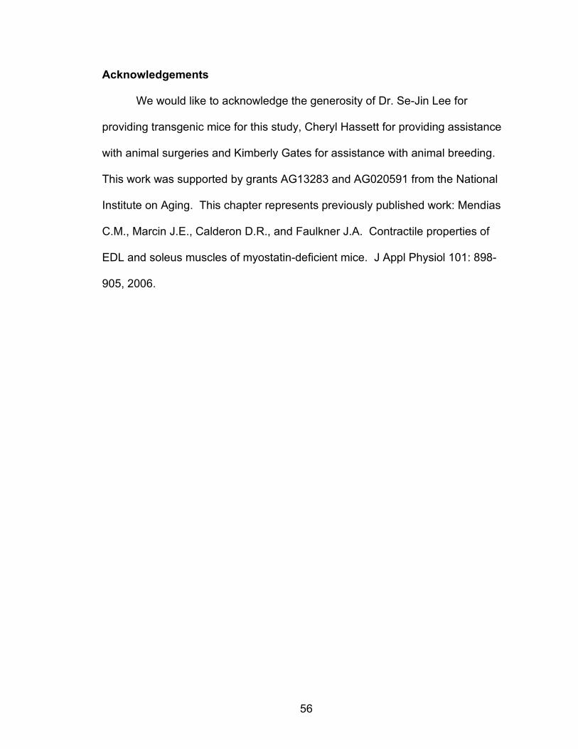
56
Acknowledgements
We would like to acknowledge the generosity of Dr. Se-Jin Lee for
providing transgenic mice for this study, Cheryl Hassett for providing assistance
with animal surgeries and Kimberly Gates for assistance with animal breeding.
This work was supported by grants AG13283 and AG020591 from the National
Institute on Aging. This chapter represents previously published work: Mendias
C.M., Marcin J.E., Calderon D.R., and Faulkner J.A. Contractile properties of
EDL and soleus muscles of myostatin-deficient mice. J Appl Physiol 101: 898-
905, 2006.
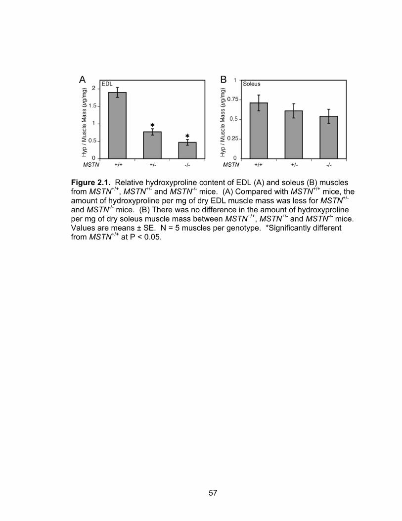
57
Figure 2.1. Relative hydroxyproline content of EDL (A) and soleus (B) muscles from MSTN+/+, MSTN+/- and MSTN-/- mice. (A) Compared with MSTN+/+ mice, the amount of hydroxyproline per mg of dry EDL muscle mass was less for MSTN+/- and MSTN-/- mice. (B) There was no difference in the amount of hydroxyproline per mg of dry soleus muscle mass between MSTN+/+, MSTN+/- and MSTN-/- mice. Values are means ± SE. N = 5 muscles per genotype. *Significantly different from MSTN+/+ at P < 0.05.
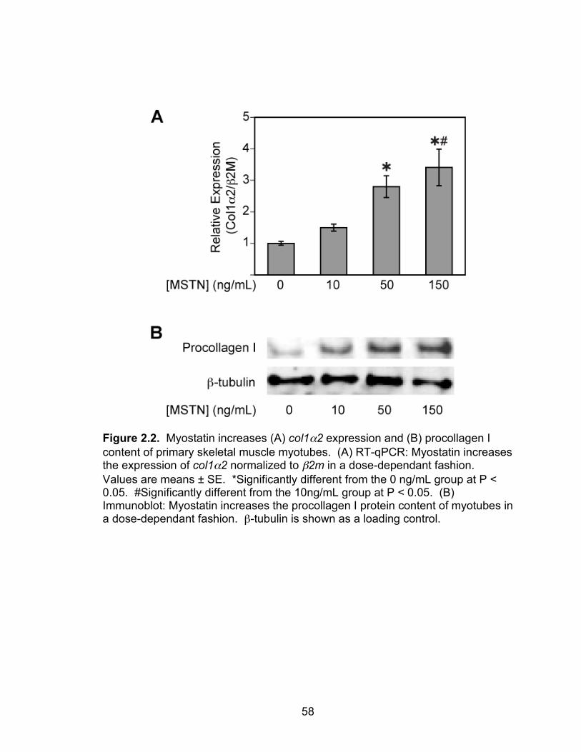
58
Figure 2.2. Myostatin increases (A) col1α2 expression and (B) procollagen I content of primary skeletal muscle myotubes. (A) RT-qPCR: Myostatin increases the expression of col1α2 normalized to β2m in a dose-dependant fashion. Values are means ± SE. *Significantly different from the 0 ng/mL group at P < 0.05. #Significantly different from the 10ng/mL group at P < 0.05. (B) Immunoblot: Myostatin increases the procollagen I protein content of myotubes in a dose-dependant fashion. β-tubulin is shown as a loading control.
59
Figure 2.3. Force values for EDL muscles (A, B) and soleus muscles (C,D) from MSTN+/+, MSTN+/- and MSTN-/- mice. (A) The Po of EDL muscles of MSTN-/- mice is greater than the Po of MSTN+/+ and MSTN+/- mice. (B) When Po is normalized to CSA, the sPo of EDL muscles of MSTN-/- mice is less than the sPo of MSTN+/+ and MSTN+/- mice. (C) The Po of soleus muscles of MSTN-/- mice is greater than the Po of MSTN+/+ and MSTN+/- mice. (D) When Po is normalized to CSA, there is no difference in sPo of soleus muscles. Values are means ± SE. N = 6 muscles per genotype. *Significantly different from MSTN+/+ at P < 0.05. #Significantly different from MSTN+/- at P < 0.05.
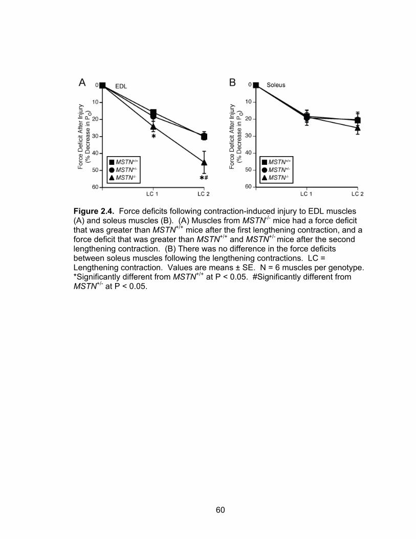
60
Figure 2.4. Force deficits following contraction-induced injury to EDL muscles (A) and soleus muscles (B). (A) Muscles from MSTN-/- mice had a force deficit that was greater than MSTN+/+ mice after the first lengthening contraction, and a force deficit that was greater than MSTN+/+ and MSTN+/- mice after the second lengthening contraction. (B) There was no difference in the force deficits between soleus muscles following the lengthening contractions. LC = Lengthening contraction. Values are means ± SE. N = 6 muscles per genotype. *Significantly different from MSTN+/+ at P < 0.05. #Significantly different from MSTN+/- at P < 0.05.
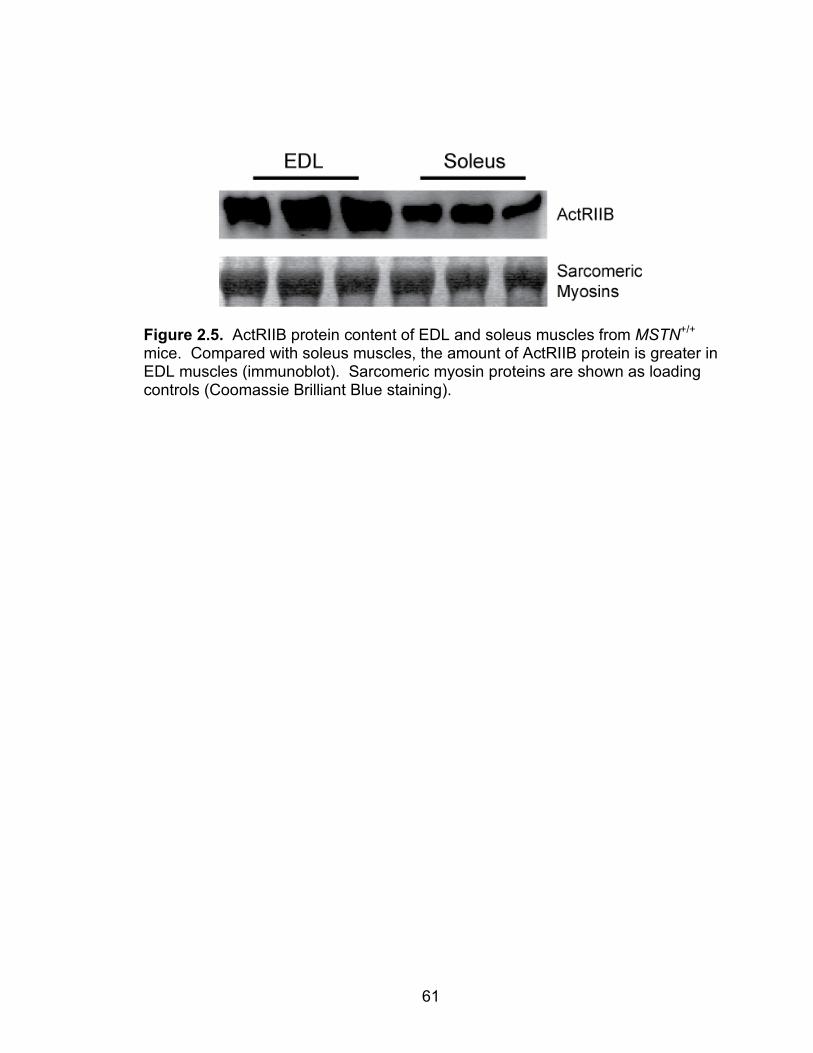
61
Figure 2.5. ActRIIB protein content of EDL and soleus muscles from MSTN+/+ mice. Compared with soleus muscles, the amount of ActRIIB protein is greater in EDL muscles (immunoblot). Sarcomeric myosin proteins are shown as loading controls (Coomassie Brilliant Blue staining).
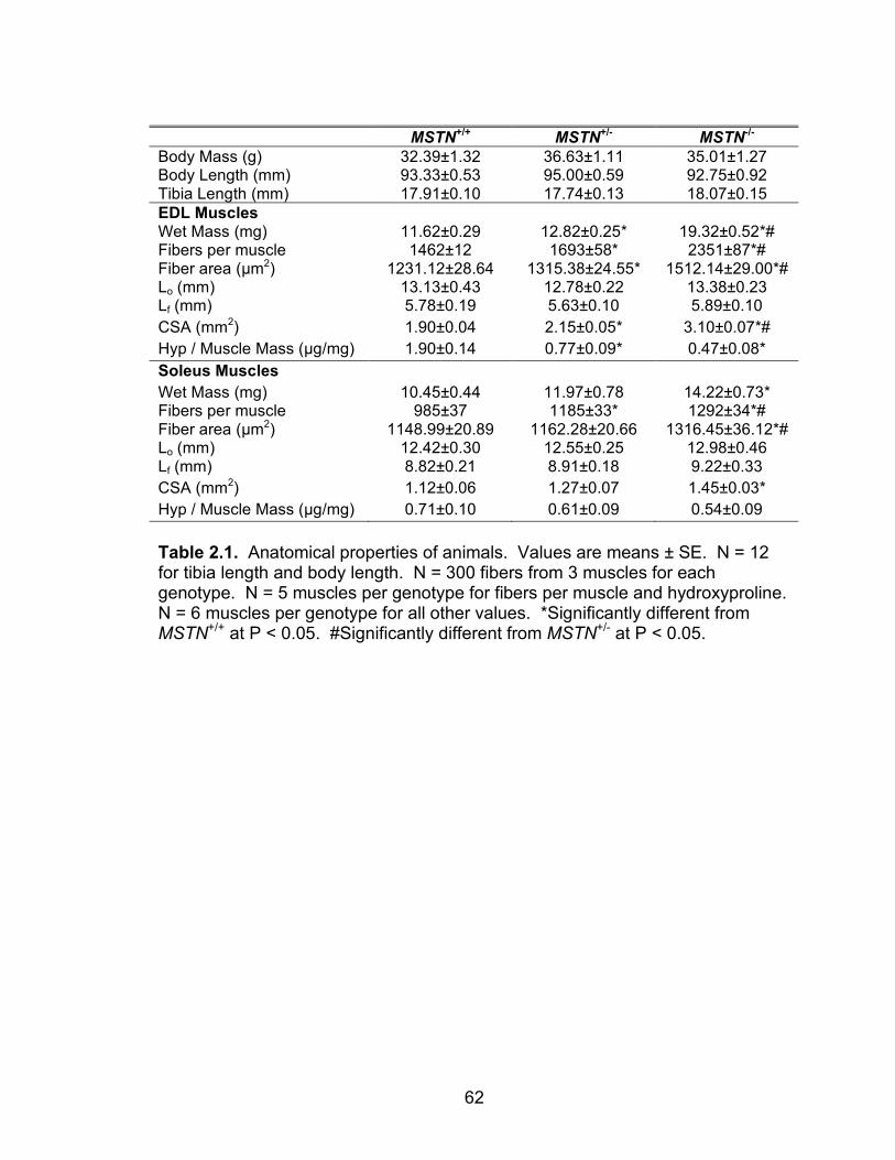
62
MSTN+/+ MSTN+/- MSTN-/- Body Mass (g) 32.39±1.32 36.63±1.11 35.01±1.27 Body Length (mm) 93.33±0.53 95.00±0.59 92.75±0.92 Tibia Length (mm) 17.91±0.10 17.74±0.13 18.07±0.15 EDL Muscles Wet Mass (mg) 11.62±0.29 12.82±0.25* 19.32±0.52*# Fibers per muscle 1462±12 1693±58* 2351±87*# Fiber area (µm2) 1231.12±28.64 1315.38±24.55* 1512.14±29.00*# Lo (mm) 13.13±0.43 12.78±0.22 13.38±0.23 Lf (mm) 5.78±0.19 5.63±0.10 5.89±0.10 CSA (mm2) 1.90±0.04 2.15±0.05* 3.10±0.07*# Hyp / Muscle Mass (µg/mg) 1.90±0.14 0.77±0.09* 0.47±0.08* Soleus Muscles Wet Mass (mg) 10.45±0.44 11.97±0.78 14.22±0.73* Fibers per muscle 985±37 1185±33* 1292±34*# Fiber area (µm2) 1148.99±20.89 1162.28±20.66 1316.45±36.12*# Lo (mm) 12.42±0.30 12.55±0.25 12.98±0.46 Lf (mm) 8.82±0.21 8.91±0.18 9.22±0.33 CSA (mm2) 1.12±0.06 1.27±0.07 1.45±0.03* Hyp / Muscle Mass (µg/mg) 0.71±0.10 0.61±0.09 0.54±0.09 Table 2.1. Anatomical properties of animals. Values are means ± SE. N = 12 for tibia length and body length. N = 300 fibers from 3 muscles for each genotype. N = 5 muscles per genotype for fibers per muscle and hydroxyproline. N = 6 muscles per genotype for all other values. *Significantly different from MSTN+/+ at P < 0.05. #Significantly different from MSTN+/- at P < 0.05.
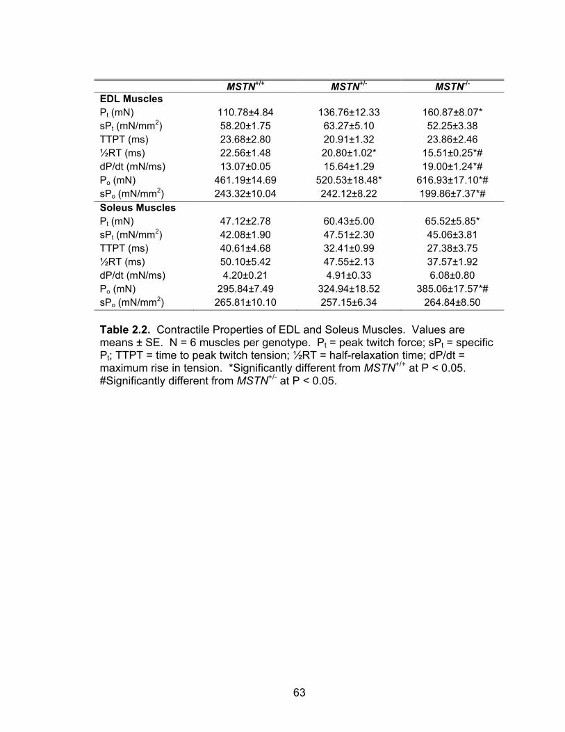
63
MSTN+/+ MSTN+/- MSTN-/- EDL Muscles Pt (mN) 110.78±4.84 136.76±12.33 160.87±8.07* sPt (mN/mm2) 58.20±1.75 63.27±5.10 52.25±3.38 TTPT (ms) 23.68±2.80 20.91±1.32 23.86±2.46 ½RT (ms) 22.56±1.48 20.80±1.02* 15.51±0.25*# dP/dt (mN/ms) 13.07±0.05 15.64±1.29 19.00±1.24*# Po (mN) 461.19±14.69 520.53±18.48* 616.93±17.10*# sPo (mN/mm2) 243.32±10.04 242.12±8.22 199.86±7.37*# Soleus Muscles Pt (mN) 47.12±2.78 60.43±5.00 65.52±5.85* sPt (mN/mm2) 42.08±1.90 47.51±2.30 45.06±3.81 TTPT (ms) 40.61±4.68 32.41±0.99 27.38±3.75 ½RT (ms) 50.10±5.42 47.55±2.13 37.57±1.92 dP/dt (mN/ms) 4.20±0.21 4.91±0.33 6.08±0.80 Po (mN) 295.84±7.49 324.94±18.52 385.06±17.57*# sPo (mN/mm2) 265.81±10.10 257.15±6.34 264.84±8.50 Table 2.2. Contractile Properties of EDL and Soleus Muscles. Values are means ± SE. N = 6 muscles per genotype. Pt = peak twitch force; sPt = specific Pt; TTPT = time to peak twitch tension; ½RT = half-relaxation time; dP/dt = maximum rise in tension. *Significantly different from MSTN+/+ at P < 0.05. #Significantly different from MSTN+/- at P < 0.05.

64
MSTN+/+ MSTN+/- MSTN-/- EDL Muscles Stretch 1 Average Force (mN) 648.07±8.21 644.48±15.74 664.56±8.63 Work (J/kg) 129.00±3.66 113.05±1.85* 81.31±2.65*# Force Deficit (% of Pre-injury Po) 15.74±0.81 18.19±2.78 24.14±3.03* Stretch 2 Average Force (mN) 631.03±8.43 632.78±14.93 651.86±9.00 Work (J/kg) 125.58±3.39 111.01±1.76* 79.76±2.65*# Force Deficit (% of Pre-injury Po) 30.10±1.71 29.59±2.46 45.23±6.52*# Soleus Muscles Stretch 1 Average Force (mN) 509.53±26.93 537.37±33.60 620.89±28.49* Work (J/kg) 173.21±11.68 160.60±5.56 161.15±5.39 Force Deficit (% of Pre-injury Po) 18.21±3.63 19.18±4.38 18.60±2.52 Stretch 2 Average Force (mN) 455.83±14.49 480.13±29.67 569.29±31.98*# Work (J/kg) 154.89±8.35 143.33±3.96 147.60±6.21 Force Deficit (% of Pre-injury Po) 20.68 ± 4.93 20.21 ± 3.45 25.08 ± 3.76 Table 2.3. Mechanical Injury of EDL and Soleus Muscles. Values are means ± SE. N = 6 muscles per genotype. *Significantly different from MSTN+/+ at P < 0.05. #Significantly different from MSTN+/- at P < 0.05.
65
References
1. Allen RE, Temm-Grove CJ, Sheehan SM, and Rice G. Skeletal muscle satellite cell cultures. Methods Cell Biol 52: 155-176, 1997. 2. Amthor H, Nicholas G, McKinnell I, Kemp CF, Sharma M, Kambadur R, and Patel K. Follistatin complexes Myostatin and antagonises Myostatin-mediated inhibition of myogenesis. Dev Biol 270: 19-30, 2004. 3. Bernasconi P, Torchiana E, Confalonieri P, Brugnoni R, Barresi R, Mora M, Cornelio F, Morandi L, and Mantegazza R. Expression of transforming growth factor-beta 1 in dystrophic patient muscles correlates with fibrosis. Pathogenetic role of a fibrogenic cytokine. J Clin Invest 96: 1137-1144, 1995. 4. Bogdanovich S, Krag TO, Barton ER, Morris LD, Whittemore LA, Ahima RS, and Khurana TS. Functional improvement of dystrophic muscle by myostatin blockade. Nature 420: 418-421, 2002. 5. Bogdanovich S, Perkins KJ, Krag TO, Whittemore LA, and Khurana TS. Myostatin propeptide-mediated amelioration of dystrophic pathophysiology. Faseb J 19: 543-549, 2005. 6. Brooks SV and Faulkner JA. Contractile properties of skeletal muscles from young, adult and aged mice. J Physiol 404: 71-82, 1988. 7. Brooks SV and Faulkner JA. The magnitude of the initial injury induced by stretches of maximally activated muscle fibres of mice and rats increases in old age. J Physiol 497 ( Pt 2): 573-580, 1996. 8. Brooks SV, Zerba E, and Faulkner JA. Injury to muscle fibres after single stretches of passive and maximally stimulated muscles in mice. J Physiol 488 ( Pt 2): 459-469, 1995. 9. Byron CD, Hamrick MW, and Wingard CJ. Alterations of temporalis muscle contractile force and histological content from the myostatin and Mdx deficient mouse. Arch Oral Biol, 2005. 10. Carlson CJ, Booth FW, and Gordon SE. Skeletal muscle myostatin mRNA expression is fiber-type specific and increases during hindlimb unloading. Am J Physiol 277: R601-606, 1999. 11. Consolino CM and Brooks SV. Susceptibility to sarcomere injury induced by single stretches of maximally activated muscles of mdx mice. J Appl Physiol 96: 633-638, 2004. 12. Contri MB, Guerra D, Vignali N, Taparelli F, Marcuzzi A, Caroli A, and Ronchetti IP. Ultrastructural and immunocytochemical study on normal human palmar aponeuroses. Anat Rec 240: 314-321, 1994. 13. Delforge G. Musculoskeletal Trauma : implications for sports injury management. Champaign, IL: Human Kinetics, 2002. 14. Dellorusso C, Crawford RW, Chamberlain JS, and Brooks SV. Tibialis anterior muscles in mdx mice are highly susceptible to contraction-induced injury. J Muscle Res Cell Motil 22: 467-475, 2001. 15. Donovan CM and Faulkner JA. Muscle grafts overloaded by ablation of synergistic muscles. J Appl Physiol 61: 288-292, 1986.
66
16. Ducomps C, Mauriege P, Darche B, Combes S, Lebas F, and Doutreloux JP. Effects of jump training on passive mechanical stress and stiffness in rabbit skeletal muscle: role of collagen. Acta Physiol Scand 178: 215-224, 2003. 17. Engvall E and Wewer UM. The new frontier in muscular dystrophy research: booster genes. Faseb J 17: 1579-1584, 2003. 18. Friden J and Lieber RL. Segmental muscle fiber lesions after repetitive eccentric contractions. Cell Tissue Res 293: 165-171, 1998. 19. Gosselin LE, Adams C, Cotter TA, McCormick RJ, and Thomas DP. Effect of exercise training on passive stiffness in locomotor skeletal muscle: role of extracellular matrix. J Appl Physiol 85: 1011-1016, 1998. 20. Grobet L, Martin LJ, Poncelet D, Pirottin D, Brouwers B, Riquet J, Schoeberlein A, Dunner S, Menissier F, Massabanda J, Fries R, Hanset R, and Georges M. A deletion in the bovine myostatin gene causes the double-muscled phenotype in cattle. Nat Genet 17: 71-74, 1997. 21. Grobet L, Pirottin D, Farnir F, Poncelet D, Royo LJ, Brouwers B, Christians E, Desmecht D, Coignoul F, Kahn R, and Georges M. Modulating skeletal muscle mass by postnatal, muscle-specific inactivation of the myostatin gene. Genesis 35: 227-238, 2003. 22. Hanyu T, Tajima T, Takagi T, Sasaki S, Fujimoto D, Isemura M, and Yosizawa Z. Biochemical studies on the collagen of the palmar aponeurosis affected with Dupuytren's disease. Tohoku J Exp Med 142: 437-443, 1984. 23. Huijing P. Muscular force transmission: a unified, dual or multiple system? A review and some explorative experimental results. Arch Physiol Biochem 107: 292-311, 1999. 24. Kambadur R, Sharma M, Smith TP, and Bass JJ. Mutations in myostatin (GDF8) in double-muscled Belgian Blue and Piedmontese cattle. Genome Res 7: 910-916, 1997. 25. Kandarian SC and White TP. Mechanical deficit persists during long-term muscle hypertrophy. J Appl Physiol 69: 861-867, 1990. 26. Khurana TS and Davies KE. Pharmacological strategies for muscular dystrophy. Nat Rev Drug Discov 2: 379-390, 2003. 27. Kjaer M. Role of extracellular matrix in adaptation of tendon and skeletal muscle to mechanical loading. Physiol Rev 84: 649-698, 2004. 28. Langley B, Thomas M, Bishop A, Sharma M, Gilmour S, and Kambadur R. Myostatin inhibits myoblast differentiation by down-regulating MyoD expression. J Biol Chem 277: 49831-49840, 2002. 29. Lee SJ and McPherron AC. Regulation of myostatin activity and muscle growth. Proc Natl Acad Sci U S A 98: 9306-9311, 2001. 30. Lesch M, Parmley WW, Hamosh M, Kaufman S, and Sonnenblick EH. Effects of acute hypertrophy on the contractile properties of skeletal muscle. Am J Physiol 214: 685-690, 1968. 31. Light N and Champion AE. Characterization of muscle epimysium, perimysium and endomysium collagens. Biochem J 219: 1017-1026, 1984. 32. Livak KJ and Schmittgen TD. Analysis of relative gene expression data using real-time quantitative PCR and the 2(-Delta Delta C(T)) Method. Methods 25: 402-408, 2001.
67
33. Macpherson PC, Dennis RG, and Faulkner JA. Sarcomere dynamics and contraction-induced injury to maximally activated single muscle fibres from soleus muscles of rats. J Physiol 500 ( Pt 2): 523-533, 1997. 34. Macpherson PC, Schork MA, and Faulkner JA. Contraction-induced injury to single fiber segments from fast and slow muscles of rats by single stretches. Am J Physiol 271: C1438-1446, 1996. 35. Mahoney DJ, Carey K, Fu MH, Snow R, Cameron-Smith D, Parise G, and Tarnopolsky MA. Real-time RT-PCR analysis of housekeeping genes in human skeletal muscle following acute exercise. Physiol Genomics 18: 226-231, 2004. 36. Martyn JK, Bass JJ, and Oldham JM. Skeletal muscle development in normal and double-muscled cattle. Anat Rec A Discov Mol Cell Evol Biol 281: 1363-1371, 2004. 37. Matsakas A, Friedel A, Hertrampf T, and Diel P. Short-term endurance training results in a muscle-specific decrease of myostatin mRNA content in the rat. Acta Physiol Scand 183: 299-307, 2005. 38. Maxwell LC, Faulkner JA, and Hyatt GJ. Estimation of number of fibers in guinea pig skeletal muscles. J Appl Physiol 37: 259-264, 1974. 39. McCroskery S, Thomas M, Maxwell L, Sharma M, and Kambadur R. Myostatin negatively regulates satellite cell activation and self-renewal. J Cell Biol 162: 1135-1147, 2003. 40. McCroskery S, Thomas M, Platt L, Hennebry A, Nishimura T, McLeay L, Sharma M, and Kambadur R. Improved muscle healing through enhanced regeneration and reduced fibrosis in myostatin-null mice. J Cell Sci 118: 3531-3541, 2005. 41. McMahon CD, Popovic L, Oldham JM, Jeanplong F, Smith HK, Kambadur R, Sharma M, Maxwell L, and Bass JJ. Myostatin-deficient mice lose more skeletal muscle mass than wild-type controls during hindlimb suspension. Am J Physiol Endocrinol Metab 285: E82-87, 2003. 42. McPherron AC, Lawler AM, and Lee SJ. Regulation of skeletal muscle mass in mice by a new TGF-beta superfamily member. Nature 387: 83-90, 1997. 43. Mendler L, Zador E, Ver Heyen M, Dux L, and Wuytack F. Myostatin levels in regenerating rat muscles and in myogenic cell cultures. J Muscle Res Cell Motil 21: 551-563, 2000. 44. Neuman RE and Logan MA. The determination of hydroxyproline. J Biol Chem 184: 299-306, 1950. 45. Patel K and Amthor H. The function of Myostatin and strategies of Myostatin blockade-new hope for therapies aimed at promoting growth of skeletal muscle. Neuromuscul Disord 15: 117-126, 2005. 46. Philip B, Lu Z, and Gao Y. Regulation of GDF-8 signaling by the p38 MAPK. Cell Signal 17: 365-375, 2005. 47. Pirottin D, Grobet L, Adamantidis A, Farnir F, Herens C, Daa Schroder H, and Georges M. Transgenic engineering of male-specific muscular hypertrophy. Proc Natl Acad Sci U S A 102: 6413-6418, 2005. 48. Prado LG, Makarenko I, Andresen C, Kruger M, Opitz CA, and Linke WA. Isoform diversity of giant proteins in relation to passive and active contractile properties of rabbit skeletal muscles. J Gen Physiol 126: 461-480, 2005.
68
49. Prentice WE. Rehabilitation techniques in sports medicine and athletic training. New York: McGraw-Hill, 2004. 50. Rebbapragada A, Benchabane H, Wrana JL, Celeste AJ, and Attisano L. Myostatin signals through a transforming growth factor beta-like signaling pathway to block adipogenesis. Mol Cell Biol 23: 7230-7242, 2003. 51. Rehfeldt C, Ott G, Gerrard DE, Varga L, Schlote W, Williams JL, Renne U, and Bunger L. Effects of the Compact mutant myostatin allele Mstn (Cmpt-dl1Abc) introgressed into a high growth mouse line on skeletal muscle cellularity. J Muscle Res Cell Motil, 2005. 52. Reisz-Porszasz S, Bhasin S, Artaza JN, Shen R, Sinha-Hikim I, Hogue A, Fielder TJ, and Gonzalez-Cadavid NF. Lower skeletal muscle mass in male transgenic mice with muscle-specific overexpression of myostatin. Am J Physiol Endocrinol Metab 285: E876-888, 2003. 53. Roth SM and Walsh S. Myostatin: a therapeutic target for skeletal muscle wasting. Curr Opin Clin Nutr Metab Care 7: 259-263, 2004. 54. Schuelke M, Wagner KR, Stolz LE, Hubner C, Riebel T, Komen W, Braun T, Tobin JF, and Lee SJ. Myostatin mutation associated with gross muscle hypertrophy in a child. N Engl J Med 350: 2682-2688, 2004. 55. Sharma M, Langley B, Bass J, and Kambadur R. Myostatin in muscle growth and repair. Exerc Sport Sci Rev 29: 155-158, 2001. 56. Taylor WE, Bhasin S, Artaza J, Byhower F, Azam M, Willard DH, Jr., Kull FC, Jr., and Gonzalez-Cadavid N. Myostatin inhibits cell proliferation and protein synthesis in C2C12 muscle cells. Am J Physiol Endocrinol Metab 280: E221-228, 2001. 57. Thies RS, Chen T, Davies MV, Tomkinson KN, Pearson AA, Shakey QA, and Wolfman NM. GDF-8 propeptide binds to GDF-8 and antagonizes biological activity by inhibiting GDF-8 receptor binding. Growth Factors 18: 251-259, 2001. 58. Thomas M, Langley B, Berry C, Sharma M, Kirk S, Bass J, and Kambadur R. Myostatin, a negative regulator of muscle growth, functions by inhibiting myoblast proliferation. J Biol Chem 275: 40235-40243, 2000. 59. Tobin JF and Celeste AJ. Myostatin, a negative regulator of muscle mass: implications for muscle degenerative diseases. Curr Opin Pharmacol 5: 328-332, 2005. 60. Verrecchia F and Mauviel A. TGF-beta and TNF-alpha: antagonistic cytokines controlling type I collagen gene expression. Cell Signal 16: 873-880, 2004. 61. Wagner KR. Muscle regeneration through myostatin inhibition. Curr Opin Rheumatol 17: 720-724, 2005. 62. Wagner KR, Liu X, Chang X, and Allen RE. Muscle regeneration in the prolonged absence of myostatin. Proc Natl Acad Sci U S A 102: 2519-2524, 2005. 63. Wagner KR, McPherron AC, Winik N, and Lee SJ. Loss of myostatin attenuates severity of muscular dystrophy in mdx mice. Ann Neurol 52: 832-836, 2002.
69
64. Wanke T, Toifl K, Merkle M, Formanek D, Lahrmann H, and Zwick H. Inspiratory muscle training in patients with Duchenne muscular dystrophy. Chest 105: 475-482, 1994. 65. Whittemore LA, Song K, Li X, Aghajanian J, Davies M, Girgenrath S, Hill JJ, Jalenak M, Kelley P, Knight A, Maylor R, O'Hara D, Pearson A, Quazi A, Ryerson S, Tan XY, Tomkinson KN, Veldman GM, Widom A, Wright JF, Wudyka S, Zhao L, and Wolfman NM. Inhibition of myostatin in adult mice increases skeletal muscle mass and strength. Biochem Biophys Res Commun 300: 965-971, 2003. 66. Woessner JF, Jr. The determination of hydroxyproline in tissue and protein samples containing small proportions of this imino acid. Arch Biochem Biophys 93: 440-447, 1961. 67. Wolfman NM, McPherron AC, Pappano WN, Davies MV, Song K, Tomkinson KN, Wright JF, Zhao L, Sebald SM, Greenspan DS, and Lee SJ. Activation of latent myostatin by the BMP-1/tolloid family of metalloproteinases. Proc Natl Acad Sci U S A 100: 15842-15846, 2003. 68. Yang J, Ratovitski T, Brady JP, Solomon MB, Wells KD, and Wall RJ. Expression of myostatin pro domain results in muscular transgenic mice. Mol Reprod Dev 60: 351-361, 2001. 69. Zhu X, Hadhazy M, Wehling M, Tidball JG, and McNally EM. Dominant negative myostatin produces hypertrophy without hyperplasia in muscle. FEBS Lett 474: 71-75, 2000. 70. Zhu X, Topouzis S, Liang LF, and Stotish RL. Myostatin signaling through Smad2, Smad3 and Smad4 is regulated by the inhibitory Smad7 by a negative feedback mechanism. Cytokine 26: 262-272, 2004. 71. Zimmers TA, Davies MV, Koniaris LG, Haynes P, Esquela AF, Tomkinson KN, McPherron AC, Wolfman NM, and Lee SJ. Induction of cachexia in mice by systemically administered myostatin. Science 296: 1486-1488, 2002.
70
Chapter III
Tendons of Myostatin-Deficient Mice are Small, Brittle and Hypocellular Abstract
Tendons play a significant role in the modulation of forces transmitted
between bones and skeletal muscles and consequently protect muscle fibers
from contraction-induced, or high strain, injuries. Myostatin (GDF-8) is a
negative regulator of muscle mass. Inhibition of myostatin not only increases the
mass and maximum isometric force of muscles, but also increases the
susceptibility of muscle fibers to contraction-induced injury. Furthermore, the
expression of myostatin and the myostatin receptors, ACVR2B and ACVRB,
were detectable in the tendons. We hypothesized that myostatin would regulate
the morphology and mechanical properties of tendons. Surprisingly, compared
with wild type (MSTN+/+) mice, the tendons of myostatin-null mice (MSTN-/-) were
smaller, had a decrease in fibroblast density and a decrease in the expression of
type I collagen. Tendons of MSTN-/- mice also had a decrease in the expression
of two genes that promote tendon fibroblast proliferation, scleraxis and
tenomodulin. Treatment of tendon fibroblasts with myostatin activated the p38
MAPK and Smad2/3 signaling cascades, increased cell proliferation and
increased the expression of type I collagen, scleraxis and tenomodulin.
Compared with the tendons of MSTN+/+ mice, the mechanical properties of tibialis
71
anterior tendons from MSTN-/- mice had a greater peak stress, lower peak strain
and a increased stiffness. We conclude that in addition to the regulation of
muscle mass and force, myostatin regulates the structure and function of tendon
tissues.
Introduction
Tendons are a critical component of the musculoskeletal system.
Situated, as tendons are, between bones and skeletal muscles, tendons are in a
position to transmit forces generated within skeletal muscle fibers to bone and
conversely transmit to skeletal muscle external loads placed on bone. The
extracellular matrix (ECM) of tendon tissue is composed primarily of type I
collagen, as well as type III collagen, elastin and various proteoglycans and
mucopolysaccharides. Tendons are in series with both the contractile and non-
contractile elements of skeletal muscles as well as with bone. Consequently,
tendons are able to both store elastic energy during locomotion and protect
muscle fibers from stretch-induced and contraction-induced injuries (15, 28).
While considerable research has been conducted on the effects of exercise,
immobilization and aging on the structure and function of tendons (19, 20), much
less is known about the specific cytokines that regulate the structure and function
of tendons.
During embryonic development of the limb, early tendon development can
be categorized into three phases, with each phase corresponding to an
upregulation of the bHLH transcription factor scleraxis (13, 40). Scleraxis is a
marker of the tendon cell lineage (38, 40) and mice deficient in scleraxis display
72
severe tendon defects, have impaired locomotion and a complete inability to use
their tails (34). Scleraxis promotes the formation of tendon ECM by inducing the
expression of type I collagen (23) and increases tendon fibroblast proliferation by
the upregulation of the expression of the type II transmembrane protein,
tenomodulin (41). During the final stages of early tendon development, FGF-4
and FGF-8, are secreted by adjacent myogenic cells and induce the expression
of scleraxis (5, 14). While FGF-4 and FGF-8 can induce scleraxis expression in
the final phase of early tendon development, they do not appear to be
responsible for the first and second phases of scleraxis expression (13). In fact,
another member of the GDF family may be a candidate for the regulation of
scleraxis expression during the first and second phases of early tendon
development (13), but the specific member of the GDF family has not been
identified.
Three members of the GDF family have been reported to influence the
development of tendon tissue. The placement of matrices coated with GDF-5,
GDF-6 and GDF-7 into skeletal muscle induce the ectopic formation of tendon-
like tissue (50). The tendons of GDF5-/- mice are smaller and display decreases
in type I collagen content, peak stress, stiffness and energy absorption to yield
(32). The GDF5-/- mice also have severe bone and joint defects (32), but
whether these changes in tendon mechanical properties arise due to a direct
effect of GDF-5 on tendon cells or the associated skeletal defects is not clear.
Compared with wild type mice, GDF7-/- mice have a minor tendon phenotype,
with a decrease in proteoglycan content and smaller collagen fibrils, but no
73
differences in Achilles tendon mechanical properties, type I collagen content, or
gross morphology (31). Some evidence supports roles for GDF-5, GDF-6 and
GDF-7 in tendon development, but whether other members of the GDF family
influence tendon development has not been established.
Myostatin (GDF-8) is a member of the TGF-ß superfamily of cytokines and
is a negative regulator of skeletal muscle mass. Myostatin binds to the activin
type IIB (ACVR2B) and type IB (ACVRB) receptors and activates the Smad2/3,
p38 MAPK and Erk1/2 signal transduction pathways (22, 37, 39, 51, 53).
Myostatin regulates muscle mass by inhibiting the proliferation and differentiation
of satellite cells (21, 26, 45), in addition to decreasing myofibrillar protein
synthesis (49) and increasing the expression of the E3 ubiquitin ligase atrogin-
1/MAFbx (27). Myostatin has a well established role in regulation of the structure
and function of skeletal muscle, but the contribution of myostatin to the regulation
of the structure and function of tendon has not been established.
In addition to the regulation of muscle mass, myostatin has a profound
impact on the contractile properties of skeletal muscles. Inhibition of myostatin
increases the maximum isometric force of skeletal muscles (3, 4, 30) and the
susceptibility of muscles to contraction-induced injury (30). During a lengthening
contraction, the series elastic component (aponeurosis and tendon) of a muscle
protects muscle fibers from damage by reducing the strain on fibers (15). The
MSTN-/- mice are much more susceptible to contraction-induced injury than the
MSTN+/+ mice, an observation that is consistent with the possibility that myostatin
might play a role in the regulation of the structural and functional properties of the
74
tendons. Our prior study (30) focused on the mechanical and contractile
properties of the muscle and aponeurosis, but did not investigate the mechanical
properties of the tendons of MSTN-/- mice directly. The overall aim of this
investigation was to determine the role of myostatin in regulation of the
mechanical and morphological properties of tendons. We hypothesized that a
deficiency in myostatin results in smaller, stiffer, and more brittle tendons.
Methods
Animals. All experiments were conducted in accordance with the
guidelines of the University of Michigan Committee on the Use and Care of
Animals. Mice were housed in specific-pathogen-free conditions and were
provided food and water ad libidum. The line of MSTN-/- mice used in this study
are of a C57Bl/6 background and were a kind gift of Dr. Se-Jin Lee. The null
MSTN allele was generated by replacing a portion of the third exon of the MSTN
gene that encodes the C-terminal region of the mature myostatin protein with a
neo cassette (29). The wild type (MSTN+/+) littermates of the MSTN-/- mice
served as controls. The genotype of mice was determined by PCR-based
analysis of DNA samples obtained via tail biopsy.
Mechanical Testing of Tendons. To evaluate the mechanical properties of
the tibialis anterior tendon, the entire tendon unit (from the myotendinous junction
to the base of the first metatarsal bone) was used. The tibialis anterior tendon
was chosen based on its relative uniformity of diameter, minimal aponeurosis
and long gauge length (2). Six month old male mice were anesthetized with
intraperitoneal injection of Avertin (400 mg/kg). Braded silk sutures were tied
75
around the distal end of the tibialis anterior muscle just superior to the
myotendinous junction and at the very distal end of the tendon, just superior to
the first metatarsal. The length (Lo) of the tibialis anterior tendon was measured
using digital calipers while the ankle was placed in maximal plantarflexion. The
tendon was removed by cutting the muscle just superior to the proximal suture
and by removing the first metatarsal bone that was inferior to the distal suture.
The tendon was immediately submerged in PBS maintained at 25º C. The
tendon was held at Lo and cross-sectional area (CSA) was calculated from 10
evenly spaced width and depth measurements from high resolution digital
photographs of both top and side views of the tendon. Side views were obtained
using a 90º prism embedded in the side of the bath. These measurements were
fitted to an ellipse and the ellipse area was used as the tendon CSA.
The proximal end of the tendon was attached to a dual-mode servo
motor/force transducer (model 305C, Aurora Scientific) and the distal end was
attached to a fixed post. Custom designed software (LabVIEW 7.1, National
Instruments) controlled the servo motor motion and recorded force and strain
data at a sampling rate of 20 kHz. The tendon was stretched to a 100% strain
relative to Lo at a velocity of 1 Lo × s-1. Peak stress was defined as the stress
that further increases in length resulted in a rupture of the tendon or the point at
which yield strength had been reached without a frank rupture of the tendon.
Peak strain was defined as the strain at which peak stress was reached. The
data were fitted by either a fourth or fifth order polynomial function with an R2 ≥
0.9995. Peak tendon stiffness was calculated by differentiating the fitted
76
polynomial and then determining its maximum value between Lo and peak strain.
Average tendon stiffness was calculated as the mean value of the differentiated
polynomial between Lo and peak strain. The energy absorption to the yield point
was calculated by integrating the force-displacement function from Lo through
peak strain and normalizing this value by the mass of the tendon.
Histology. To determine the density of fibroblasts in tendon tissue, tibialis
anterior tendons were removed from 6 month old anesthetized mice, placed in
embedding media and snap frozen in using isopentane cooled with dry ice.
Sections were obtained from the proximal, middle and distal thirds of the tendon
and stained with hematoxylin and eosin. Fibroblast density for the entire tendon
was calculated by taking the mean value of the counts from each of the three
regions of the tendon.
Tendon Fibroblast Isolation, Culture And Treatment With Myostatin.
Hindlimb and forelimb tendons were isolated from anesthetized 4 month old male
MSTN+/+ mice, carefully trimmed of muscle and fat tissue, finely minced and
placed in DMEM + 0.05% Type II Collagenase (Invitrogen) in a shaking water
bath for 2 h at 37º C. Following dissociation, fibroblasts were pelleted by
centrifugation, resuspended in DMEM + 2% fetal bovine serum (FBS) + 1%
antibiotic-antimycotic (AbAm) and expanded in 100 mm culture dishes coated
with type I collagen (BD Biosciences). Fibroblasts were passaged twice upon
reaching 70% confluence. Following the last passage, 2 × 10⁴ fibroblasts were
plated in 35 mm culture dishes coated with type I collagen and expanded until
reaching 80% confluence. Fibroblasts were then starved of serum for 24 hours
77
prior to treatment by replacing serum containing media with DMEM + 1% AbAm
+ 1× Insulin-Transferrin-Selenium supplement (ITS, Invitrogen). Recombinant
murine myostatin, produced in NS0 mouse myeloma cells (R & D Systems), was
dissolved into the serum-free media at a final concentration of 500 or 1000
ng/mL. Stock solutions of the p38 MAPK inhibitor SB-203580 (10) and the
Smad2/3 inhibitor SB-431542 (17) were prepared by dissolving these solid
anhydrous compounds in DMSO at a concentration of 10mM. These stock
solutions were then added to serum-free media containing 1% DMSO at a final
concentration of 10µM for SB-203580 and 5µM for SB-431542. Fibroblasts were
pretreated with SB-203580 or SB-431542 for 1 hour prior to treatment with
myostatin.
Cell Proliferation and Immunocytochemistry. Following serum starvation,
fibroblasts were incubated in serum free media containing 20µM of the thymidine
analog 5-bromo-2′-deoxyuridine (BrdU, Sigma) for 3 hours. Fibroblasts were
rinsed twice with serum free media and treated with myostatin, SB-203580 and
SB-431542 as described above. Following 24 hours of treatment, fibroblasts
were rinsed with PBS, fixed in ice cold methanol and permeabilized with 0.5%
Triton X-100. The BrdU epitope was exposed by digesting DNA with 200U/mL of
EcoRI and denaturing DNA with 2N HCl. BrdU was visualized using an anti-
BrdU antibody (G3G4, SJ Kaufman, Developmental Studies Hybridoma Bank)
and a Cy3-conjugated secondary antibody (Jackson ImmunoResearch). DAPI
(Sigma) was used as a non-specific nuclear stain. Twenty five random fields
78
were counted per dish. Cell proliferation data presented are the mean ± SE
values of three independent experiments.
RT-PCR. RNA was isolated from samples using an RNeasy Mini Kit
(Qiagen). When isolating RNA from whole tendons, the tissue was treated with
type II collagenase and proteinase K prior to vigorous homogenization in
guanidine thiocyanate buffer. Due to the smaller size of the tibialis anterior
tendons, we were unable to consistently obtain adequate quantities of RNA
with an A260/A280 ratio between 1.8 and 2.0. We instead used RNA from Achilles
tendons, as we were able to obtain RNA with A260/A280 ratios between 1.8 and
2.0 from these tendons. RNA was treated with DNase I and reverse
transcribed using an Omniscript RT kit (Qiagen) and oligo(dT)15 primers.
Primers for PCR reactions (Table 3.1) were designed to generate amplicons that
span multiple exons. For standard PCR, 250ng of cDNA underwent 42 rounds of
amplification using GoTaq Green (Promega). PCR products were separated
using a 2% agarose gel. For real-time PCR, cDNA was amplified using a
QuantiTect SYBR Green I PCR system (Qiagen) with Uracil-N-Glycosylase
(Invitrogen) in an Opticon 2 real-time thermal cycler (Bio-Rad). qPCR reactions
were conducted in quadruplicate for each sample. We used the methods of
Livak and Schmittgen (25) to determine optimal loading quantities of cDNA and
in the validation of GAPDH as a housekeeping gene. Gene expression was
normalized to GAPDH expression using the 2-ΔΔC(t) method (25). The presence of
single amplicons from qPCR reactions was verified by melting curve analysis as
79
well as electrophoresis using a 2% agarose gel. qPCR data presented is the
combined means ± SE of three independent experiments.
Immunoblot. Tendon fibroblasts, prepared as described above and
starved of serum for 24 hours, were treated for two hours with myostatin, SB-
203580 and SB-431542 for 2 hours. Following treatment, cells were rinsed with
PBS and scraped and homogenized in Laemmli's sample buffer with 1:20 ß-
mercaptoethanol, 1:20 protease inhibitor cocktail (Sigma) and 1:40 phosphatase
inhibitor cocktail (Sigma), and then placed in boiling water for 5 minutes. Protein
concentration of the samples was determined using an RC DC Protein Assay
(Bio-Rad). Equal amounts of protein were loaded into polyacrylamide gels and
subjected to electrophoresis using a 4% stacking, 10% resolving gel. Proteins
were transferred to a 0.45 µm nitrocellulose membrane, stained with Ponceau S
to verify equal protein transfer and blocked using casein (Vector Labs).
Antibodies against p38 MAPK and phospho-p38 MAPK were purchased from
Cell Signaling. Primary antibodies against Smad2/3 and phospho-Smad2/3 were
purchased from Millipore. Biotinylated secondary antibodies were purchased
from Pierce Biotechnology. Avidin-HRPO conjugates were purchased from
Vector Labs. Membranes were developed using SuperSignal West Dura
enhanced chemiluminescent reagents (Pierce Biotechnology) and visualized
using a FluorChem chemiluminescent documentation system (Alpha Innotech).
Statistical Analysis. Results are presented as means ± SE. KaleidaGraph 4.02
software was used to conduct statistical tests. For gene expression and cell
proliferation data from cell culture experiments, differences between groups were
80
tested with a one-way ANOVA with α = 0.05. Fisher’s least significant post hoc
test was used to identify specific differences when significance was tested. For
all other data, differences between MSTN+/+ and MSTN-/- mice was tested with
Student’s t-test with α = 0.05.
Results
MSTN-/- mice have greater muscle masses but smaller tendons. We first
determined the impact of myostatin deficiency on the mass of tendons. Although
the mass of the tibialis anterior (TA) muscles of MSTN-/- mice were 72% greater
than MSTN+/+ mice, the TA tendons of the MSTN-/- mice were 40% smaller
(Table 3.2). When the tendon mass was normalized by the muscle mass, the
MSTN-/- mice had a 64% decrease in the tendon/muscle mass ratio. Similar
results were observed for soleus muscles and Achilles tendons. The mass of the
soleus muscles of MSTN-/- mice were 82% greater than MSTN+/+ mice, but the
Achilles tendons of the soleus muscles of MSTN-/- mice were 44% smaller than
those of MSTN+/+ mice. Consequently, for MSTN-/- mice the Achilles
tendon/soleus muscle mass ratio was decreased by 69%. Furthermore, for the
MSTN-/- mice, the CSA of the TA tendons were 50% smaller than those of the
MSTN+/+ mice (Table 3.3). The lengths and densities of the TA tendons of
MSTN-/- and MSTN+/+mice were not different. CSA and the lengths and densities
of Achilles tendons were not determined, as the Achilles tendons were not used
in testing of mechanical properties.
Tendon fibroblasts express the myostatin receptors and activate the p38
MAPK and Smad2/3 signaling pathways in response to myostatin treatment.
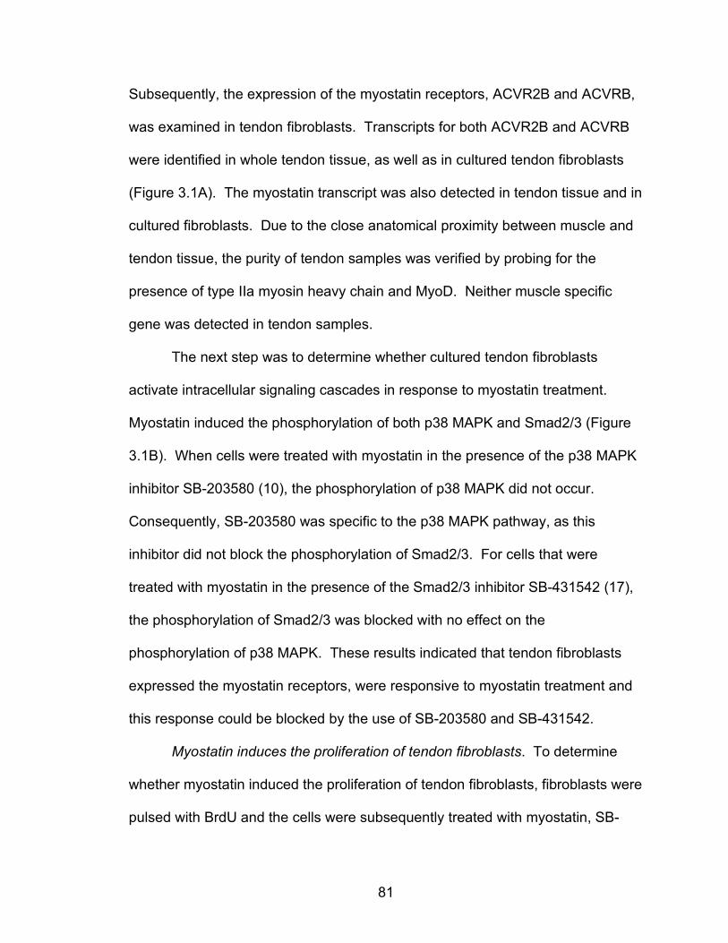
81
Subsequently, the expression of the myostatin receptors, ACVR2B and ACVRB,
was examined in tendon fibroblasts. Transcripts for both ACVR2B and ACVRB
were identified in whole tendon tissue, as well as in cultured tendon fibroblasts
(Figure 3.1A). The myostatin transcript was also detected in tendon tissue and in
cultured fibroblasts. Due to the close anatomical proximity between muscle and
tendon tissue, the purity of tendon samples was verified by probing for the
presence of type IIa myosin heavy chain and MyoD. Neither muscle specific
gene was detected in tendon samples.
The next step was to determine whether cultured tendon fibroblasts
activate intracellular signaling cascades in response to myostatin treatment.
Myostatin induced the phosphorylation of both p38 MAPK and Smad2/3 (Figure
3.1B). When cells were treated with myostatin in the presence of the p38 MAPK
inhibitor SB-203580 (10), the phosphorylation of p38 MAPK did not occur.
Consequently, SB-203580 was specific to the p38 MAPK pathway, as this
inhibitor did not block the phosphorylation of Smad2/3. For cells that were
treated with myostatin in the presence of the Smad2/3 inhibitor SB-431542 (17),
the phosphorylation of Smad2/3 was blocked with no effect on the
phosphorylation of p38 MAPK. These results indicated that tendon fibroblasts
expressed the myostatin receptors, were responsive to myostatin treatment and
this response could be blocked by the use of SB-203580 and SB-431542.
Myostatin induces the proliferation of tendon fibroblasts. To determine
whether myostatin induced the proliferation of tendon fibroblasts, fibroblasts were
pulsed with BrdU and the cells were subsequently treated with myostatin, SB-
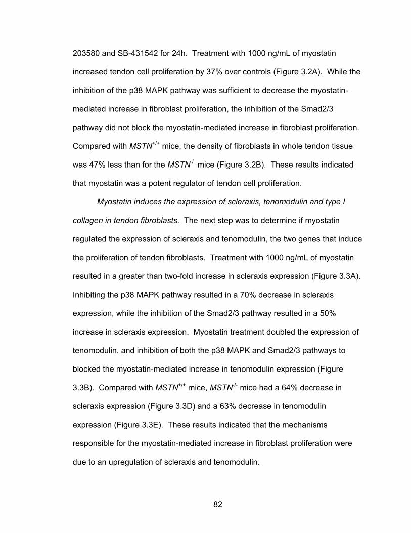
82
203580 and SB-431542 for 24h. Treatment with 1000 ng/mL of myostatin
increased tendon cell proliferation by 37% over controls (Figure 3.2A). While the
inhibition of the p38 MAPK pathway was sufficient to decrease the myostatin-
mediated increase in fibroblast proliferation, the inhibition of the Smad2/3
pathway did not block the myostatin-mediated increase in fibroblast proliferation.
Compared with MSTN+/+ mice, the density of fibroblasts in whole tendon tissue
was 47% less than for the MSTN-/- mice (Figure 3.2B). These results indicated
that myostatin was a potent regulator of tendon cell proliferation.
Myostatin induces the expression of scleraxis, tenomodulin and type I
collagen in tendon fibroblasts. The next step was to determine if myostatin
regulated the expression of scleraxis and tenomodulin, the two genes that induce
the proliferation of tendon fibroblasts. Treatment with 1000 ng/mL of myostatin
resulted in a greater than two-fold increase in scleraxis expression (Figure 3.3A).
Inhibiting the p38 MAPK pathway resulted in a 70% decrease in scleraxis
expression, while the inhibition of the Smad2/3 pathway resulted in a 50%
increase in scleraxis expression. Myostatin treatment doubled the expression of
tenomodulin, and inhibition of both the p38 MAPK and Smad2/3 pathways to
blocked the myostatin-mediated increase in tenomodulin expression (Figure
3.3B). Compared with MSTN+/+ mice, MSTN-/- mice had a 64% decrease in
scleraxis expression (Figure 3.3D) and a 63% decrease in tenomodulin
expression (Figure 3.3E). These results indicated that the mechanisms
responsible for the myostatin-mediated increase in fibroblast proliferation were
due to an upregulation of scleraxis and tenomodulin.
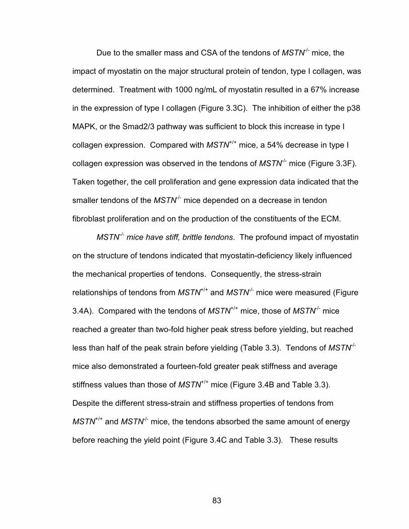
83
Due to the smaller mass and CSA of the tendons of MSTN-/- mice, the
impact of myostatin on the major structural protein of tendon, type I collagen, was
determined. Treatment with 1000 ng/mL of myostatin resulted in a 67% increase
in the expression of type I collagen (Figure 3.3C). The inhibition of either the p38
MAPK, or the Smad2/3 pathway was sufficient to block this increase in type I
collagen expression. Compared with MSTN+/+ mice, a 54% decrease in type I
collagen expression was observed in the tendons of MSTN-/- mice (Figure 3.3F).
Taken together, the cell proliferation and gene expression data indicated that the
smaller tendons of the MSTN-/- mice depended on a decrease in tendon
fibroblast proliferation and on the production of the constituents of the ECM.
MSTN-/- mice have stiff, brittle tendons. The profound impact of myostatin
on the structure of tendons indicated that myostatin-deficiency likely influenced
the mechanical properties of tendons. Consequently, the stress-strain
relationships of tendons from MSTN+/+ and MSTN-/- mice were measured (Figure
3.4A). Compared with the tendons of MSTN+/+ mice, those of MSTN-/- mice
reached a greater than two-fold higher peak stress before yielding, but reached
less than half of the peak strain before yielding (Table 3.3). Tendons of MSTN-/-
mice also demonstrated a fourteen-fold greater peak stiffness and average
stiffness values than those of MSTN+/+ mice (Figure 3.4B and Table 3.3).
Despite the different stress-strain and stiffness properties of tendons from
MSTN+/+ and MSTN-/- mice, the tendons absorbed the same amount of energy
before reaching the yield point (Figure 3.4C and Table 3.3). These results
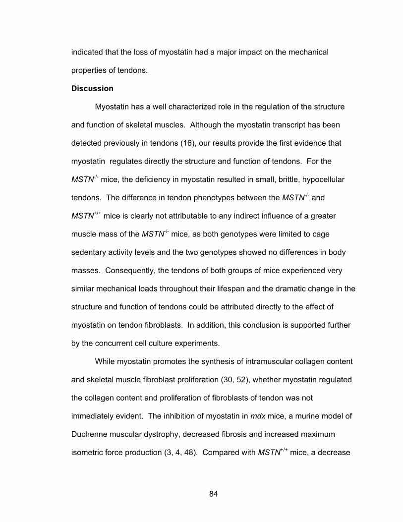
84
indicated that the loss of myostatin had a major impact on the mechanical
properties of tendons.
Discussion
Myostatin has a well characterized role in the regulation of the structure
and function of skeletal muscles. Although the myostatin transcript has been
detected previously in tendons (16), our results provide the first evidence that
myostatin regulates directly the structure and function of tendons. For the
MSTN-/- mice, the deficiency in myostatin resulted in small, brittle, hypocellular
tendons. The difference in tendon phenotypes between the MSTN-/- and
MSTN+/+ mice is clearly not attributable to any indirect influence of a greater
muscle mass of the MSTN-/- mice, as both genotypes were limited to cage
sedentary activity levels and the two genotypes showed no differences in body
masses. Consequently, the tendons of both groups of mice experienced very
similar mechanical loads throughout their lifespan and the dramatic change in the
structure and function of tendons could be attributed directly to the effect of
myostatin on tendon fibroblasts. In addition, this conclusion is supported further
by the concurrent cell culture experiments.
While myostatin promotes the synthesis of intramuscular collagen content
and skeletal muscle fibroblast proliferation (30, 52), whether myostatin regulated
the collagen content and proliferation of fibroblasts of tendon was not
immediately evident. The inhibition of myostatin in mdx mice, a murine model of
Duchenne muscular dystrophy, decreased fibrosis and increased maximum
isometric force production (3, 4, 48). Compared with MSTN+/+ mice, a decrease
85
in the type I collagen content of EDL muscles of MSTN-/- mice was observed
(30). Myostatin also induced the expression of type I collagen in skeletal muscle
myotubes (30) and muscle-derived fibroblasts (52) and increased the
proliferation of cultured muscle-derived and NIH/3T3 fibroblasts (52). The
current investigation indicated that myostatin also promotes fibroblast
proliferation and type I collagen synthesis in tendons, both in vivo and in vitro.
A rapidly growing body of literature supports the role of scleraxis and
tenomodulin in the embryonic development of tendons (5, 12-14, 23, 34, 38, 40,
41), but little is known regarding their function in adult tendons. FGF-4 and FGF-
8 directly induced the expression of scleraxis in tendon fibroblasts (5, 6, 14, 43).
Furthermore, TGF-ß induced the expression of scleraxis in osteosarcoma cells
(24) . Overexpression of scleraxis in tendon fibroblasts either directly or
indirectly resulted in the upregulation of tenomodulin (41). The relative
expression of scleraxis and tenomodulin in the tendons of MSTN+/+ and MSTN-/-
mice in this study were in good agreement with the data on tendon mass and cell
density. Similar to tenomodulin deficient mice, the tendons of MSTN-/- mice were
hypocellular (12). The results from the current study indicate that scleraxis and
tenomodulin are expressed in adult tendons and that both genes are downstream
targets of myostatin, that activates similar signal transduction cascades as TGF-
ß and FGF.
During several stages of embryonic development, myogenic cells and
tendon precursor cells interact with each other to ensure proper spatial alignment
and proper timing of differentiation events (13, 18). While myostatin was
86
expressed in myogenic cells and decreased the expression of the myogenic
genes MyoD, Myf-5 and Pax3 in these cells, myostatin was also expressed in
non-myogenic cells of the ectoderm (1) but the reason for the expression of
myostatin in non-muscle cells was not known. Scleraxis expression occurred in
three phases of tendon development (13, 40), with the final phase initiated by
FGF-4 and FGF-8 (5, 14) produced in adjacent myogenic cells. The signal that
initiated the first phase of scleraxis expression in tendon progenitor cells is not
known, but this signal does not come from muscle cells, as removal of myogenic
cells does not alter scleraxis expression (14). The ectoderm appeared critical to
the induction of the first stage of scleraxis expression, as ablation of the
ectoderm abolished the first phase of scleraxis expression (40). Myostatin was
expressed in the ectoderm around the time of the first phase of scleraxis
expression (1) and removal of the ectoderm resulted in a downregulation of
scleraxis. Consequently, myostatin may play a direct role in the development of
tendons by regulating the initial expression of scleraxis.
One of the most striking differences between the mechanical properties of
MSTN+/+ and MSTN-/- mice was the fourteen-fold increase in the stiffness of
tendons in MSTN-/- mice. The stiffness of tendons is a critical factor in
determining the damage to muscle fibers during lengthening contractions.
Immediately following a two-stretch lengthening contraction protocol, the EDL
muscles of MSTN-/- mice had a 15% greater force deficit than MSTN+/+ mice (30).
The force deficit following a contraction-induced injury is directly related to the
strain on the muscle fibers during the lengthening contraction (7). Consequently,
87
the series elastic component of a muscle protects the sarcomeres from damage
by limiting the strain of the muscle fibers during the contraction (15). Similarly,
the increased stiffness of tendons increases the strain on the muscle fibers plays
a direct role in the greater force deficit observed for the muscles of MSTN-/- mice
compared with MSTN+/+ mice following a lengthening contraction protocol (30).
While the mechanisms responsible for the increased stiffness of tendons from
MSTN-/- mice are not known, the stiffening of tendons is thought to arise as a
result of the increased crosslinking between type I collagen molecules (47).
Future studies that evaluate the biochemical and molecular differences between
the tendons of MSTN+/+ and MSTN-/- mice are necessary to determine the
mechanisms behind the regulation of tendon mechanical properties by myostatin.
Considerable interest has focused on the potential use of myostatin
inhibitors in the treatment of muscle wasting diseases such as Duchenne
muscular dystrophy (8, 35, 46). Muscles of mdx mice (9, 11, 33, 36, 44) and
patients with Duchenne muscular dystrophy (42) are highly susceptible to
contraction-induced injury. A profound increase in the stiffness of tendons would
likely further increase the susceptibility of dystrophic muscles to contraction-
induced injury and exacerbate the symptoms of muscular dystrophy. A careful
evaluation of the long term impact of myostatin suppression on the mechanical
properties of tendons of dystrophic muscles, and the susceptibility of dystrophic
muscles to contraction-induced injury, is warranted.

88
Acknowledgements
The authors wish to thank Dennis Claflin, Keith Baar, Cheryl Hassett and
Kimberly Gates for providing technical assistance. This work was supported by
the grants AG-13283 and AG-20591 from the National Institute on Aging.
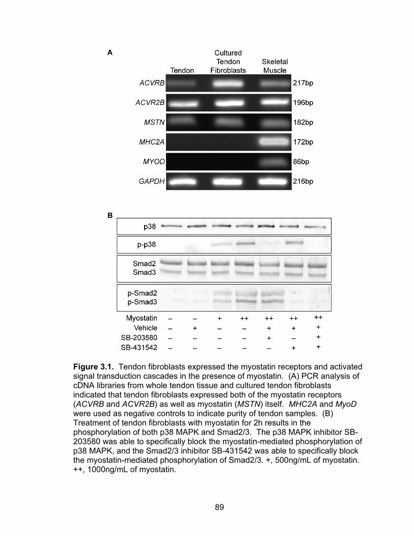
89
Figure 3.1. Tendon fibroblasts expressed the myostatin receptors and activated signal transduction cascades in the presence of myostatin. (A) PCR analysis of cDNA libraries from whole tendon tissue and cultured tendon fibroblasts indicated that tendon fibroblasts expressed both of the myostatin receptors (ACVRB and ACVR2B) as well as myostatin (MSTN) itself. MHC2A and MyoD were used as negative controls to indicate purity of tendon samples. (B) Treatment of tendon fibroblasts with myostatin for 2h results in the phosphorylation of both p38 MAPK and Smad2/3. The p38 MAPK inhibitor SB-203580 was able to specifically block the myostatin-mediated phosphorylation of p38 MAPK, and the Smad2/3 inhibitor SB-431542 was able to specifically block the myostatin-mediated phosphorylation of Smad2/3. +, 500ng/mL of myostatin. ++, 1000ng/mL of myostatin.
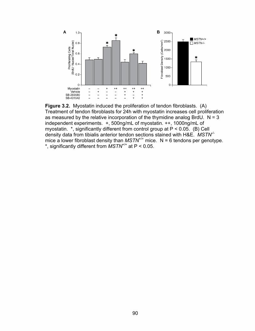
90
Figure 3.2. Myostatin induced the proliferation of tendon fibroblasts. (A) Treatment of tendon fibroblasts for 24h with myostatin increases cell proliferation as measured by the relative incorporation of the thymidine analog BrdU. N = 3 independent experiments. +, 500ng/mL of myostatin. ++, 1000ng/mL of myostatin. *, significantly different from control group at P < 0.05. (B) Cell density data from tibialis anterior tendon sections stained with H&E. MSTN-/- mice a lower fibroblast density than MSTN+/+ mice. N = 6 tendons per genotype. *, significantly different from MSTN+/+ at P < 0.05.
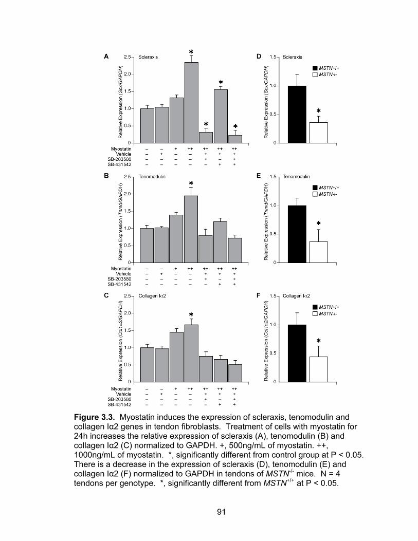
91
Figure 3.3. Myostatin induces the expression of scleraxis, tenomodulin and collagen Iα2 genes in tendon fibroblasts. Treatment of cells with myostatin for 24h increases the relative expression of scleraxis (A), tenomodulin (B) and collagen Iα2 (C) normalized to GAPDH. +, 500ng/mL of myostatin. ++, 1000ng/mL of myostatin. *, significantly different from control group at P < 0.05. There is a decrease in the expression of scleraxis (D), tenomodulin (E) and collagen Iα2 (F) normalized to GAPDH in tendons of MSTN-/- mice. N = 4 tendons per genotype. *, significantly different from MSTN+/+ at P < 0.05.
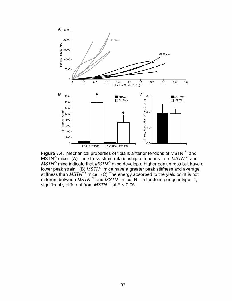
92
Figure 3.4. Mechanical properties of tibialis anterior tendons of MSTN+/+ and MSTN-/- mice. (A) The stress-strain relationship of tendons from MSTN+/+ and MSTN-/- mice indicate that MSTN-/- mice develop a higher peak stress but have a lower peak strain. (B) MSTN-/- mice have a greater peak stiffness and average stiffness than MSTN+/+ mice. (C) The energy absorbed to the yield point is not different between MSTN+/+ and MSTN-/- mice. N = 5 tendons per genotype. *, significantly different from MSTN+/+ at P < 0.05.
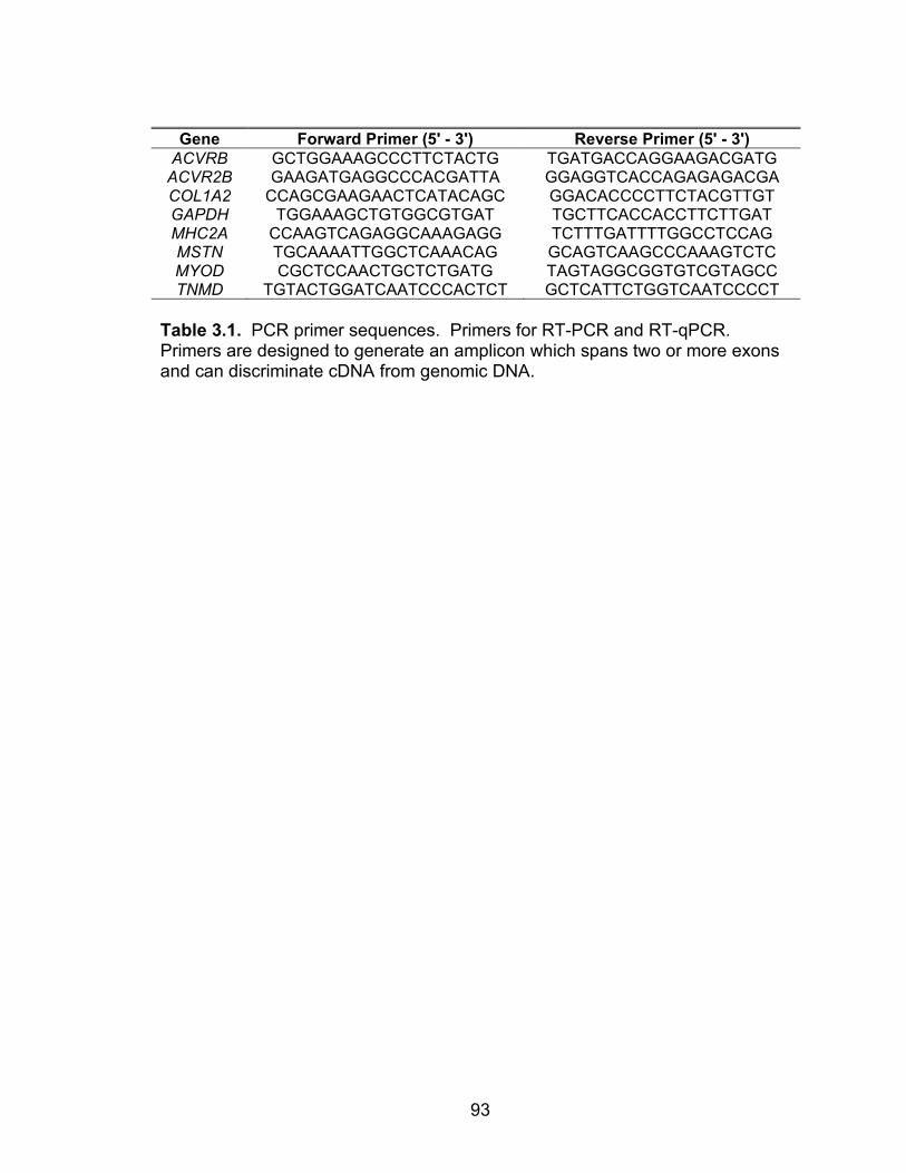
93
Gene Forward Primer (5' - 3') Reverse Primer (5' - 3')
ACVRB GCTGGAAAGCCCTTCTACTG TGATGACCAGGAAGACGATG ACVR2B GAAGATGAGGCCCACGATTA GGAGGTCACCAGAGAGACGA COL1A2 CCAGCGAAGAACTCATACAGC GGACACCCCTTCTACGTTGT GAPDH TGGAAAGCTGTGGCGTGAT TGCTTCACCACCTTCTTGAT MHC2A CCAAGTCAGAGGCAAAGAGG TCTTTGATTTTGGCCTCCAG MSTN TGCAAAATTGGCTCAAACAG GCAGTCAAGCCCAAAGTCTC MYOD CGCTCCAACTGCTCTGATG TAGTAGGCGGTGTCGTAGCC TNMD TGTACTGGATCAATCCCACTCT GCTCATTCTGGTCAATCCCCT
Table 3.1. PCR primer sequences. Primers for RT-PCR and RT-qPCR. Primers are designed to generate an amplicon which spans two or more exons and can discriminate cDNA from genomic DNA.
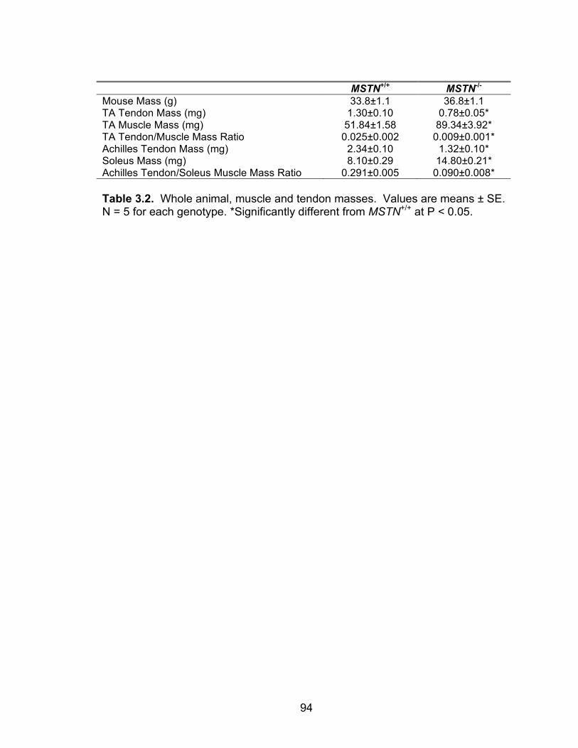
94
MSTN+/+ MSTN-/- Mouse Mass (g) 33.8±1.1 36.8±1.1 TA Tendon Mass (mg) 1.30±0.10 0.78±0.05* TA Muscle Mass (mg) 51.84±1.58 89.34±3.92* TA Tendon/Muscle Mass Ratio 0.025±0.002 0.009±0.001* Achilles Tendon Mass (mg) 2.34±0.10 1.32±0.10* Soleus Mass (mg) 8.10±0.29 14.80±0.21* Achilles Tendon/Soleus Muscle Mass Ratio 0.291±0.005 0.090±0.008* Table 3.2. Whole animal, muscle and tendon masses. Values are means ± SE. N = 5 for each genotype. *Significantly different from MSTN+/+ at P < 0.05.
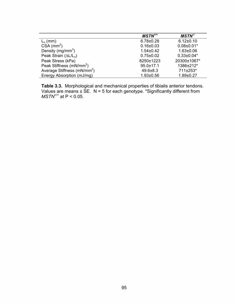
95
MSTN+/+ MSTN-/- Lo (mm) 6.78±0.28 6.12±0.10 CSA (mm2) 0.16±0.03 0.08±0.01* Density (mg/mm3) 1.54±0.42 1.63±0.06 Peak Strain (ΔL/Lo) 0.75±0.02 0.33±0.04* Peak Stress (kPa) 8250±1223 20300±1067* Peak Stiffness (mN/mm2) 95.0±17.1 1386±212* Average Stiffness (mN/mm2) 49.6±8.3 711±253* Energy Absorption (mJ/mg) 1.93±0.56 1.89±0.27 Table 3.3. Morphological and mechanical properties of tibialis anterior tendons. Values are means ± SE. N = 5 for each genotype. *Significantly different from MSTN+/+ at P < 0.05.
96
References 1. Amthor H, Huang R, McKinnell I, Christ B, Kambadur R, Sharma M, and Patel K. The regulation and action of myostatin as a negative regulator of muscle development during avian embryogenesis. Dev Biol 251: 241-257, 2002. 2. Arruda E, Calve S, Dennis R, Mundy K, and Baar K. Regional variation of tibialis anterior tendon mechanics is lost following denervation. J Appl Physiol 101: 1113-1117, 2006. 3. Bogdanovich S, Krag T, Barton E, Morris L, Whittemore L, Ahima R, and Khurana T. Functional improvement of dystrophic muscle by myostatin blockade. Nature 420: 418-421, 2002. 4. Bogdanovich S, Perkins K, Krag T, Whittemore L, and Khurana T. Myostatin propeptide-mediated amelioration of dystrophic pathophysiology. FASEB J 19: 543-549, 2005. 5. Brent A, Schweitzer R, and Tabin C. A somitic compartment of tendon progenitors. Cell 113: 235-248, 2003. 6. Brent A, and Tabin C. FGF acts directly on the somitic tendon progenitors through the Ets transcription factors Pea3 and Erm to regulate scleraxis expression. Development 131: 3885-3896, 2004. 7. Brooks SV, Zerba E, and Faulkner JA. Injury to muscle fibres after single stretches of passive and maximally stimulated muscles in mice. J Physiol 488 ( Pt 2): 459-469, 1995. 8. Chakkalakal JV, Thompson J, Parks RJ, and Jasmin BJ. Molecular, cellular, and pharmacological therapies for Duchenne/Becker muscular dystrophies. Faseb J 19: 880-891, 2005. 9. Consolino CM, and Brooks SV. Susceptibility to sarcomere injury induced by single stretches of maximally activated muscles of mdx mice. J Appl Physiol 96: 633-638, 2004. 10. Cuenda A, Rouse J, Doza Y, Meier R, Cohen P, Gallagher T, Young P, and Lee J. SB 203580 is a specific inhibitor of a MAP kinase homologue which is stimulated by cellular stresses and interleukin-1. FEBS Lett 364: 229-233, 1995. 11. Dellorusso C, Crawford RW, Chamberlain JS, and Brooks SV. Tibialis anterior muscles in mdx mice are highly susceptible to contraction-induced injury. J Muscle Res Cell Motil 22: 467-475, 2001. 12. Docheva D, Hunziker E, Fässler R, and Brandau O. Tenomodulin is necessary for tenocyte proliferation and tendon maturation. Mol Cell Biol 25: 699-705, 2005. 13. Edom-Vovard F, and Duprez D. Signals regulating tendon formation during chick embryonic development. Dev Dyn 229: 449-457, 2004. 14. Edom-Vovard F, Schuler B, Bonnin M, Teillet M, and Duprez D. Fgf4 positively regulates scleraxis and tenascin expression in chick limb tendons. Dev Biol 247: 351-366, 2002. 15. Griffiths RI. Shortening of muscle fibres during stretch of the active cat medial gastrocnemius muscle: the role of tendon compliance. J Physiol 436: 219-236, 1991.
97
16. Heinemeier K, Olesen J, Schjerling P, Haddad F, Langberg H, Baldwin K, and Kjaer M. Short-term strength training and the expression of myostatin and IGF-I isoforms in rat muscle and tendon: differential effects of specific contraction types. J Appl Physiol 102: 573-581, 2007. 17. Inman G, Nicolás F, Callahan J, Harling J, Gaster L, Reith A, Laping N, and Hill C. SB-431542 is a potent and specific inhibitor of transforming growth factor-beta superfamily type I activin receptor-like kinase (ALK) receptors ALK4, ALK5, and ALK7. Mol Pharmacol 62: 65-74, 2002. 18. Kardon G. Muscle and tendon morphogenesis in the avian hind limb. Development 125: 4019-4032, 1998. 19. Kjaer M. Role of extracellular matrix in adaptation of tendon and skeletal muscle to mechanical loading. Physiol Rev 84: 649-698, 2004. 20. Kjaer M, Magnusson P, Krogsgaard M, Boysen Møller J, Olesen J, Heinemeier K, Hansen M, Haraldsson B, Koskinen S, Esmarck B, and Langberg H. Extracellular matrix adaptation of tendon and skeletal muscle to exercise. J Anat 208: 445-450, 2006. 21. Langley B, Thomas M, Bishop A, Sharma M, Gilmour S, and Kambadur R. Myostatin inhibits myoblast differentiation by down-regulating MyoD expression. J Biol Chem 277: 49831-49840, 2002. 22. Lee S, and McPherron A. Regulation of myostatin activity and muscle growth. Proc Natl Acad Sci USA 98: 9306-9311, 2001. 23. Léjard V, Brideau G, Blais F, Salingcarnboriboon R, Wagner G, Roehrl M, Noda M, Duprez D, Houillier P, and Rossert J. Scleraxis and NFATc regulate the expression of the pro-alpha 1(I) collagen gene in tendon fibroblasts. J Biol Chem 2007. 24. Liu Y, Cserjesi P, Nifuji A, Olson E, and Noda M. Sclerotome-related helix-loop-helix type transcription factor (scleraxis) mRNA is expressed in osteoblasts and its level is enhanced by type-beta transforming growth factor. J Endocrinol 151: 491-499, 1996. 25. Livak K, and Schmittgen T. Analysis of relative gene expression data using real-time quantitative PCR and the 2(-Delta Delta C(T)) Method. Methods 25: 402-408, 2001. 26. McCroskery S, Thomas M, Maxwell L, Sharma M, and Kambadur R. Myostatin negatively regulates satellite cell activation and self-renewal. J Cell Biol 162: 1135-1147, 2003. 27. McFarlane C, Plummer E, Thomas M, Hennebry A, Ashby M, Ling N, Smith H, Sharma M, and Kambadur R. Myostatin induces cachexia by activating the ubiquitin proteolytic system through an NF-kappaB-independent, FoxO1-dependent mechanism. J Cell Physiol 209: 501-514, 2006. 28. McHugh M, and Pasiakos S. The role of exercising muscle length in the protective adaptation to a single bout of eccentric exercise. Eur J Appl Physiol 93: 286-293, 2004. 29. McPherron A, Lawler A, and Lee S. Regulation of skeletal muscle mass in mice by a new TGF-beta superfamily member. Nature 387: 83-90, 1997.
98
30. Mendias C, Marcin J, Calerdon D, and Faulkner J. Contractile properties of EDL and soleus muscles of myostatin-deficient mice. J Appl Physiol 101: 898-905, 2006. 31. Mikic B, Bierwert L, and Tsou D. Achilles tendon characterization in GDF-7 deficient mice. J Orthop Res 24: 831-841, 2006. 32. Mikic B, Schalet B, Clark R, Gaschen V, and Hunziker E. GDF-5 deficiency in mice alters the ultrastructure, mechanical properties and composition of the Achilles tendon. J Orthop Res 19: 365-371, 2001. 33. Moens P, Baatsen PH, and Marechal G. Increased susceptibility of EDL muscles from mdx mice to damage induced by contractions with stretch. J Muscle Res Cell Motil 14: 446-451, 1993. 34. Murchison N, Price B, Conner D, Keene D, Olson E, Tabin C, and Schweitzer R. Regulation of tendon differentiation by scleraxis distinguishes force-transmitting tendons from muscle-anchoring tendons. Development 2007. 35. Patel K, Macharia R, and Amthor H. Molecular mechanisms involving IGF-1 and myostatin to induce muscle hypertrophy as a therapeutic strategy for Duchenne muscular dystrophy. Acta Myol 24: 230-241, 2005. 36. Petrof BJ, Shrager JB, Stedman HH, Kelly AM, and Sweeney HL. Dystrophin protects the sarcolemma from stresses developed during muscle contraction. Proc Natl Acad Sci U S A 90: 3710-3714, 1993. 37. Philip B, Lu Z, and Gao Y. Regulation of GDF-8 signaling by the p38 MAPK. Cell Signal 17: 365-375, 2005. 38. Pryce B, Brent A, Murchison N, Tabin C, and Schweitzer R. Generation of transgenic tendon reporters, ScxGFP and ScxAP, using regulatory elements of the scleraxis gene. Dev Dyn 236: 1677-1682, 2007. 39. Rebbapragada A, Benchabane H, Wrana J, Celeste A, and Attisano L. Myostatin signals through a transforming growth factor beta-like signaling pathway to block adipogenesis. Mol Cell Biol 23: 7230-7242, 2003. 40. Schweitzer R, Chyung J, Murtaugh L, Brent A, Rosen V, Olson E, Lassar A, and Tabin C. Analysis of the tendon cell fate using Scleraxis, a specific marker for tendons and ligaments. Development 128: 3855-3866, 2001. 41. Shukunami C, Takimoto A, Oro M, and Hiraki Y. Scleraxis positively regulates the expression of tenomodulin, a differentiation marker of tenocytes. Dev Biol 298: 234-247, 2006. 42. Siegel IM. The management of muscular dystrophy: a clinical review. Muscle Nerve 1: 453-460, 1978. 43. Smith T, Sweetman D, Patterson M, Keyse S, and Münsterberg A. Feedback interactions between MKP3 and ERK MAP kinase control scleraxis expression and the specification of rib progenitors in the developing chick somite. Development 132: 1305-1314, 2005. 44. Stevens ED, and Faulkner JA. The capacity of mdx mouse diaphragm muscle to do oscillatory work. J Physiol 522 Pt 3: 457-466, 2000. 45. Thomas M, Langley B, Berry C, Sharma M, Kirk S, Bass J, and Kambadur R. Myostatin, a negative regulator of muscle growth, functions by inhibiting myoblast proliferation. J Biol Chem 275: 40235-40243, 2000.
99
46. Tobin J, and Celeste A. Myostatin, a negative regulator of muscle mass: implications for muscle degenerative diseases. Current opinion in pharmacology 5: 328-332, 2005. 47. Tuite D, Renström P, and O'Brien M. The aging tendon. Scandinavian journal of medicine & science in sports 7: 72-77, 1997. 48. Wagner K, McPherron A, Winik N, and Lee S. Loss of myostatin attenuates severity of muscular dystrophy in mdx mice. Ann Neurol 52: 832-836, 2002. 49. Welle S, Bhatt K, and Pinkert C. Myofibrillar protein synthesis in myostatin-deficient mice. Am J Physiol Endocrinol Metab 290: E409-415, 2006. 50. Wolfman N, Hattersley G, Cox K, Celeste A, Nelson R, Yamaji N, Dube J, DiBlasio-Smith E, Nove J, Song J, Wozney J, and Rosen V. Ectopic induction of tendon and ligament in rats by growth and differentiation factors 5, 6, and 7, members of the TGF-beta gene family. J Clin Invest 100: 321-330, 1997. 51. Yang W, Chen Y, Zhang Y, Wang X, Yang N, and Zhu D. Extracellular signal-regulated kinase 1/2 mitogen-activated protein kinase pathway is involved in myostatin-regulated differentiation repression. Cancer Res 66: 1320-1326, 2006. 52. Zhu J, Li Y, Shen W, Qiao C, Ambrosio F, Lavasani M, Nozaki M, Branca M, and Huard J. Relationships between TGF-beta -1, myostatin, and decorin: Implications for skeletal muscle fibrosis. J Biol Chem 2007. 53. Zhu X, Topouzis S, Liang L, and Stotish R. Myostatin signaling through Smad2, Smad3 and Smad4 is regulated by the inhibitory Smad7 by a negative feedback mechanism. Cytokine 26: 262-272, 2004.


101
Structure. The genetic deficiency of myostatin leads to both hypertrophy
and hyperplasia of fast-fibered muscles (17). In prior studies, the measurements
of the number of fibers in the skeletal muscles were determined from cross
sections of muscles that might not provide an accurate indication of the true
number of fibers (15). By digesting the ECM of EDL and soleus muscles and
counting all of the fibers present in the muscle, we have now demonstrated that
the deficiency of myostatin leads to a bona fide hyperplasia.
In addition to a greater number of fibers, myostatin deficiency leads to an
increase in the CSAs of fibers. While myostatin inhibition increases satellite cell
proliferation (12, 16, 27), this satellite cell inhibition does not appear to be wholly
sufficient to explain the hypertrophic response that occurs with the inhibition of
myostatin. Administering mice an inhibitory antibody against myostatin resulted
in a 14% increase in muscle mass in only two weeks (29). This increase in
muscle mass is unlikely to occur simply by modulating satellite cell activity. The
preliminary study presented in Chapter I demonstrated that the deficiency of
myostatin leads to a marked decrease in ubiquitinated myosin heavy chain and a
decrease in atrogin-1 expression. No difference in relative myofibrillar protein
content was observed between the muscles of MSTN+/+ and MSTN-/- mice.
These results suggest that the inhibition of myostatin leads to muscle fiber
hypertrophy by the inhibition of protein degradation.
The deficiency of myostatin reduced collagenous fibrosis in mdx mice (1,
2, 28), but whether myostatin regulate the expression of type I collagen directly
was not clear. The cell culture studies from Chapter II demonstrated that

102
myostatin increased directly the expression of type I collagen. Additionally,
myostatin-deficient EDL muscles showed a decrease in whole muscle collagen
content. These results indicated that, in addition to regulating the numbers of
fibers in a muscle, myostatin regulated the ECM of skeletal muscle.
Function. In addition to the profound impact on the structure of skeletal
muscle, myostatin had an impact on the function of skeletal muscle through the
hypertrophy and hyperplasia present in the skeletal muscles of myostatin-
deficient mice, and the impact these changes had on the contractile properties of
muscles. The studies from Chapter II demonstrated that the deficiency of
myostatin increased the Po of both EDL and soleus muscles. When Po was
normalized to the CSA of the muscles to calculate sPo, an unexpected finding
was observed. As a muscle undergoes hypertrophy and is able to generate a
greater Po, the sPo will usually decrease due to the increase in θ (15). Despite
having a greater number of fibers, fiber CSA, Po and θ, the EDL muscles from
MSTN+/- and soleus muscles from MSTN-/- mice did not show a decrease in sPo.
In contrast, the EDL muscles of the MSTN-/- mice, that had a greater number of
fibers, fiber CSA, Po and θ did suffer from a decrease in sPo. For a muscle that
undergoes hypertrophy, a threshold appears to exist above which a decrease in
sPo occurs. In contrast, for small increases in muscle CSA, mass and Po, sPo
does not change.
Furthermore, the deficiency of myostatin was found to increase the
susceptibility of muscles to contraction-induced injury. Following contraction-
induced injury, the EDL muscles of MSTN-/- mice had greater relative force

103
deficits than those of MSTN+/+ mice. Despite having a greater force deficit
following injury, the work done to stretch the muscles of MSTN-/- mice was less
than MSTN+/+ and MSTN+/- mice. As the series elastic component of muscles
can protect fibers from injury (9), we initially hypothesized that the series elastic
component of muscles of the MSTN-/- mice were more compliant, exceeded their
elastic limit during the lengthening contractions, and therefore no longer acted as
mechanical springs. This lead to a subsequent study of tendon structure and
function, presented in Chapter III. After completion of the investigation of the
properties of tendon structure and function, the initial hypothesis as to why the
EDL muscles of MSTN-/- mice are more susceptible to contraction-induced injury
was rejected. The rejection was based on the observation that the tendons of
MSTN-/- mice were fourteen times stiffer than the tendons of MSTN+/+ mice and
the peak stiffness occurred at an early strain value. These findings suggested
that the series elastic component of the tendons of the MSTN-/- mice were so stiff
that they acted as a rigid body and did not take up much, if any, strain as the
muscle was stretched. Extremely stiff tendons appear to be the reason why the
deficiency of myostatin increases the susceptibility of the fast-fibered EDL
muscles to contraction-induced injury.
Future Studies. An interesting finding from the studies presented in
Chapter II is that the EDL muscles of MSTN+/- mice developed a greater Po than
the MSTN+/+ mice, but unlike the MSTN-/- mice, no difference was observed in
sPo, or force deficit, after the injury. Even though the collagen content of the EDL
muscles of MSTN+/- mice was less than that of MSTN+/+ mice, this did not

104
increase the susceptibility of these muscles to contraction-induced injury. As the
MSTN+/- mice have a 20% decrease in circulating myostatin levels compared to
MSTN+/+ mice, this suggests that the partial suppression of myostatin is able to
increase force production of muscles without making the muscles more
susceptible to injury. Based on the preliminary data in Chapter I that showed that
myostatin increased protein degradation and the data in Chapter II that showed
that myostatin increased type I collagen expression, the use of a pharmaceutical
inhbitor of myostatin might be beneficial in the treatment of severe contraction-
induced injuries. Following the induction of a severe lengthening contraction
injury to the muscles of a mouse, the administration of a pharmaceutical inhibitor
such as a monoclonal antibody against myostatin, the propeptide of myostatin, or
a small peptide that blocks the interaction of myostatin with its receptors, the
effect of myostatin inhibition on the recovery from a severe contraction induced
injury could be determined. Based on the findings from Chapters I and II, the
hypothesis that inhibition of myostatin promotes the long-term recovery of muscle
from severe injury by blocking protein degradation and reducing fibrosis appears
promising.
Discussion of Findings and Future Directions - Tendon
The study described in Chapter III demonstrated, for the first time, that
myostatin has a profound impact on the structure and function of tendon tissue.
The study not only described and characterized the phenotype of tendons from
myostatin-deficient mice, but also demonstrated that myostatin can directly
activate intracellular signal transduction cascades in tendon fibroblasts and

105
explored the possible cellular and molecular mechanisms behind the tendon
phenotype. In addition to showing that myostatin can directly regulate tendon
structure and function, Chapter III demonstrated for the first time that the bHLH
transcription factor scleraxis continues to be expressed in adult tendon
fibroblasts and that myostatin increases the expression of scleraxis directly.
Tenomodulin was also shown to be a downstream target of myostatin signaling.
While tenomodulin deficient mice displayed hypocellular tendons with no change
in ECM structure or size (3), the mechanisms behind the control of cell
proliferation by tenomodulin are not known. Three future studies are suggested
that would provide further insight into the regulation of tendon physiology.
Study 1 – Myostatin-mediated regulation of scleraxis expression during
development. During the development of tendon structures in the limb, tendon
development can be categorized into three phases, each phase corresponding to
an upregulation of scleraxis (4, 25). While FGF appears to be responsible for the
third phase of scleraxis expression, FGF does not appear to be responsible for
the first and second phases of scleraxis expression (4). Edom-Vovard and
Duprez (4) suggested that a member of the GDF family is a likely candidate for
the regulation of scleraxis expression during the first and second phases of early
tendon development. To determine if myostatin induces the expression of
scleraxis during the first and second phases of early tendon development, a
double transgenic animal model could be utilized. Crossing the myostatin
deficient mice with the ScxGFP line of mice that contain a GFP reporter linked to
the scleraxis promoter (23) to obtain MSTN-/-ScxGFP mice would create a model

106
organism that would allow for the visualization of scleraxis expression in the
absence of myostatin at various stages of development. Comparing GFP
intensity between MSTN+/+ScxGFP and MSTN-/-ScxGFP mice would be a useful
first step to determine if myostatin is the cytokine that induces the first two
phases of scleraxis expression. The most likely interpretation is that compared
with MSTN+/+ScxGFP mice, the GFP intensity in the tendon primordia of MSTN-/-
ScxGFP mice is severely depressed.
Study 2 – Myostatin-mediated biochemical modifications of tendons.
Compared with MSTN+/+ mice, the tendons of MSTN-/- mice had a two-fold
greater peak stress, less than half of the peak strain, and a fourteen-fold greater
peak stiffness. Furthermore, the fibroblast density of the tendons of the MSTN-/-
mice was only half that of the MSTN+/+ mice. While the hypocellularity may
explain the increase in stiffness, as fibroblasts can break the spontaneous cross-
links that form between collagen molecules, changes in the activities of other
enzymes might also occur that regulate tendon mechanical properties. Lysyl
oxidase is the chief enzyme that can form cross-links between collagen
molecules (6). In addition, elastin is a structural protein that allows tendon to
have a greater peak stress (8). Determining the expression of lysyl oxidase and
elastin in tendon might reveal the molecular mechanism behind the myostatin-
mediated regulation of tendon mechanical properties. Based on the stiffness and
peak strain data, myostatin might promote the expression of lysyl oxidase and
elastin in tendon tissue.

107
Study 3 – Regulation of Tendon Fibroblast Proliferation by Tenomodulin.
Tenomodulin is a type II transmembrane protein that promotes the proliferation of
tendon fibroblasts (3), but the molecular mechanisms behind the actions of
tenomodulin are not known. The tenomodulin protein consists of two domains,
an N-terminal transmembrane anchor domain and a C-terminal extracellular
domain (26). The C-terminal peptide is cleaved and presumably interacts with
other receptors on the plasma membrane, but the identities of these receptors
are not known. To determine if the C-terminal peptide of tenomodulin interacts
with receptors on the plasma membrane, recombinant c-myc tagged C-terminal
tenomodulin peptide could be incubated with tendon fibroblast cells followed by
treatment with a crosslinking reagent. The cell homogenates would then be
subjected to immunoprecipitation with an antibody against c-myc. The pull down
would then be treated with a reagent to reverse the crosslinking and samples
would be separated using 2D SDS-PAGE. Proteins would be removed from the
gel and subjected to N-terminal sequencing. The amino acid sequence data
could then be used to search for possible receptors with which the C-terminal
peptide of tenomodulin interacts. The underlying mechanism could be that the
C-terminal tenomodulin peptide interacts with a receptor that controls cell
proliferation, but currently there is no basis for the selection of a particular
receptor. Identifying the receptors with which tenomodulin interacts is the first
step in determining the molecular mechanisms behind the tenomodulin-mediated
increase in tendon fibroblast proliferation.

108
Summary
The studies described in this thesis demonstrate that myostatin not only
has a profound impact on the structure and function of muscle tissue, but also on
the structure and function of tendon tissue. While initially characterized as a
cytokine that regulates skeletal muscle function, myostatin also has important
roles in tendon, cardiac, bone, mammary and adipose tissue (5, 11, 18, 20, 30).
From an evolutionary perspective, an interesting question is: Why is there
a gene for myostatin? The presence of larger, stronger muscles would appear to
be beneficial for many aspects of everyday life. All vertebrate species have a
myostatin gene (22), and fish have two copies (14). While myostatin has a
controversial role in metabolism, one potential role of myostatin in metabolism is
to divert food energy to fat, instead of to muscle (5, 7, 13, 18, 19, 30, 31).
Diverting food energy to fat instead of muscle would presumptively better enable
an animal to survive during periods of low food availability. A recent study
explored the adaptive evolution of myostatin in the human genome (24). The
authors of this study concluded that positive natural selection has acted on the
myostatin gene, but acknowledged that the amount and rate of polymorphisms at
the myostatin locus may suggest an evolutionarily recent decline in natural
selective pressure for myostatin. While not all polymorphisms lead to a loss of
function mutation, the greater the rate of polymorphisms, the greater the chance
of developing a loss of function mutation. The authors rejected this decline in
selective pressure at the myostatin locus based entirely on the long history of
conservation of the myostatin gene across all vertebrate species (24). The

109
results of this thesis argue against a decline in the natural selective pressure at
the myostatin gene. The studies in Chapter II indicate that the MSTN+/- mice
generate greater forces, without incurring an increased susceptibility to injury. In
the Whippet breed of racing dogs, a natural loss of the functional mutation in the
myostatin gene has arisen (21). The MSTN+/- and MSTN-/- whippets are known
as "Bully Whippets" and are considerably faster than their MSTN+/+ counterparts
(21). While we did not directly assess the amount of adipose tissue, the MSTN+/-
mice appear to be leaner than their MSTN+/+ littermates. We are currently
conducting a longevity study of MSTN+/+, MSTN+/-, and MSTN-/- mice. While the
study is still ongoing, our preliminary results indicate that the MSTN+/- mice have
an 8 month longer maximum longevity than either the MSTN+/+ or MSTN-/- mice.
While the long history of selective pressure being applied at the myostatin locus
may have helped our ancestors survive through periods of famine, in the
developed world, currently most humans have access to a virtually unlimited
source of food energy. The increased rate of polymorphisms does suggest a
decline in selective pressure at the myostatin locus. With an unlimited access to
food energy, the "larger, faster, stronger, leaner, longer-lived" phenotype of
MSTN+/- organisms has clear advantages over the wild type phenotype. A
pharmaceutical inhibitor of myostatin, Stamulumab (Myo-029, Wyeth), is
currently in clinical trials in the USA and Europe. Stamulumab is not yet
available for prescription in the USA, but is available for purchase in China and
Korea. Inhibition of myostatin appears to be a practical and potentially useful
treatment for many muscle wasting conditions and to counteract the decline in

110
muscle mass that occurs with aging. Future studies that determine the long term
safety and efficacy of the use of myostatin inhibitors are warranted. Doping
control policies and procedures for myostatin inhibitors must also be developed
to ensure equality in athletic performance.

111
References 1. Bogdanovich S, Krag T, Barton E, Morris L, Whittemore L, Ahima R, and Khurana T. Functional improvement of dystrophic muscle by myostatin blockade. Nature 420: 418-421, 2002. 2. Bogdanovich S, Perkins K, Krag T, Whittemore L, and Khurana T. Myostatin propeptide-mediated amelioration of dystrophic pathophysiology. FASEB J 19: 543-549, 2005. 3. Docheva D, Hunziker E, Fässler R, and Brandau O. Tenomodulin is necessary for tenocyte proliferation and tendon maturation. Mol Cell Biol 25: 699-705, 2005. 4. Edom-Vovard F, and Duprez D. Signals regulating tendon formation during chick embryonic development. Dev Dyn 229: 449-457, 2004. 5. Feldman B, Streeper R, Farese R, and Yamamoto K. Myostatin modulates adipogenesis to generate adipocytes with favorable metabolic effects. Proc Natl Acad Sci USA 103: 15675-15680, 2006. 6. Gerriets J, Curwin S, and Last J. Tendon hypertrophy is associated with increased hydroxylation of nonhelical lysine residues at two specific cross-linking sites in type I collagen. J Biol Chem 268: 25553-25560, 1993. 7. Gonzalez-Cadavid N, and Bhasin S. Role of myostatin in metabolism. Current opinion in clinical nutrition and metabolic care 7: 451-457, 2004. 8. Gosline J, Lillie M, Carrington E, Guerette P, Ortlepp C, and Savage K. Elastic proteins: biological roles and mechanical properties. Philos Trans R Soc Lond, B, Biol Sci 357: 121-132, 2002. 9. Griffiths RI. Shortening of muscle fibres during stretch of the active cat medial gastrocnemius muscle: the role of tendon compliance. J Physiol 436: 219-236, 1991. 10. Grobet L, Martin L, Poncelet D, Pirottin D, Brouwers B, Riquet J, Schoeberlein A, Dunner S, Ménissier F, Massabanda J, Fries R, Hanset R, and Georges M. A deletion in the bovine myostatin gene causes the double-muscled phenotype in cattle. Nat Genet 17: 71-74, 1997. 11. Hamrick M, Pennington C, Webb C, and Isales C. Resistance to body fat gain in 'double-muscled' mice fed a high-fat diet. International journal of obesity (2005) 30: 868-870, 2006. 12. Langley B, Thomas M, Bishop A, Sharma M, Gilmour S, and Kambadur R. Myostatin inhibits myoblast differentiation by down-regulating MyoD expression. J Biol Chem 277: 49831-49840, 2002. 13. Lin J, Arnold HB, Della-Fera MA, Azain MJ, Hartzell DL, and Baile CA. Myostatin knockout in mice increases myogenesis and decreases adipogenesis. Biochem Biophys Res Commun 291: 701-706, 2002. 14. Maccatrozzo L, Bargelloni L, Cardazzo B, Rizzo G, and Patarnello T. A novel second myostatin gene is present in teleost fish. FEBS Lett 509: 36-40, 2001. 15. Maxwell L, Faulkner J, and Hyatt G. Estimation of number of fibers in guinea pig skeletal muscles. Journal of applied physiology 37: 259-264, 1974.

112
16. McCroskery S, Thomas M, Maxwell L, Sharma M, and Kambadur R. Myostatin negatively regulates satellite cell activation and self-renewal. J Cell Biol 162: 1135-1147, 2003. 17. McPherron A, Lawler A, and Lee S. Regulation of skeletal muscle mass in mice by a new TGF-beta superfamily member. Nature 387: 83-90, 1997. 18. McPherron A, and Lee S. Suppression of body fat accumulation in myostatin-deficient mice. J Clin Invest 109: 595-601, 2002. 19. Milan G, Dalla Nora E, Pilon C, Pagano C, Granzotto M, Manco M, Mingrone G, and Vettor R. Changes in muscle myostatin expression in obese subjects after weight loss. J Clin Endocrinol Metab 89: 2724-2727, 2004. 20. Morissette M, Cook S, Foo S, McKoy G, Ashida N, Novikov M, Scherrer-Crosbie M, Li L, Matsui T, Brooks G, and Rosenzweig A. Myostatin regulates cardiomyocyte growth through modulation of Akt signaling. Circ Res 99: 15-24, 2006. 21. Mosher D, Quignon P, Bustamante C, Sutter N, Mellersh C, Parker H, and Ostrander E. A mutation in the myostatin gene increases muscle mass and enhances racing performance in heterozygote dogs. PLoS Genet 3: e79, 2007. 22. Pie M, and Alvares L. Evolution of myostatin in vertebrates: is there evidence for positive selection? Mol Phylogenet Evol 41: 730-734, 2006. 23. Pryce B, Brent A, Murchison N, Tabin C, and Schweitzer R. Generation of transgenic tendon reporters, ScxGFP and ScxAP, using regulatory elements of the scleraxis gene. Dev Dyn 236: 1677-1682, 2007. 24. Saunders M, Good J, Lawrence E, Ferrell R, Li W, and Nachman M. Human adaptive evolution at Myostatin (GDF8), a regulator of muscle growth. Am J Hum Genet 79: 1089-1097, 2006. 25. Schweitzer R, Chyung J, Murtaugh L, Brent A, Rosen V, Olson E, Lassar A, and Tabin C. Analysis of the tendon cell fate using Scleraxis, a specific marker for tendons and ligaments. Development 128: 3855-3866, 2001. 26. Shukunami C, Oshima Y, and Hiraki Y. Molecular cloning of tenomodulin, a novel chondromodulin-I related gene. Biochem Biophys Res Commun 280: 1323-1327, 2001. 27. Thomas M, Langley B, Berry C, Sharma M, Kirk S, Bass J, and Kambadur R. Myostatin, a negative regulator of muscle growth, functions by inhibiting myoblast proliferation. J Biol Chem 275: 40235-40243, 2000. 28. Wagner K, McPherron A, Winik N, and Lee S. Loss of myostatin attenuates severity of muscular dystrophy in mdx mice. Ann Neurol 52: 832-836, 2002. 29. Whittemore L, Song K, Li X, Aghajanian J, Davies M, Girgenrath S, Hill J, Jalenak M, Kelley P, Knight A, Maylor R, O'Hara D, Pearson A, Quazi A, Ryerson S, Tan X, Tomkinson K, Veldman G, Widom A, Wright J, Wudyka S, Zhao L, and Wolfman N. Inhibition of myostatin in adult mice increases skeletal muscle mass and strength. Biochem Biophys Res Commun 300: 965-971, 2003. 30. Zhao B, Wall R, and Yang J. Transgenic expression of myostatin propeptide prevents diet-induced obesity and insulin resistance. Biochem Biophys Res Commun 337: 248-255, 2005.

113
31. Zimmers T, Davies M, Koniaris L, Haynes P, Esquela A, Tomkinson K, McPherron A, Wolfman N, and Lee S. Induction of cachexia in mice by systemically administered myostatin. Science 296: 1486-1488, 2002.