regulation and properties of glucose-6-phosphate ... · siddhartha singh1,2, asha anand1 and pramod...
TRANSCRIPT

International Journal of Plant Physiology and Biochemistry Vol. 4(1), pp. 1-19, 2 January, 2012 Available online at http://www.academicjournals.org/IJPPB DOI: 10.5897/IJPPB11.045 ISSN-2141-2162 ©2012 Academic Journals
Review
Regulation and properties of glucose-6-phosphate dehydrogenase: A review
Siddhartha Singh1,2, Asha Anand1 and Pramod K. Srivastava1*
1Department of Biochemistry, Faculty of Science, Banaras Hindu University, Varanasi-221005, Uttar Pradesh, India.
2Department of Basic Science and Humanities, College of Horticulture and Forestry, Central Agricultural University,
Pasighat-791102, Arunachal Pradesh, India.
Accepted 28 December, 2011
Glucose-6-phosphate dehydrogenase (G6PD) is the key enzyme of the pentose phosphate pathway that catalyzes the conversion of glucose-6-phosphate to 6-phosphogluconolactone in presence of NADP
+.
G6PD is an enzyme of vital importance because of its role in various haemolytic disorders and its potential as a regulator for various biosynthetic pathways. NADPH is the important product of the reaction and is used for the reductive biosynthesis of fatty acids, isoprenoids, aromatic amino acids, etc. NADPH produced also plays important function in the protection of the cell against oxidative agents by transferring its reductive power to glutathione disulphide via glutathione disulphide reductase. The present review deals with the importance, occurrence, structure, physico-chemical properties, genetics, and regulation of G6PD. Key words: Glucose-6-phosphate dehydrogenase, NADPH, pentose phosphate pathway, oxidative stress.
INTRODUCTION
Glucose-6-phosphate dehydrogenase (G6PD; D-glucose-6-phosphate: NADP
+ 1-oxidoreductase; EC 1.1.1.49) was
first described by Warburg and Christian in1931. It catalyzes the transformation of glucose-6-phosphate to 6-phosphogluconolactone concomitant with conversion of NADP to NADPH (Figure 1a). This enzyme has aroused considerable attention because of its important role in pentose phosphate pathway, its involvement in various haemolytic disorders, its activity variation under different nutritional and hormonal conditions (Tepperman and Tepperman, 1958; Kletzien and Berdanier, 1993; Kletzien et al., 1994; Hodge and Salati, 1997; Farhud and Yazdanpanah, 2008) and its potential as a regulator for the availability of the reduced NADPH required for *Corresponding author. E-mail: [email protected]. Tel: +91-9415811822. Abbreviations: G6PD, Glucose-6-phosphate dehydrogenase; NADP
+, Nicotinamide adenine dinucleotide phosphate; NAD
+,
Nicotinamide adenine dinucleotide; ATP, Adenosine triphosphate.
various biosynthetic processes (Chung and Langdon, 1963). The reducing power produced is necessary for the reductive biosynthesis of fatty acids, isoprenoids and aromatic amino acids in the dark and for nitrogen assimilation in heterotrophic tissues (Turner and Turner, 1980; Copeland and Turner, 1987; Bowsher et al., 1992; Graeve, Schaewen and Scheibe, 1994; Hauschild and Schaewen, 2003). The NADPH and pentose phosphates produced also serves as the route of entry of 3-5 carbon sugars to the glycolytic pathway (Danisan et al., 2004). NADPH also plays important functions in the protection of the cell against oxidative agents by transferring its reductive power to glutathione disulphide (GSSG) via glutathione disulphide reductase (Figure 1b) (Levy, 1979; Debnam et al., 2004; Farhud and Yazdanpanah, 2008; Machida et al., 2010). The reducing power for nitrogen assimilation in root cells and algae is provided through G6PD activity (Bowsher et al., 1992; Wright et al., 1997; Jin et al., 1998; Espoito et al., 2001; Espoito et al., 2003; Wakao et al., 2008). Apart from nitrogen assimilation, the enzyme is also an important source of NADPH for non photosynthetic tissues (Emes and Neuhaus, 1997; Wakao et al., 2008) and fat producing tissues such as

2 Int. J. Plant Physiol. Biochem.
H C OH NADP+ NADPH + H+ C=O
H C OH O Mg2+ H C OH O
H O C H Glucose-6-Phosphate Dehydrogenase H O C H
H C OH H C OH
H C H C
CH2OPO32- CH2OPO3
2-
Glucose-6-Phosphate 6-Phospho glucono-δ-lactone Figure 1a. Reaction catalyzed by glucose-6-phosphate dehydrogenase. Glucose-
6-phosphate is converted to 6-phosphogluconolactone, an intramolecular ester. NADP
+ is the electron acceptor.
H2O2
OH.
Oxidative damage to 2H2O
proteins, lipids, DNA, etc.
NADP+ NADPH + H+
Glucose-6-Phosphate 6-Phosphogluconolactone
Fatty Acids NADP+
Precursors NADPH
+ H+
Ribulose-5-Phosphate
Ribose-5-Phosphate
Nucleotides, Coenzymes,
Nucleic Acids
GSH GSSG
Glutathione Peroxidase
GR
Glucose-6-Phosphate Dehydrogenase
Reductive Biosynthesis
X
Figure 1b. Role of NADPH and glutathione in protecting cells against reactive oxygen species. Reduced glutathione (GSH) is regenerated from its oxidized form (GSSG) with
the help of NADPH produced in the glucose-6-phosphate dehydrogenase catalyzed reaction. Reduced glutathione protects the cell from oxidative damage by destroying hydrogen peroxide (H2O2) and peroxide free radicals (OH
.).

pollen and oil seeds (Eastmond and Rawsthorne, 1998; Niewiadomski et al., 2005; Wakao et al., 2008). The enzyme is widely distributed and has been isolated from microorganisms, plants and various mammalian tissues (Danisan et al., 2004; Ulusu et al., 2005; Ibraheem et al., 2005; Igoillo-Esteve and Cazzulo, 2006; Demir et al., 2009; Cardi et al., 2011). In higher plants, G6PD isoforms has been reported in cytosol, plastidic stroma and peroxisomes (Corpas et al., 1998). Chloroplastic G6PD is reported to be under the control of photosynthetic redox modulation (Fickenscher and Scheibe, 1986). Unlike other target enzymes like fructose-1, 6-bisphosphatase, sedoheptulose-1, 7-bisphosphatase, hosphoribulokinase, NADP-malate dehydrogenase, ATPase; chloroplast G6PD is inactivated in presence of light (Lendzian and Ziegler, 1970) via reduced thioredoxin (Scheibe and Anderson, 1981; Buchanan, 1991). G6PD is a housekeeping enzyme critical in the redox metabolism of all aerobic cells but it is easily inducible in different conditions (Kletzien et al., 1994; Luzzatto, 2006). An estimated 400 million people worldwide are affected by common human metabolic disorder of G6PD deficiency (Beutler, 1996; Miwa and Fujii, 1996). However, all the individuals with G6PD deficiency are not anaemic but it can cause several other disorders such as neonatal jaundice, mild haemolytic anaemia to chronic non-spherocytic haemolytic anaemia with attacks of severe anaemia induced by infections, specific drugs or consumption of fava beans (Yan et al., 2006). Importance of glucose-6-phosphate dehydrogenase Hydrogen peroxides, hydroxyl radicals and super oxides are known as reactive oxygen species (ROS) which causes oxidative stress and damages cell membranes (Shihabi et al., 2002; Leopold and Loscalzo, 2005). Decreased antioxidative enzyme activity plays an important role in oxidative injuries of different organs, tissues and cells including vascular cells, heart, brain, (Leopold and Loscalzo, 2005) and causes Alzheimer‟s and Parkinson‟s disease and also contribute to the aging process (Halliwell, 1992; Tsun-Yee Chiu and Liu, 1997; Savitha et al., 2005; Farhud and Yazdanpanah, 2008). Antioxidative enzymes like glutathione reductase, superoxide dismutase, catalase, peroxidase and G6PD protects the cells against oxidative damage. G6PD is said to be an essential modulator in the body‟s antioxidative defence system that plays a very important role in all cells especially in red blood cells (Leopold and Loscalzo, 2005). Glucose-6 Phosphate Dehydrogenase is a key enzyme for maintenance of redox potential in cells (Farhud and Yazdanpanah, 2008). G6PD leads to NADPH production through pentose phosphate pathway which is important as a central reductant and regulator of redox potential (Tian et al., 1999). It also acts as a cofactor for other anti-oxidant enzymes like glutathione reductance (Fico et al., 2004; Leopold and Loscalzo, 2005;
Singh et al. 3
Farhud and Yazdanpanah, 2008). Reduced glutathione acts as a cofactor for the glutathione peroxidase and is crucial for neutralization of peroxides and also protects protein sulphahydryl groups against oxidation (Beutler, 1994). Regeneration of reduced glutathione from its oxidized form requires the NADPH produced in the G6PD reaction (Figure 1b). NADPH is critical for conversion of inactive form of catalase into active form (Beutler, 1994). Researchers have shown that stimulation of cell growth is associated with increased G6PD activity. It has been found that epidermal growth factor and insulin stimulated cell growth in rat liver cell culture is closely associated with G6PD activity (Yoshimoto et al., 1983; Burdon et al., 1995). Growth hormone induced cell growth, cancer and cultured tumor cells also exhibit increased G6PD activity (Sulis, 1972; Schaffer, 1985; Weber, 1987; Kletzien et al., 1994). Cell growth can be stimulated by over expression of glucose-6-phophate dehydrogenase (Tian et al., 1998). Necrosis and apoptosis are the two described patterns of cell death. Necrosis is associated with inflammation whereas the regulated apoptosis have association with nuclear fragmentation and chromatin condensation (Orrenius, 1995). Deletion of G6PD gene leads to death of cells exposed to oxidative stress (Filosa et al., 2003). Inhibition of G6PD activity leads to hydrogen peroxide induced cell death, significant increase in apoptosis, loss of protein thiols and changes in mitogen activated protein kinase phosphorylation (Tian et al., 1999). Glucose 6-phosphate dehydrogenase plays a crucial role in the protection from redox stress induced apoptosis (Fico et al., 2004). Hence, G6PD plays an important role in cell death by affecting the redox potential. G6PD deficiency is associated with a large number of clinical manifestations like acute haemolytic anaemia (Mehta et al., 2000; Prchal and Gregg, 2005), neonatal jaundice (Dennery et al., 2001), malaria (Luzzatto and Bienzle, 1979; Luzzatto 1979; Ruwende and Hill, 1998), etc. Distribution of glucose-6-phosphate dehydrogenase
Glucose-6-phosphate dehydrogenase is widely distributed among microorganisms (Banerjee and Fraenkel, 1972; Vander Wyk and Lessie, 1974; Reuter et al., 1990; Jeffery et al., 1993; Ragunathan and Levy, 1994; Anderson and Anderson, 1995; Moritz et al., 2000; Hansen et al., 2002; Ibraheem et al., 2005; Igoillo-Esteve and Cazzulo, 2006), plants (Graeve, Schaewen and Scheibe, 1994; Espoito et al., 2001; Espoito et al., 2003; Demir et al., 2004; Demir et al., 2009; Cardi et al., 2011) and in different tissues of animals (Holten, 1972; Lee et al., 1979; Ozer et al., 2001; Ozer et al., 2002; Ciftci et al., 2003; Danisan et al., 2004; Ulusu et al., 2005). G6PD has been reported in a number of bacteria such as Zymomonas mobilis (Scopes et al., 1985), Pseudomonas multivorans (Vander Wyk and Lessie, 1974), Pseudomonas W6 (Reuter et al., 1990), Escherichia coli (Banerjee and Fraenkel, 1972), Acetobacter xylinum

4 Int. J. Plant Physiol. Biochem. (Ragunathan and Levy, 1994), Azotobacter vinelandii (Anderson and Anderson, 1995), Corynebacterium glutamicum (Moritz et al., 2000), Thermotoga maritima (Hansen et al., 2002). Mammalian form of G6PD was isolated from a number of species (Kanji et al., 1976; Lee et al., 1979; Pittalis et al., 1992; Ozer et al., 2001; Ozer et al., 2002; Beydemir et al., 2002; Beydemir et al., 2003; Yilmaz et al., 2003; Ciftci et al., 2003; Ciftci et al., 2004; Danisan et al., 2004; Ulusu et al., 2005). Studies about regulation of G6PD has been carried out in plants, eukaryotic algae and cyanobacteria but glucose-6-phophate dehydrogenase encoding gene has been isolated from only few of these sources (Srivastava and Anderson, 1983; Fickenscher and Scheibe, 1986; Graeve, Schaewen and Scheibe, 1994; Gleason, 1996). Two isoforms (cytosolic and chloroplastic isoforms) of G6PD has been reported in green algae Chlorella vulgaris C-27(Honjoh et al., 2003). Several isoforms of G6PDH have been reported in cytosol, plastidic stroma, and peroxisomes of higher plants (Corpas et al., 1998). Cytosolic G6PD has been reported in spinach (Schnarrenberger et al., 1973), pea leaves (Fickenscher and Scheibe, 1986) and potato tubers (Graeve et al., 1994). Chloroplastic isoforms has been reported in spinach (Schnarrenberger et al., 1973), pea leaves (Srivastava and Anderson, 1983) potato leaves (von Shaewen et al., 1995) and tobacco (Knight et al., 2001). cDNA sequence of both cytosolic and plastidic isoenzymes have been reported in potato and tobacco (Graeve et al., 1994). However, expression of specific G6PD gene has been reported in wheat (Nemoto and Sasakuma, 2000). Genetics of glucose-6-phosphate dehydrogenase Glucose-6-phosphate dehydrogenase encoding genes are located at the telomeric region of the distal long arm of X chromosome (Xq28) from base pairs 153,759,605 to 153,775,786; which also contains the gene for haemophilia A (Oberle et al., 1987), fragile X syndrome and colour vision (Filosa et al., 1993). G6PD gene consists of 13 exons and 12 introns and is 18.5 Kb long (Luzzatto, 2006; Peters and Van Noorden, 2009). The promoter of G6PD is embedded in the CpG island which is found to be conserved from mice to humans (Tonio et al., 1991). The G6PD gene promoter contains a TATA like sequence (TTAAAT, but no CAAT element) and numerous stimulatory protein 1 elements (Tonio et al., 1991; Philippe et al., 1994; Rank et al., 1994). Three DNase1 hypersensitivity sites (Hss-1, Hss-2 and Hss-3) have been localized in the 5‟ end of the G6PD gene. Hss-3 located in introns 2 is liver specific whereas the other two Hss-1 and Hss-2 are ubiquitously present in all tissues (Hodge et al., 1998). The translation start site is located in exon 2 and has been mapped in rats and humans (Rank et al., 1994; Franze et al., 1998). The gene
is remarkably conserved through evolution. The regions flanking the G6PD coding sequences have been analyzed and compared between species. The 5‟ untranslated flank is 71 bp in rodent and humans (Toniolo et al., 1991) and 3‟ untranslated flank is 608 bp and contains noncanonical polyadenylation site which is preserved in a processed pseudogene of rat genome (Kletzien and Berdanier, 1993). The entire gene sequence for G6PD has been reported (Chen et al., 1991). More than 450 G6PD variants have been differentiated on the basis of different parameters ( Luzzatto and Battistuzzi, 1985; Chen et al., 1991; Peters and Van Noorden, 2009), out of which the World Health Organisation have acknowledged about 300 variants (WHO, 1967; Peters and Van Noorden, 2009). The variants have been categorized into approximately 30 mutant groups (Beutler, 1992; Vulliamy, 1992). The mutations are present all over the gene being found in all exons except 3 and 13 (Kletzien et al., 1994). All but one mutation are point mutations and small deletion which causes enzyme structural defects, with over 50% being C to G transitions (Kletzien et al., 1994; Peters and Van Noorden, 2009). DNA sequence of more than 140 mutations has been established (Beutler and Vulliamy 2002). Mutation leads to instability of the enzyme by decreasing its affinity towards its substrates (Luzzatto, 2006). cDNA library from potato leaves were also screened (Graeve, Schaewen and Scheibe, 1994) and the homology of the plant sequence with G6PD sequences from animals and yeast was found to be rather high (52%), whereas there was significantly lower homology with sequences of bacterial origin (37%). The 5‟ untranslated sequence comprises good “Kozak consensus” around the start codon. This region does not encode a transit peptide, since there is a UGA stop codon at position -9 upstream of the ATG whereas the 3‟-untranslated trailer carries putative polyadylation signals within 100-200 nt downstream of the termination codon (Graeve, Schaewen and Scheibe, 1994). Properties Extraction and purification Vigorous contentions concerning the purity and activity of G6PD have been hallmark. G6PD has been extracted and purified from various animals, plants, bacteria and fungi utilizing various methods of extraction and purification (Table 1).
D-G6PD (Zwischenferment) was first discovered in 1931 by Warburg and Christian. An active preparation was then obtained from horse erythrocyte and then from the Lebdev juice made from brewers‟ yeast (Warburg and Christian, 1932). Warburg and Christian (Warburg and Christian, 1931a; Warburg and Christian, 1931b; Warburg
and Christian, 1932; Warburg and Christian, 1933;

Singh et al. 5 Table 1. Purification strategies of glucose-6-phosphate dehydrogenase from various sources.
Source Purification strategy Fold purification/
% yield References
Brewer‟s Yeast
Ammonium sulphate precipitation, calcium phosphate gel adsorption, ethyl alcohol fractionation, Alumina Cγ adsorption and chromatography on starch celite
600-900/19% Glaser and Brown (1955)
Human erythrocyte DEAE Cellulose fractionation, Ammonium sulphate fractionation, Affinity and anion exchange chromatography
47.2/35%
-/70%
Chung and Langdon (1963)
Pittalis et al. (1992)
Leucocyte of Sardinian Mutants (2 Mutants)
Three step ammonium sulphate fractionation along with Calcium phosphate gel adsorption after first ammonium sulphate fractionation
In presence of NADP-306 & 318 fold / 12.2 & 10.8%
In absence of NADP- 246&280 fold/ 3.8 & 4.7%
Bonsignore et al. (1966)
Neurospora crassa
Ammonium sulphate precipitation, calcium phosphate gel adsorption, DEAE-Cellulose, hydroxylapatite and bio-gel A column chromatography
2400/10% Scott and Tatum (1971)
Spinach 70% ammonium sulphate precipitation - Schnarrenberger et al. (1973)
Pig Liver Acid denaturation, Triton X-100 treatment, Sephadex G-200 and DEAE-cellulose ion exchange chromatography
1130/40 Kanji et al. (1976)
Methylomonas M15
N-acetyl-N, N, N-trimethylammonium bromide precipitation, ion-exchange chromatography and affinity chromatography on blue-dextran-Sepharose
192/- Steinbach et al. (1978)
Pseudomonas W6 Dye ligand affinity chromatography on Cibacron blue F3G-A-Sephadex and Procion Red HE-3B-Sepharose
- Reuter et al.(1990)
Potato tuber Ammonium sulphate fractionation and affinity chromatography
850/- Graeve and Schaewen (1994)
Azotobacter vinelandii
Ion exchange and matrix dye chromatography
81/- Anderson and Anderson (1995)
Dog liver
2‟,5‟-ADP-Sepharose 4B affinity gel chromatography followed by Sephadex G-100 SF gel filtration and rechromatography on 2‟,5‟-ADP-Sepharose 4B
2000/18% Ozer et al.(2002)
Sheep erythrocyte Ammonium sulphate fractionation and 2‟,5‟-ADP-Sepharose 4B affinity gel chromatography
-/37.1% Beydemir et al. (2002)
Bubalus bublis (Buffalo)
2‟,5‟-ADP-Sepharose 4B affinity gel chromatography
650/31% Ciftci et al.(2003)
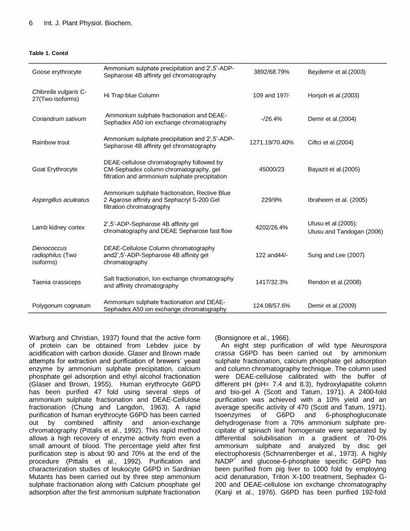
6 Int. J. Plant Physiol. Biochem. Table 1. Contd
Goose erythrocyte Ammonium sulphate precipitation and 2‟,5‟-ADP-Sepharose 4B affinity gel chromatography
3892/68.79% Beydemir et al.(2003)
Chlorella vulgaris C-27(Two isoforms)
Hi Trap blue Column 109 and 197/- Honjoh et al.(2003)
Coriandrum sativum Ammonium sulphate fractionation and DEAE-Sephadex A50 ion exchange chromatography
-/26.4% Demir et al.(2004)
Rainbow trout Ammonium sulphate precipitation and 2‟,5‟-ADP-Sepharose 4B affinity gel chromatography
1271.19/70.40% Ciftci et al.(2004)
Goat Erythrocyte DEAE-cellulose chromatography followed by CM-Sephadex column chromatography, gel filtration and ammonium sulphate precipitation
45000/23 Bayazit et al.(2005)
Aspergillus aculeatus Ammonium sulphate fractionation, Rective Blue 2 Agarose affinity and Sephacryl S-200 Gel filtration chromatography
229/9% Ibraheem et al. (2005)
Lamb kidney cortex 2‟,5‟-ADP-Sepharose 4B affinity gel chromatography and DEAE Sepharose fast flow
4202/26.4% Ulusu et al.(2005);
Ulusu and Tandogan (2006)
Dienococcus radiophilus (Two isoforms)
DEAE-Cellulose Column chromatography and2‟,5‟-ADP-Sepharose 4B affinity gel chromatography
122 and44/- Sung and Lee (2007)
Taenia crassiceps Salt fractionation, Ion exchange chromatography and affinity chromatography
1417/32.3% Rendon et al.(2008)
Polygonum cognatum Ammonium sulphate fractionation and DEAE-Sephadex A50 ion exchange chromatography
124.08/57.6% Demir et al.(2009)
Warburg and Christian, 1937) found that the active form of protein can be obtained from Lebdev juice by acidification with carbon dioxide. Glaser and Brown made attempts for extraction and purification of brewers‟ yeast enzyme by ammonium sulphate precipitation, calcium phosphate gel adsorption and ethyl alcohol fractionation (Glaser and Brown, 1955). Human erythrocyte G6PD has been purified 47 fold using several steps of ammonium sulphate fractionation and DEAE-Cellulose fractionation (Chung and Langdon, 1963). A rapid purification of human erythrocyte G6PD has been carried out by combined affinity and anion-exchange chromatography (Pittalis et al., 1992). This rapid method allows a high recovery of enzyme activity from even a small amount of blood. The percentage yield after first purification step is about 90 and 70% at the end of the procedure (Pittalis et al., 1992). Purification and characterization studies of leukocyte G6PD in Sardinian Mutants has been carried out by three step ammonium sulphate fractionation along with Calcium phosphate gel adsorption after the first ammonium sulphate fractionation
(Bonsignore et al., 1966). An eight step purification of wild type Neurospora
crassa G6PD has been carried out by ammonium sulphate fractionation, calcium phosphate gel adsorption and column chromatography technique. The column used were DEAE-cellulose calibrated with the buffer of different pH (pH= 7.4 and 8.3), hydroxylapatite column and bio-gel A (Scott and Tatum, 1971). A 2400-fold purification was achieved with a 10% yield and an average specific activity of 470 (Scott and Tatum, 1971). Isoenzymes of G6PD and 6-phosphogluconate dehydrogenase from a 70% ammonium sulphate pre-cipitate of spinach leaf homogenate were separated by differential solubilisation in a gradient of 70-0% ammonium sulphate and analyzed by disc gel electrophoresis (Schnarrenberger et al., 1973). A highly NADP
+ and glucose-6-phosphate specific G6PD has
been purified from pig liver to 1000 fold by employing acid denaturation, Triton X-100 treatment, Sephadex G-200 and DEAE-cellulose ion exchange chromatography (Kanji et al., 1976). G6PD has been purified 192-fold

from Methylomonus M15, grown on methanol as sole source for carbon and energy (Steinbach et al., 1978). The purification procedure involved N-acetyl-N, N, N-trimethylammonium bromide precipitation, ion-exchange chromatography and affinity chromatography on blue-dextran-Sepharose (Steinbach et al., 1978). The final enzyme preparations were homogeneous as judged by acrylamide gel electrophoresis and by sedimentation in an ultracentrifuge (Steinbach et al., 1978). A partial purification of G6PD from cyanobacteria Anabaena sp. ATCC 27893 was carried out by using Sepharose 4B blue dextran column (Schaeffer and Stanier, 1978). The enzyme has also been purified from methanol grown Pseudomonas W6 by a simple procedure involving dye-ligand affinity chromatography on Cibacronblue F3G-A-Sephadex and Procion Red HE-3B-Sepharose having high specific activity of 390 and 500 units/mg with NADP and NAD, respectively (Reuter et al., 1990). G6PD has been purified from potato tuber at least 850 fold to apparent homogeneity and with a specific activity of about 200 U/mg protein by ammonium sulphate fractionation and affinity chromatography (Graeve, Schaewen and Scheibe, 1994). The enzyme was found to be stable for at least 6 months when stored at -20°C in a buffer containing 0.02 M Tris-HCl, pH 8.0, 0.1 mM NADP, 5mM 2-mercaptoethanol, 1 mM PMSF, 2mM benzamidine and 1mM 6-amino-n-caproic acid (Graeve, Schaewen and Scheibe, 1994). The enzyme was also purified from a basidiomyceteous yeast Cryptococcus neoformans which is an opportunistic pathogen of AIDS patients with a specific activity of 50 U/mg (Niehaus and Mallett, 1994). A. vinelandii G6PD isolated from cell sonicates was purified 81 fold to electrophoretic homogeneity and a specific activity of 73 units/mg protein using ion-exchnage and Matrex Dye chromatography (Anderson and Anderson, 1995). Some methods have been given for studying the G6PD activity in brain areas (Ninfali et al., 1997a) and higher G6PD activity was reported in the olfactory lobe (Ninfali et al., 1997b) but no attempts has been made for the extraction and purification studies of G6PD from brain. A 250 fold purified enzyme was obtained from Schizosaccharomyces pombe (Tsai and Chen, 1998). G6PD was also purified from dog liver with a specific activity of 130 U mg
−1 and a yield of 18%. PAGE showed
two bands on protein staining; only the slower moving band had G6PD activity (Ozer et al., 2002). Sheep erythrocyte G6PD has been purified with a yield of 37.1% and specific activity of 4.64 U/mg proteins by using ammonium sulphate fractionation and 2‟, 5‟-ADP Sepharose 4B affinity chromatography (Beydemir et al., 2002). Glucose 6-phosphate dehydrogenase (G6PD) was purified from buffalo (Bubalus bubalis) erythrocytes in two steps: hemolysate preparation and 2
′, 5
′-ADP–Sepharose
4B affinity gel chromatography. The two consecutive processes gives 650 fold purified enzyme having a specific activity of 69.7 EU/mg proteins with a yield of
Singh et al. 7 31% (Ciftci et al., 2003). Goose erythrocyte G6PD has been purified to 3892 fold with a specific activity of 36.2 EU/mg protein and 68.79% yield by using ammonium sulphate precipitation and 2‟, 5‟-ADP Sepharose 4B affinity gel chromatography (Beydemir et al., 2003). Two isoforms of G6PD from cells of a freezing tolerant strain of C. vulgaris C-27, by sequential steps of chroma-tography on five kinds of columns, including a Hi Trap Blue column which showed excellent separation of the isoforms from each other (Honjoh et al., 2003). The two isoforms were purified up to 109 and 197 fold respectively with specific activity of 14.4 and 26 U/mg proteins, respectively (Honjoh et al., 2003). Rat small intestine G6PD was purified with a yield of 19.2% and specific activity of 53.8 U/mg proteins (Danisan et al., 2004). A74 fold purified G6PD with a yield of 26.4% and had a specific activity of 1.826 U/mg proteins was obtained from coriander (Coriandrum sativum) leaves by using ammonium sulfate fractionation, and DEAE-Sephadex A50 ion exchange chromatography (Demir et al., 2004). Glucose 6-phosphate dehydrogenase from rainbow trout (Oncorhynchus mykiss) erythrocytes was purified, using a simple and rapid method which involves hemolysate preparation, ammonium sulphate precipitation and 2‟, 5‟-ADP Sepharose 4B affinity gel chromatography. The enzyme was1271.19 fold purified with a specific activity of 14.51 EU/mg proteins and 70.40% yield (Ciftci et al., 2004). The enzyme was also purified from a filamentous fungus, Aspergillus aculeatus previously isolated from infected tongue of a patient (Ibraheem et al., 2005). The purified enzyme was apparently homogeneous with a specific activity of 220 U/mg proteins (Ibraheem et al., 2005). Partial purification of G6PD by aqueous two-phase poly (ethylene glycol)/phosphate systems were also reported (Ribeiro et al., 2007). Continuous counter-current purification of glucose-6-phospahte dehydrogenase using liquid-liquid extraction by reverse micelles was reported (Hasmann et al., 2007). In this method, a biocompatible reverse micellar system consisting of 0.05 M soybean lecithin in isooctane with hexanol was employed to study the influence of different flow rates on G6PD purification (Hasmann et al., 2007). The enzyme recovery yield varied from 32 to 115% and G6PD purification factor from 1.0 to 2.2 (Hasmann et al., 2007). Two isoforms of glucose-6-phsophate dehydrogenase was also purified from an extraordinarily UV-resistant bacterium Dienococcus radiophilus (Sung and Lee, 2007). The two isoforms were purified 122 and 44 fold respectively by using DEAE-Cellulose column chromatography and 2‟, 5‟-ADP Sepharose 4B affinity chromatography (Sung and Lee, 2007). The enzyme has also been purified from lamb kidney cortex by using 2‟, 5‟-ADP Sepharose 4B affinity chromatography (Ulusu et al., 2005; Ulusu and Tandogan, 2006). The enzyme has also been purified to homogeneity from the soluble fraction of larval Taenia crassiceps by salt fractionation, ion exchange and affinity

8 Int. J. Plant Physiol. Biochem. chromatography (Rendon et al., 2008). A 124.08 fold purified enzyme with a specific activity of 1.896 EU/mg proteins and 57.6% yield was also obtained from Polygonum cognatum Meissn leaves by ammonium sulphate precipitation and DEAE- Sephadex A50 ion-exchange chromatography (Demir et al., 2009). Structure and function of glucose-6-phosphate dehydrogenase Glucose-6-phosphate dehydrogenase is the first enzyme of pentose phosphate pathway and catalyzes the conversion of glucose into pentose sugar required for various biosynthetic reactions like nucleotide synthesis in both plants and animals. In addition, the pathway also provides reducing power in the form of NADPH (Figure 1b) by the action of G6PD and 6-phosphogluconate dehydrogenase which serves as an electron donor for many enzymatic reactions of biosynthetic pathways. NADPH produced plays an important role in protecting the cells from oxidative stress (Pandolfi et al., 1995). G6PD also helps in regeneration of reduced glutathione along with formation of a molecule of NADPH (Luzzatto, 1995; Tsai et al., 1998). Almost all the G6PD isoforms that have been studied so far are highly specific for their substrate, glucose-6-phosphate and NADP
+, for this
reason many of the properties of the enzyme have been evolutionary preserved (Levy 1979). However, some of the bacterial isoforms are also reported for utilising NAD
+
instead of NADP+ (Anderson et al., 1997). G6PDs
reported so far have single subunit type with native form existing as dimer, tetramer or hexamer. However, the enzyme is active as dimer or tetramer (Luzzatto, 2006) in a pH dependent equilibrium. The single subunit of enzyme consists of 515 amino acids and molecular weight of 59096 Da (Luzzatto et al., 2001). The enzyme monomer consists of two domains which are linked by an α-helix, which contains conserved eight residue peptide that acts as substrate binding site (Mason, 1996; Naylor et al., 1996; Au et al., 2000). The structure of the enzyme reveals one NADP
+ molecule in every subunit of tetramer,
away from active site but close to dimer interface (Boyer and Graham, 1965). Complete primary structure of the human enzyme has been deduced from the sequence of a full length complementary DNA clone (Persico et al., 1986). Human G6PD enzyme has two NADP
+ binding
sites per subunit. One is the “catalytic” and other is “structural” site. However, the bound NADP
+ at the
structural site is not involved in enzyme catalysis but many enzyme deficiency mutations are located in the dimer interface and close to the “structural” NADP
+ site
(Au et al., 2000). The auxiliary cofactor is said to be crucial for the stability and integrity of the active form of enzyme (Wang et al., 2008). NADP
+ is required for
monomer hybridization and refolding (Beutler and Collins, 1965; Yoshida et al., 1967; Gomez-Gallego et al., 1996;
Gomez-Gallego et al., 2000; Wang and Engel, 2009). Most of the characterized enzyme shows hyperbolic kinetics, however enzymes isolated from cyanobacteria shows sigmoidal kinetics (Schaeffer and Stanier, 1978). Molecular weight and subunits There have been several attempts to determine the molecular weight using various techniques. G6PD has been reported to exist in monomer, dimer, tetramer and hexamer, however only dimeric and tetrameric form was found to be enzymatically active. Although, G6PD was first identified in 1931 (Warburg and Christian, 1931a; Warburg and Christian, 1931b), the physical characteri-zation was first attempted in 1962 by Kirkman and Henderickson (Kirkman, 1962; Kirkman and Hendrickson, 1962). The molecular weight of human erythrocyte G6PD dimer was reported to be 105 KDa (Kirkman and Hendrickson, 1962). In 1963, Chung and Langdon made an attempt for the calculation of human erythrocyte G6PD from the diffusion and sedimentation constants of the enzyme by Svedberg equation and the molecular weight calculated was 190 KDa.
The molecular mass of the Pseudomonas W6 native enzyme was estimated to be 123 ± 5 KDa (Reuter et al., 1990). For the polypeptide chain after SDS denaturation, a molecular mass of about 61 KDa was calculated (Reuter et al., 1990). The G6PD isozymes from spinach were reported to have very similar molecular weight at about 105 ± 10% KDa as estimated by sedimentation velocity measurements (Schnarrenberger et al., 1973). G6PD from pig liver occurs as dimer with molecular weight of 133 KDa with monomeric unit having a molecular weight of 67.5 KDa (Kanji et al., 1976). The molecular weight of an obligate methanol utilizing bacterium Methylomonas M15 was determined by sedimentation equilibrium method and was found to be 108 ± 5 KDa (Steinbach et al., 1978). Sucrose gradient centrifugation and polyacrylamide gel electrophoresis reveal three principal forms of G6PD in Anabaena sp. with approximate molecular weights of 120, 240 and 345 KDa (Schaeffer and Stanier, 1978). Although the molecular weight distributions were very similar, about 50% activity was lost if the gradient did not contain glucose-6-phosphate and centrifugation in presence of 0.5 mM NADP
+ resulted in a markedly different molecular
weight distribution (Schaeffer and Stanier, 1978). A 112 KDa dimeric G6PD was also reported from Schizosaccharomyces pombe (Tsai and Chen, 1998). G6PD had a subunit molecular weight of 50 KDa in Cryptococcus neoformans (Niehaus and Mallett, 1994), the sodium dodecyl sulphate polyacrylamide gel electrophoresis and molecular exclusion chromatography indicated the presence of a tetrameric enzyme from A. vinelandii with subunit molecular weight of 52 KDa (Anderson and Anderson, 1995). Monomeric enzyme

has been reported from dog liver with a molecular weight of 52.5 KDa (Ozer et al., 2002). Two isoforms of G6PD were reported from a free tolerant strain of C. vulgaris C-27 with an apparent molecular weight of 200 KDa a with a subunit molecular weight of about 58 KDa (Honjoh et al., 2003). The apparently homogeneous enzyme from A. aculeatus had a molecular weight of 105± 5 KDa with a subunit molecular weight of 52±1.1 KDa (Ibraheem et al., 2005), whereas the enzyme from Aspergillus parasiticus had a molecular weight of 180 KDa and was composed of four subunits of apparently equal size (Niehaus and Dilts, 1984). The molecular weight of G6PD from turkey erythrocyte was determined by Rf –logM.W graph and by gel filtration method and was found 73.117 and 75.46 KDa respectively (Yilamz et al., 2003). Interestingly, the enzyme was found to be active in its monomeric form (Yilamz et al., 2003). The molecular mass of glucose-6-phospahte dehydrogenase from coriander was estimated to be 74.4 KDa by SDS-PAGE and 73.2 KDa by Sephadex G-200 gel filtration column chromatography (Demir et al., 2004). The molecular weight of the G6PD from rainbow trout was estimated to be 64.26 KDa by using SDS –PAGE and 66.22 KDa by gel filtration (Ciftci et al., 2004). An interesting fact about the rainbow trout enzyme is that the enzyme is active in its monomeric form whereas the active forms reported were only dimers or tetramers (Ciftci et al., 2004).Goat erythrocyte contains a tetrameric enzyme with subunit molecular weight of 52 KDa. The enzyme from lamb kidney cortex is reported to have a molecular weight of 67 KDa (Ulusu and Tandogan, 2006). Two isoforms of G6PD has been isolated from Deinococcus radiophilus with subunit molecular weight of 35 KDa (tetramer) and 60 KDa (dimer) respectively (Sung and Lee, 2007). Substrate and coenzyme specificity G6PD was earlier regarded to be specific for glucose-6-phosphate. However, later on it has been claimed that other phosphorylated monosaccharides were also oxidized by G6PD. Scott and Tatum (1971) have examined various phosphorylated and non-phosphorylated monosaccharides for the oxidation by N. crassa G6PD and they found that in addition to glucose-6-phosphate only galactose-6-phosphate and 2-deoxyglucose-6-phosphate were oxidized by the enzyme (Scott and Tatum, 1971). However, the relative rates of oxidation were low as compared to glucose-6-phosphate.
Fructose-6-phosphate was also oxidized but its effectiveness as substrate was speculative, since it was known to be contaminated with glucose-6-phosphate.
However, no activity was observed with glucose-1-phosphate, glucose, glucosamine-6-phosphate, 6-phosphogluconic acid, mannose-6-phsophate and fructose. Similar observations were observed in case of G6PD from other sources also. The coenzyme NADP
+
Singh et al. 9 shows more affinity towards the enzyme as compared to the substrate, glucose-6-phosphate. The substrate binding enhances the affinity of the enzyme for coenzyme (Ibraheem et al., 2005). G6PD isolated so far from different sources can be divided on the basis of their nucleotide specificity. One group exemplified by the enzymes from brewers‟ yeast (Warburg and Christian, 1931), E. coli (Banerjee and Fraenkel, 1972), N. crassa (Scott and Tatum, 1971), spinach leaf (Schnarrenberger et al., 1973) Anabaena sp. (Schaeffer and Stanier, 1978; Gleason, 1996), goose erythrocyte (Beydemir et al., 2003), A. aculeatus (Ibraheem et al., 2005), reacts exclusively with NADP. The second group, which appears to include animal G6PD, in general reacts with NADP but also gives weak activity with NAD. The third group of the enzyme can react approximately equally well with NAD and NADP. This class includes the enzyme from a number of bacterial strains such as Leuconostoc mesenteroides (Demoss et al., 1953; Olive et al., 1971), Pseudomonas aeruginosa (Ju-Fang et al., 1998), Pseudomonas W6 (Reuter et al., 1990), Streptomyces aureofaciens (Haghighi et al., 2005), Methylomonas M15 (Steinbach et al., 1978). In dog liver G6PD NADP was replaced by d-NADP (Deamino NADP). The latter exhibit more affinity as well as catalytic efficiency than NADP (Ozer et al., 2002). The human placental G6PD oxidizes a variety of substrates other than glucose-6-phosphate, such as galactose-6 - phosphate and deoxyglucose -6 -phosphate. The affinity of the enzyme for glucose-6-phosphate and galactose-6-phosphate were nearly equal and Deamino- NADP was also found to act as coenzyme for the human placental enzyme (Ozer et al., 2001). Km and Vmax The rate of G6PD catalyzed reaction varies with the condition of measurement, method of assay, nature of buffer and its pH. Therefore, wide variations in Km and Vmax values have been reported by various workers. The Km value for glucose-6-phosphate is found to be more than NADP
+ except in some cases indicating that G6PD
shows more affinity towards NADP+. The values of
maximum velocity Vmax is reported in few sources only. Km and Vmax values in some of the sources are given in Table 2. Optimum pH The pH optima of maximal catalytic activity appear to depend on the nature of the buffer and source of enzyme. G6PD from rat liver has been found to have pH optima of 7.6 in glycineglycine buffer whereas in veronal buffer the pH optimum was 8.5 (Glock and McLean, 1953). The dried brewers‟ yeast enzyme shows maximal activity at pH 8.5 (Glaser and Brown, 1955). N. crassa G6PD was

10 Int. J. Plant Physiol. Biochem.
Table 2. Km and Vmax values as reported in some sources.
Source Km(µM) Vmax (U/mg)
References
G6P
NADP+
NAD+
G6P
NADP+
NAD+
Rat liver
13 a
-----
-----
-----
-----
-----
Glock and McLean (1953)
Leukocyte
pH
5.0 11 a
80
----
----
----
----
Bonsignore et al. (1966)
6.0 16 a
82
7.5 31 a
81
8.5 34 a 80
9.5 39 a 83
Leuconostoc mesenteroides 81
a 5.69 106
----
----
----
Olive et al. (1971)
52.7b
Neurospora Crassa 37a 12 -----
-----
-----
-----
Scott and Tatum (1971)
Thiobacillus ferrooxidans 53.9 a 24.3 2080 ---- ---- ---- Tabita and Lundgren (1971)
Spinach 400
a and
330a (Two
isozymes) ----- ----- ----- ----- -----
Schnarrenberger
et al.(1973)
Pig liver 36±3 a 4.8±0.5 ---- ---- ---- ---- Kanji et al. (1976)
Baker‟s Yeast 51 a 19 ---- ---- ---- ---- Gould and Goheer (1976)
Methylomonas M15 110
a 14 200 ---- ---- ---- Steinbach et al. (1978)
290b
Mouse 50 a 10 ---- ---- ---- ---- Lee et al. (1979)
Soybean Nodules
Hong and Copeland (1991)
G6PD I
pH 7.2 69 a 12.3 --- 166 175 ----
pH 8.3 68 a 9.1 257 280
G6PD II
pH 7.2 98a 8.5 ---- 198 228 ----
pH 8.3 76 a 6.8 341 372
Cryptococcus neoformans 24 a 1.6 ---- ---- ---- ---- Niehaus and Mallet (1994)
Acetobacter hansenii 89
a 340 104 ---- ---- ----
Ragunathan and Levy (1994) 71
b
Potato tuber 260 a 6 ---- ---- ---- ---- Graeve et al. (1994)
Azotobacter vinelandii ---- 50 220 ---- ---- ---- Anderson and Anderson (1995)
Rat liver 329 a 100 ---- ---- ---- ---- Corpas et al. (1995)
Rat Kidney-cortex 206 a 25 ---- ---- ---- ---- Corpas et al. (1995)
Pea leaves 2000 a 500 ---- ---- ---- ---- Semenikhina et al.(1999)
Trypanosoma brucei. 138 a 5.3 ----- ----- ----- ----- Heise and Opperdoes (1999)
Bovine lens 35±13 a 8±2 ---- ---- ---- ---- Ulusu et al. (1999)
Trypanosoma brucei. 138 a 35 ----- ----- ----- ----- Diffieux et al. (2000)
Human placenta 40±8 a 20±10 ---- ---- ---- ---- Ozer et al. (2001)

Singh et al. 11 Table 2. Contd.
Thermotoga maritima 150 a 30 12 x10
3 20 20 12 Hansen et al. (2002)
Dog liver 122±18 a 10±1 ----- ----- ----- ----- Ozer et al. (2002)
Goose Erythrocyte 24.3 a 7.4 ----
0.28 (U/ml)
0.286 (U/ml)
---- Beydemir et al. (2003)
Turkey erythrocytes 49.7 a 17.1 ----
0.5 (U/ml)
0.37
(U/ml) ---- Yilmaz et al. (2003)
Rat small Intestine 70.1±20.8a 23.2± 7.6 ---- 53.8 53.8 ---- Danisan et al. (2004)
Coriandrum Sativum 116a 26 ----
0.038 (U/ml)
0.035 (U/ml)
---- Demir et al. (2004)
Rainbow Trout
Erythrocyte 500
a 166 ----
1.352
(U/ml)
0.275
(U/ml) ---- Ciftci et al. (2004)
Aspergillus Aculeatus 75 ±6 a 6±1 ---- ---- ---- ---- Ibraheem et al. (2005)
Streptomyces aureofaciens
790 a 75 ---- ---- ---- ---- Haghighi et al. (2005)
Goat erythrocyte 334 a
--------
--------
642
µM/min
--------
-------- Bayazit et al. (2005)
Lamb kidney Cortex 39±6a 18±2 ---- ---- ---- ---- Ulusu et al. (2005)
Sheep kidney cortex 41±4.3 a 14.7± 1 ----- 28.23 ±0.86 ----- ----- Ulusu et al. (2006)
Taenia crassiceps
(cysticerci) 14 ± 1.7
a 1.3± 0.4 20.3±3.2 x 10
3 33.8± 2.1 33.8± 2.1 4.1±0.6
Rendon et al. (2008)
a using NADP
+; b
using NAD+
found to exhibit maximal activity in Tris-HCl buffer. The pH of the enzyme in Tris-HCl is broad, ranging from about pH 7.4 to pH 8.2 (Scott and Tatum, 1971). However, in malate-NaOH buffer, activity was 75% of that observed in Tris-HCl buffer at comparable pH values (Scott and Tatum, 1971). In Thiobacillus ferrooxidans, when the enzyme was assayed with NADP at different pH values of Tris buffer, a relatively sharp optimum was reported at pH 7.8 to 7.9, whereas, enzyme activity with NAD as coenzyme differed in that no activity was obtained at pH values below 7.5; a relatively broad pH optimum was observed from pH 8.0 to 8.5 (Tabita and Lundgren, 1971).The two isoenzymes of G6PD from spinach have been found to have pH optima of 9.2 and 9 (Schnarrenberger et al., 1973). Kanji et al. (1976) have studied the effect of pH on activity in pig liver. They performed their studies in glycine/NaOH and Tris/Malate/NaOH buffers and observed a slow rise in activity with increasing pH to a maximum at pH 8.5 (Kanji et al., 1976). G6PD from rainbow trout was more stable
at pH 8.9 than at other pH values for the duration of 48 h (Ciftci et al., 2004).The two NADP
+-specific G6PD
isoenzymes from soybean nodules shows optimum activity at 8.5 and 8.1(Hong and Copeland, 1991). A broad pH optimum between pH 7.5 and 9 has been reported in potato tuber (Graeve, Schaewen and Scheibe, 1994). G6PD from A. vinelandii exhibit a pH optimum of 8.5 (Anderson and Anderson, 1995), from pea
from pea leaves pH optimum of 8.0 (Semenikhina et al., 1999), from dog liver pH optimum of 7.8 (Ozer et al., 2002). The optimal pH of goose erythrocyte G6PD was found to be 7.0 using Tris-HCl buffer; however, the stable pH of the enzyme was reported to be 9.0 in the same buffer (Beydemir et al., 2003). pH optimum of the enzyme from rat small intestine was found to be 8.1 (Danisan et al., 2004). The enzyme from coriander had an optimum pH at 8.5 and was stable at pH 8.0 in 0.1 M Tris-HCl buffer (Demir et al., 2004). The optimum pH of G6PD from turkey erythrocytes has been found to be 9.0, whereas, the enzyme is stable at pH 8.0 (Yilmaz et al.,

12 Int. J. Plant Physiol. Biochem. 2003). The enzyme from buffalo erythrocyte shows optimum pH of 8.0 using 1 M Tris-HCl (Ciftci et al., 2003). However, the stable pH of the enzyme was reported to be 9.0 in Tris-HCl. (Ciftci et al., 2003). In S. aureofaciens the optimum pH was 6.6 for NAD
+ and 7.4 for NADP
+
dependent activity (Haghighi et al., 2005) The two isoforms of G6PD from D. radiophilus show optimum activity at pH 8.0 (Sung and Lee, 2007). The enzyme from Taenia crassiceps shows maximal activity between pH 6.7 and 7.8 (Rendon et al., 2008).
Optimum temperature Studies on determination of optimum temperature have not been so practicable in the case of G6PD. Very few reports on optimum temperature are available. A wide range of optimum temperature was reported in different sources. The range varies in between 30 - 92°C. Goose erythrocyte G6PD shows highest activity point at 50°C (Beydemir et al., 2003). Similar reports were found for rat liver and kidney-cortex (Corpas et al., 1995) and bovine lens (Ulusu et al., 1999). The rainbow trout enzyme was found to show the highest activity at 45°C after tried between 10–80°C (Ciftci et al., 2004). The optimum temperature of turkey erythrocyte was found to be 50°C (Yilmaz et al., 2003). The most thermoactive G6PD was reported from hyperthermophilic bacterium T. maritima whose temperature optimum was reported to be 92°C (Hansen et al., 2002). Rat small intestine G6PD shows an optimum temperature of 38°C (Danisan et al., 2004). Optimum temperature in coriander was found to be 30°C (Demir et al., 2004). The two isoforms of G6PD from D. radiophilus show optimum activity at 30°C (Sung and Lee, 2007). The activities of both the enzymes decreases above 40°C, but at 70°C, the activity of G6PD-I decreases more profoundly than the activity of G6PD-II (Sung and Lee, 2007). This indicates that G6PD-I was more sensitive than G6PD-II (Sung and Lee, 2007). Inhibition Product inhibition Product inhibition studies of G6PD were conducted with NADPH. NADPH is a potent inhibitor of NAD-linked G6PD and a competitive inhibitor of NADP-linked G6PD (Levy et al., 1966). In case of L. mesenteroides when NADP
+ was the varied substrate, linear competitive inhibi-
tion was obtained, whereas with glucose-6-phosphate as varied substrate the inhibition was linear non competitive (Olive et al., 1971). For NAD
+ linked reaction, the
inhibition was linear non competitive when NAD+ was
used as varied substrate and glucose-6-phosphate was non saturating and when glucose-6-phosphate was used as varied substrate the inhibition by NADH was non-competitive without any correlation with NAD
+ being
saturating or non saturating (Olive et al., 1971). The enzyme isolated from obligate methylotrophic bacterium Pseudomonas W6 is not inhibited by NADPH but by NADH (Miethe and Babel, 1976), however the purified enzyme from Methylomonas M15 was reported to be inhibited by both NADPH and NADH (Steinbach et al., 1978). Grossman and McGowan (1975) reported that NADPH inhibits the activity of cyanobacterial G6PD, which was later on confirmed by Pelroy et al. (1976). Inhibition of G6PD in the cyanobacterium Anabaena sp. has also been reported (Schaeffer and Stanier, 1978). Human placental G6PD was also reported to be competitively inhibited by NADPH (Ozer et al., 2001). Several other reports have been shown that G6PD from yeast (Gould and Goheer, 1976; Niehaus and Mallett, 1994; Tsai and Chen, 1998), bacteria (Raghunathan and Levy, 1994; Hansen et al., 2002), fungi (Ibraheem et al., 2005), animals (Ozer et al., 2002) and plants (Demir et al., 2004) are strongly and competitively inhibited by NADPH. S. aureofaciens G6PD was inhibited by NADH and NADPH (Haghighi et al., 2005). Both NADH and NADPH inhibited the enzyme competitively and noncom-petitively with respect to the corresponding oxidized coenzymes and glucose 6-phosphate, respectively (Haghighi et al., 2005). Inhibition by various inhibitors
Several classes of G6PD inhibitors are reported. G6PD from brewers‟ yeast was reported to be competitively inhibited by D-glucosamine-6-phosphate with a Ki value of 7.2 X 10
-4 M (Glaser and Brown, 1955).
Dehydroisoandrosterone, pregnenolone, and certain related steroids, at concentrations of 10
-6 M or less,
inhibit mammalian G6PD activity. However, these steroids do not inhibit spinach or yeast G6PD (Marks and Banks, 1960). G6PD was found to be highly sensitive to inhibitory effect of long chain acyl coenzyme A (Eger-Neufeldt et al., 1965). Inhibition of partially purified Leuconostoc G6PD from iodoacetate has been reported (Demoss et al., 1953) but later studies on Leuconostoc G6PD inhibition shows no inhibition by iodoacetate, iodoacetamide and p-hydroxymercuribenzoate (Olive et al., 1971). A pH and concentration dependent inhibition of G6PD by pyridoxal 5‟-phosphate was also reported (Olive et al., 1971). Pyridoxal 5‟-phosphate inhibits the enzyme at 0.1mM and 7.66 and 6.32 pH values (Olive et al., 1971). Pyridoxal 5‟-phosphate behaves as competitive inhibitor when the glucose-6-phosphate was varied at a saturating concentration of NAD
+ and behaves as non
competitive inhibitor when NAD+ was varied at a
saturating concentration of glucose-6-phosphate (Olive et al., 1971). The activity of G6PD in leaf extract of barley and spinach was reported to be 20-35% inhibited by dithiothreitol (Johnson, 1972). However, the inhibition is reversible. Cyanate inhibition of erythrocyte G6PD has been reported by Glader and Conrad (1972). Palmitoyl
coenzyme A is reported to reversibly inhibit the yeast G6PD (Kawaguchi and Bloch, 1974). Phosphoenol pyruvate inhibits the G6PD activities of different bacteria, in contrast the enzyme from methanol grown Methylomonas M15 was not inhibited by this metabolite but ATP inhibits the enzyme non-competitively with respect to NAD, NADP and glucose-6-phosphate (Steinbach et al., 1978). Zinc ion was reported to be a powerful inhibitor of G6PD, inhibition being competitive with respect to glucose-6-phosphate with Ki about 2.5 µM (Niehaus and Dilts, 1984; Niehaus and Mallett, 1994; Ibraheem et al., 2005). Human erythrocyte G6PD was reported to be inhibited by some antibiotics (Ciftci et al., 2000). Sodium cefoperazone, Gentamicin sulphate and Netilmicin sulphate were found to be competitive inhibitor of human erythrocyte enzyme, whereas, Sodium ceftizoxime, Sodium ampicillin, Sodium cefuroxime, Sodium cefazolin and Streptomycin sulphate were found to be non-competitive inhibitor of the enzyme (Ciftci et al., 2000). Inhibition of human erythrocyte G6PD by aluminium, nickel and cadmium is also reported (Haghighi and Ilghari, 2004). Aluminium causes 60% inhibition, nickel causes 70% inhibition and cadmium cause 65% inhibition of the enzyme (Haghighi and Ilghari, 2004). It was reported that cadmium binds either to the enzyme or enzyme substrate complex producing binary and ternary complexes and hence resulting in confor-mational changes leading to enzyme inactivation, aluminium leads to a pure competitive inhibition with respect to glucose-6-phosphate surprisingly, as the two molecules are not substrate analogues (Haghighi and Ilghari, 2004). The inhibition might be due to metal induced conformational change that prevents proper binding of substrate. Nickel shows a mixed type inhibition of the enzyme (Haghighi and Ilghari, 2004). Phosphoenol pyruvate, ATP, fructose-6-phosphate and cobalt ion was also reported to inhibit the enzyme (Ibraheem et al., 2005). ATP and oleic acid inhibits the two isoforms of enzyme from D. radiophilus (Sung and Lee, 2007). Oleic acid was found to be more profound inhibitor for G6PD-I than G6PD-II (Sung and Lee, 2007). The profound inhibit-tion of both enzymes by β-naphthoquinone-4-sulphonic acid suggests that lysine is present at the active site of enzyme (Sung and Lee, 2007). Cu
2+ was found to be a
potent inhibitor of G6PD-II but a lesser degree to G6PD-I (Sung and Lee, 2007). Regulation of glucose-6-phosphate dehydrogenase Regulation of G6PD by nutritional and hormonal factors For the production of NADPH and for control of carbon flow through pentose phosphate pathway, G6PD is required in each cell. As a provider of NADPH for fatty acid biosynthesis, G6PD has long been considered as a
Singh et al. 13 member of lipogenic enzymes family (Kletzien and Berdanier, 1993; Kletzien et al., 1994). Animal tissues with high levels of fatty acid biosynthesis control the oxidative pentose phosphate pathway at the level of G6PD regulation. In hepatocytes, G6PD is induced in response to excess dietary carbohydrate and is repressed in response to starvation (Levy, 1979; Iritani, 1992). The molecular basis of the elevated activity is not well understood but seems to be dependent upon the presence of insulin, glucocorticoids and a high carbohydrate; low fat diet (Kletzien and Berdanier, 1993; Kletzien et al., 1994). The activity of the enzyme is also affected by the type of dietary carbohydrate. Higher enzyme activity was found when the rats are fed on a diet with higher percentage of glucose to starch and induction by fructose is higher as compared to glucose (Kastrouni et al., 1984). Glucose-6-phospahte dehydrogenase activity is inhibited by a high fat, low carbohydrate diet both in intact animal and in primary hepatocytes in culture (Salati et al., 1988; Stabile et al., 1996; Stabile et al., 1998). The inhibition of G6PD gene expression is induced by presence of polyunsaturated fatty acid in diet. The inhibition of expression is through a posttranscriptional mechanism which involves decrease in the efficiency of splicing of the pre-mRNA (Tao et al., 2002). The signalling pathway that causes increased expression of G6PD involves insulin activation of phosphatidylinositol 3-kinase pathway resulting in accumulation of glucose-6-phospahte dehydrogenase mRNA (TaluKDar et al., 2005). Polyunsaturated fatty acids such as arachidonic acid inhibits accumulation of glucose-6-phospahte dehydrogenase mRNA by inhibiting insulin signalling through the activation of p38 mitogen activated protein kinase (p38 MAPK) and the subsequent Ser
307 phosphorylation of insulin receptor substrate-1
(IRS-1) (TaluKDar et al., 2005). In rat hepatocytes, arachidonic acid activation of AMP activated protein kinase (AMPK) is involved in the glucose-6-phsophate dehydrogenase expression (Kohan et al., 2009). First activation of AMPK inhibits the insulin induction of glucose-6-phospahte dehydrogenase mRNA whereas second activation of AMPK by AICAR (aminoimidazole carboxamide ribonucleotide) activates p38MAPK resulting in Ser
307 phosphorylation of insulin receptor
substrate-1 (IRS-1) and inhibits AKT phosphorylation similar to the action of arachidonic acid (Kohan et al., 2009). Inhibition of p38 MAPK blocks the effect of AICAR on accumulation of glucose-6-phospahte dehydrogenase mRNA (Kohan et al., 2009). The inhibition of G6PD gene expression is mainly due to polyunsaturated fatty acids and not due to saturated or monounsaturated fatty acids.
Regulation of glucose-6-phsophate dehydrogenase by hormonal factors has been observed in hepatocytes in primary culture. In hepatocytes primary culture role of insulin and glucocorticoids has been examined for the expression of G6PD (Kletzien and Berdanier, 1993; Kletzien et al., 1994). Role of insulin in induction of G6PD
14 Int. J. Plant Physiol. Biochem. activity in hepatocytes primary culture was given by Kurts and Wells in 1981. A 3-4 fold induction in G6PD activity is seen on incubation of hepatocytes with insulin which is also accompanied with changes in rate of synthesis and mRNA abundance (Manos et al., 1991). Several other studies have led to the conclusion that insulin is a primary hormone in upregulating G6PD expression at the level of transcription (Manos et al., 1991; Fukuda et al., 1992). However, the molecular mechanism of insulin action on transcription of G6PD gene and other insulin regulated genes is not well understood. Glucocorticoids are also positive regulators of G6PD activity in rat hepatocytes.
They clearly increase G6PD mRNA in the presence or absence of insulin (Manos et al., 1991; Fukuda et al., 1992) without affecting the relative rate of enzyme synthesis which indicates that it may be due to translational control which can be activated by insulin but not by the glucocorticoids (Kletzien et al., 1994). How-ever, maximum increase in G6PD mRNA was found in the presence of insulin and dexamethasone suggesting that both hormones are capable of increasing transcription (Kletzien et al., 1994; Manos et al., 1991; Fukuda et al., 1992).
G6PD expression by hormones, nutrients and some growth factors has also been observed in tissues other than liver. Carbohydrate refeeding has shown a 9 fold increase in G6PD mRNA in rat epididymal fat pads (Louie et al., 1990). Increase in G6PD mRNA is also observed as 3T3-L1cells differentiate into adipocytes or upon insulin treatment of confluent, non proliferating brown adipose cells (Kletzien et al., 1992; Valverde et al., 1992). Estrogen has been shown to increase G6PD in uterus (Smith and Barker, 1974). Epidermal growth factor has also been shown to induce G6PD mRNA and relative rate of enzyme synthesis indicating that growth stimulation of cells is linked to induction of glucose-6-phsophate dehydrogenase gene (Yoshimoto et al., 1983). Additional examples of endocrine regulation of glucose-6-phsophate dehydrogenase include norepinephrine induction of mRNA and activity in heart (Zimmer et al., 1992), elevation of activity due to vitamin D3 in gut epithelium (Nasr et al., 1989). The above discussion provides evidence that variety of tissues exhibit the capacity for adaptive regulation of glucose-6-phsophate dehydrogenase. Regulation of glucose-6-phosphate dehydrogenase by oxidative stress G6PD induction by a number of non hormonal agents indicates that the gene acts as a guard for oxidant stress and shows quick response to the need of NADPH for maintenance of redox state in the cells. G6PD plays an important role in the cellular response to the oxidant stress in E. coli (Greenberg et al., 1990). Agents responsible for production of oxygen radicals induce
G6PD through soxR regulon, which regulates G6PD and eight other genes required to protect cells from free radical damage (Greenberg et al., 1990). In Saccharomyces cerevisiae, a mutation (Par1) that renders the cells which are sensitive to oxidant stress leads to decreased expression of G6PD as well as other enzymes required for oxygen detoxification (Kletzien et al., 1994; Schnell et al., 1992). In mammalian lung, exposure of alveolar type II cells from neonatal rats to hyperoxia in vitro resulted in increased G6PD activity (Kennedy et al., 1989). The adaptive response of the G6PD gene in several tissues makes up a part of the coordinated cellular response to oxidant stress. Hepatic cells are highly exposed to a number of oxidants from dietary, environmental and pharmaceuticals sources.
Hepatic G6PD is induced by chemicals such as diquat (Cramer et al., 1993), ethanol (Stumpo and Kletzien, 1985). Studies have revealed that ethanol induces G6PD activity, relative rate of G6PD enzyme synthesis and mRNA in primary cultures of rat hepatocytes (Stumpo and Kletzien, 1985). The oxidative stress experiments in Trypanosoma cruzi showed that metacyclic trypomastigotes and amastigotes have high G6PD activity (Igoillo-Esteve and Cazzulo, 2006). The mechanism of enzyme induction is similar to that of reported in S. cerevisiae, E. coli and mammalian cells (Igoillo-Esteve and Cazzulo, 2006). However, in plants oxidative stress does not affect the cytosolic G6PD activity (Hauschild and Schaewen, 2003). Regulation of glucose-6-phosphate dehydrogenase by other factors Glucose-6-phosphate dehydrogenase regulation in higher plants occurs at the transcription and translation level. In alfalfa, induction in G6PD transcription and activity in response to fungal elicitors in roots and nodules has been reported (Fahrendorf et al., 1995). Induction in plastidic G6PD expression has been observed in response to illumination of light deprived potato shoots (Schaewen et al., 1995). Role of ferredoxin/thioredoxin system has been examined under light dependent inactivation of G6PD. The ferredoxin/thioredoxin system consists of ferredoxin, thioredoxin and ferredoxin-thioredoxin reductase (Buchanan et al., 1994). Ferrodoxin is the terminal electron acceptor of photosynthetic electron transport chain. Excitation of Photosystem-I by light, transfers the electrons to ferrodoxin and then to ferrodoxin dependent enzymes and NADP
+ which are reduced. Electrons are then
passed to ferredoxin-thioredoxin reductase reducing a disulphide bond to its dithiol form. Reduced ferredoxin-thioredoxin reductase then reduces thioredoxin which regulates the activity of variety of enzymes by changing the redox status. A dark activation and light inactivation cycle as a way of activity regulation has been observed in

isolated spinach chloroplasts (Scheibe et al., 1989). G6PD is also inactivated by dithiothreitol (DTT), a sulphahydryl reagent that imitates the effect of thioredoxin, however, inactivation by DTT and light may not be achieved through same mechanism (Scheibe et al., 1989). All chloroplast G6PD homologues are inactivated by redox modification of the ferredoxin-thioredoxin system (Buchanan 1991). G6PD is kept in reduced state in light and becomes active only in its oxidized state in the dark. The molecular basis of this regulation is disulphide-dithiol interchange of certain regulatory cysteine residues in the target enzymes.
Transfer of algal cultures to nitrogen free media is reported to induce glucose-6-phsophate dehydrogenase activity (Huppe and Turpin, 1996). Elevated enzyme activity may be linked with assimilation of inorganic nitrogen by the unicellular green algae.
However, the information regarding G6PD isoform regulation is limited. Several studies decscribing conditions that stimulate G6PD activity did not distinguish the different isoenzymes nor analyze possible mechanisms involved (Daniel et al., 1990). In potato, regulations of cytosolic and chloroplastic G6PD are governed by distinct mechanisms. Transcription of cytosolic G6PD gene via sugar-mediated signalling to the nucleus is triggered by high sugar levels in the cytosol (Hauschild and Schaewen, 2003). Plastidic G6PD can be stimulated by low NADPH to NADP ratio, most probably via dephosphorylation of the existing enzyme pool (Hauschild and Schaewen, 2003). CONCLUSION
G6PD is an important enzyme which plays a significant role in oxidative pentose-phosphate pathway. It provides NADPH, the reducing power required for various biosynthetic processes and plays an important role in the protection of cells against oxidative damage. Its deficiency is said to be preventive against malaria caused by Plasmodium falciparum. The enzyme exists as monomer, dimer, tetramer and hexamer, of which dimeric and tetrameric forms are active, however, some reports regarding active monomeric enzyme is also present.. The enzyme shows a wide range of pH optima. Inhibition of the enzyme has been an interesting area of study. The enzyme is strongly inhibited by NADPH, the reduced form of coenzyme. The enzyme is regulated by nutritional and hormonal factors at post translational and post transcriptional level and also by oxidant stress. Redox regulation of enzyme is through ferredoxin-thioredoxin system. The X-ray crystallographic studies and studies about the mutation of G6PD gene will give an insight to structure-function relationship and will help to elucidate the precise mechanisms of structural alterations in mutant forms. The studies of G6PD deficiency and regulation of enzyme is an important aspect and will provide a better understanding of its role in several
Singh et al. 15 human metabolic disorders of G6PD deficiency. REFERENCES
Anderson BM, Wise DJ, Anderson CD (1997) Azotobacter vinelandii
glucose-6-phosphate dehydrogenase properties of NAD- and NADP-linked reactions. Biochim. Biophys. Acta 1340: 268-276.
Anderson BM, Anderson CD (1995). Purification and characterization of Azotobacter vinelandii glucose-6-phosphate dehydrogenase: dual
coenzyme activity. Arch. Biochem. Biophys., 321(1): 94-100.
Au SWN, Gover S, Lam VM, Adams MJ (2000). Human glucose-6-phosphate dehydrogenase: the crystal structure reveals a structural NADP
+ molecule and
provides insights into enzyme deficiency. Struct., 8: 293-303. Banerjee S, Fraenkel (1972). Glucose-6-Phosphate Dehydrogenase
from Escherichia coli and from a “High-Level” Mutant. J. Bacteriol.,
110(1):155–160. Bayazit V, Cayci M K, Khan K M (1995). Purification and Kinetic
Properties of Glucose-6-Phosphate dehydrogenase from Goat (Cpraaegagrus hicrus) Erythrocytes. J. Chem Soc. Pak., 27(5):518-
526. Beutler E (1994). G6PD Deficiency. Blood 84(11): 3613-3636.
Beutler E (1992). The molecular biology of G6PD variants and other red cell enzyme defects. Annu. Rev. Med., 43: 47-59.
Beutler E, Vulliamy TJ (2002). Hematologically important mutations:
glucose-6-phosphate dehydrogenase. Blood Cells Mol. Dis., 28; 93-103.
Beutler, E (1996) G6PD: population genetics and clinical
manifestations. Blood Rev., 10:45-52. Beutler E, Collins Z. (1965). Hybridization of glucose-6-phosphate
dehydrogenase from rat and human erythrocytes, Science 150:1306-
1307. Beydemir S, Ciftci M, Kufrevioglu OI (2002). Purification and
characterization of glucose-6-phosphate dehydrogenase from sheep
erythrocytes and inhibitory effects of some antibiotics on enzyme activity. J Enzyme Inhib Med Chem. 17 (4):217-277
Beydemir S, Yilmaz H, Ciftci M, Bakan E, Kufrevioglu OI (2003).
Purification of Glucose-6-phosphate dehydrogenase from goose erythrocytes and kinetic properties. Turk J. Vet. Anim. Sci., 27:1179-1185.
Bonsignore A Fornaini G, Leoncini G, Fantoni A, Segni P (1966). Characterization of Leukocyte Glucose-6-phosphate dehydrogenase in Sardinian Mutants. J. Clin. Invest., 45(12):1865-1874.
Bowsher CG Boulton EL, Rose J, Nayagam S, Emes MJ (1992). Reductant for glutamate synthase is generated by the oxidative pentose phosphate pathway in non photosynthetic root plastids. Plant
J., 2:893-898. Boyer SH, Graham JB (1965). Linkage between the X chromosome loci
for glucose-6-phosphate dehydrogenase electrophoretic variation and
haemophilia A. Am. J. Hum. Genet., 17:320-324. Buchanan BB (1991). Regulation of CO2 assimilation in oxygenic
photosynthesis: the ferredoxin/thioredoxin system. Arch. Biochem.
Biophys., 288:1-9. Buchanan BB, Schurmann P, Decottignies P, Lozano RM (1994)
Thioredoxin: Amultifunctional protein with a bright future in
technology and medicine. Arch Biochem. Biophys., 314: 257-260. Burdon RH, Alliangana D, Gill V (1995). Hydrogen peroxide and the
proliferation of BHK-21 cells. Free Radical Res., 23: 471-486. Cardi M, Chibani K, Cafasso D, Rouhier N, Jacquot JP, Esposito S
(2011). Abscisic acid effects on activity and expression of barley (Hordeum vulgare) plastidial glucose-6-phosphate dehydrogenase. J.
Exp. Botany. DOI: 10.1093/jxb/err100 (http://jxb.oxfordjournals.org).
Chen EY, Cheng A, Lee A, Kuang WJ, Hillier L, Green P, Schlessinger D, Ciccodicola A, D'Urso M (1991). Sequence of human glucose-6-phosphate dehydrogenase cloned in plasmids and a yeast artificial
chromosome. Genomics 10: 792-800. Chung AE, Langdon RG (1963). Human Erythrocyte glucose-6-
phosphate dehydrogenase I. Isolation and properties of the enzyme.
J. Biol. Chem., 238(7): 2309-2316. Ciftci M, Ciltas A, Erdogan O (2004). Purification and characterization of
glucose-6-phosphate dehydrogenase from rainbow trout

16 Int. J. Plant Physiol. Biochem. (Oncorhynchus mykiss) erythrocytes. Vet. Med. – Czech, 49(9): 327–
333. Ciftci M, Kufrevioglu OI, Gundogdu M, Ozmen I (2000). Effects of some
antibiotics on enzyme activity of glucose-6-phosphate dehydrogenase from human erythrocytes. Pharmacol. Res., 41(1):109-113.
Ciftci M, Beydemir S, Yilmaz H, Altikat S (2003). Purification of glucose 6-phosphate dehydrogenase from Buffalo (Bubalus bubalis)
erythrocytes and investigation of some kinetic properties. Prot. Exp.
Purif., 29:304-310. Copeland L, Turner JF (1987). The regulation of glycolysis and the
pentose phosphate pathway. In the Biochemistry of Plants, (Stumpf,
P. K. and Conn, E. E., eds) New York: Academic Press, 11:107-125 Corpas FJ, Barroso JB, Sandalio LM, Distefano S, Palma JM, Lupianez
JA, Del Rio LA (1998). A dehydrogenase-mediated recycling system
of NADPH in plant peroxisomes. Biochem. J., 330 (7):777–784. Corpas FJ, Garcia- Salguero L, Peragon J, Lupianez JA (1995). Kinetic
properties of hexose monophosphate dehydrogenase. Isolation and
partial purification of glucose-6-phosphate dehydrogenase from rat liver and kidney-cortex. Life Sci., 56: 179-189.
Cramer CT, Ginsberg LC, Stapleton SR, Kletzien RF, Ulrich RG (1993). Induction of G6PDH in rat hepatocytes under oxidative stress conditions. The Toxicologist, 13:20.
Daniel S Tiemann K, Wittkampf U, Bless W, Hinderer W, Barz W
(1990). Elicitor-induced metabolic changes in cell cultures of chick pea (Cicer arietinum L.) cultivars resistant and susceptible to Ascochyta rabie: I. Investigations of enzyme activities involved in
isoflavone and pterocarpan phytoalexin biosynthesis. Planta, 182:270-278.
Danisan A, Ceyhan D, Ogus I H, Ozer N (2004). Purification and
characterization of glucose-6-phosphate dehydrogenase from rat small intestine. Protein J., 23(5): 317-324.
Debnam PM, Fernie AR, Leisse A, Golding A, Bowsher CG, Grimshaw
C, Knight JS, Emes MJ (2004). Altered activity of the P2 isoform of plastidic glucose-6-phosphate dehydrogenase in tobacco (Nicotiana tabacum cv. Samsun) causes changes in carbohydrate metabolism
and response to oxidative stress in leaves. Plant J., 38:49-59. Demir H, Beydemir S, Ciftci M, Kufrevioglu OC (2004). Purification and
Properties of Glucose 6-phosphate dehydrogenase from Coriander (Coriandrum Sativum) leaves. J. Food Biochem., 28(2):155-168.
Demir H, Beydemir S, Ciftci M (2009). Purification, characterization and kinetic properties of glucose 6-phosphate dehydrogenase from Polygonum cognatum Meissn leaves. AsianJ. Chem., 21(1):517-527.
Demoss RD, Gunsalus IC, Bard RC (1953). A Glucose-6-Phosphate Dehydrogenase from Leuconostoc mesenteroides. J. Bacteriol., 66:
10-16. Dennery PA, Seidman DS, Stevenson DK (2001). Neonatal
Hyperbilirubinemia. N. Engl. J. Med., 344: 581-590. Duffieux F, Van Roy J, Michels PA, Opperdoes FR (2000). Molecular
Characterization of the first two enzymes of the pentose phosphate pathway of Trypanosoma brucei. J. Biol. Chem. 275 (36):27559-
27565.
Eastmond PJ, Rawsthorne S (1998). Comparison of the metabolic properties of plastids isolated from developing leaves or embryos of Brassica napus L. J. Exp. Bot., 49:1105-1111
Eger-Neufeldt I, Teinzer A, Weiss L, Wieland O (1965) Inhibition of glucose-6-phosphate dehydrogenase by long chain acyl coenzyme A. Biochem. Biophys.Res. Comm., 19(1):43-48.
Emes MJ, Neuhaus HE (1997). Metabolism and tramnsport in non photosynthetic plastids. J. Exp. Bot., 48: 1995-2005.
Esposito S, Carfagna S, Massaro G, Vona V, Di Martino Rigano V
(2001). Glucose-6-phosphate dehydrogenase in barley roots: kinetic properties and localization of the isoforms. Planta 212: 627-634.
Esposito S, Massaro G, Vona V, Di Martino Rigano V, Carfagna S
(2003). Glutamate synthesis in barley roots: the role of the plastidic glucose-6-phosphate dehydrogenase, Planta 216: 639-647.
Fahrendorf T, Ni W, Shorrosh BS, Dixon RA (1995) Stress responses in
alfalfa (Medicago sativa L.) XIX. Transcriptional activation of the oxidative pentose phosphate pathway genes at the onset of the isoflavinoid phytoalexin response. Plant Mol. Biol., 28:885-900.
Farhud DD, Yazdanpanah L (2008). Glucose-6-phosphate dehydrogenase (G6PD) Deficiency. Iranian J. Publ. Health 37(4): 1-
18. Fickenscher K, Scheibe R (1986). Purification and properties of the
cytoplasmic glucose-6-phosphate dehydrogenase from pea leaves.
Arch. Biochem. Biophys., 247: 393-402. Fico A, Paglialunga F, Cigliano L, Abrescia P, Verde P, Martini G,
Iaccarino I, Filosa S (2004) Glucose-6-phosphate dehydrogenase
plays a crucial role in the protection from redox-stress induced apoptosis. Cell Death Differ., 11: 823-831.
Filosa S, Calabrò V, Lania G, Vulliamy TJ, Brancati C, Tagarelli A,
Luzzatto L, Martini G (1993). G6PD haplotypes spanning Xq28 from F8C to red/green color vision. Genomics 17: 6-14.
Filosa S, Fico A, Paglialunga F, Balestrieri M, Crooke A, Verde P,
Abrescia P, Bautista JM, Martini G (2003) Failure to increase glucose consumption through the pentose phosphate pathway results in the death of glucose-6-phosphate dehydrogenase gene-deleted mouse
embryonic stem cells subjected to oxidative stress. Biochem. J., 370: 935-943.
Franze A, Ferrante MI, Fusco F, Santoro A, Sanzari E, Martini G, Ursini
MV (1998). Molecular anatomy of the human glucose-6-phosphate dehydrogenase core promoter. FEBS Lett., 437: 313-318.
Fukuda H, Katsurada A, Iritani N (1992). Nutritional and hormonal
regulation of Mrna levels of lipogenic enzymes in primary cultures of rat hepatocytes. J. Biochem., 111:25-30.
Glader BE, Conrad ME (1972). Cyanate inhibition of erythrocyte
glucose-6-phosphate dehydrogenase. Nat., 237: 336-338. Glaser L, Brown DH (1955). Purification and Properties of D-Glucose-6-
phosphate dehydrogenase. J. Biol. Chem., 216: 67-79.
Gleason F (1996). Glucose-6-phosphate dehydrogenase from the cyanobacterium Anabaena sp. PCC7120: Purification and kinetics of
redox modulation. Arch. Biochem. Biophys., 334: 277-283.
Glock GE, McLean P (1953). Further studies on the properties and assay of glucose-6-phosphate dehydrogenase and 6-phosphogluconate dehydrogenase of rat liver. Biochem. J., 55:400-
408. Gomez-Gallego F, Garrido-Pertierra A, Bautista JM (2000). Structural
defects underlying protein dysfunction in human glucose-6-phosphate
dehydrogenase A(-) deficiency, J. Biol. Chem., 275:9256-9262. Gomez-Gallego F, Garrido-Pertierra A, Mason PJ, Bautista JM (1996).
Unproductive folding ofthe human G6PD-deficient variant A-, FASEB
10: 153-158. Gould BJ, Goheer MA (1976). Kinetic Mechanism from Steady State
Kinetcs of the Reaction Catalysed by Baker‟s Yeast Glucose-6-
Phosphate Dehydrogenase in Solution and covalently attached to Sepharose. Biochem. J., 157: 389-393.
Graeve K, von Schaewen A and Scheibe, R (1994). Purification,
characterization and cDNA sequence of glucose-6-phosphate dehydrogenase from potato (Solanum tuberosum L.). The Plant J.,
5(3):353-361 Greenberg JT, Monach P, Chou JH, Josephy PD, Demple B (1990).
Positive control of a global antioxidant defense regulon activated by superoxide-generating agents in Escherichia coli. Proc. Natl. Acad.
Sci. USA., 87:6181-6185.
Grossman A, McGowan RE (1975). Regulation of glucose-6-phosphate dehydrogenase in blue green algae. Plant Physiol., 55:658-662.
Haghighi B, Ilghari D (2004). Inhibition of human erythrocyte glucose-6-
phosphate dehydrogenase by cadmium, nickel and aluminium. J. Sci. Islamic Rep. of Iran 15(4): 315-320.
Haghighi B, Aghatabar AM, Shahsavari G (2005). Glucose-6-phosphate dehydrogenase from Streptomyces aureofaciens: Ligand Induced
conformational change. Iran. J. Sci. Tech., 29:71-78. Halliwell B (1992). Reactive Oxygen species and the Central Nervous
System. J. Neurochem., 59(5): 1609-1623. Hansen T, Schlichting B, Schoneit P (2002). Glucose-6-phosphate
dehydrogenase from the hyperthermophilic bacterium Thermotoga
maritima: expression of the g6pd gene and characterization of an
extremely thermophilic enzyme. FEMS Microbiol Lett., 216(2): 249-253.
Hasmann FA, Cortez DV, Gurpilhares DB, Santos VC, Roberto IC, Pessoa-Junior A (2007). Continuous counter-current purification of glucose-6-phosphate dehydrogenase using liquid-liquid extraction by
reverse micelles. Biochem. Engin. J., 34: 236-241. Hauschild R, von Schaewen A (2003). Differential regulation of glucose-

6-phosphate dehydrogenase isoenzymes activities in potato. Plant
Physiol., 133:47-62. Heise N, Opperdoes FR (1999). Purification, localisation and
characterisation of glucose-6-phosphate dehydrogenase from Trypanosoma brucei. Mol. Biochem. Parasitol., 99:21-32
Hodge DL, Salati LM (1997). Nutritional Regulation of glucose-6-
phosphate dehydrogenase gene is mediated by a nuclear posttranscriptional mechanism. A. Biochem. Biophys., 348 (2):303-312.
Hodge DL, Charron T, Stabile LP, Klautky SA, Salati LM (1998). Structural characterization and tissue specific expression of the mouse glucose-6-phosphate dehydrogenase gene. DNA Cell Biol.,
17:283-291. Holten D (1972). Relationships among the multiple molecular forms of
rat liver glucose-6-phosphate dehydrogenase. Biochim. Biophys.
Acta 268: 4-12. Hong ZQ, Copeland L (1991). Isozymes of glucose-6-phosphate
dehydrogenase from the plant fraction of soybean nodules. Plant
Physiol 96: 862-867. Honjoh K, Mimura A, Kuroiwa E, Hagisako T, Suga K, Shimizu H,
Dubey RS, Miyamoto T, Hatano S, Iio M (2003). Purification and
characterization of two isoforms of glucose 6-phosphate dehydrogenase (G6PDH) from Chlorella vulgaris C-27. Biosci.
Biotechnol. Biochem., 67(9):1888-1896.
Huppe HC, Turpin DH (1996). Appearance of novel glucose-6-phosphate dehydrogenase isoforms in Chlamydomonas reinhardtii
during growth on nitrate. Plant Physiol., 110: 1431-1433.
Ibraheem O, Adewale IO, Afolayan A (2005). Purification and properties of glucose-6-phosphate dehydrogenase from Aspergillus aculeatus.
J. Biochem. Mol. Biol., 38:584-590.
Igoillo-Esteve M, Cazzulo JJ (2006). The glucose-6-phosphate dehydrogenase from Trypanosoma cruzi: Its role in the defense of
the parasite against oxidative stress. Mol. Biochem. Parasitol.,
149:170-181. Iritani N (1992). Nutritional and hormonal regulation of lipogenic-
enzyme gene expression in rat liver. Eur. J. Biochem., 205:433-442.
Jeffery J, Persson B, Wood I, Bergman T, Jeffery R, Jörnvall H (1993). Glucose-6-phosphate dehydrogenase. Structure function relationship and the Pichia jadini enzyme structure. Eur. J. Biochem. 212:41-49.
Jin T, Huppe HC, Turpin DH (1998). Invitro reconstitution of electron transport from glucose-6-phosphate and NADPH to nitrite. Plant Physiol 117: 303-309.
Johnson HS (1972). Dithiothreitol: An Inhibitor of Glucose-6-Phosphate Dehydrogenase activity in Leaf extracts and Isolated Chloroplasts. Planta 106:273-277.
Ju-Fang M, Hager PW, Howell ML, Phibbs PV, Hassett DJ (1998). Cloning and Characterization of the Pseudomonas aeruginosa zwf
Gene Encoding Glucose-6-Phosphate Dehydrogenase, an Enzyme Important in Resistance to Methyl Viologen (Paraquat). J. Bacteriol., 180(7):1741-1749.
Kanji MI, Toews M L, Carper WR (1976). Glucose-6-phosphate dehydrogenase. Purification and partial characterization. J. Biol
Chem. 251 (8): 2255-2257. Kastrouni E, Pegiou T, Gardiki P, Trakatellis A (1984). Activity changes
of glucose-6-phosphate dehydrogenase in response to diet and
insulin. Int. J. Biochem., 16:1353-1358. Kawaguchi A, Bloch K (1974). Inhibition of glucose-6-phosphate
dehydrogenase by palmitoyl coenzyme A. J. Biol. Chem., 249(18):
5793-5800. Kennedy KA, Crouch LS, Warshaw JB (1989). Effect of hyperoxia on
antioxidants in neonatal rat type II cells in vitro and in vivo. Pediatr.
Res., 26: 400-403. Kirkman HN (1962). Glucose 6-Phosphate Dehydrogenase from Human
Erythrocytes. I. Further Purification and Characterization. J. Biol.
Chem., 237: 2364-2370. Kirkman HN, Hendrickson EM (1962). Glucose 6-Phosphate
Dehydrogenase from Human Erythrocytes. II. Subactive States of the
Enzyme from Normal Persons. J. Biol. Chem., 237:2371-2376. Kletzien RF, Berdanier CD (1993). Glucose-6-phosphate
dehydrogenase: diet and hormonal influences on de novo enzyme
synthesis. In Nutrition and Gene Expression (Berdanier C D and Hargrove J L eds). CRC press, Boca Raton, 187-206.
Singh et al. 17 Kletzien RF, Clarke SD, Ulrich RG (1992). Enhancement of adipocyte
differentiation by an insulin sensitizing agent. Mol. Pharmacol.,
41:393-398.
Kletzien RF, Harris PK, Foellmi LA (1994). Glucose-6-phosphate dehydrogenase: a „housekeeping‟ enzyme subject to tissue specific regulation by hormones, nutrients and oxidant stress. FABES J., 8:
174-181. Knight JS, Emes MJ, Debnam PM (2001). Isolation and characterisation
of a fulllength genomic clone encoding a plastidic glucose 6-phosphate dehydrogenase from Nicotiana tobaccum. Planta. 212(4):
499-507. Kohan AB, Talukdar I, Walsh CM, Salati LM (2009). A role for AMPK in
the inhibition of glucose-6-phsophate dehydrogenase by polyunsaturated fatty acids. Biochem. Biophys.Res. Commun., 388:117-121.
Kurts JW, Wells WW (1981). Induction of glucose-6-phosphate dehydrogenase in primary culture of rat hepatocytes: requirement for insulin and dexamethasone. J. Biol. Chem., 256:10870-10877.
Lee CY, Yuan JH, Moser D, Kramer JM (1979). Purification and characterization of mouse glucose-6-phosphate dehydrogenase. Mol.Cell Biochem., 24(2): 67-73.
Lendzian K, Ziegler H (1970). Ober die Regulation der Glucose-6- phosphat-Dehydrogenase in Spinatchloroplasten durch Licht. Planta
94:27-36.
Leopold JA, Loscalzo J (2005). Oxidative Enzymopathies and Vascular Disease. Arterioscler Thromb. Vasc. Biol., 25: 1332. Levy HR (1979). In Meister A (ed).Advances in Enzymology. Rel. Areas
Mol. Biol., 48: 97-192. Levy HR, Raineri RR, Nevaldine BH (1966). On the structure and
catalytic function of mammary glucose-6-phosphate dehydrogenase.
J. Biol. Chem., 241(10):2181-2187. Louie P, Nakayama R, Holten D (1990). Solution hybridization
quantitation of G6PD mRNA in rat epididymal fat pads. Biochim.
Biophys. Acta 1087:25-30. Luzzatto L, Mehta AB, Vulliamy T (2001). Glucose-6-phosphate
dehydrogenase deficiency. In: Scriver C R, Beaudet AL, Sly WS, et.
al, eds. The metabolic and molecular bases of inherited disease, 8th
edn. Columbus; McGraw-Hill, 4517-53. Luzzatto L (1995). Glucose-6-phosphate dehydrogenase deficiency and
the pentose phosphate pathway. In: Handin RI, Lux SE, Stossel TP, eds. Blood: principles and practice of hematology. Philadelphia: Lippincott, Williams and Wilkins. 1897-1902.
Luzzatto L (1979). Genetics of red cells and susceptibility to malaria. Blood 54: 961-976.
Luzzatto L, Bienzle U (1979). The malaria/G6PD hypothesis. Lancet
1:1183-1184. Luzzatto L, Battistuzzi G (1985). Glucose-6-phoshate dehydrogenase.
Adv. Hum. Genet., 14: 217-218. Luzzatto L (2006), Glucose-6-phosphate dehydrogenase deficiency:
from genotype to phenotype. Hematologica / The Hematol. J., 91(10):1303-1306.
Machida T, Ohashi N, Mimura A, Honjoh K, Iio M, Miyamoto T (2010). Chloroplastic glucose-6-phosphate dehydrogenase from Chlorella vulgaris alleviates freezing and menadione-induced oxidative stresses in Saccharomyces cerevisiae. J. Fac. Agr. Kyushu Univ.,
55(1):29-38. Manos P, Nakayama R, Holten D (1991). Regulation of glucose-6-
phosphate dehydrogenase synthesis and mRNA abundance in
cultured rat hepatocytes. Biochem. J., 276:245-250. Marks PA, Banks J (1960). Inhibition of mammalian glucose-6-
phosphate dehydrogenase by steroids. Proc. Natl. Acad. Sci.,
46(4):447-452. Mason PJ (1996). New insights into G6PD deficiency. Br. J. Haematol.,
94:585-591.
Mehta A, Mason PJ, Vulliamy TJ (2000). Glucose-6-phosphate dehydrogenase deficiency. Best Prac. Res. Clin. aematol., 13: 21-38.
Miethe D Babel W (1976). Regulation of glucose-6-phosphate
dehydrogenase in the obligate methyltrophic bacterium Pseudomonas W6. Z. Allg. Mikrobiol., 16:289-299.
Miwa S Fujii H (1996) Molecular basis of erythroenzymopathies
associated with hereditary haemolytic anemia: tabulation of mutant enzymes. Am. J. Hematol., 51: 122-132.

18 Int. J. Plant Physiol. Biochem. Moritz B, Striegel K, De Graaf AA, Sahm H (2000). Kinetic properties of
the glucose-6-phsophate dehydrogenase and 6-phosphogluconate dehydrogenases from Corynebacterium glutamicum and their
application for predicting pentose phosphate pathway flux in vivo.
Eur. J. Biochem., 267:3442-3452. Nasr LB, Monet JD, Lucas P, Bader CA (1989). Vitamin D3 and
glucose-6-phsophate dehydrogenase in rat duodenal epithelial cells. Am. J. Physiol., 193:G760-G765.
Naylor CE, Rowland P, Basak AK, Gover S, Mason PJ, Bautista JM,
Vulliamy TJ, Luzzatto L, Adams MJ (1996). Glucose-6-phosphate dehydrogenase mutations causing enzyme deficiency in a model of tertiary structure of the human enzyme. Blood 87:2974-2982.
Nemoto Y, Sasakuma T (2000). Specific expression of glucose-6-phosphate dehydrogenase (G6PDH) gene by salt stress in wheat (Triticum aestivum L.) Plant Sci., 158: 53-60.
Niehaus WG, Mallett, TC (1994). Purification and characterization of glucose-6-phosphate dehydrogenase from Cryptococcus neoformans: identification as “nothing dehydrogenase”.Arc. Biochem.
Biophys., 313(2): 304-309. Niehaus WG-Jr, Dilts RP-Jr (1984). Purification and characterization of
glucose-6-phosphate dehydrogenase from Aspergillus parasiticus.
Arch Biochem. Biophys., 228(1):113-119. Niewiadomski P, Knappe S, Geimer S, Fischer K, Schulz B, Unte US,
Rosso M G, Ache P , Flügge UI, Schneider A (2005). The Arabidopsis
plastidic glucose-6-phosphate/ phosphate translocator GPT1 is essential for pollen maturation and embryo sac development. Plant Cell 7:2139-2149.
Ninfali P, Aluigi G, Pompella A (1997a). Methods for studying the glucose-6-phosphate
dehydrogenase activity in brain areas. Brain Res. Protocols 1:357-363.
Ninfali P, Aluigi G, Balduini W, Pompella A (1997b). Glucose-6-phosphate dehydrogenase activity is higher in the olfactory lobe than in other brain areas. Brain Res., 744:138-142.
Oberle I, Camerino G, Wrogemann K, Arveiler B, Hanauer A, Raimondi E, Mandel JL (1987). Multipoint genetic mapping of the Xq26-q28 region in families with fragile X mental retardation and in
normal families reveals tight linkage of markers in q26-q27. Hum. Genet., 77:60-65.
Olive C, Geroch ME, Levy HR (1971). Glucose-6-phosphate dehydrogenase from Leuconostoc mesenteroides. J. Biol. Chem.,
246(7):2047-2057. Orrenius S (1995). Apoptosis: molecular mechanisms and implications
for human disease. J. Internat. Med., 237:529-536.
Ozer N, Aksoy Y, Ogus IH (2001). Kinetic properties of human placental glucose-6 -phosphate dehydrogenase. Internat. J. Biochem. Cell
Biol.,.33(3): 221-226. Ozer N, Bilgi C, Ogus H (2002). Dog liver glucose-6-phosphate
dehydrogenase: purification and kinetic properties. The Internat. J. Biochem. Cell Biol., 34(3): 263-262.
Pandolfi PP, Sonati F, Rivi R, Mason P, Grosveld F, Luzzatto L (1995) Targeted disruption of the housekeeping gene encoding glucose 6-phosphate dehydrogenase (G6PD): G6PD is dispensable for pentose
synthesis, but essential for defense against oxidative stress. EMBO J., 14:5209-5215.
Pelroy RA, Kirk MR, Bassham JA (1976). Photosystem II regulation of macromolecule synthesis in blue green alga Aphanocapsa 6714. J.
Bacteriol., 128:623-632. Persico MG, Viglietto G, Martini G, Toniolo D, Paonessa G, Moscatelli
C, Dono R, Vulliamy T, Luzzatto L, D'Urso M (1986). Isolation of human glucose-6-phosphate dehydrogenase (G6PD) Cdna clones:primary structure of the protein and unusual 5‟ non-coding
region. Nucleic Acids Res., 14: 2511-2522. Peters AL, Van Noorden CJF (2009). Glucose-6-phosphate
Dehydrogenase Deficiency and Malaria: Cytochemical Detection of
Heterozygous G6PD Deficiency in Women. J. Histochem. Cytochem., 57(11):1003–1011
Philippe M, Larondelle Y, Lemaigre F, Mariamé B, Delhez H, Mason P,
Luzzatto L, Rousseau GG (1994). Promoter function of the human glucose-6-phosphate dehydrogenase gene depends on two GC boxes that are cell specifically controlled. Eur. J. Biochem., 226:377-
384. Pittalis S, Martinez di Montemuros F, Tavazzi D, Fiorelli G (1992).
Rapid isolation of glucose-6-phosphate dehydrogenase from human
erythrocytes by combined affinity and anion exchange chromatography for biochemical characterization of variants. J.
Chromatogr., 573:29-34. Prchal JT, Gregg XT (2005). Red Cell Enzymes. Hematol., 1:19-23.
Ragunathan S, Levy HR (1994). Purification and Characterization of the
NAD-preferring glucose-6-phosphate dehydrogenase from Acetobacter hansenii (Acetobacter xylinum). Arch. Biochem.
Biophys., 310(2): 360-366.
Rank KB, Harris PK, Ginsberg LC, Stapleton SR (1994). Isolation and sequence of a rat glucose-6-phosphate dehydrogenase promoter. Biochim. Biophys. Acta 1217:90-92.
Rendon JL, Del Arenal IP, Guevara-Flores A, Mendoza-Hernández G, Pardo JP (2008). Glucose-6-Phosphate Dehydrogenase from larval Taenia crassiceps (cysticerci): purification and properties. Parasitol.
Res., 102:1351-1357. Reuter R, Naumann M, Metz P, Kopperschläger G (1990). Purification
and Characterization of glucose-6-phosphate dehydrogenase from Pseudomonas W6. Biomed. Biochim. Acta 47(7):539-546.
Ribeiro MZ, Silva DP, Vitolo M, Roberto IC, Pessoa-Jr A (2007). Partial purification of glucose-6-phosphate dehydrogenase by aqueous two-
phase poly(ethylene glycol)/phosphate systems. Braz. J. Microbiol., 38(1):78-83.
Ruwende C, Hill A (1998). Glucose 6-phosphate dehydrogenase
deficiency and malaria. J. Mol. Med., 76:581-588. Salati LM, Adkins-Finke B, Clarke SD (1988). Free fatty acid inhibition
of the insulin induction of glucose-6-phsophate dehydrogenase in rat
hepatocytes monolayers. Lipids 23:36-41. Savitha S, Tamilselvan J, Anusuyadevi M, Panneerselvam C (2005).
Oxidative stress on mitochondrial antioxidant defence system in the
aging process: role of DL-alpha-lipoic acid and L-carnitine. Clin. Chim. Acta, 355:173-180.
Schaeffer F, Stanier RY (1978) Glucose-6-phosphate dehydrogenase of Anabaena sp.:Kinetic and molecular properties. Arch. Microbiol., 116,
9-19. Schaffer WT (1985). Effects of growth hormone on lipogenic enzyme
activities in cultured rat hepatocytes Am. J. Physiol., 248 (6): E719-E725.
Scheibe R, Anderson LE (1981). Dark modulation of NADP-dependent
malate dehydrogenase and glucose-6-phosphate dehydrogenase. Biochim. Biophys. Acta 636:58-64.
Scheibe R, Geissler A, Fickenscher K (1989). Chloroplast glucose-6-
phosphate dehydrogenase: Km shift upon light modulation and reduction. Arch. Biochem. Biophy., 274:290-297.
Schnarrenberger C, Oeser A, Tolbert NE (1973). Two isoenzymes each
of glucose-6-phosphate dehydrogenase and 6-phosphogluconate dehydrogenase in spinach leaves. Arch. Biochem. Biophys., 154 (1): 438-448.
Schnell N, Krems B, Entian KD (1992). The Par1 Yapl-snq3 gene of Saccharomyces cerevisiae, a c-jun homologue, is involved in oxygen
metabolism. Curr. Genet., 21:269-273. Scopes RK, Testolin V, Stoter A, Griffiths-Smith K, Algar EM (1985).
Simultaneous purification and characterization of glucokinase, fructokinase and glucose-6-phosphate dehydrogenase from Zymomonas mobilis. Biochem. J., 228(3): 627-634.
Scott WA, Tatum, EL (1971). Purification and partial characterization of glucose-6-phosphate dehydrogenase from Neurospora crassa. J.
Biol. Chem., 246(20):6347-6352.
Semenikhina AV, Popova TN, Matasova LV (1999). Catalytic properties of glucose-6-phosphate dehydrogenase from pea leaves. Biochem., (Mosc). 64(8): 863-866.
Shihabi A, Li WG, Miller FJ, Weintraub NL (2002). Antioxidant therapy for atherosclerotic vascular disease: the promise and the pitfalls. Am J Physiol Heart Circ Physiol, 282: H797-H802.
Smith ER, Barker KL (1974). Effects of estradiol and NADP on the rate of synthesis of uterine glucose-6-phosphate dehydrogenase. J. Biol. Chem., 249:6541-6547.
Srivastava DK, Anderson LE (1983). Isolation and characterization of light- and dithiothreitol- modulatable glucose-6-phosphate dehydrogenase from pea chloroplasts. Biochim. Biophys. Acta 724:
359-369 Stabile LP, Klautky SA, Minor SM, Salati LM (1998). Polyunsaturated

fatty acids inhibit the expression of the glucose-6-phosphate
dehydrogenase gene in primary rat hepatocytes by a nuclear posttranscriptional mechanism.J. Lipid Res., 39:1951-1963.
Stabile LP, Hodge DL, Klautky SA, Salati LM (1996). Post transcriptional regulation of glucose-6-phosphate dehydrogenase by dietary polyunsaturated fat. Arch. Biochem. Biophys., 332:269-279.
Steinbach RA, Sahm H, Schutte H (1978). Purification and regulation of glucose-6-phosphate dehydrogenase from obligate methanol utilizing bacterium Methylomonas M15. Eur. J. Biochem., 87:409-415.
Stumpo DJ, Kletzien RF (1985). The effect of ethanol, alone and in combination with the glucocorticoids and insulin, on glucose-6-phosphate dehydrogenase synthesis and mRNA in primary cultures
of hepatocytes. Biochem. J., 226:123-130. Sulis E (1972). G6PD deficiency and cancer (letter). Lancet 1: 1185. Sung JY, Lee YN (2007). Isoforms of Glucose-6-Phosphate
Dehydrogenase in Dienococcus radiophilus. J. Microbiol., 45:318-
325. Tabita R, Lundgren DG (1971). Glucose-6-phosphate dehydrogenase
from the chemolithotroph Thiobacillus ferrooxidan. J. Bacteriol.,
108:343-352. TaluKDar I, Szeszel-Fedorowicz W, Salati LM (2005). Arachidonic acid
inhibits the insulin induction of glucose-6-phosphate dehydrogenase via p38 MAP kinase. J. Biol. Chem., 280 (49): 40660-40667.
Tao H, Szeszel-Fedorowicz W, Amir-Ahmady B, Gibson MA, Stabile
LP, Salati LM (2002). Inhibition of the splicing of glucose-6-phosphate dehydrogenase precursor mRNA by polyunsaturated fatty acids. J. Biol. Chem., 277:31270-31278.
Tepperman J, Tepperman HM (1958). Effects of antecedent food intake on hepatic lipogenesis. Am. J. Physiol., 193: 55
Tian WN, Braunstein LD, Pang J, Stuhlmeier KM, Xi QC, Tian X,
Stanton RC (1998). Importance of glucose-6-phosphate dehydrogenase activity for cell growth. J. Biol. Chem., 273(17):10609-10617.
Tian WN, Braunstein LD, Apse K, Pang J, Rose M, Tian X, Stanton RC (1999). Importance of glucose-6-phosphate dehydrogenase activity for cell death. Am. J. Physiol-Cell Physiol., 276: C1121-C1131.
Toniolo D, Filippi M, Dono R, Lettieri T, Martini G (1991). The CpG island in the 5‟ region of the G6PD gene of man and mouse. Gene
102: 197-203.
Tsai CS, Chen Q (1998). Purification and kinetic characterization of hexokinase and glucose-6-phosphate dehydrogenase from Schizosaccharomyces pombe. Biochem. Cell Biol., 76(1): 107-113.
Tsai KJ, Hung IJ, Chow CK, Stern A, Chao SS, Tsun-Yee Chiu D (1998). Impaired production of nitric oxide, superoxide and hydrogen peroxide in glucose-6-phosphate dehydrogenase deficient
granulocytes. FEBS Lett., 436:411-414. Tsun-Yee CD, Liu TZ (1997). Free radicals and oxidative damage in
human blood cells. J. Biochem. Sci., 4:256-259.
Turner JF, Turner DH (1980). The regulation of glycolysis and the
pentose phosphate pathway. In the Biochemistry of Plants, (Stumpf, P. K. and Conn, E. E., eds) New York: Academic Press, 2: 279-316.
Ulusu NN, Kus MS, Acan NL, Tezcan EF (1999). A rapid method for the
purification of glucose-6-phosphate dehydrogenase from bovine lens. Int. J. Biochem. Cell. B., 31:787-796.
Ulusu NN, Tandogan B, Tezcan FE (2005). Kinetic properties of
glucose-6-phosphate dehydrogenase from lamb kidney cortex. Biochimie 87: 187-190.
Ulusu NN, Tandogan B (2006). Purification and kinetics of sheep kidney
cortex glucose-6-phosphate dehydrogenase. Comp. Biochem. Physiol., 143: 249-255.
Valverde AM, Benito M, Lorenzo M (1992). Hormonal regulation of
malic enzyme and G6PDH expression in fetal brown adipocyte primary cultures under non proliferative conditions. Eur. J. Biochem., 203:313-319.
Singh et al. 19 Vander Wyk JC, Lessie TG (1974). Purification and Characterization of
the Pseudomonas multivorans Glucose-6-Phosphate Dehydrogenase
Active with Nicotinamide Adenine Dinucleotide. J. Bacteriol.,
120(3):1033-1042. von Shaewen A, Langenkämper G, Graeve K, Wenderoth I, Scheibe R
(1995).Molecular characterization of the plastidic glucose-6-
phosphate dehydrogenase from potato in comparison to its cytosolic counterpart. Plant Physiol., 109: 1327-1335.
Vulliamy T, Mason P, Luzzatto L (1992). The molecular basis of
glucose-6-phosphate dehydrogenase deficiency. Curr. Trends Genet.. 8:138-143.
Wakao S, Andre C, Benning C (2008). Functional analyses of cytosolic
glucose-6-phosphate dehydrogenase and their contribution to seed oil accumulation in Arabidopsis. Plant Physiol., 146: 277-288.
Wang XT, Engel PC (2009). An optimised system for refolding of human glucose 6-phosphate dehydrogenase, BMC Biotechnology 9:19.
Wang XT, Chan TF, Lam V M S, Engel PC (2008). What is the role of the second "structural" NADP+-binding site in human glucose 6-
phosphate dehydrogenase? Protein Sci., 17:1403-1411. Warburg O, Christian H (1931a). Aktivierung von Kohlehydrat in roten
Blutzellen. Biochem. Z., 238:131
Warburg O, Christian H (1931b). Uber Aktivierung der Robinsonschen Hexose- Monophosphorsaure in roten Blutzellen und die Gewinnung aktivierender Fermentlosungen. Biochem. Z., 242:206
Warburg O, Christian H (1932). Uber ein neues Oxydationsferment und sein Absorptionsspektrum Biochem. Z., 254:438.
Warburg O, Christian H (1933). Uber das gelbe Ferment und seine
Wirkungen. Biochem. Z., 266: 377. Warburg O, Christian H (1937). Abbau von Robison-Ester durch
Triphospho-Pyridin- Nucleotid. Biochem. Z., 292: 287.
Weber G (1987). In Modulation of Liver Cell Expression (Reutter W, Popper H, Arias I M, Heinrich P C, Keppler D, Landmann L eds.) MTP Press Ltd. Lancaster, PA, 43:303-314.
World Health Organization (1967). Standardization of the procedures for the study of glucose-6-phosphate dehydrogenase. World Health Organ Tech Rep Ser 1-53.
Wright DP, Huppe HC, Turpin DH (1997). In vivo and invitro studies of glucose-6-phosphate dehydrogenase from barley root plastids in relation to reductant supply for NO2
- assimilation. Plant Physiol.,
114:1413-1419. Yan T, Cai R, Mo O, Zhu D, Ouyang H, Huang L, Zhao M, Huang F, Li
L, Liang X, Xu X (2006). Incidence and complete molecular
characterization of glucose-6-phosphate dehydrogenase deficiency in the Guangxi Zhuang autonomous region of Southern China: description of four novel mutations. Hematologica / the Hematol. J.,
91(10):1321-1328. Yilmaz H, Ciftci M, Beydemir S, Bakan E, Kufrevioglu OI (2003).
Purification and properties of glucose-6-phosphate dehydrogenase from turkey erythrocytes. Indian J. Biochem. Biophys., 40: 62-65.
Yoshida A, Steinmann L, Harbert P (1967). In vitro hybridization of
normal and variant human glucose-6-phosphate dehydrogenase, Nat., 216: 275-276.
Yoshimoto K, Nakamura T, Ichihara A (1983) Reciprocal effects of epidermal growth factor on key lipogenic enzymes in primary cultures of adult rat hepatocytes. J. Biol. Chem., 258: 12355-12361.
Zimmer HG, Lankat-Butlgereit B, Kolbeck-Ruhmkorff C, Nagano T, Zierhut W (1992). Effects of norepinephrine on the oxidative pentose phosphate pathway in the rat heart. Circ. Res., 71:451-459.