reggies/flotillins regulate cytoskeletal remodeling during ... · pdf filereggies/flotillins...
TRANSCRIPT

�CorrespondKonigsallee 9 2
Tel.: +49551 5
E mail addr1Both author
Reggies/flotillins regulate cytoskeletal remodeling during neuronal
differentiation via CAP/ponsin and Rho GTPases
Matthias F. Langhorsta,�, Friederike A. Jaegera,1, Stephanie Muellera,1,L. Sven Hartmannb, Georg Luxenhoferc, Claudia A.O. Stuermera
aDepartment of Biology, University of Konstanz, Developmental Neurobiology Group, Universitatsstraße 10,
D 78457 Konstanz, GermanybInstitute of Neural Signaltransduction, Centre for Molecular Neurobiology, Falkenried 94, D 20251 Hamburg, GermanycInstitute of Physiology, University of Hohenheim, Garbenstraße 30, D 70593 Stuttgart, Germany
Abstract
The reggies/flotillins were discovered as proteins upregulated during axon regeneration. Here, we show thatexpression of a trans-negative reggie-1/flotillin-2 deletion mutant, R1EA, which interferes with oligomerization of thereggies/flotillins, inhibited insulin-like growth factor (IGF)-induced neurite outgrowth in N2a neuroblastoma cells andimpaired in vitro differentiation of primary rat hippocampal neurons. Cells expressing R1EA formed only short andbroad membrane protrusions often with abnormally large growth cones. R1EA expression strongly perturbed thebalanced activation of the Rho-family GTPases Rac1 and cdc42. Furthermore, focal adhesion kinase (FAK) activitywas also enhanced by R1EA expression, while other signaling pathways like ERK1/2, PKC or PKB signaling wereunaffected. These severe signaling defects were caused by an impaired recruitment of the reggie/flotillin-associatedadaptor molecule CAP/ponsin to focal contacts at the plasma membrane. Thus, the reggies/flotillins are crucial forcoordinated assembly of signaling complexes regulating cytoskeletal remodeling.
Keywords: Reggies/flotillins; CAP/ponsin; Rho GTPases; Focal adhesion kinase; Focal adhesions; Cytoskeletal remodeling; Actin;
Neurite outgrowth
Introduction
Axon regeneration after lesion depends on two majorfactors: a permissive surrounding and the re-expression ofgrowth-associated proteins (Stuermer et al., 1992). Whilethe glial cell environment surrounding lesioned axons
ing author at: Carl Zeiss MicroImaging GmbH,
1, D 37081 Gottingen, Germany.
060 583; fax: +49 551 5060 574.
ess: [email protected] (M.F. Langhorst).
s contributed equally.
of the mammalian central nervous system stronglyinhibits axon outgrowth (Caroni and Schwab, 1993),the Schwann cells of the peripheral nervous system ofmammals and oligodendrocytes of the central nervoussystem of fish promote axon outgrowth (Stuermer et al.,1992). This difference is one of the major causes leadingto failure of regeneration in the central nervous system ofmammals. On the other hand, a neuron has to re-initiateaxon outgrowth upon injury and therefore has to re-express growth-associated proteins, which represent theneuron-intrinsic determinants of successful regeneration(Fawcett, 1992; Stuermer et al., 1992).
922
Reggie-1 and reggie-2 were discovered in our lab asproteins upregulated in retinal ganglion cells after opticnerve injury in goldfish and rat (Schulte et al., 1997;Lang et al., 1998). They were independently described asproteins abundant in the floating, detergent-resistantmembrane fraction prepared from mouse lung tissueand therefore named flotillin-2 and -1, respectively(Bickel et al., 1997). The reggies/flotillins are evolution-arily highly conserved from fly to man (Galbiati et al.,1998; Malaga-Trillo et al., 2002). Via acylations at theirN-terminus, they associate with cellular membranes(Neumann-Giesen et al., 2004), where they form clustersof 50–100 nm by homo- and hetero-oligomerization,which is mediated by the C-terminal flotillin domain(Neumann-Giesen et al., 2004; Solis et al., 2007). Theoligomeric reggie/flotillin clusters serve as membranemicrodomain scaffolds for the regulated assemblyof multiprotein signaling complexes (reviewed inLanghorst et al., 2005). Accordingly, the reggies wereimplicated in a variety of signaling pathways, e.g. inGlut4 translocation (Baumann et al., 2000), src-kinasesignaling (Stuermer et al., 2001) or ABCA-1 function(Bared et al., 2004). Several reports linked the reggie/flotillin proteins to cytoskeletal remodeling. Overexpres-sion of reggie-1/flotillin-2 induced filopodia formation inepithelial cell lines (Hazarika et al., 1999; Neumann-Giesen et al., 2004) and increased metastatic potential inmelanoma cells (Hazarika et al., 2004). In T lympho-cytes, the reggies/flotillins form preassembled, polarizedplatforms, upon which the T cell receptor signalingcomplex assembles after activation (Rajendran et al.,2003; Slaughter et al., 2003). The guanine-nucleotideexchange factor (GEF) Vav is constitutively associatedwith the reggie/flotillin scaffolds, and inhibition ofreggie/flotillin function using a trans-negative reggie-1/flotillin-2 deletion mutant perturbed specifically cyto-skeletal remodeling after stimulation, while other earlysignaling pathways (Ca2+ signaling or ZAP-70 phos-phorylation) were not affected (Langhorst et al., 2006b).
Having established a role of the reggies/flotillins in Tcell actin remodeling, we suspected that they might playa similar role in neurons – as their discovery during axonregeneration suggests. To build up the complex mor-phology of a mature neuron, the original round shape ofthe undifferentiated cell has to change dramaticallyduring neurite extension and differentiation of the axonand dendrites. These processes are highly dependent onregulated remodeling of the cytoskeleton. Actin is thedriving force of newly formed membrane protrusions,and microtubules stabilize neurites thereafter (da Silvaand Dotti, 2002). The actual actin remodeling duringneurite outgrowth is controlled by actin-binding pro-teins like profilin, cofilin, Arp2/3, the WASP complex,and filamin (reviewed in Revenu et al., 2004). Theiractivity in turn is regulated by various signalingcascades, among which the Rho-family GTPases are
well established key players (Hall, 1998; Burridge andWennerberg, 2004).
We show here that the reggies/flotillins are crucial forcontrolled and balanced cytoskeletal remodeling duringneuronal differentiation. Expression of a trans-negativereggie-1/flotillin-2 mutant R1EA inhibited neurite out-growth after IGF-1 stimulation in N2a neuroblastomacells and perturbed in vitro differentiation of primaryrat hippocampal neurons. Recruitment of CAP/ponsinto focal contacts was impaired in R1EA-expressing cells,leading to an imbalanced activation of Rho GTPasesand an enhanced activity of FAK, while other signalingpathways were not affected.
Material and methods
Antibodies and reagents
Anti-reggie-1/flotillin-2 (ESA), anti-FAK, anti-paxil-lin and anti-Rac1 monoclonal antibodies (mAB) werepurchased from BD Transduction Laboratories (Heidel-berg, Germany), anti-CAP/ponsin polyclonal antibodieswere from Upstate (Charlottesville, USA), anti-RhoAand anti-cdc42 mAB were from Santa Cruz (Santa Cruz,USA), anti-Ras mAB from Oncogene/Calbiochem (BadSoden, Germany), and anti-HA mAB (rat) from Roche(Mannheim, Germany). Phosphorylation-specific anti-bodies against PKB (Ser473), pan-PKC (Ser660 andhomologues residues), ERK1/2 (Thr202/Tyr204), JNK(Thr183/Tyr185), FAK (Tyr576/577), and p38 (Thr180/Tyr182) were from Cell Signaling Technology (Beverly,MA, USA). Secondary antibodies coupled to HRP orCy3 were from Jackson ImmunoResearch (Soham,UK), and secondary antibodies coupled to Alexa488were from Molecular Probes (Leiden, The Netherlands).
Reggie/flotillin full-length and deletion constructswere described earlier (Neumann-Giesen et al., 2004;Langhorst et al., 2006b).
Cell culture and transfection
N2a neuroblastoma cells were cultivated and trans-fected as described previously (Langhorst et al., 2006a),transfection efficiency under these conditions was�70%. For microscopic analysis, cells were grown on25-mm coverslips coated with poly-L-lysine and laminin(Sigma) and mounted in an Attofluor chamber (Invitro-gen, Karlsruhe, Germany). For differentiation, cellswere plated on laminin-coated coverslips and cultured inMEM containing 50 ng/ml IGF-1 (Biomol, Hamburg,Germany) but no FCS. IGF is known to preventapoptosis and induce neurite outgrowth in N2a and avariety of other neuroblastoma cells (Recio-Pinto et al.,1986; Kim et al., 1997).
923
Primary rat hippocampal neurons were prepared atembryonic day 18 (E18). Briefly, Wistar rats wereanaesthetized by inhalation of CO2 and decapitated.Embryos were removed and isolated hippocampal tissuedigested and homogenized. Cells were plated on poly-L-lysine-coated coverslips in plating medium (MEMsupplemented with 0.6% glucose and 10% horse serum),after 3 h the medium was changed to Neurobasalcontaining B27 supplement and 0.5mM L-glutamine(enriched Neurobasal, all Invitrogen). Glutamate(25 mM) was included for the first 3 days after plating.Neurons were transfected using the calcium phosphatemethod as described previously (Fuhrmann et al., 2002).Twenty-four hours after transfection cells were collectedby trypsinization and plated on new coverslips. Seventy-two hours post transfection cells were fixed usingHistofix (Roth, Karlsruhe, Germany) and mounted.
Cell lysates, GTPase assays and Western blotting
Cells were starved overnight, stimulated with 50 ng/mlIGF-1 and lysed in ice-cold kinase lysis buffer (20mMTris-HCl, pH 7.5, 2mM EDTA, 100mM NaCl, 5mMMgCl2, 1% (v/v) Triton-X-100, 10% (v/v) glycerol,supplemented with phosphatase inhibitor cocktail II(Calbiochem) and Roche mini complete proteaseinhibitors (Roche, Mannheim, Germany)). Clearedlysates were either directly used for Western blottingor incubated with glutathione-agarose coupled to GST-Pak1-RBD, GST-Raf-RBD or GST-Rhotekin-RBD(Upstate) for the precipitation of GTP-Rac1/GTP-cdc42, GTP-Ras or GTP-RhoA, respectively. Beadswere extensively washed in kinase lysis buffer, boundproteins were eluted by boiling in SDS sample bufferand analyzed by Western blotting. Western blotting wasperformed according to standard procedures usingImmobilon-P PVDF membranes (Millipore, Billerica,MA, USA), HRP-coupled secondary antibodies andSuperSignal chemiluminescence detection (Pierce, Rock-ford, USA). Co-immunoprecipitations were carried outas described previously (Langhorst et al., 2006b).
Microscopy
For widefield imaging an Axiovert 200M with a 40� /1.3 Plan-Neofluar or a 63� /1.4 Plan-Apochromatobjective was used in combination with an AxioCamMRm with a full resolution of 1388� 1040 pixels. Fortotal internal reflection fluorescence (TIRF) illumina-tion the TIRF slider system with a multi-line argon laserwas used in combination with a 100� /1.45 aPlan-Fluarobjective (all Carl Zeiss, Jena, Germany). Interferencereflection microscopy was carried out using a 565/30bandpass filter, a 50R/50T mirror and no emission filter.Confocal images were acquired on an LSM 510 META
with a 63� /1.4 Plan-Apochromat or a 100� /1.45aPlan-Fluar objective. Images were processed using theLSM 510 software, AxioVision 4.5 (both Carl Zeiss) andImageJ (Abramoff et al., 2004).
Results
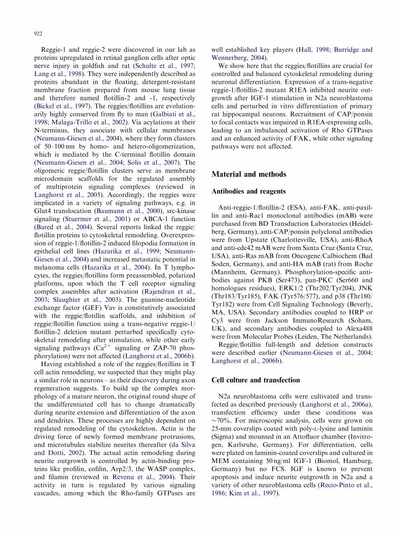
We have previously shown that expression of theisolated oligomerization domain of reggie-1/flotillin-2(named R1EA, Fig. 1A) has a trans-negative effect onreggie/flotillin function in T cells (Langhorst et al.,2006b). By interfering with the oligomerization of theendogenous proteins, R1EA impaired correct position-ing of the reggies/flotillins in T cells (Langhorst et al.,2006b). In N2a cells, expression of R1EA similarlyimpeded membrane-association of e.g. reggie-2/flotillin-1. Upon co-expression with R1EA, reggie-2/flotillin-1-EGFP was retained to a large extent in the cytosol ofN2a cells, while in cells co-expressing reggie-2/flotillin-1-EGFP and DsRed as a control, reggie-2/flotillin-1-EGFP clearly associated with the plasma membrane(Fig. 1B and C). To quantify this effect, we calculatedthe ratio between the mean fluorescence intensity of aregion of interest (ROI) at the plasma membrane overthe mean fluorescence intensity of an ROI in the cytosolas indicated in Fig. 1B and C. For control cells this ratiowas 3.770.4, while for R1EA-expressing cells, it was1.470.6 (23 and 27 cells, respectively, po0.001 Stu-dent’s t-test). Thus, as shown previously in T cells,R1EA expression severely impaired correct localizationof full-length reggies/flotillins.
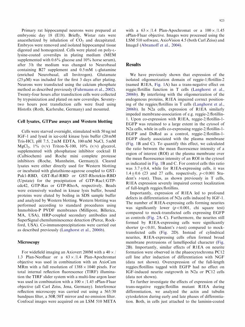
Importantly, expression of R1EA led to profounddefects in differentiation of N2a cells induced by IGF-1.The number of R1EA-expressing cells forming neuriteswas significantly lower (po0.001, chi square test)compared to mock-transfected cells expressing EGFPas controls (Fig. 2A–C). Furthermore, the neurites stillformed by R1EA-expressing cells were significantlyshorter (po0.01, Student’s t-test) compared to mock-transfected cells (Fig. 2D). Instead of cylindricalneurites, R1EA-expressing cells often formed broadmembrane protrusions of lamellipodial character (Fig.2B). Importantly, similar effects of R1EA on neuriteformation were observed in the phaeocytochroma PC12cell line after induction of differentiation with NGF(data not shown). Overexpression of the full-lengthreggies/flotillins tagged with EGFP had no effect onIGF-induced neurite outgrowth in N2a or PC12 cells(data not shown).
To further investigate the effects of expression of thetrans-negative reggie/flotillin mutant R1EA duringdifferentiation, we analyzed the actin and tubulincytoskeleton during early and late phases of differentia-tion. Both, in cells just attached to the laminin-coated
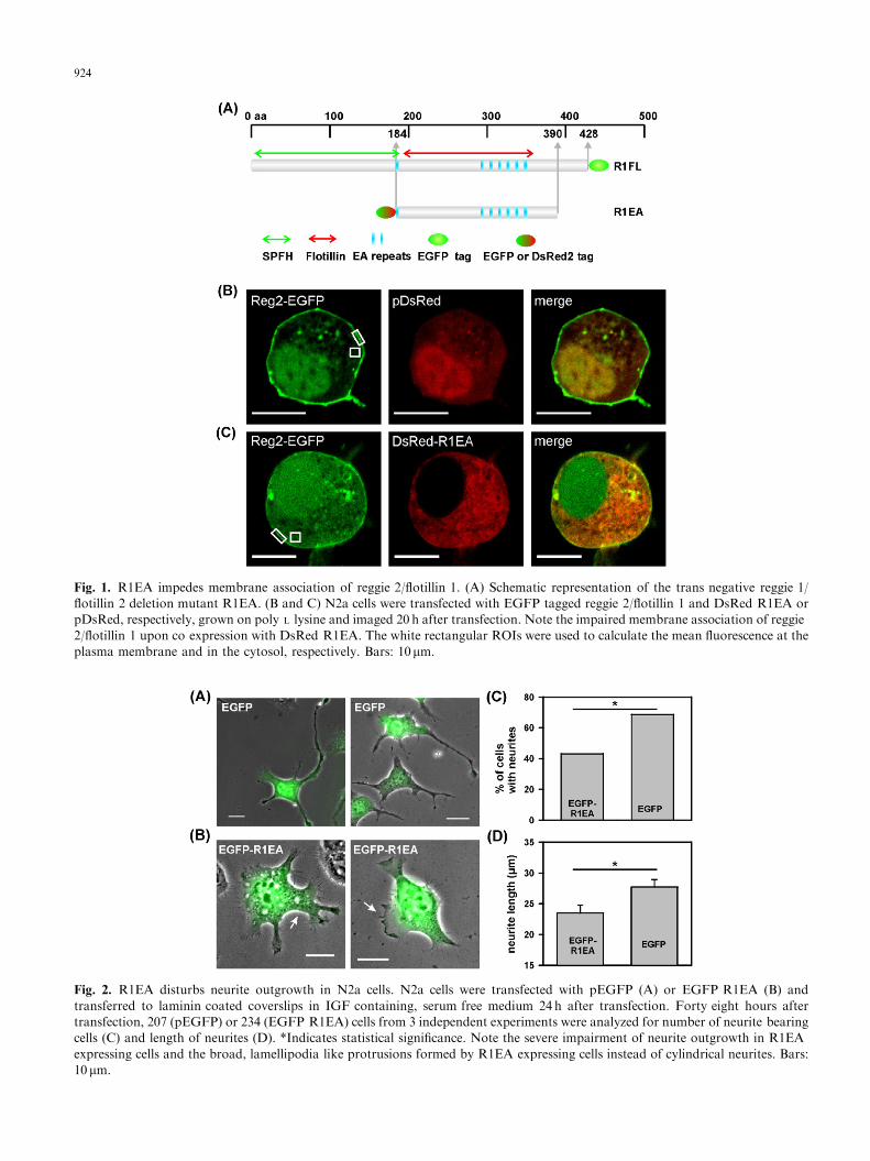
Fig. 1. R1EA impedes membrane association of reggie 2/flotillin 1. (A) Schematic representation of the trans negative reggie 1/
flotillin 2 deletion mutant R1EA. (B and C) N2a cells were transfected with EGFP tagged reggie 2/flotillin 1 and DsRed R1EA or
pDsRed, respectively, grown on poly L lysine and imaged 20 h after transfection. Note the impaired membrane association of reggie
2/flotillin 1 upon co expression with DsRed R1EA. The white rectangular ROIs were used to calculate the mean fluorescence at the
plasma membrane and in the cytosol, respectively. Bars: 10mm.
Fig. 2. R1EA disturbs neurite outgrowth in N2a cells. N2a cells were transfected with pEGFP (A) or EGFP R1EA (B) and
transferred to laminin coated coverslips in IGF containing, serum free medium 24 h after transfection. Forty eight hours after
transfection, 207 (pEGFP) or 234 (EGFP R1EA) cells from 3 independent experiments were analyzed for number of neurite bearing
cells (C) and length of neurites (D). *Indicates statistical significance. Note the severe impairment of neurite outgrowth in R1EA
expressing cells and the broad, lamellipodia like protrusions formed by R1EA expressing cells instead of cylindrical neurites. Bars:
10 mm.
924

Fig. 3. R1EA severely disturbs cytoskeletal remodeling during differentiation. N2a cells were transfected with EGFP or EGFP
R1EA and transferred to laminin coated coverslips in IGF containing, serum free medium 24h after transfection. (A) 29 h and (B)
48 h post transfection, the actin cytoskeleton was stained with phalloidin Alexa 555. A large proportion of R1EA expressing cells
exhibited numerous lamellipodia in both stages of differentiation. (C) Similarly, the tubulin cytoskeleton was stained by indirect
immunofluorescence 29 h post transfection, which was disordered in R1EA expressing cells. Bars: 10mm.
925
coverslip and starting to differentiate (Fig. 3A) and inalready largely differentiated cells (Fig. 3B), we observedthe formation of numerous lamellipodia on R1EA-expressing cells, but clearly less on control cells.Filopodia formed by R1EA-expressing cells were onaverage significantly shorter than those formed by
control cells (5.3 mm (R1EA) vs. 6.4 mm (EGFP),po0.01, 121 and 137 cells, respectively, from 5independent experiments). These results suggest thatactin remodeling during differentiation is severelyaffected by expression of the trans-negative reggie/flotillin mutant. Furthermore, the tubulin cytoskeleton
926
was altered; in many R1EA-expressing cells it appearedless organized. No stabilizing dense tubulin networkcould be found in R1EA-expressing cells, only thickbundles (Fig. 3C). The positioning and orientation ofthe microtubule-organizing centre (MTOC) was, how-ever, not changed by R1EA expression (data notshown).
To verify the physiological importance of the reggies/flotillins for neuronal differentiation, we tested whetherR1EA affects in vitro differentiation of primary rathippocampal neurons. The majority of cells fully
Fig. 4. R1EA inhibits in vitro differentiation of primary hippocampa
detailed in Materials and methods. Cells were transfected with EG
transfection and differentiated in the presence of enriched Neuroba
from three independent experiments were evaluated. Note the short
(arrowhead) of R1EA expressing cells, while control transfected cells
three categories (cells in (A) are examples of full differentiatio
differentiation, respectively). R1EA expression significantly shifted
test). Bars: 10mm.
differentiated in the presence of enriched Neurobasalwithin 48 h when transfected with pEGFP as controls(Fig. 4A and C). Cells transfected with R1EA, however,exhibited significantly impaired differentiation andstriking phenotypes (Fig. 4B and C). The majority ofR1EA-expressing cells formed only short axons, oftenwith abnormally large growth cones (Fig. 4B, arrows).The bases of dendrites were also often broadened(Fig. 4B, arrowhead). Only few R1EA-expressing cellsreached a fully differentiated morphology (Fig. 4C). Thephenotypes observed in primary neurons correlate well
l neurons. Rat primary hippocampal neurons were prepared as
FP R1EA or pEGFP, transferred to new coverslips 24 h post
sal. For quantitative analysis, 80 (EGFP) and 75 (R1EA) cells
axons, large growth cones (white arrows) and broad dendrites
differentiated normally (A and B). (C) Cells were grouped into
n, cells in (B) are examples of impaired and intermediate
the distribution towards impaired differentiation (po0.001, w2

Fig. 6. MAP kinase, PKB and PKC signaling in IGF
stimulated N2a cells expressing R1EA. N2A cells were
transfected with pEGFP and EGFP R1EA, serum deprived
overnight, and stimulated with 50 ng/ml IGF 1 for 5min.
Phosphorylation of key signaling molecules was assessed using
phospho specific antibodies. Representative blots of 5 8
independent experiments are shown. Total ERK1/2, total
PKB and total FAK were used as loading controls. FAK
phosphorylation was elevated by R1EA expression (A);
ERK1/2, JNK phosphorylation (B) and PKC and PKB
signaling (C) was not affected by R1EA.
927
with our results from neuroblastoma cells, thus con-firming the essential role of the reggies/flotillins inneuronal differentiation.
The phenotypes observed in R1EA-expressing cellssuggested a severe impairment of cytoskeletal remodel-ing caused by the expression of the trans-negativereggie-1/flotillin-2 mutant. Therefore, we next investi-gated the effects of R1EA expression on signalingpathways known to be involved in cytoskeletonremodeling. We assayed the activation of small GTPasesof the Rho family by GST-pulldown with minimalbinding domains of effector proteins specific for theGTP-bound form of the GTPase – as pioneered for Rasusing the minimal Ras-binding domain of Raf coupledto GST (de Rooij and Bos, 1997). In accordance withthe severe defects observed in neurite outgrowth, theseassays revealed a drastic alteration of the activationpatterns of the small GTPases Rac1 and cdc42.Activation of Rac1 by IGF-1 was almost completelyblocked by expression of R1EA, while in mock-transfected cells IGF-1 induced a strong activation ofRac1 (Fig. 5). In contrast, cdc42 activity was stronglyenhanced in resting cells expressing R1EA, and IGF-1stimulation of these cells induced only a small increasein cdc42-GTP levels. In control cells, nearly no cdc42-GTP could be detected in resting cells but IGF-1induced a strong increase in cdc42-GTP levels (Fig. 5).Activation of Ras was undistinguishable in R1EA-expressing cells and mock-transfected cells (Fig. 5),similarly RhoA activation was not significantly alteredin R1EA-expressing cells (data not shown). Thus,expression of the trans-negative reggie-1/flotillin-2 dele-tion mutant R1EA leads to an imbalanced activation ofsmall GTPases of the Rho family, while Ras activationis not affected.
Using phospho-specific antibodies against some keysignaling proteins we tested whether R1EA also inter-fered with activation of other signaling pathways. Thegeneral level of FAK tyrosine phosphorylation atresidues 567 and 577 was strongly enhanced in R1EA-
Fig. 5. Rho GTPase signaling after IGF stimulation in N2a cells ex
EGFP R1EA, serum deprived overnight, and stimulated with 50 n
assayed as detailed in Materials and methods. Western blots of cru
shown is representative of 4 8 independent experiments. R1EA e
activation and strongly enhanced cdc42 activation in resting cells, w
expressing cells (Fig. 6A) while the relative response toIGF was not changed (data not shown). Expression ofR1EA had no effect on the phosphorylation of the MAPkinases ERK 1/2 and JNK by IGF-1 (Fig. 6B).Phosphorylation of PKB and PKC were also notaffected by R1EA expression (Fig. 6C). siRNA-mediated knockdown of reggie-1/flotillin-2 had similareffects on signaling and morphology of neuronal cells (C.Munderloh and C.A.O. Stuermer, unpublished results).
pressing R1EA. N2A cells were transfected with pEGFP and
g/ml IGF 1 for 5min. GTP loading of small GTPases was
de lysates are shown as controls for equal loading. The result
xpression almost completely inhibited IGF stimulated Rac1
hile Ras activation was unaffected.

928
These results suggest that interference with reggie/flotillin function selectively interferes with signalingpathways to the cytoskeleton. Reggie-2/flotillin-1 isknown to interact with proteins of the vinexin family(Kimura et al., 2001), which are crucial adaptors insignaling pathways to actin remodeling (Kioka et al.,2002). The vinexin-family member CAP/ponsin isexpressed in N2a cells, predominantly in a known splicevariant of 100 kDa, while expression of vinexin-a and -bcould not be detected (data not shown). CAP/ponsinclearly co-localized both with reggie-1/flotillin-2 andreggie-2/flotillin-1 in clusters at the basal plasmamembrane of N2a cells as observed by TIRF micros-copy (Fig. 7A and B). These clusters were points of closecontact of the cell with the substrate as shown byinterference reflection microscopy (data not shown).Additionally, FAK (Fig. 7C) and paxillin (data notshown) could be found in these clusters, identifyingthem as focal adhesions. To formally prove theinteraction of CAP/ponsin with the reggies/flotillins,we carried out co-immunoprecipitation experiments ofendogenous CAP/ponsin and the endogenous reggies/flotillins. After immunoprecipitation of CAP/ponsin,reggie-1/flotillin-2 could be reliably detected in theprecipitate (Fig. 7D), thus proving that not onlyreggie-2/flotillin-1 but also reggie-1/flotillin-2 can inter-act with CAP/ponsin.
Most interestingly, expression of the trans-negativereggie-1/flotillin-2 mutant R1EA significantly inhibitedthe recruitment of CAP/ponsin to the plasma mem-brane. CAP/ponsin staining of the basal plasmamembrane as observed by TIRF microscopy was lowand largely diffuse in R1EA-expressing cells instead ofclustered in focal adhesions as in control cells (Fig. 8A).The clustering of paxillin (Fig. 8B) and FAK (data notshown), however, was not affected by R1EA expression.R1EA expression did not alter the expression level ofCAP/ponsin (Fig. 8C). This suggests that reggie/flotillinlocalization to focal contacts is essential for therecruitment of CAP/ponsin, but not for focal contactassembly itself.
Discussion
The results of this study can be summarized asfollows: In neurons, the reggies/flotillins are necessaryfor the recruitment of vinexin-family adaptor proteins tothe plasma membrane and especially to focal contacts.Failure of membrane recruitment, as in cells expressinga trans-negative reggie-1/flotillin-2 mutant, leads toenhanced FAK activity and an imbalanced activity ofRho-family GTPases. This strongly impairs cytoskeletalremodeling and consequently neurite outgrowth both inneuroblastoma cells and in primary rat hippocampal
neurons, while other signaling pathways, such as PKBor ERK1/2 signaling are unaffected. In most of theseexperiments, we have worked with cells early indifferentiation, but we strongly believe that the resultsobtained can be extrapolated to later stages of neuriteoutgrowth and axon extension as shown by our resultsdemonstrating impaired differentiation of hippocampalneurons.
The vinexin-family adaptor proteins vinexin, CAP/ponsin and ArgBP2 act as integrators of growth factorsignaling and signaling from cell-substrate contact sites,like focal adhesions (Kioka et al., 2002; Suwa et al.,2002; Mitsushima et al., 2004). CAP/ponsin was shownto localize to focal adhesions of 3T3 fibroblasts, where itinteracts with FAK (Ribon et al., 1998). Overexpressionof CAP/ponsin in these cells led to decreased tyrosinephosphorylation of FAK (Ribon et al., 1998). This is ingood agreement with our data showing an increasedtyrosine phosphorylation of FAK after loss of CAP/ponsin membrane recruitment. Interestingly, FAK is akey integrator of signaling during neurite outgrowth:FAK activity not only integrates integrin and growthfactor signaling, but also signaling from guidance cueslike ephrins or netrins (Davy and Robbins, 2000; Renet al., 2004).
In addition to its role in regulating FAK activity, ourresults show that reggie/flotillin-mediated recruitment ofCAP/ponsin to focal contacts is crucial for the balancedactivation of the Rho-family GTPases. CAP/ponsin isknown to interact directly with Sos and indirectly withother GEFs via the adaptor protein Grb4 (Kioka et al.,2002), which makes it a key adaptor and regulatorcontrolling a variety of GTPases. The regulated inter-action of different Rho GTPases is critical in neuriteoutgrowth. As mentioned above, a variety of studiessuggests that RhoA activity generally inhibits, whilecdc42 and Rac activity generally enhances neuriteoutgrowth. But recent studies suggested that the picturemight be more complex. Both constitutive-active anddominant-negative Rac1 mutants inhibited neurite out-growth in chick embryo motor neurons (Kuhn et al.,1998). Furthermore, live imaging of Rho GTPaseactivation in neurons using FRET reporter constructsshowed that all three classical Rho GTPases, RhoA,Rac1 and cdc42, undergo repetitive activation andinactivation cycles at motile protrusions, with differentlocalizations of their respective peak activity (Aoki etal., 2004; Kurokawa et al., 2005; Nakamura et al., 2005).Thus, a finely tuned interplay of different Rho GTPasesis apparently necessary for successful neurite outgrowth.The Rho GTPases are not only necessary for the actin-dependent formation of the initial membrane protru-sions, but also for the control of microtubuli dynamicsduring stabilization of the outgrowing neurite (da Silvaand Dotti, 2002). Thus, the broad, lamellipodia-likeprotrusions formed by R1EA-expressing cells reflect
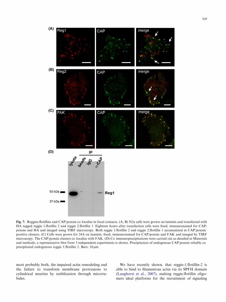
Fig. 7. Reggies/flotillins and CAP/ponsin co localize in focal contacts. (A, B) N2a cells were grown on laminin and transfected with
HA tagged reggie 1/flotillin 2 and reggie 2/flotillin 1. Eighteen hours after transfection cells were fixed, immunostained for CAP/
ponsin and HA and imaged using TIRF microscopy. Both reggie 1/flotillin 2 and reggie 2/flotillin 1 accumulated in CAP/ponsin
positive clusters. (C) Cells were grown for 24 h on laminin, fixed, immunostained for CAP/ponsin and FAK and imaged by TIRF
microscopy. The CAP/ponsin clusters co localize with FAK. (D) Co immunoprecipitations were carried out as detailed in Materials
and methods, a representative blot from 3 independent experiments is shown. Precipitation of endogenous CAP/ponsin reliably co
precipitated endogenous reggie 1/flotillin 2. Bars: 10mm.
929
most probably both, the impaired actin remodeling andthe failure to transform membrane protrusions tocylindrical neurites by stabilization through microtu-bules.
We have recently shown, that reggie-1/flotillin-2 isable to bind to filamentous actin via its SPFH domain(Langhorst et al., 2007), making reggie/flotillin oligo-mers ideal platforms for the recruitment of signaling

Fig. 8. R1EA inhibits CAP/ponsin recruitment to the plasma
membrane. (A, B) Cells were transfected with pDsRed or
DsRed R1EA, transferred to laminin coated coverslips 24 h
post transfection, fixed and stained for CAP (A) and paxillin
(B) 48 h post transfection. R1EA expression inhibited CAP/
ponsin clustering at the plasma membrane. (C) R1EA
expression did not lead to a change in CAP/ponsin protein
levels as judged by Western blotting. Bars: 10 mm.
930
complexes regulating cytoskeletal dynamics. Scaffoldingproteins contribute to the specificity of GTPase signal-ing by bringing the GTPases, their regulators andspecific effectors in close proximity. The involvementof reggie/flotillin scaffolds in GTPase signaling, asshown in this study for neurons, was also suggested bystudies in two other cell types. Upon insulin stimulationof adipocytes, reggie-2/flotillin-1 was shown to recruit amultiprotein complex to the membrane, which finallyresulted in the activation of the small GTPase TC10 viaCAP/ponsin and c-Cbl-mediated recruitment of CrkIII-C3G (Baumann et al., 2000). We have previously shownthat in T lymphocytes, reggie-1/flotillin-2-dependentpositioning of the GEF Vav is essential for actinremodeling after activation, while other early signalingpathways were unaffected (Langhorst et al., 2006b).Thus, it is well conceivable that regulating the assemblyof Rho GTPase signaling complexes and thus cytoskel-eton remodeling is a general function of reggie/flotillinmicrodomains in many different cell types.
Acknowledgments
This work was supported by grants from the DeutscheForschungsgemeinschaft DFG (SFB-TR11), the Minis-terium Forschung, Wissenschaft und Kunst Baden-Wurttemberg (TSE program) and the Fonds derChemischen Industrie.
References
Abramoff, M.D., Magelhaes, P.J., Ram, S.J., 2004. Image
processing with imageJ. Biophoton. Int. 11, 36 42.
Aoki, K., Nakamura, T., Matsuda, M., 2004. Spatio temporal
regulation of Rac1 and Cdc42 activity during nerve growth
factor induced neurite outgrowth in PC12 cells. J. Biol.
Chem. 279, 713 719.
Bared, S.M., Buechler, C., Boettcher, A., Dayoub, R.,
Sigruener, A., Grandl, M., Rudolph, C., Dada, A.,
Schmitz, G., 2004. Association of ABCA1 with syntaxin
13 and flotillin 1 and enhanced phagocytosis in tangier
cells. Mol. Biol. Cell 15, 5399 5407.
Baumann, C.A., Ribon, V., Kanzaki, M., Thurmond, D.C.,
Mora, S., Shigematsu, S., Bickel, P.E., Pessin, J.E., Saltiel,
A.R., 2000. CAP defines a second signalling pathway
required for insulin stimulated glucose transport. Nature
407, 202 207.
Bickel, P.E., Scherer, P.E., Schnitzer, J.E., Oh, P., Lisanti,
M.P., Lodish, H.F., 1997. Flotillin and epidermal surface
antigen define a new family of caveolae associated integral
membrane proteins. J. Biol. Chem. 272, 13793 13802.
Burridge, K., Wennerberg, K., 2004. Rho and Rac take center
stage. Cell 116, 167 179.
Caroni, P., Schwab, M.E., 1993. Oligodendrocyte and myelin
associated inhibitors of neurite growth in the adult nervous
system. Adv. Neurol. 61, 175 179.
da Silva, J.S., Dotti, C.G., 2002. Breaking the neuronal sphere:
regulation of the actin cytoskeleton in neuritogenesis. Nat.
Rev. Neurosci. 3, 694 704.
Davy, A., Robbins, S.M., 2000. Ephrin A5 modulates cell
adhesion and morphology in an integrin dependent man
ner. EMBO J. 19, 5396 5405.
de Rooij, J., Bos, J.L., 1997. Minimal Ras binding domain of
Raf1 can be used as an activation specific probe for Ras.
Oncogene 14, 623 625.
Fawcett, J.W., 1992. Intrinsic neuronal determinants of
regeneration. Trends Neurosci. 15, 5 8.
Fuhrmann, J.C., Kins, S., Rostaing, P., El Far, O., Kirsch, J.,
Sheng, M., Triller, A., Betz, H., Kneussel, M., 2002.
Gephyrin interacts with dynein light chains 1 and 2,
components of motor protein complexes. J. Neurosci. 22,
5393 5402.
Galbiati, F., Volonte, D., Goltz, J.S., Steele, Z., Sen, J.,
Jurcsak, J., Stein, D., Stevens, L., Lisanti, M.P., 1998.
Identification, sequence and developmental expression of
invertebrate flotillins from drosophila melanogaster. Gene
210, 229 237.
Hall, A., 1998. Rho GTPases and the actin cytoskeleton.
Science 279, 509 514.
931
Hazarika, P., Dham, N., Patel, P., Cho, M., Weidner, D.,
Goldsmith, L., Duvic, M., 1999. Flotillin 2 is distinct from
epidermal surface antigen (ESA) and is associated with
filopodia formation. J. Cell. Biochem. 75, 147 159.
Hazarika, P., McCarty, M.F., Prieto, V.G., George, S., Babu,
D., Koul, D., Bar Eli, M., Duvic, M., 2004. Up regulation
of flotillin 2 is associated with melanoma progression and
modulates expression of the thrombin receptor protease
activated receptor 1. Cancer Res. 64, 7361 7369.
Kim, B., Leventhal, P.S., Saltiel, A.R., Feldman, E.L., 1997.
Insulin like growth factor I mediated neurite outgrowth in
vitro requires mitogen activated protein kinase activation.
J. Biol. Chem. 272, 21268 21273.
Kimura, A., Baumann, C.A., Chiang, S.H., Saltiel, A.R., 2001.
The sorbin homology domain: a motif for the targeting of
proteins to lipid rafts. Proc. Natl. Acad. Sci. USA 98,
9098 9103.
Kioka, N., Ueda, K., Amachi, T., 2002. Vinexin, CAP/ponsin,
ArgBP2: a novel adaptor protein family regulating cyto
skeletal organization and signal transduction. Cell Struct.
Funct. 27, 1 7.
Kuhn, T.B., Brown, M.D., Bamburg, J.R., 1998. Rac1
dependent actin filament organization in growth cones is
necessary for beta1 integrin mediated advance but not for
growth on poly D lysine. J. Neurobiol. 37, 524 540.
Kurokawa, K., Nakamura, T., Aoki, K., Matsuda, M., 2005.
Mechanism and role of localized activation of Rho family
GTPases in growth factor stimulated fibroblasts and
neuronal cells. Biochem. Soc. Trans. 33, 631 634.
Lang, D.M., Lommel, S., Jung, M., Ankerhold, R., Petrausch,
B., Laessing, U., Wiechers, M.F., Plattner, H., Stuermer,
C.A., 1998. Identification of reggie 1 and reggie 2 as
plasmamembrane associated proteins which cocluster with
activated GPI anchored cell adhesion molecules in non
caveolar micropatches in neurons. J. Neurobiol. 37,
502 523.
Langhorst, M.F., Reuter, A., Stuermer, C.A., 2005. Scaffold
ing microdomains and beyond: the function of reggie/
flotillin proteins. Cell. Mol. Life Sci. 62, 2228 2240.
Langhorst, M.F., Genisyuerek, S., Stuermer, C.A., 2006a.
Accumulation of FlAsH/Lumio green in active mitochon
dria can be reversed by beta mercaptoethanol for specific
staining of tetracysteine tagged proteins. Histochem. Cell
Biol. 125, 743 747.
Langhorst, M.F., Reuter, A., Luxenhofer, G., Boneberg,
E.M., Legler, D.F., Plattner, H., Stuermer, C.A., 2006b.
Preformed reggie/flotillin caps: stable priming platforms
for macrodomain assembly in T cells. FASEB J. 20,
711 713.
Langhorst, M.F., Solis, G.P., Hannbeck, S., Plattner, H.,
Stuermer, C.A., 2007. Linking membrane microdomains to
the cytoskeleton: regulation of the lateral mobility of
reggie 1/flotillin 2 by interaction with actin. FEBS Lett.
581, 4697 4703.
Malaga Trillo, E., Laessing, U., Lang, D.M., Meyer, A.,
Stuermer, C.A., 2002. Evolution of duplicated reggie genes
in zebrafish and goldfish. J. Mol. Evol. 54, 235 245.
Mitsushima, M., Suwa, A., Amachi, T., Ueda, K., Kioka, N.,
2004. Extracellular signal regulated kinase activated by
epidermal growth factor and cell adhesion interacts with
and phosphorylates vinexin. J. Biol. Chem. 279,
34570 34577.
Nakamura, T., Aoki, K., Matsuda, M., 2005. FRET imaging
in nerve growth cones reveals a high level of RhoA activity
within the peripheral domain. Brain Res. Mol. Brain Res.
139, 277 287.
Neumann Giesen, C., Falkenbach, B., Beicht, P., Claasen, S.,
Luers, G., Stuermer, C.A., Herzog, V., Tikkanen, R., 2004.
Membrane and raft association of reggie 1/flotillin 2: role
of myristoylation, palmitoylation and oligomerization and
induction of filopodia by overexpression. Biochem. J. 378,
509 518.
Rajendran, L., Masilamani, M., Solomon, S., Tikkanen, R.,
Stuermer, C.A., Plattner, H., Illges, H., 2003. Asymmetric
localization of flotillins/reggies in preassembled platforms
confers inherent polarity to hematopoietic cells. Proc. Natl.
Acad. Sci. USA 100, 8241 8246.
Recio Pinto, E., Rechler, M.M., Ishii, D.N., 1986. Effects of
insulin, insulin like growth factor II, and nerve growth
factor on neurite formation and survival in cultured
sympathetic and sensory neurons. J. Neurosci. 6,
1211 1219.
Ren, X.R., Ming, G.L., Xie, Y., Hong, Y., Sun, D.M., Zhao,
Z.Q., Feng, Z., Wang, Q., Shim, S., Chen, Z.F., Song, H.J.,
Mei, L., Xiong, W.C., 2004. Focal adhesion kinase in
netrin 1 signaling. Nat. Neurosci. 7, 1204 1212.
Revenu, C., Athman, R., Robine, S., Louvard, D., 2004. The
co workers of actin filaments: from cell structures to
signals. Nat. Rev. Mol. Cell Biol. 5, 635 646.
Ribon, V., Herrera, R., Kay, B.K., Saltiel, A.R., 1998. A role
for CAP, a novel, multifunctional Src homology 3 domain
containing protein in formation of actin stress fibers and
focal adhesions. J. Biol. Chem. 273, 4073 4080.
Schulte, T., Paschke, K.A., Laessing, U., Lottspeich, F.,
Stuermer, C.A., 1997. Reggie 1 and reggie 2, two cell
surface proteins expressed by retinal ganglion cells during
axon regeneration. Development 124, 577 587.
Slaughter, N., Laux, I., Tu, X., Whitelegge, J., Zhu, X., Effros,
R., Bickel, P., Nel, A., 2003. The flotillins are integral
membrane proteins in lipid rafts that contain TCR
associated signaling components: implications for T cell
activation. Clin. Immunol. 108, 138 151.
Solis, G.P., Hoegg, M., Munderloh, C., Schrock, Y., Malaga
Trillo, E., Rivera Milla, E., Stuermer, C.A., 2007. Reggie/
flotillin proteins are organized into stable tetramers in
membrane microdomains. Biochem. J. 403, 313 322.
Stuermer, C.A., Bastmeyer, M., Bahr, M., Strobel, G.,
Paschke, K., 1992. Trying to understand axonal regenera
tion in the CNS of fish. J. Neurobiol. 23, 537 550.
Stuermer, C.A., Lang, D.M., Kirsch, F., Wiechers, M.,
Deininger, S.O., Plattner, H., 2001. Glycosylphosphatidyl
inositol anchored proteins and fyn kinase assemble in
noncaveolar plasma membrane microdomains defined by
reggie 1 and 2. Mol. Biol. Cell 12, 3031 3045.
Suwa, A., Mitsushima, M., Ito, T., Akamatsu, M., Ueda, K.,
Amachi, T., Kioka, N., 2002. Vinexin beta regulates the
anchorage dependence of ERK2 activation stimulated by
epidermal growth factor. J. Biol. Chem. 277, 13053 13058.