reconstruction of devital teeth using direct fiber ... · reconstruction of devital teeth using...
TRANSCRIPT
Vol 7, No 2, 2005 1
Reconstruction of Devital Teeth Using Direct Fiber-reinforced Composite Resins: A Case Report
Simone Deliperia/David N. Bardwellb/Carlo Coianac
a Visiting Instructor and Research Associate, Postgraduate Esthetic Dentistry,Tufts University School of Dental Medicine, Boston, MA, USA; Dentist inprivate practice, Cagliari, Italy.
b Associate Clinical Professor of Restorative Dentistry; Director, PostgraduateEsthetic Dentistry, Tufts University School of Dental Medicine, Boston, MA,USA.
c Periodontist in private practice, Cagliari, Italy.
Summary: Nonrestored devitalized teeth are structurally compromised and represent one of the greatest challeng-es for the clinician. Restoration of endodontically treated teeth has been associated with the use of posts. Variouspost materials and designs have been introduced over the years; however, motivated by the desire to conserve theremaining sound tooth structure and thanks to properties of modern adhesive systems, clinicians have re-evaluat-ed the dogma of traditional restorative dentistry and seek alternative methods to build up devitalized teeth. Theuse of direct Ultra High Molecular Weight Polyethylene (UHMWPE) fiber-reinforced post systems is becoming popu-lar among clinicians because enlargement of the root canal space is not required and the risk of root perforationeliminated. This article presents an experimental clinical technique to reconstruct severely damaged endodonticallytreated posterior teeth using direct fiber reinforced post systems. Particular attention is paid to the incrementaland curing techniques adopted to build up the restoration. The problems that clinicians can encounter in bondingto teeth that have undergone endodontic treatment are also analyzed. Questions that have yet to be answered byscientific research are presented.
Key words: adhesive system, composite resin, Class II restoration, devital teeth, post and core.
J Adhes Dent 2005; 7: xx–xx. Submitted for publication: 04.03.04; accepted for publication: 16.08.04.
Reprint requests: Dr. Simone Deliperi, Via G. Baccelli,10/b, 09126 Cagliari,Italy. Tel:+39-347-953-3259, Fax: +39-070-497293. e-mail: [email protected]
arlier than 15 years ago, the restoration of devitalteeth was automatically associated with a combina-
tion of prefabricated or custom-made metallic post andcores and full crowns.38 In this way, a considerableamount of coronal and radicular sound tooth structurewas sacrificed with increasing risk of root perforation orfracture.16,41 Moreover, patients had to accept treat-ment which was costly in terms of both time andmoney.43 The continuous development of total-etch ad-hesive systems45,47-49 and the improvement of physicaland mechanical properties of resin bonded composite(RBC)22,23 were responsible for a complete revolution inrestorative dentistry. Adhesive restorations allow clini-cians to create minimally invasive preparations, thus
E conserving sound tooth structure. The increased predict-ability of RBC has encouraged clinicians to progressivelyabandon amalgam.3 Patient demands for esthetic resto-rations and their increasing desire to save remainingsound tooth structure are pushing dentists to stretchclinical indications for direct RBC restorations.6,28 Thissituation may be further influenced by patients’ inabilityto afford the ideal indirect restoration in large posteriorand anterior defects. As a consequence, clinical indica-tions for anterior and posterior RBC restorations are pro-gressively expanding; practitioners are looking for newmaterial and techniques to further enhance the clinicalperformance of direct RBC when placed in severelydestroyed vital or nonvital teeth.6,28
Tooth-colored fiber posts were introduced in the 1990sand have several advantages over conventional metalposts.35 They are esthetic, bond to tooth structure, havea modulus of elasticity similar to that of dentin but stillrequire dentin preparation to fit into the canal.
Lately, fiber reinforcement systems have been intro-duced in the attempt to increase RBC durability anddamage tolerance.12,19,46 Ultra High Molecular WeightPolyethylene (UHMWPE) fiber reinforcement systems aregaining popularity and have various clinical applications(Table 1). Being bondable reinforcement fibers, they canbe used to build up endodontic post and cores, as theyadapt to the root canal walls without requiring additional
Deliperi.fm Seite 1 Freitag, 29. April 2005 9:50 09

Deliperi et al
2 The Journal of Adhesive Dentistry
enlargement of the root canal after endodontic treatment.These woven fibers have a modulus of elasticity similar tothat of dentin and are supposed to create a monoblockdentin-post-core system able to better distribute forcesalong the root.32 Recently, improved UHMWPE fiber sys-tems have been introduced onto the market. They havegreater strength properties than conventional UHMWPEfibers because of unidirectional fibers with braided fila-ments inserted between them.26
Increased interest and attention have also been devot-ed to contemporary light-curing techniques as a possiblemethod for controlling stress.7,13,18,25,44 The advantagesof using a combination of pulse and progressive curingtechniques to counteract polymerization shrinkage havebeen previously reported.6,8 However, clinicians mayprefer using a conventional curing method to save time,although this may increase the possibility of RBC postop-erative sensitivity and reduced longevity. This trend isalso influenced by manufacturers resisting the use ofalternative modes of polymerization in their light-curingsystems. Clinicians cannot program their curing lights formode of polymerization and may be constrained by curingintensity and time provided by manufacturers.
The purpose of this article is to provide a simplifiedclinical approach to reconstruct severely damaged endo-dontically treated posterior teeth and discuss the bene-fits and problematic issues related to a similar proce-dure.
CASE REPORT
Case PresentationA 46-year-old female patient presented with a previouslyendodontically treated right maxillary premolar (15),which had been restored with a MOD amalgam restora-tion and suffered a fracture of the palatal cusp after a fewyears of clinical service (Fig 1). As the fracture line wasbelow the CEJ, invading the biological width, it wasexplained to the patient that the placement of a dentalimplant could be indicated if an unfavorable crown-to-rootratio would result after completing crown lengthening.The patient expressed the desire to maintain tooth 15and restore it with a direct RBC restoration, due to the
lower cost compared to an indirect restoration. If a rootfracture occurred, then a dental implant would be placed.The treatment plan was accepted and an informed con-sent was secured.
Restorative ProcedureOnce crown lengthening was completed and tissue heal-ing was accomplished, rubber-dam was placed in order tocomplete isolation and expose the cavosurface gingivalmargins. The existing amalgam restoration was complete-ly removed, being careful to preserve as much toothstructure as possible. Cavity preparation was completedby placing a gingival butt joint with no bevel on the axialor occlusal surface, rounding sharp angles with #2 and#4 burs (Brasseler, Savannah, GA, USA), and trying toselectively remove the superficial sclerotic dentin (Fig 2).Three to 4 mm of gutta-percha were removed from thefacial and palatal canals to expose dentin and increasemicroretention when using enamel dentin adhesive sys-tems (Fig 3). A circular matrix (Automatrix, Dentsply/Caulk, Milford, DE, USA) was placed around the tooth andtightened. Interproximal matrix adaptation was securedusing wooden wedges. Enamel and dentin were etchedfor 30 s using 34% phosphoric acid (Tooth ConditionerGel, Dentsply/Caulk) (Fig 4). The etchant was removedand the cavity was water sprayed for 30 s, being carefulto maintain a moist surface. A fifth generation nanofilledacetone-based adhesive system (Prime & Bond NT,Dentsply/Caulk) was placed in the preparation, gently airthinned to evaporate solvent (Fig 5), and light cured for20 s at 800 mW/cm2 from the occlusal surface using aQuartz Tungsten Halogen curing light (Spectrum 800,Dentsply/Caulk).
In order to avoid microcrack formation on the remain-ing facial wall, the authors used a previously describedtechnique,6,7 which is based on a combination of pulseand progressive curing technique. A particular compositeplacement technique was also selected to build up sucha challenging restoration; a combination of RBC wedgeshaped increments and UHMWPE fiber reinforcement sys-tem was considered of paramount importance to furtherreduce polymerization shrinkage, better support the RBC,reinforce the remaining tooth structure, and reduce thetotal composite volume.
Table 1 Clinical indications of bondable reinforcement ribbon fibers
Clinical indications
Endodontic post and corePeriodontal splintingOrthodontic retainersProvisional metal-free bridgesTreatment of split-tooth syndrome
Fig 1 Preoperative view of tooth 15 with fracture of the palatal wall.
Deliperi.fm Seite 2 Freitag, 29. April 2005 9:50 09
Deliperi et al
Vol 7, No 2, 2005 3
Esthet-X microhybrid RBC (Dentsply/Caulk) was con-sidered the material of choice to restore tooth 15because of its variety of enamel shades and excellentmechanical properties. However, different microhybridcomposites based on the natural layering technique10
may also be utilized (eg, Point 4, Kerr, Orange, CA; Vit-l-es-cence and Amelogen, Ultradent Products, South Jordan,UT, USA). The matrix was burnished against the adjacentteeth. Tooth buildup was started using 2-mm triangu-lar-shaped (wedge-shaped) gingivo-occlusally placed lay-ers of amber (AE) and clear (CE) enamel shades to recon-struct the proximal and palatal surfaces. This uncuredcomposite was condensed and sculptured against thecavosurface margins and circular matrix, and each incre-ment was pulse cured for 3 s at 300 mW/cm2 to avoid mi-crocrack formation. Final polymerization of the AE and CEproximal and palatal composite walls was then completedat 300 mW/cm2 for 40 s (Fig 6). The enamel peripheralskeleton of the restoration was built up, yielding addition-al spatial references to create a correct occlusal anatomy.An increased C-factor resulted as a consequence of thislayering technique. The C-factor was defined as the ratiobetween bonded and unbonded surfaces; increasing thisratio resulted in increased polymerization stresses.14 Inthis context, the application of wedge-shaped increments
of composite resin was of paramount importance, be-cause it helped in decreasing the C-factor ratio. Dentinstratification of the facial, palatal, and proximal walls wasinitiated placing 2-mm wedge-shaped increments of A3RBC at each enamel wall, avoiding any contact of freshincrements (Fig 7). Meanwhile, a UHMWPE triaxial fiber(Ribbond Triaxial, Ribbond, Seattle, WA, USA) was select-ed, and the dental assistant manipulated it according tomanufacturer’s instructions. Successive A3 incrementswere placed until a central area resulted for placement ofa resin-impregnated fiber composite system and fabrica-tion of a direct post and core. Each dentin increment wascured using a progressive curing technique (40 s at300 mW/cm2 instead of a conventional continuous irradi-ation mode of 20 s at 600 mW/cm2).6,8 Triaxial fiberswere wetted with an unfilled resin (D/E resin, Bisco,Schaumburg, IL, USA) (Fig 8), excess resin was removed,and fibers were completely covered with a B1 light-curedflowable composite resin (X-Flow, Dentsply/Caulk) andplaced in the central area of the restoration. UHMWPEtriaxial fibers were folded and each end pushed in the twocanals using a thin composite spatula. B1 flowable com-posite resin was used to fill any composite void, then apolymerization cycle of both the fiber-resin complex andcomposite resin was started. The polymerization process
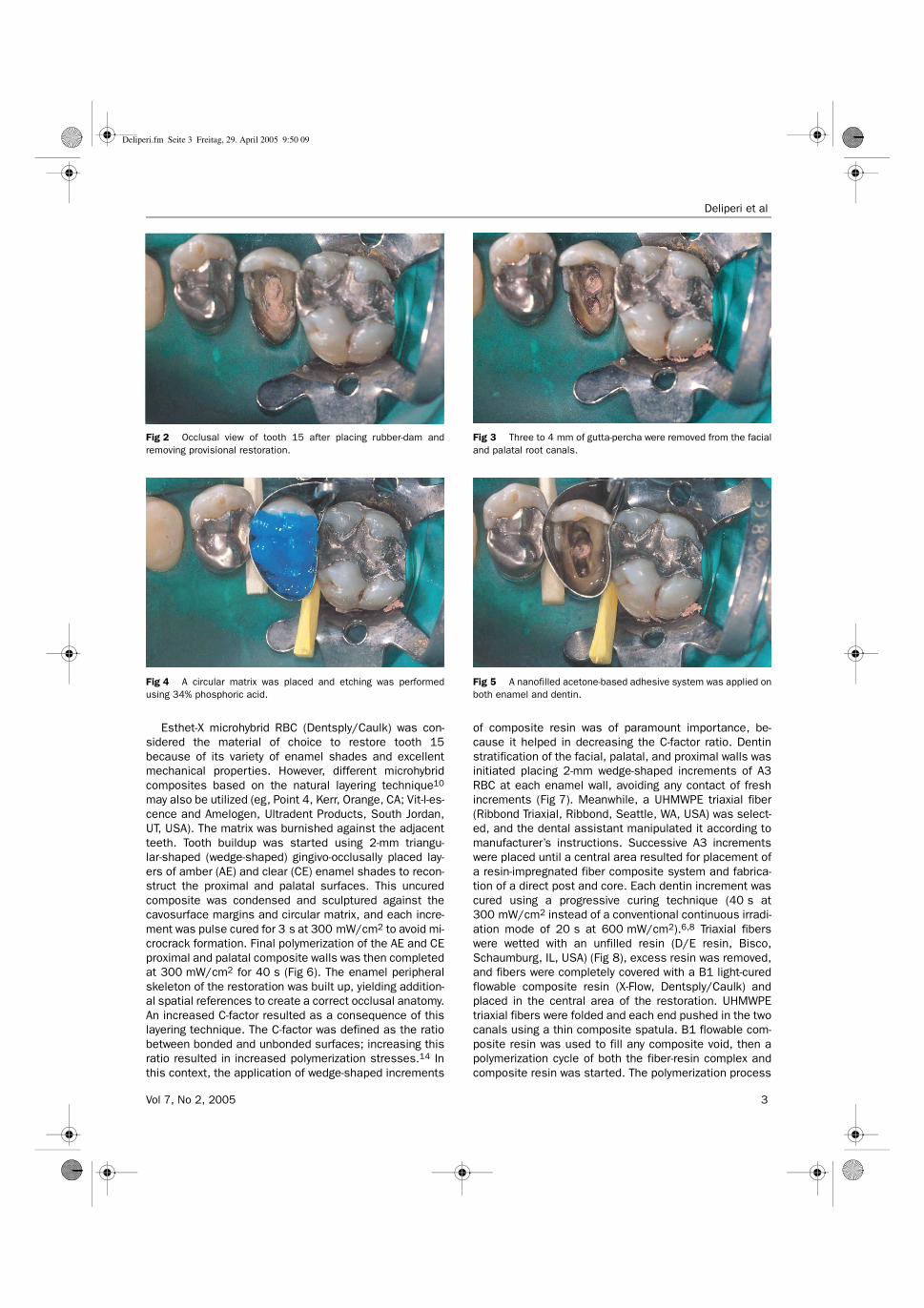
Fig 2 Occlusal view of tooth 15 after placing rubber-dam andremoving provisional restoration.
Fig 3 Three to 4 mm of gutta-percha were removed from the facialand palatal root canals.
Fig 4 A circular matrix was placed and etching was performedusing 34% phosphoric acid.
Fig 5 A nanofilled acetone-based adhesive system was applied onboth enamel and dentin.
Deliperi.fm Seite 3 Freitag, 29. April 2005 9:50 09
Deliperi et al
4 The Journal of Adhesive Dentistry
included an initial polymerization using a progressive cur-ing technique (40 s at 300 mW/cm2) followed by a con-tinuous irradiation at 800 mW/cm2 for 240 s to assurecomplete polymerization of the fiber-complex down intothe canal. At this point, the middle third of the dentin res-toration was built up using a combination of B5/DY andB3 to increase the chroma, unnaturally reduced by previ-
ously using B1 flowable composite (Fig 9). Enamel layersof CE were applied to the final contour of the occlusal sur-face according to a successive cusp buildup technique(Fig 10). This final layer was pulse cured for 3 s at300 mW/cm2. A waiting period of 3 min was observed toallow for stress relief before polymerizing at a higherintensity (30 s at 800 mW/cm2) (Table 2).
Subsequently, wedges and matrix were removed andthe final polymerization cycle was completed by irradiat-ing the restored tooth through the facial and palatalsurface for 30 s each at 800 mW/cm2.
Rubber-dam was removed, occlusion checked and therestoration was finished using the Raptor system (Bisco).Final polishing was performed using a one-step diamondmicropolisher system (Pogo, Dentsply/Caulk) (Figs 11and 12). The same restoration was evaluated at a 2-yearrecall (Fig 13).
DISCUSSION
Cuspal coverage is considered fundamental to avoid cuspfracture in endodontically treated teeth.39,40 However, arecent in vitro study reported no difference in cuspal frac-ture when large inlay and onlay restorations were complet-
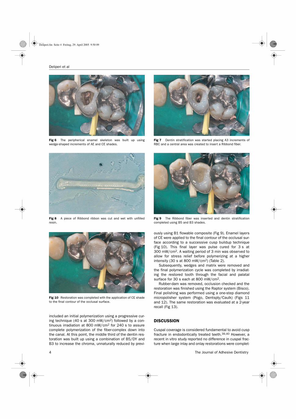
Fig 6 The peripherical enamel skeleton was built up usingwedge-shaped increments of AE and CE shades.
Fig 7 Dentin stratification was started placing A3 increments ofRBC and a central area was created to insert a Ribbond fiber.
Fig 8 A piece of Ribbond ribbon was cut and wet with unfilledresin.
Fig 9 The Ribbond fiber was inserted and dentin stratificationcompleted using B5 and B3 shades.
Fig 10 Restoration was completed with the application of CE shadeto the final contour of the occlusal surface.
Deliperi.fm Seite 4 Freitag, 29. April 2005 9:50 09
Deliperi et al
Vol 7, No 2, 2005 5
ed on devital teeth, even though the restored cavity hada remaining buccal and lingual wall that was very thin.27
Although we performed a direct inlay-onlay RBC restora-tion, results may be the consequence of increased reli-ability of dentin adhesion in the last decade.45,47-49 Itwould be interesting to compare indirect and directinlay-onlay restorations to evaluate any differences in therate of cuspal fracture. Some laboratory studies havedemonstrated that modern adhesive systems in combina-tion with RBC can further reinforce remaining tooth struc-ture.2,21 It has yet to be explained to what extent this canoccur for both direct and indirect resin restorations, andfurther biomechanical studies are required. Certainly,adhesive systems have been researched intensively andimprovement has been dramatic, possibly superior tothose for composite resin. Lately, the mechanism ofbonding to normal and sclerotic, coronal and radiculardentin has been researched, allowing clinicians to per-form more predictable composite resin restorations withquartz and carbon fiber post-supported buildups.15,30,50
Conversely, the mechanism of bonding to endodonticallytreated teeth needs to be further researched. The poten-tial benefits of adhesive dentistry in this field have not
been fully explored.5 Devital dentin is deprived of its od-ontoblastic process and collagen layer, so a differenthybrid layer is created due to absence of resin-impregnat-ed collagen fibrils. The influence of endodontic sealers incontact with root dentin should be evaluated, as well asthe interference of conventional irrigants (sodium hypo-chlorite and hydrogen peroxide) on dentin permeability
Table 2 Recommended photocuring times and intensities for enamel, dentin, and post-and-core buildup
Buildup Composite shade(Esthet-X)
Polymerizationtechnique
Intensity(mW/cm2)
Time(s)
Proximal and palatal enamel AE-CE pulse 300+300
3+40
Dentin A3-B3-B5 progressive curing 300 40
Ribbond post-and-core buildup B1 progressive curing+continuous curing
300+800
40+240
Occlusal enamel AE-CE pulse 300+800
3+30
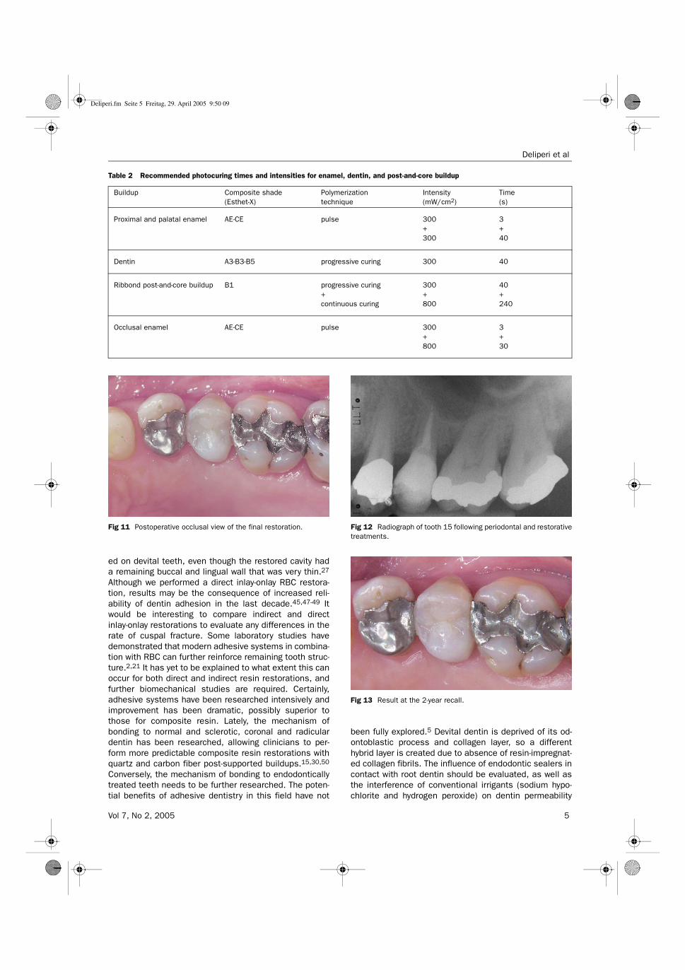
Fig 11 Postoperative occlusal view of the final restoration. Fig 12 Radiograph of tooth 15 following periodontal and restorativetreatments.
Fig 13 Result at the 2-year recall.
Deliperi.fm Seite 5 Freitag, 29. April 2005 9:50 09

Deliperi et al
6 The Journal of Adhesive Dentistry
and bond strength of current adhesive systems.24,33,36
Both biomechanical and ultrastructural studies are alsorequired to better explain the behavior of adhesive sys-tems on this particular substrate.
This case presents an example of resin-compositelongevity in the posterior region. At the 2-year recall, nomarginal discoloration, recurrent decay, chipping, or com-posite clefts were detected. Although the observationtime was limited to only 2 years and just one case reportwas considered, the clinical performance of Esthet-Xmicrohybrid composite was more than acceptable. Eventhough the clinical technique applied still has an experi-mental character, the clinical performance of UHMWPfibers placed on devital premolars and molars with miss-ing cusps is under investigation in our clinic. The resultsseem to be promising after 12 months of clinical service,and are comparable to those presented in this study (un-published data). However, before recommending a similartreatment on a regular basis, a longer follow-up period isrequired in addition to further in vitro and clinical studies.
A recent clinical study reported excellent clinical per-formance of direct RBC restorations used to reconstructendodontically bleached teeth in extensive anterior resto-rations after a 2-year evaluation period.9 A combination ofprogressive and pulse-curing technique was also adoptedin the former study. The curing technique could have influ-enced the clinical performance of direct RBC restora-tions, because efforts were concentrated to delay the gelpoint in the attempt to give composite particles moretime to flow in the direction of cavity walls, thus relievingstress from polymerization shrinkage. Resin compositegoes from a pre-gel state (early setting) to a post-gel state(final setting) during polymerization; once the gel point isachieved, flow cannot occur because of increased stiff-ness of RBC.
It was demonstrated that a pulse-curing technique canreduce stress development at the cavosurface margins,avoiding the formation of microcracks.8,25,44 If a conven-tional, continuous, fast curing technique is adopted, thebonding interface may remain intact, but microcracks maydevelop just outside the cavosurface margins, due to thestress of polymerization shrinkage.20,34 Furthermore,lower light intensity and longer curing time has demon-strated an improvement in marginal adaptation whilemaintaining the excellent physical properties of compos-ite resin.31,37 The progressive curing technique used topolymerize dentinal increments may be critical in trans-mitting lower stress at the cavosurface margins.
It has become clear in recent literature reviews thatposts do not strengthen endodontically treated teeth, andtheir use is justified only for retention of the coronal res-toration.4,43 Post preparation may be responsible for thedestruction of sound tooth structure, and tooth perfora-tion may also occur during this procedure. Some authorsfind that teeth with remaining coronal structure may notrequire the cementation of a post if adhesive restorationsare used.9,17,27 However, this assumption needs to beconfirmed by further laboratory and longitudinal clinicalstudies. A similar procedure cannot be routinely recom-
mended, due to the distinction between anterior andposterior tooth thickness and number of remaining cavitywalls after tooth preparation.
UHMWPE fibers may represent a good compromise interms of sound radicular tooth structure preservation andincreased strength and retention of the composite build-up. However, the post-and-core buildup using woven fibersmay be a technique-sensitive procedure. Manufacturersrecommend the use of a combination of a visible-light-cur-ing (VLC) unfilled resin, self- or dual-curing adhesive sys-tem and resin cement to assure complete polymerizationof the post-and-core system. This same technique hasbeen described in some recent clinical reports.12,19,46
The use of dual or self-curing adhesive systems and resincement may be responsible for some chemical incompat-ibility with the light-cured unfilled resin which is used towet the ribbon fiber; then clinicians may not have enoughtime to insert and mold the fibers into the canal whenbuilding up a post. This procedure is based on the empir-ical assumption that the unfilled resin is used just tomoisten the fibers; with the resin being very thin, thedual-curing flowable composite would cause its polymer-ization (Rudo DN, personal communication). The use of adual or self-curing unfilled resin to wet the polyethylenefiber may be recommended, but it may render the proce-dure more technique sensitive.
Conversely, we used a VLC unfilled resin, a VLC adhe-sive system, and flowable composite to bond the poly-ethylene fibers to the tooth structure and composite resincore. To assure complete polymerization, a polymeriza-tion cycle of 240 s at 800 mW/cm2 was completed, andfurther light energy was provided during the core buildupand final polymerization procedure. A lower total energymay be used to assure polymerization of the polyethylenefibers, but further research needs to be conducted on thissubject. The introduction of composite resin with lowerpolymerization shrinkage and increased photosensitivityalong with improved LED curing lights may dramaticallyreduce this curing time.11,42 The long curing time used tobuild up the restoration may not be acceptable for mostgeneral practitioners, but dentist should keep in mindthat considerable effort is needed to adapt the use ofcurrent direct resin composite to such challenging clinicalsituations. Single-appointment direct RBC restorationsshould ideally be restricted to small to medium-sizedintracoronal lesions.1 Alternatively, large multisurfacedefects can best be restored with indirect laboratory-pro-cessed restorations.29 However, the higher cost of indi-rect restorations, patients’ desire to maintain remainingsound tooth structure, and unfavorable anatomical condi-tions may render the direct restoration the first choice inmany clinical situations.
CONCLUSION
The clinical case presented may be considered challeng-ing even for experts in esthetic dentistry. Further in vitroand clinical studies are required before recommending
Deliperi.fm Seite 6 Freitag, 29. April 2005 9:50 09
Deliperi et al
Vol 7, No 2, 2005 7
such treatment on a regular basis. However, continuousimprovement of adhesive systems, composite resin, andcuring techniques may make the use of direct RBC inreconstructing severely damaged teeth commonplace inthe near future. The demand for indirect restorations maybe reduced, and costs for both patient and dentist maybe dramatically cut while saving remaining sound toothstructure.
REFERENCES
1. ADA council on scientific affairs. Statement on posterior resin basedcomposite. ADA council on dental benefit program. J Amer Dent Assoc1998;129:1627-1628.
2. Ausiello P, De Gee AJ, Rengo S, Davidson CL. Fracture resistance ofendodontically treated maxillary premolars, adhesively restored withvarious materials. Am J Dent 1997;10:237-241.
3. Christensen GJ. Amalgam vs. composite resin. J Amer Dent Assoc 1998;129:1757-1759.
4. Christensen GJ. Posts: necessary or unnecessary? J Amer Dent Assoc1996;127:1522-1524.
5. Degrange M. Nonvital teeth: a list of questions. J Adhes Dent 2001;3:291.6. Deliperi S, Bardwell DN, Congiu MD. A clinical challenge: Reconstruction
of severely damaged endo/bleached teeth using a microhybrid compositeresin. Two year case report. Pract Proced Aesthet Dent 2003;15:221-226.
7. Deliperi S, Bardwell DN. An alternative method to reduce polymerizationshrinkage in direct posterior composite restorations. J Amer Dent Assoc2002;133:1387-1398.
8. Deliperi S, Bardwell DN, Papathanasiou A. In vitro evaluation of compositemicroleakage using different methods of polymerization. Am J Dent 2003;16:73A-76A.
9. Deliperi S, Bardwell DN. Clinical evaluation of a microhybrid composite toreconstruct endo-bleached teeth [abstract 1376]. J Dent Res 2004;83(special issue A).
10. Dietschi D. Layering concepts in anterior composite restorations. J AdhesDent 2001;3:71-80.
11. Ernst CP, Meyer GR, Müller J, Ahlers MO, Willershausen B. Depth of cureof LED vs QTH light curing devices at a distance of 7 mm. J Adhes Dent2004;6:141-150.
12. Eskitascioglu G, Belli S. Use of bondable reinforcement fiber forpost-and-core buildup in an endodontically treated tooth: a case report.Quintessence Int 2002;33:549-551.
13. Feilzer AJ, Dooren LH, De Gee AJ, Davidson CL. Influence of light intensityon polymerization shrinkage and integrity of restoration-cavity interface.Eur J Oral Sci 1995;103:322-326.
14. Feilzer AJ, De Gee AJ, Davidson CL. Setting stress in composite resin inrelation to configuration of the restoration. J Dent Res 1987;66:1636-1639.
15. Ferrari M, Mannocci F, Vichi A, Cagidiaco MC, Mjör IA. Bonding to root canal:structural characteristics of the substrate. Am J Dent 2000;13:255-260.
16. Fuss Z, Lustig J, Katz A, Tamse A. An evaluation of endodontically treatedvertical root fractured teeth: impact of operative procedures. J Endod2001;27:46-48.
17. Göhring TN, Peters OA. Restorations of endodontically treated teethwithout posts. Am J Dent 2003;16:313-318.
18. Goracci G, Mori G, Casa de Martinis L. Curing light intensity and marginalleakage of resin composite restorations. Quintessence Int 1996;27:355-362.
19. Gluskin AH, Ahmad I, Herrero DB. The aesthetic post and core: unifyingradicular form and structure. Pract Proced Aesthet Dent 2002;14:313-321.
20. Han L, Okamoto A, Iwaku M. The effect of various clinical factors onmarginal enamel microcracks produced around composite restoration.Dent Mater 1990;6:26-37.
21. Hernandez R, Bader S, Boston D, Trope M. Resistance to fracture ofendodontically treated premolars restored with new generation dentinbonding systems. Int Endodont J 1994;27:281-284.
22. Hickel R, Manhart J, Garcìa Godoy F. Clinical results and new developmentsof direct posterior restorations. Am J Dent 2000;13:41D-54D.
23. Hickel R, Manhart J. Longevity of restorations in posterior teeth andreasons for failure. J Adhes Dent 2001;3:45-64.
24. Inaba D, Ruben J, Takagi O, Arends J. Effect of sodium hypoclorite treatmenton remineralization of human dentin in vitro. Caries Res 1996;30:218-224.
25. Kanca J, Suh BI. Pulse activation: reducing resin-based composite con-traction stresses at the cavosurfce margins. Am J Dent 1999;12:107-112.
26. Karbhari VM, Rudo DN, Strassler HE. Designing fiber reinforcements thatsurvive in the real world of the damaging oral environment. Society forBiomaterials Annual Meeting 2003;poster 529.
27. Krejci I, Duc O, Dietschi D, de Campos E. Marginal adaptation, retentionand fracture resistance of adhesive composite restorations on devitalteeth with and without posts. Oper Dent 2003;28:127-135.
28. Liebenberg WH. Assuring restorative integrity in extensive posterior resinrestorations: Pushing the envelope. Quintessence Int 2000;31:153-164.
29. Liebenberg WH. Partial coverage posterior ceramic restorations. Part 1:a return to diligence. J Esthet Rest Dent 2001;13:296-303.
30. Mannocci F, Sheriff M, Ferrari M, Watson TF. Microtensile bond strengthand confocal microscopy of dental adhesives bonded to root canal dentin.Am J Dent 2001;14:200-204.
31. Miyazaki M, Yoshida Y, Moore K, Onose H. Effect of light exposure onfracture toughness and flexural strength of light-cured composites. DentMater 1996:12:328-332.
32. Newman MP, Yaman P, Dennison J, Rafter M, Billy E. Fracture resistanceof endodontically treated teeth restored with composite posts. J ProsthetDent 2003;89:360-367.
33. Perdigão J, Lopes M, Geraldeli S, Lopes GC, Garcìa-Godoy F. Effect of asodium hypochlorite gel on dentin bonding. Dent Mater 2000;16:311-323.
34. Prati C, Simpson M, Mitchem J, Tao L, Pashley DH. Relationship betweenbond strength and microleakage measured in the same class I restora-tions. Dent Mater 1992;8:37-41.
35. Qualtrough AJE, Mannocci F. Tooth-colored post systems: a review. OperDent 2003;28:86-91.
36. Rueggeberg FA, Margeson DH. The effect of oxygen inhibition on anunfilled/filled composite system. J Dent Res 1990;69:1652-1658.
37. Sakaguchi RL, Berge HX. Reduced light energy density decreases post gelcontraction while maintaining degree of conversion. J Dent 1998;26:695-700.
38. Shillingburg HT, Hobo S, Whitsett LD, Jacobi R, Brackett SE. Fundamentalsof fixed prosthodotics. Chicago: Quintessence, 1997.
39. Smith CT, Schuman N. Restoration of endodontically treated teeth: a guidefor the restorative dentist. Quintessence Int 1997;28:457-462.
40. Sorensen JA, Martinoff JT. Intracoronal reinforcement and coronal cover-age: a study of endodontically treated teeth. J Prosthet Dent 1984;51:780-784.
41. Sornkul E, Stannard JG. Strength of root before and after endodontictreatment and restoration. J Endod 1992;18:440-443.
42. Stansbury JW. Curing dental resins and composites by photopolymeriza-tion. J Esthet Dent 2000;12:300-308.
43. Stockton L, Lavelle CLB, Suzuki M. Are posts mandatory for the restorationof endodontically treated teeth? Endod Dent Traumatol 1998;14:59-63.
44. Suh BI. Controlling and understanding the polymerization shrinkage-in-duced stresses in light cured composites. Comp Cont Edu Dent 1999;20:34-41.
45. Swift EJ Jr, Perdigao J, Wilder AD, Heymann HO, Sturdevant JR, Bayne SC.Clinical evaluation of two one-bottle dentin adhesives at three years. J AmDent Assoc 2001;132:1117-1123.
46. Terry DA, Triolo PT, Swift EJ. Fabrication of direct fiber-reinforced posts: astructural design concept. J Esthet Restor Dent 2001;13:228-240.
47. Van Meerbeek B, Peumans M, Verschueren M, Gladys S, Braem M,Lambrechts P, Vanherle G. Clinical status of ten dentin adhesive systems.J Dent Res 1994;73:1690-1702.
48. Van Meerbeek B, Perdigao J, Lambrechts P, Vanherle G. The clinicalperformance of adhesives. J Dent 1998;26:1-20.
49. Van Meerbeek B, Vargas M, Inoue S, Yoshida Y, Peumans M, LambrechtsP, Vanherle G. Adhesives and cements to promote preservation dentistry.Oper Dent 2001;(supplement 6):119-144.
50. Yoshiyama M, Sano H, Ebisu S, Tagami J, Ciucchi B, Carvalho RM, JohnsonMH, Pashley DH. Regional strengths of bonding agents to cervical scleroticroot dentin. J Dent Res 1996;75:1404-1413.
Clinical relevance: Success of direct composite resinrestorations is influenced by selection of materialsand techniques. The dentist should also keep in mindthat the diligence and skill of clinicians play a veryimportant role particularly when reconstructing se-verely damaged teeth.
Deliperi.fm Seite 7 Freitag, 29. April 2005 9:50 09