recombination signal sequence binding protein jk is
TRANSCRIPT
MOLECULAR AND CELLULAR BIOLOGY,0270-7306/97/$04.0010
July 1997, p. 3733–3743 Vol. 17, No. 7
Copyright © 1997, American Society for Microbiology
Recombination Signal Sequence Binding Protein Jk IsConstitutively Bound to the NF-kB Site of the Interleukin-6
Promoter and Acts as a Negative Regulatory FactorSTEPHANE PLAISANCE, WIM VANDEN BERGHE, ELKE BOONE, WALTER FIERS,
AND GUY HAEGEMAN*
Laboratory of Molecular Biology, Flanders Interuniversity Institute for Biotechnology and University of Ghent,B-9000 Ghent, Belgium
Received 4 November 1996/Returned for modification 6 December 1996/Accepted 21 April 1997
Analysis by electrophoretic mobility shift assays (EMSA) of the different proteins associated with the kBsequence of the interleukin-6 (IL-6) promoter (IL6-kB) allowed us to detect a specific complex formed with therecombination signal sequence binding protein Jk (RBP-Jk). Single-base exchanges within the oligonucleotidesequence defined the critical base pairs involved in the interaction between RBP-Jk and the IL6-kB motif.Binding analysis suggests that the amount of RBP-Jk protein present in the nucleus is severalfold higher thanthe total amount of inducible NF-kB complexes but that the latter bind DNA with a 10-fold-higher affinity. Areporter gene study was performed to determine the functional implication of this binding; we found that theconstitutive occupancy of the IL6-kB site by the RBP-Jk protein was responsible for the low basal levels of IL-6promoter activity in L929sA fibrosarcoma cells and that RBP-Jk partially blocked access of NF-kB complexesto the IL-6 promoter. We propose that such a mechanism could be involved in the constitutive repression ofthe IL-6 gene under normal physiological conditions.
Although interleukin-6 (IL-6) was originally described as apleiotropic cytokine released by activated monocytes whichplays a crucial role in the immune response, it can also beinduced in many other cell types, including fibroblasts (for areview, see reference 23). IL-6 stimulates differentiation of,among others, T and B cells, production of immunoglobulins(Igs), proliferation of thymocytes, differentiation of PC12 cells,and expression of acute-phase proteins in the liver. The IL-6gene is induced in response to bacterial endotoxins, viral in-fection, phytohemagglutinin, or a variety of other mediators ofinflammation, including cytokines such as tumor necrosis fac-tor (TNF) and IL-1 (reviewed in reference 1).
Loss of regulation of the IL-6 gene is involved in certainpathological conditions such as rheumatoid arthritis and dif-ferent types of tumors including myelomas or after viral infec-tion by human T-cell leukemia virus type 1 or human immu-nodeficiency virus type 1 (for a review, see reference 3).
It has also been reported that IL-6 exerts a negative effect onthe growth of some tumor types, e.g., melanoma at the earlystage and breast carcinoma (32, 43).
A 1.2-kb fragment of the 59-flanking region of the IL-6 genecontains all of the elements necessary for its induction by avariety of stimuli (42). Promoter deletion analysis revealed thepresence and the functional involvement of an NF-kB elementbetween positions 273 and 263 (31, 52, 63). A multiple re-sponse element (MRE) between 2173 and 2145 has also beendefined (40). This region contains a consensus binding site forthe transcription factor C/EBPb/NF-IL6, which is involved inthe tissue-specific transcription of the IL-6 gene in response toTNF, lipopolysaccharides, IL-1, or IL-6 (see reference 2; for areview, see reference 1). Finally, various other elements, in-cluding an AP-1 site located between 2283 and 2277 (56),have been proposed on the basis of sequence analysis.
In L929sA cells, electrophoretic mobility shift assaying
(EMSA) with synthetic oligonucleotides revealed the constitu-tive nuclear localization of all of the transcription factors men-tioned above, except NF-kB (data not shown). An NF-kBcomplex is released from its cytoplasmic form and translocatedto the nucleus after induction by TNF or IL-1. While NF-kBinduction is not the only event necessary to induce expressionof the IL-6 gene in L929sA cells, stimulation by TNF leads toa rapid NF-kB induction followed by detectable levels of IL-6mRNA after 2 to 3 h (39).
EMSA with an IL6-kB probe revealed constitutive bindingof a nuclear protein present in all cell extracts tested (includingL929sA and many other cell types of lymphoid and nonlym-phoid origin). This complex, which has a mobility higher thanthat of the NF-kB complex, does not contain any of the Relproteins, as shown by supershift experiments. There have beenseveral reports showing the binding of non-Rel peptides tokB-responsive sequences. Among these factors, the high-mo-bility-group (HMG) family of DNA-binding proteins are in-volved in the cooperative activation of several inducible pro-moters including beta interferon (IFN-b) and E-selectin (seereferences 30 and 57; for a review, see reference 58). Theconstitutive protein, which binds to the IL6-kB oligonucleo-tide, turned out to be identical to the KBF2 protein that waspreviously identified with oligonucleotides specific for the kBsites present in the promoter of several genes including mouseclass I major histocompatibility complex (MHC-CI) (reference27 and personal communication) and b2-microglobulin (59).KBF2 is identical to the 60-kDa recombination signal sequencebinding protein Jk (RBP-Jk) found in the nuclear compart-ment of all mammalian cell lines tested so far (21). RBP-Jk wasoriginally identified in vitro through its binding to the immu-noglobulin recombination signal sequence flanking the k-typeJ segment of the immunoglobulin gene (34). However, Tun etal. (59) demonstrated that this observation was due to anartifactual binding of the protein to the synthetic oligonucleo-tide used in EMSA. Furthermore, they analyzed the RBP-Jk
* Corresponding author. Phone: (32) 9.264.51.66. Fax: (32)9.264.53.48, E-mail: [email protected].
3733
Dow
nloa
ded
from
http
s://j
ourn
als.
asm
.org
/jour
nal/m
cb o
n 14
Nov
embe
r 20
21 b
y 12
3.24
.231
.160
.

sequence requirement and defined the consensus binding siteas (T/C)GTGGGAA.
In Drosophila, Suppressor of Hairless [Su(H)], which is aprotein homologous to RBP-Jk, is required for the normaldevelopment of sensory organs (18, 51). This result was con-firmed for mice, in which knockout experiments of the RBP-Jkgene resulted in early embryonic death (38). Cloning ofcDNAs from different species revealed that Drosophila, rats,and humans have a high degree of RBP-Jk sequence identity(4).
Several proteins, including the Drosophila hairless product(10), the Epstein-Barr virus nuclear antigen 2 (EBNA-2), andthe Notch protein, have recently been shown to interact in vitroand in vivo with the RBP-Jk protein. In Epstein-Barr virus(EBV)-infected cells, EBNA-2 is responsible for viral geneexpression and for cellular transformation (12). EBNA-2 reg-ulates its own expression as well as the expression of severalother genes through indirect binding to a consensus promotersequence identical to the RBP-Jk site. This sequence is foundin a number of cellular genes, including CD21, CD23, and c-gfr,and in EBV and adenovirus pIX viral promoters. EBNA-2,which does not directly bind to DNA, was shown to exert itstransactivating function through interaction with RBP-Jk,which was used as a docking protein (22, 62).
The membrane-bound receptor Notch, which is found at thesurfaces of vertebrate and invertebrate cells, has been impli-cated in cell fate decisions in the course of development. Sig-nalling through the Notch pathway leads to gene regulation,possibly by association of the intracellular domain of Notchwith RBP-Jk and migration of this complex to the nucleus (forreviews, see references 24, 26, and 55). A truncated form ofNotch (NotchDC) is found in some human T-lymphoblasticneoplasms, and its mouse cellular homolog Int-3 is able totransform mammalian epithelial cells, suggesting that Notchmay be considered as a protooncogene (17, 45).
On the other hand, a splice variant of RBP-Jk, RBP-2N, actsas a repressor of transcription of the adenovirus pIX gene andof SP-1 or Gal4/VP16 synthetic enhancers; this effect is posi-tion independent up to a distance of 0.4 kb from the start siteof transcription (16). EBNA-2 and Notch-1 can mask the neg-ative regulatory domain of RBP-Jk and convert this repressorinto an activator, suggesting that EBNA-2 mimics the Notchcellular pathway to induce some cellular genes, which are nor-mally suppressed by RBP-Jk and necessary for EBV replica-tion (25, 26).
In conclusion, data from viral, tumoral, and developmentalstudies all indicate that the DNA-docking protein RBP-Jk is animportant gene regulatory factor. This led us to analyze theimplication of the presence of an RBP-Jk binding site in theIL-6 promoter. Here, we show that RBP-Jk is a constitutivesilencer of the IL-6 gene in the absence of NF-kB factors andfunctions as a modulator of NF-kB binding and transactivationafter stimulation of L929sA cells by TNF, presumably by lim-iting the access of NF-kB complexes to the DNA.
MATERIALS AND METHODS
Cell culture. L929sA fibrosarcoma cells (60) were cultured in Dulbecco mod-ified Eagle medium (DMEM) supplemented with antibiotics (50-mg/ml penicil-lin, 50-mg/ml streptomycin, 100-mg/ml neomycin), 2 mM L-glutamine, 5% new-born calf serum, and 5% fetal calf serum (all from GIBCO-BRL). Semiconfluentcultures were routinely detached with 0.25% trypsin–0.5 mM EDTA (GIBCO-BRL) and seeded at 2 3 105 cells/75-cm2 flask. Murine recombinant TNF alpha(mTNF-a) or murine recombinant IL-1b (mIL-1b), both produced and purifiedin our laboratory, were added to culture media up to final concentrations of 500to 2,000 IU/ml and 10 ng/ml, respectively. The biological IL-6/7TD1 growth assaywas performed as originally described (61).
Plasmid DNA and oligonucleotides. Plasmid DNA was prepared with Qiagen(Chatsworth, Calif.) or PZ-522 (5prime-3prime, Boulder, Colo.) columns. Syn-
thetic oligonucleotides (381A synthesizer; Applied Biosystems, Foster City, Cal-if.) were made in the laboratory, purified, and desalted by high-performanceliquid chromatography.
The pCMV/RBP3 vector containing the cDNA specific for the human RBP-Jksplicing variant RBP-3 was generously provided by A. Israel and has beenpreviously described (10). The pRSV vector was constructed by replacing theBglII-HindIII cytomegalovirus (CMV) promoter fragment of pCDNA3 with thecorresponding Rous sarcoma virus (RSV) fragment from pRc/RSV (both fromInvitrogen). RBP-Jk was cloned in this vector as a HindIII-XhoI fragment frompCMV/RBP3. Mutagenesis was performed with the Modifier kit (Clontech); the[EEF238AAA] mutation was carried out with the oligonucleotide CAT CTCGGA CTG TGG CCG CGG CTC CTT CTG ATT C introducing a unique NotIsite.
The p1168huIL6P-luc1 reporter plasmid contains 1,168 bp of the human IL-6(hIL-6) promoter (19), cloned as a HindIII-XhoI fragment at the HindIII site ofthe pGL3 basic reporter plasmid (Promega Biotec, Madison, Wis.). The234huIL6P-luc1 plasmid resulted from deletion of the XbaI-NheI upstreampromoter fragment followed by religation. The mini-IL6P-luc1 reporter plasmidwas derived from this vector by removing the BamHI-to-SspI promoter fragment,resulting in a 50-bp minimal IL-6 promoter coupled to luciferase. The twosynthetic reporter constructs (IL6-kB)3-luc1 and (T5C-kB)3-luc1 were obtainedby replacing the PstI-SspI promoter fragment by a 59-PstI-blunt-39 syntheticdouble-stranded DNA, leaving the proximal 50 bp of the IL-6 promoter.(IL6kB)3-luc1 refers to a concatenated trimer of the wild-type sequence atgtGGGATTTTCCcatg, while (T5C-kB)3-luc1 corresponds to a three-time repeatwith a T-for-C substitution at position 5 (underlined) of the IL6-kB motif atgtGGGACTTTCCcatg (capital letters indicate the IL6-kB core sequence).
The vector pAB-Gal4 was used to drive expression (under the control of theRSV long terminal repeat) of fusion proteins containing the Gal4-DNA bindingdomain (8). pAB-p65 codes for the fusion of the Gal4-DNA binding domain withthe full human RelA protein and was described previously (50). 50pAB-RBP-Jkwas constructed by cloning of the RBP3 blunted BamHI fragment from thepCMV/RBP3 vector into the blunted XmaI site of pAB-Gal4.
The (Gal4)2 reporter element [CCG GGC GGA GGA C(A/T)G TCC TCCGGA TCC GGA GGA C(A/T)G TCC TCC GC], which contains two identical17-mer sites, was synthesized as an XmaI-compatible self-complementary oligo-nucleotide and was cloned into the XmaI site of the different reporter constructs(see Fig. 8).
The pPGKbGeobpA vector constitutively expressing a [neo]r-b-galactosidasefusion protein was a kind gift of P. Soriano (54).
Restriction analysis and DNA sequencing were used to control all plasmidconstructions.
Recombinant proteins. The recombinant glutathione S-transferase (GST)–RBP-Jk fusion protein was produced in Escherichia coli MC1061, which hadbeen transformed with the GST fusion protein vector pGEX-2T (Pharmacia,Uppsala, Sweden) containing the RBP3 cDNA as an insertion within the BamHIsite. The fusion protein was purified as described by Matthews et al. (35).Alternatively, the eukaryotic RBP3 protein was obtained by in vitro transcriptionof the pCMV/RBP3 vector with T7 RNA polymerase and translation in therabbit reticulocyte system (TNT-RRL; Promega).
EMSA. Nuclear extracts were prepared according to a modification of themethod described by Dignam et al. (15). Cells were plated in 10-cm-diameterdishes at a density of 2 3 105 to 1 3 106 cells per dish for 2 days. For induction,cells were stimulated in 10 ml of pooled, conditioned medium for the indicatedtimes. After two washes with ice-cold phosphate-buffered saline, the cells wereharvested with a rubber policeman and pelleted in 15 ml of phosphate-bufferedsaline by centrifugation for 5 min at 1,100 3 g. Pellets were resuspended in 1 mlof hypotonic buffer no. 1 (10 mM HEPES [pH 7.5], 10 mM KCl, 1 mM MgCl2,5% glycerol, 0.5 mM EDTA, 0.1 mM EGTA, 2 mM Pefabloc [Pentafarm, Basal,Switzerland], 0.5 mM dithiothreitol [DTT], 0.15-IU/ml aprotinin, 10-mg/ml leu-peptin), transferred to microtubes, incubated for 15 min on ice, and vortexed for10 s with 0.6% Nonidet P-40. Nuclei were separated from the cytosol by centrif-ugation at 12,000 3 g for 60 s and were resuspended in buffer no. 2 (20 mMHEPES [pH 7.5], 1% Nonidet P-40, 10 mM KCl, 1 mM MgCl2, 400 mM NaCl,20% glycerol, 0.5 mM EDTA, 0.1 mM EGTA, 2 mM Pefabloc, 0.5 mM DTT,0.15-IU/ml aprotinin, 10-mg/ml leupeptin). After 10 min of vortexing at 8°C and5 min of centrifugation at 12,000 3 g, nuclear extracts were transferred toEppendorf tubes. Protein concentrations were measured according to themethod described by Bradford (9) with a commercial reagent (Bio-Rad, Rich-mond, Calif.).
For the binding reaction, nuclear extract (5 to 10 mg of protein) was diluted upto a total volume of 20 ml containing 20 mM HEPES (pH 7.5), 60 mM KCl, 4%Ficoll 400, 2 mM DTT, 20 mg of purified bovine serum albumin, and 2 mg ofpoly(dI/dC). Oligonucleotides containing different binding sites (Fig. 1) wereannealed, radiolabelled with the Klenow fragment of DNA polymerase I and[a-32P]dCTP in a fill-in reaction of the termini, and purified by reverse-phasechromatography with Elutip-D columns (Schleicher & Schuell). A 0.5-ng amountof oligonucleotide (i.e., ;30 fmol, corresponding to 50,000 to 100,000 cpm) wasadded to the reaction mixture followed by incubation for 30 min at room tem-perature. The protein-DNA complexes were separated by electrophoresisthrough a native 6 to 8% polyacrylamide–5% glycerol–13 Tris-borate-EDTA(TBE) gel run in 0.53 TBE buffer (13 TBE 5 89 mM Tris, 89 mM boric acid,
3734 PLAISANCE ET AL. MOL. CELL. BIOL.
Dow
nloa
ded
from
http
s://j
ourn
als.
asm
.org
/jour
nal/m
cb o
n 14
Nov
embe
r 20
21 b
y 12
3.24
.231
.160
.

2 mM EDTA). The gels were dried, and radioactivity was revealed by phospho-rimaging and computer analysis (ImageQuant software; Molecular Dynamics,Sunnyvale, Calif.).
UV cross-linking. Bromodeoxyuridine (BrdU)-substituted IL6-kB oligonucle-otide was purchased from Eurogentech (Seraing, Belgium). The sequences of theoligonucleotides were as follows: upper, 59-AGC TAB GBG GGA BBB BCCCAT GAG C-39; lower, 59-AGC TGC BCA BGG GAA AAB CCC ACA B-39(where B is 5-BrdU). The oligonucleotides were mixed, annealed, labelled withthe Klenow fragment of DNA polymerase I, and purified as described above. Afivefold-scaled-up EMSA reaction mix was set up, incubated for 20 min at roomtemperature, transferred on a piece of Parafilm laid on ice, and exposed for 10min to UV light (UV transilluminator; Bio-Rad) at a distance of 5 cm. Thereaction mixture was then denatured in Laemmli buffer and fractionated in aclassical sodium dodecyl sulfate (SDS)–8% polyacrylamide gel electrophoresis(PAGE) gel.
Methylation interference assay. The methylation interference experiment wasperformed essentially as described elsewhere (6, 7). Briefly, both strands of theIL6-kB oligonucleotide (Fig. 1) were separately labelled with [g-32P]ATP by theT4 polynucleotide kinase (GIBCO-BRL) and annealed with the unlabelled com-plementary strand. The resulting double-stranded DNA molecules were filled inwith unlabelled deoxynucleotides by using the Klenow fragment of DNA poly-merase I (Boehringer) and purified by reverse chromatography on Elutip-Dcolumns. DNA was then precipitated and desalted. A total of 106 cpm of eachprobe were incubated with dimethyl sulfate (Sigma Chemical Co.) for 5 min at25°C as described in the protocol; stop solution was then added, and dimethylsulfate was removed by two cycles of ethanol precipitation. The resulting probewas dissolved and used in a 53 scaled-up standard EMSA experiment togetherwith 50 mg of nuclear proteins from L929sA cells. After electrophoresis in a 8%polyacrylamide gel, bound and free DNA fractions were recovered by electro-transfer on DEAE-cellulose paper (DE-81; Whatman). Methylated DNA wasprocessed for 30 min in 1 M piperidine at 95°C (Sigma Chemical Co.), driedunder vacuum, and subjected to two cycles of solubilization-lyophylization in aSpeedvac (Savant). Final pellets were dissolved in 13 formamide dye and boiledfor 5 min. Duplicate samples (5,000 cpm) from each fraction were loaded in astandard 15% polyacrylamide–7 M Urea–13 TBE sequencing gel. The gel waspreheated for 30 min at 50 W before loading of the samples and was run at 30W until the bromophenol blue dye reached two-thirds of the gel length. The gelwas dried under vacuum and exposed for 72 h before phosphorimaging analysis.
Cell transfection and luciferase assay. For transient expression, murineL929sA cells were transfected with Lipofectamine according to the instructionsof the manufacturer (GIBCO-BRL). Briefly, exponentially growing cells wereseeded in 24-well plates at a density of 6 3 104 cells/well in 1 ml of completemedium and were grown for 24 h prior to transfection. On the day of transfec-tion, the cells were washed twice with Optimem medium (GIBCO-BRL). A totalamount of 0.5 mg of DNA was combined with 2.5 ml of Lipofectamine reagent for30 min, which was added to the cell layer together with Optimem medium andleft for 5 h at 37°C in a CO2 incubator. An equal volume of DMEM containing20% serum and antibiotics was then added to each well and left for 24 to 36 h.The cells were stimulated, washed, and lysed in 24-well plates, and lysates weretransferred to 96-well plates for the luciferase and protein quantification assays.
For stable expression, L929sA cells were transfected by the calcium phosphateprecipitate method (33), with 30 mg of DNA containing 1 mg of pPGKbGeobpA
selection vector (encoding a [neo]r-b-galactosidase fusion protein conferringresistance to G418 as well as constitutive b-galactosidase enzymatic activity), 5mg of luciferase reporter plasmid, and either 24 mg of carrier DNA or 30 mg ofexpression vector. After selection for 15 days in DMEM containing 400-mg/mlG418 (GIBCO-BRL), the resistant colonies were trypsinized and pooled for useas a polyclonal population in further experiments, reducing in this way individualclonal variation and yielding an average response to the inducers.
The luciferase assay was performed as described by the manufacturer, Pro-mega. The cells were lysed in buffer no. 3 (25 mM Tris phosphate [pH 7.8], 2 mMDTT, 2 mM cyclohexanediaminetetraacetic acid, 10% glycerol, 1% TritonX-100) for 15 min at room temperature, and cell extracts were transferred to a96-well plate. A 20-ml volume of each extract was tested for luciferase activity bythe addition of 50 ml of luciferase assay reagent (20 mM Tricine, 1.07 mM[(MgCO3)4Mg(OH)2 z 5H2O], 2.67 mM MgSO4, 0.1 mM EDTA, 33.3 mM DTT,270 mM coenzyme A [CoA], 470 mM D-luciferin, 530 mM ATP) and analyzed ina luminescence microplate counter (TopCount; Packard, Meriden, Conn.). Lu-ciferase activity, which was expressed in arbitrary light units, was corrected forprotein content in transient experiments or for b-galactosidase activity (Galacto-Light kit; Tropix, Bedford, Mass.) in stable transfection experiments.
RESULTS
The IL6-kB sequence is recognized by constitutive and in-ducible nuclear proteins. The protein-DNA complexes formedwith L929sA extracts were analyzed by gel shift assays (Fig. 2).Equivalent amounts of cytoplasmic and nuclear extracts fromL929sA cells, either untreated or induced for 30 min with1,000-IU/ml mTNF, were mixed with a synthetic probe identi-cal to the NF-kB site present in the Ig k-chain (Ig-kB) gene orwith the IL6-kB probe and were separated on a native gel. Anumber of complexes with different mobilities were resolved,all of which were present in nuclear extracts only. Some com-plexes were constitutive and migrated as a broad band or adoublet, while the others were detected only in TNF-inducedcells and could be resolved as doublets of variable intensities.The pattern of nuclear extracts from TNF-induced cells ob-tained with the classical Ig-kB probe (Fig. 2, lane 6) suggeststhat the upper doublet corresponded to activated NF-kB, whilethe lower complex, which was not detected with this referenceprobe, was formed by another protein.
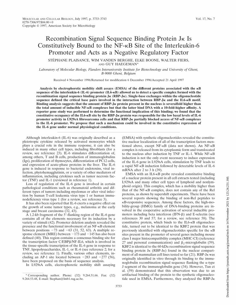
Identification of the IL6-kB binding proteins. In order toidentify the polypeptides involved in each of the complexes, weperformed supershift analyses (Fig. 3) with antisera raisedagainst p50 and p65 (44), RBP-Jk (10, 27), or other membersof the Rel family or other transcription factors (B-rel, c-Rel,
FIG. 1. Sequences and relative binding levels of different oligonucleotidesused in EMSA. Only the positive strand of the oligonucleotides is represented.Underlining refers to unchanged base compared to the IL6-kB probe. Boldface,part of the NF-kB decameric site; lowercase, the cohesive ends of the double-stranded oligonucleotides filled in with Klenow DNA polymerase. Results shownare relative to those obtained for the same complex with the IL6-kB probe.111, 100%; 1111, .100%; 11, .50%; 1/2, ,30%; 2, no binding.
FIG. 2. NF-kB EMSA experiment with L929sA extracts. SubconfluentL929sA cells grown for 48 h were assayed for NF-kB activity with oligonucleo-tides specific for the classical NF-kB motif (Ig-kB [lanes 1, 2, 5, and 6]) or withthe IL6-kB sequence (lanes 3, 4, 7, and 8; see Fig. 1). Equivalent cellular amountsof cytoplasmic (lanes 1 to 4) or nuclear (lanes 5 to 8) extracts from L929sA cellswere analyzed on an 8% EMSA gel. When indicated, mTNF had been added tothe cells at a final concentration of 1,000 IU/ml for 30 min. induc, induced; const,constitutive.
VOL. 17, 1997 RBP-Jk REPRESSES TRANSCRIPTION OF THE IL-6 GENE 3735
Dow
nloa
ded
from
http
s://j
ourn
als.
asm
.org
/jour
nal/m
cb o
n 14
Nov
embe
r 20
21 b
y 12
3.24
.231
.160
.
p52, CEBPb/NF-IL6, CEBPd, c-Jun [all from Santa Cruz Bio-technology]).
The results in Fig. 3 show that the two inducible NF-kBcomplexes (lanes 2 to 7) corresponded to the classical p50homodimer and p50-p65 heterodimer, respectively, while theconstitutive, faster-migrating complex was completely shiftedby antiserum raised against the RBP-Jk protein (lane 8). De-spite many attempts and the use of different extracts, no othernuclear factors reported to associate with Rel proteins, such asNF-IL6 or c-Jun, could be demonstrated in our extracts bysupershift experiments with the IL6-kB oligonucleotide, sug-gesting that the three identified complexes correspond to themajor IL6-kB-binding compounds in L929sA cells (lanes 12 to14).
In their study on the beta interferon (IFN-b) promoter,Thanos and Maniatis (57) have shown simultaneous binding ofHMG and NF-kB proteins to the oligonucleotide, resulting ina slower-migrating NF-kB–HMG–DNA complex. We couldnot detect a similar difference in mobility in the case of theIL6-kB oligonucleotide compared to the Ig-kB oligonucleotide(Fig. 2), suggesting that the binding of each complex with theIL6-kB sequence was mutually exclusive. Furthermore, thesupershift experiments with anti-RBP-Jk antiserum (Fig. 3,lane 8) revealed no change in the mobility of the upper NF-kBcomplexes.
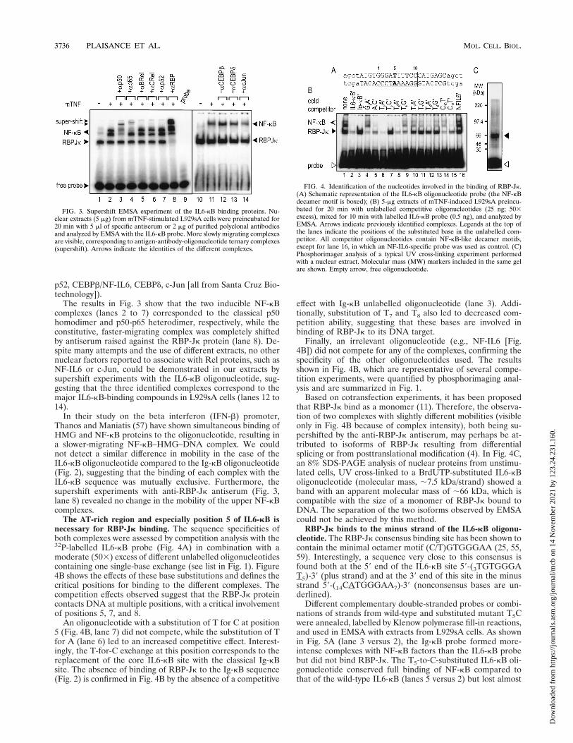
The AT-rich region and especially position 5 of IL6-kB isnecessary for RBP-Jk binding. The sequence specificities ofboth complexes were assessed by competition analysis with the32P-labelled IL6-kB probe (Fig. 4A) in combination with amoderate (503) excess of different unlabelled oligonucleotidescontaining one single-base exchange (see list in Fig. 1). Figure4B shows the effects of these base substitutions and defines thecritical positions for binding to the different complexes. Thecompetition effects observed suggest that the RBP-Jk proteincontacts DNA at multiple positions, with a critical involvementof positions 5, 7, and 8.
An oligonucleotide with a substitution of T for C at position5 (Fig. 4B, lane 7) did not compete, while the substitution of Tfor A (lane 6) led to an increased competitive effect. Interest-ingly, the T-for-C exchange at this position corresponds to thereplacement of the core IL6-kB site with the classical Ig-kBsite. The absence of binding of RBP-Jk to the Ig-kB sequence(Fig. 2) is confirmed in Fig. 4B by the absence of a competitive
effect with Ig-kB unlabelled oligonucleotide (lane 3). Addi-tionally, substitution of T7 and T8 also led to decreased com-petition ability, suggesting that these bases are involved inbinding of RBP-Jk to its DNA target.
Finally, an irrelevant oligonucleotide (e.g., NF-IL6 [Fig.4B]) did not compete for any of the complexes, confirming thespecificity of the other oligonucleotides used. The resultsshown in Fig. 4B, which are representative of several compe-tition experiments, were quantified by phosphorimaging anal-ysis and are summarized in Fig. 1.
Based on cotransfection experiments, it has been proposedthat RBP-Jk bind as a monomer (11). Therefore, the observa-tion of two complexes with slightly different mobilities (visibleonly in Fig. 4B because of complex intensity), both being su-pershifted by the anti-RBP-Jk antiserum, may perhaps be at-tributed to isoforms of RBP-Jk resulting from differentialsplicing or from posttranslational modification (4). In Fig. 4C,an 8% SDS-PAGE analysis of nuclear proteins from unstimu-lated cells, UV cross-linked to a BrdUTP-substituted IL6-kBoligonucleotide (molecular mass, ;7.5 kDa/strand) showed aband with an apparent molecular mass of ;66 kDa, which iscompatible with the size of a monomer of RBP-Jk bound toDNA. The separation of the two isoforms observed by EMSAcould not be achieved by this method.
RBP-Jk binds to the minus strand of the IL6-kB oligonu-cleotide. The RBP-Jk consensus binding site has been shown tocontain the minimal octamer motif (C/T)GTGGGAA (25, 55,59). Interestingly, a sequence very close to this consensus isfound both at the 59 end of the IL6-kB site 59-(3TGTGGGAT5)-39 (plus strand) and at the 39 end of this site in the minusstrand 59-(14CATGGGAA7)-39 (nonconsensus bases are un-derlined).
Different complementary double-stranded probes or combi-nations of strands from wild-type and substituted mutant T5Cwere annealed, labelled by Klenow polymerase fill-in reactions,and used in EMSA with extracts from L929sA cells. As shownin Fig. 5A (lane 3 versus 2), the Ig-kB probe formed more-intense complexes with NF-kB factors than the IL6-kB probebut did not bind RBP-Jk. The T5-to-C-substituted IL6-kB oli-gonucleotide conserved full binding of NF-kB compared tothat of the wild-type IL6-kB (lanes 5 versus 2) but lost almost
FIG. 3. Supershift EMSA experiment of the IL6-kB binding proteins. Nu-clear extracts (5 mg) from mTNF-stimulated L929sA cells were preincubated for20 min with 5 ml of specific antiserum or 2 mg of purified polyclonal antibodiesand analyzed by EMSA with the IL6-kB probe. More slowly migrating complexesare visible, corresponding to antigen-antibody-oligonucleotide ternary complexes(supershift). Arrows indicate the identities of the different complexes.
FIG. 4. Identification of the nucleotides involved in the binding of RBP-Jk.(A) Schematic representation of the IL6-kB oligonucleotide probe (the NF-kBdecamer motif is boxed); (B) 5-mg extracts of mTNF-induced L929sA preincu-bated for 20 min with unlabelled competitive oligonucleotides (25 ng; 503excess), mixed for 10 min with labelled IL6-kB probe (0.5 ng), and analyzed byEMSA. Arrows indicate previously identified complexes. Legends at the top ofthe lanes indicate the positions of the substituted base in the unlabelled com-petitor. All competitor oligonucleotides contain NF-kB-like decamer motifs,except for lane 16, in which an NF-IL6-specific probe was used as control. (C)Phosphorimager analysis of a typical UV cross-linking experiment performedwith a nuclear extract. Molecular mass (MW) markers included in the same gelare shown. Empty arrow, free oligonucleotide.
3736 PLAISANCE ET AL. MOL. CELL. BIOL.
Dow
nloa
ded
from
http
s://j
ourn
als.
asm
.org
/jour
nal/m
cb o
n 14
Nov
embe
r 20
21 b
y 12
3.24
.231
.160
.

all binding of RBP-Jk, as previously shown. Finally, the G2A-substituted probe had largely lost the ability to bind NF-kB,while it still bound to RBP-Jk (lane 4). Interestingly, the bind-ing of the NF-kB complexes to the T5C mutant was not com-parable to that to the Ig-kB oligonucleotide, even when thetwo probes shared the complete NF-kB consensus sequence.This difference probably reflects a higher amount of accessibleIg-kB probe in this experiment or a better binding of theNF-kB factor to the Ig-kB sequence due to the different flank-ing nucleotides. Alternatively, RBP-Jk, while binding veryweakly to the single substituted probe T5C, as shown byEMSA, may still be able to compete with NF-kB factors, yield-ing a reduced amount of NF-kB complexes.
We then used probes resulting from annealing of oligonu-cleotides with a 1-base mismatch at position 5. The probesused in this experiment are comparable to the A6-A centralmismatched probe used by Muller et al. (37) in their crystal-lographic study of p50 homodimers bound to the kB motif andreported to exert little effect on protein affinity. When theT-for-C substitution occurred in the plus strand, the binding ofNF-kB and of RBP-Jk was only marginally affected (Fig. 5A,lanes 7 versus 2). On the contrary, substitution in the minusstrand reduced the binding of RBP-Jk considerably (and, to alesser extent, that of NF-kB), leading to a similar pattern asobtained with the fully complementary (T5C) oligonucleotide(lanes 6 versus 5). In summary, these results clearly indicatethe major involvement of the A residue at position 5 in theminus strand of the IL6-kB sequence, which is absent in theIg-kB site.
Furthermore, the oligonucleotide probe, which correspondsto a single RBP-Jk site present in the Drosophila hairy en-hancer of split (HES) promoter but which does not resemblean NF-kB motif (10), forms a complex of similar mobility asobserved with the IL6-kB probe (Fig. 5A, lane 8), confirmingthat RBP-Jk binds to the latter as a monomer.
To further demonstrate that the binding of RBP-Jk occurs atthe level of the minus strand, we performed a standard meth-ylation interference experiment (Fig. 5B). As shown afterphosphorimager analysis, the lower strand and, particularly,the G residues at positions 211 to 29 are involved in RBP-Jkbinding (plain line versus dotted lines in the densitometric plotin Fig. 5). By contrast, no significant difference could be ob-served when bound and free DNA fractions labelled at theupper strand were compared.
In summary, the results of Fig. 5 demonstrate that RBP-Jkbinds only to the minus strand of the IL6-kB sequence, with anadditional requirement of A5, which is not part of the reportedminimal consensus site.
Recombinant or in vitro-synthesized RBP-Jk specificallybinds to the IL6-kB site. Different approaches were used tofurther confirm the identity of the constitutive protein bindingto IL6-kB. Figure 6A shows that unlabelled rabbit reticulocytelysate contains endogenous NF-kB and RBP-Jk activities. Inlane 5, the binding of rabbit RBP-Jk was completely abolishedby a 50-fold excess of unlabelled HES probe. The T5C muta-tion (lane 4) as well as the Ig-kB oligonucleotide (lane 2) didnot compete for RBP-Jk binding, as already shown in Fig. 4B,while the G2A-substituted oligonucleotide did (lane 3).
In Fig. 6B, analysis of the binding of 35S-Met-labelled RBP3obtained by in vitro transcription-translation (TNT-coupledrabbit reticulocyte lysate; Promega) of the human cDNA ho-molog of the mouse RBP-Jk gene (pCMV-RBP3) to unla-belled oligonucleotides showed the same sequence specificityas the cellular constitutive protein, confirming the results ob-tained with L929sA nuclear extracts.
Furthermore, an E. coli recombinant GST–RBP-Jk fusionprotein also bound specifically to IL6-kB (Fig. 6C, lane 3),while GST alone or a nonrelevant fusion protein (GST-Grb2 [akind gift of J. Vandekerckhove]) did not bind to the probe(Fig. 6C, lanes 1 and 2). The lower complex in lane 3 might bea proteolytical degradation product of full-length GST–RBP-Jk which had retained its ability to bind to immobilized gluta-thione (used for purification) as well as to the IL6-kB probeused in the EMSA.
RBP-Jk is highly expressed but binds with an affinity lowerthan that of NF-kB. Figure 7 shows a typical result obtained byanalysis of the binding of the different complexes to the IL6-kBprobe, in particular showing kinetic on and off rates of RBP-Jkand NF-kB complexes, as determined by running gel shift
FIG. 5. EMSA analysis and methylation interference assay. (A) Differentoligonucleotides or combinations of plus and minus strands from the wild-type(WT) and T5C-substituted IL6-kB oligonucleotides were annealed, labelled byKlenow DNA polymerase, and mixed with nontreated (lane 1) and TNF-induced(lanes 2 to 8) L929sA nuclear extracts before EMSA analysis. (B) Nuclearproteins from noninduced L929sA cells (50 mg) were used for methylationinterference analysis in combination with methylated IL6-kB probe labelled atone end by the T4 polynucleotide kinase. Bound (B) and free (F) DNAs weresubjected to piperidine degradation and analyzed in duplicate by electrophoresisthrough a 15% polyacrylamide–7 M urea-TBE sequencing gel. Line plots cor-responding to normalized bound and free profiles are shown next to the scan.Dotted lines, free probe samples; solid lines, bound fractions. The IL6-kB motifis boxed.
VOL. 17, 1997 RBP-Jk REPRESSES TRANSCRIPTION OF THE IL-6 GENE 3737
Dow
nloa
ded
from
http
s://j
ourn
als.
asm
.org
/jour
nal/m
cb o
n 14
Nov
embe
r 20
21 b
y 12
3.24
.231
.160
.

analysis. For on-rate analysis, nuclear proteins from TNF-in-duced L929sA cells were added to the EMSA reaction mix, andaliquots were loaded on a running 8% EMSA gel at varioustime points. In this experiment, the two types of complexesshared a large excess of probe without competition. The resultsobtained show that NF-kB binding to the probe reached equi-librium after approximately 20 min, while binding of RBP-Jkhad not reached a plateau after 35 min of incubation. Theoff-rate study (Fig. 7B) was similar, except that the protein-DNA complexes were allowed to form for 20 min before ad-dition of a 1003 excess of unlabelled IL6-kB oligonucleotideand periodical loading on the running gel. In this experiment,NF-kB dissociation was analyzed in extracts from TNF-in-duced L929sA cells, while dissociation of RBP-Jk complexeswas measured in extracts from noninduced cells. Finally, incu-bation of RBP-Jk with a 1003 excess of unrelated oligonucle-otide (AP-1 probe) did not lead to displacement, as shown bythe control curve.
We finally performed a semiquantitative binding analysis ofeach complex by the method of Scatchard (49). RBP-Jk andNF-kB binding were analyzed in the same extract from TNF-induced cells (Fig. 7C). In both cases, a large excess of unla-belled oligonucleotide (2003) was added to the reaction mix-ture. The HES oligonucleotide was used to complex RBP-Jk inthe case of NF-kB binding study, while unlabelled Ig-kB oli-gonucleotide could prevent binding of NF-kB proteins to thelabelled probe for analysis of RBP-Jk binding. Figure 7C showsbinding of the NF-kB factor with saturation for values exceed-ing 1 nM free probe. By contrast, in the case of RBP-Jk,saturation could not be obtained with a probe concentration ofas high as 1.4 nM. The affinity of RBP-Jk for its DNA site isclearly lower than that of NF-kB and could not be preciselydefined due to the insufficient amount of probe used in this
experiment. The highest probe concentration used here and,particularly, previously obtained results with higher concentra-tions of labelled probe (data not shown) suggest that the half-saturation of RBP-Jk binding is obtained in the range of 2 to3 nM probe, while the NF-kB affinity constant could be esti-mated within the range of 200 to 400 pM. On the other hand,the total amount of RBP-Jk protein present in the nucleus is atleast five- to sixfold higher than the amount of activated NF-kB, as determined by the saturation level obtained for bothcomplexes (.3,500 versus 700 arbitrary units). The facts thatthe amount of RBP-Jk is severalfold higher than that of NF-kBbut that RBP-Jk displays at least 10-fold less affinity for itstarget confirm that competition between the two factors canoccur when low levels of NF-kB are present in the nucleus
FIG. 6. Binding of rabbit or recombinant RBP-Jk proteins to wild-typeIL6-kB or substituted probes. (A) Rabbit reticulocyte extracts were used in acompetition experiment with 32P-labelled IL6-kB probe and unlabelled compet-itor oligonucleotides (compet. oligo). A probe specific for the Su(H) RBP-Jkhomolog (HES [lane 5]) was used as a control. The NF-kB and RBP-Jk arrowspoint to endogenous binding activities present in the rabbit reticulocyte extract.(B) Reverse-EMSA experiment with 35S-labelled RBP-Jk protein produced bythe in vitro transcription-translation-coupled rabbit reticulocyte system andmixed with unlabelled wild-type or mutated IL6-kB oligonucleotides. (C) EMSAwith the IL6-kB probe and 2 ml of soluble extract from E. coli expressingrecombinant GST protein, a GST–RBP-Jk fusion protein, or an irrelevant GST-Grb2 fusion protein.
FIG. 7. Complex formation as measured by EMSA as a function of incuba-tion time or oligonucleotide concentration. (A) On-rate analysis of RBP-Jk(filled squares) and NF-kB (filled triangles) complexes as determined by runningshift assaying. (B) Off-rate analysis of preformed complexes (20 min at roomtemperature), mixed with a 1003 molar excess of unlabelled competitive oligo-nucleotide and loaded on a running gel. (C) Scatchard plot analysis. Aliquotscontaining 5 mg of nuclear protein extract from nonstimulated or mTNF-stimu-lated L929sA cells (1,000-IU/ml mTNF; 30 min) were mixed for 30 min withincreasing amounts of 32P-labelled IL6-kB probe and a 2003 excess of unla-belled probe specific for one species (HES for RBP-Jk and Ig-kB for the NF-kBcomplexes) and analyzed by EMSA. The intensity of each complex (verticalscale) is expressed in arbitrary units (a.u.). Insert, an enlarged portion of theNF-kB plot, corresponding to the lower range of probe concentration.
3738 PLAISANCE ET AL. MOL. CELL. BIOL.
Dow
nloa
ded
from
http
s://j
ourn
als.
asm
.org
/jour
nal/m
cb o
n 14
Nov
embe
r 20
21 b
y 12
3.24
.231
.160
.

(e.g., early induction and suboptimal stimulation). Further-more, this suggests that the full induction of NF-kB transloca-tion leads to an influx of transactivator in the nucleus that willefficiently displace the RBP-Jk protein from the promoter.
RBP-Jk represses transcription. The presence of a bindingsite for RBP-Jk overlapping the IL6-kB site led us to postulatea functional involvement of the RBP-Jk protein in the tran-scriptional regulation of the IL-6 gene. L929sA cells weretransiently or stably transfected with different luciferase re-porter constructs and induced by mTNF or other stimuli foranalysis in a standard luciferase assay. The various constructsare described in Materials and Methods and are schematicallyrepresented in Fig. 8.
We have already shown that the exchange of a single base atparticular positions within the IL6-kB sequence is sufficient tocompletely abolish the recognition by the RBP-Jk protein,while binding of NF-kB was only marginally reduced (Fig. 4Band 5A). Figure 9 shows a typical result obtained with pooledL929sA cells stably transfected by the calcium phosphatemethod and selected for 15 days with G418 (see Materials andMethods). As can be seen in Fig. 9A, substitution of theIL6-kB core sequence by the T5C variant was sufficient toenhance the basal level of luciferase expression by a factor of13. Furthermore, after 6 h of induction with TNF or IL-1, theluciferase activity obtained with the (T5C-kB)3-50hu.IL6P syn-thetic promoter was still 6- to 10-fold higher than that with the(IL6-kB)3-50hu.IL6P reporter construct.
These results not only confirm that RBP-Jk competitivelybinds to the kB site of the IL-6 promoter, thus limiting theaccess of NF-kB factors translocated to the nucleus after cy-tokine induction, but also show that RBP-Jk acts as a repressorof basal transcription in the absence of detectable amounts ofNF-kB proteins. We conclude, therefore, that RBP-Jk is ageneral silencer of the IL-6 promoter, as further documentedbelow.
A control experiment (Fig. 9B) shows that the difference inluciferase activity observed between the two constructions(IL6-kB)3-50 hu.IL6P luc1 and (T5C-kB)3-50 hu.IL6P luc1was not due to a quantitative difference in the amounts oftransfected reporter DNA but truly reflected the stronger pro-moter activity of the point-mutated sequence. The peak ofactivity at approximately 3 mg of DNA per six-well plate cor-responds to the optimal quantity of reporter gene activity inthe cells for both constructions; the further decrease in activityis probably due to toxicity at high DNA concentrations or tosquelching effects.
FIG. 8. Schematic maps of the different constructions used in this study.Black bars, the reporter gene coding region; dotted bars, the IL-6 promoterregion between 250 and 21168 bp; white bars, the 50-bp IL-6 promoter frag-ment located upstream of the major start site of transcription and containing theputative TATA region; hatched bars, NF-kB sites; rounded blocks, UAS sites.Restriction enzyme sites used for construction are abbreviated as follows: B,BamHI; H, HindIII; N, NcoI; Nh, NheI; P, PstI; S, SspI; Xb, XbaI; Xh, XhoI; andXm, XmaI. bl, blunt end generated by restriction or after fill in with KlenowDNA polymerase.
FIG. 9. Effect of RBP-Jk binding on gene expression. (A) Pools of trans-fected cells were stimulated (when indicated) with 10-ng/ml IL-1 or 500-IU/mlmTNF for 6 h before extraction and luciferase assaying. The results representmore than six experiments and three different transfections. Results are normal-ized to the b-galactosidase activity present in the extracts (see Materials andMethods), and the relative induction factors are summarized. a.u., arbitraryunits. (B) Effect of the amount of transfected plasmid on luciferase activity.L929sA cells were transiently transfected in six-well plates with Lipofectamineand increasing amounts of reporter DNA plasmid. Cells were left for 72 h beforechanging of the medium or stimulation with 2,000-IU/ml TNF for 6 h. Results areexpressed in arbitrary light units. (C) Effect of the single-base exchange withinthe 234-bp promoter fragment. The T5C base substitution was introduced in the234-bp IL-6 promoter fragment coupled to luciferase. Results are the averageresults and deviations for four independent assays. n.i., noninduced.
VOL. 17, 1997 RBP-Jk REPRESSES TRANSCRIPTION OF THE IL-6 GENE 3739
Dow
nloa
ded
from
http
s://j
ourn
als.
asm
.org
/jour
nal/m
cb o
n 14
Nov
embe
r 20
21 b
y 12
3.24
.231
.160
.

The observations made with the synthetic reporter construc-tions could be reproduced with a 234-bp fragment of the IL-6promoter bearing the single-base exchange of T for C in the kBsite (Fig. 9C). The more important level of luciferase activityobtained with the mutant (;4-fold-higher) promoter probablyreflects both higher basal transcription in the absense of therepressor protein and earlier induction of transcription afterTNF treatment of the cells, leading to a higher accumulation ofmRNA and luciferase enzyme (Fig. 9C).
To confirm the ability of RBP-Jk to repress transcription ofthe IL-6 promoter, cotransfection experiments with differentreporter constructs under the control of two Gal4 upstreamactivated sequences (UAS) in combination with eukaryoticexpression vectors for Gal4-fusion proteins were performed.We cloned the two UAS sites upstream of the minimal orfull-length IL-6 promoters (Fig. 8) and used Gal4 fusion vec-tors for expression of RBP-Jk or for the p65 NF-kB subunit. Inagreement with previously reported data on the repressor ac-tivity of RBP-Jk (25), the results shown in Fig. 10A confirmthat RBP-Jk is a powerful repressor of basal transcription evenwhen it is positioned more than 1 kb upstream of the start siteof transcription (73 and 55% repression for the 50- and1,168-bp constructs, respectively). On the other hand, the p65transactivation potential became very weak at the longer dis-tance (17- and 1.8-fold induction, respectively). These resultsindicate that in addition to its competitor effects with theactivated NF-kB complexes for access to the IL6-kB sequencein the IL-6 gene promoter, the RBP-Jk protein may also func-tion on its own to repress or downmodulate IL-6 promoteractivity in general.
RBP-Jk was already shown by others to repress transcriptionpromoted by the VP16 acidic transactivator (16). Figure 10Bshows that the Gal4 N-terminal 147-amino-acid domain is re-sponsible for a two- to threefold transactivation compared tomock-transfected cells. Others have already made this obser-vation (25). By contrast, transient transfection of as little as 50ng of Gal4–RBP-Jk fusion vector led to complete repression ofthe promoter activity, confirming the result shown by Dou etal. with a similar construction (16). Our result confirms thehigh inhibitory potential of RBP-Jk on basal transcription.Furthermore, we show that this inhibition is strong enough torepress the 234-bp IL-6 promoter after TNF induction and inthe presence of upstream regulatory sequences including NF-IL6, MRE, and NF-kB binding sites, strongly suggesting thatRBP-Jk is indeed able to silence the IL-6 promoter as long asit can bind to the DNA and that this inhibition occurs even inthe presence of nuclear NF-kB factor bound to a downstreamsequence.
Overexpression of an inactive RBP-Jk positively modulatesendogenous IL-6 secretion. We described above that a muta-tion of the IL6-kB site, which strongly reduced RBP-Jk bind-ing, was associated with higher promoter activity and proposedthat this was due to loss of binding of the repressor protein.
To confirm this hypothesis, we stably transfected L929sAcells with constructions coding for wild-type RBP-Jk or itsdominant-negative mutant EEF-238-AAA (RBP-AAA), whichwas reported by others to have lost its repression activity butwhich still binds to the DNA (25). Analysis of the pooledpopulation of each transfection showed (Fig. 11) that expres-sion of wild-type RBP-Jk did not lead to an enhanced repres-sion of IL-6 synthesis, probably because of the already highlevel of endogenous protein, but may have delayed it (see 4-hpoint). By contrast, constitutive expression of RBP-JkEEF-238-AAA
led to a higher amount of secreted IL-6 for noninducedL929sA cells as well as to an approximately doubled amount ofIL-6 protein in the supernatant after 6 h of TNF induction.
These data are complementary to the result obtained with apoint mutation of the IL6-kB site (Fig. 9C). The more pro-nounced effect obtained by mutation of the DNA site could bedue to the fact that it completely abolished binding of endog-enous RBP-Jk, while the constitutively expressed mutant pro-tein still had to compete with the endogenous pool of wild-typeRBP-Jk.
Despite several attempts and the use of CMV and RSVpromoters, overexpression of an epitope-tagged RBP-Jk or ofits inactive mutant RBP-JkEEF-238-AAA did not lead to an in-creased level of DNA binding activity, while the transfectedprotein could be shown by immunoblotting (not shown). Thissuggests that the overall quantity of RBP-Jk protein in the cellmight be limited by regulatory events or that the higher-level-
FIG. 10. One-hybrid analysis of RBP-Jk repressor effect. (A) Transienttransfection experiment showing the effect of expression of Gal4-fusion proteinson the minimal or full IL-6 promoters coupled to two Gal4 binding sites. Lucif-erase (luc) activity was measured 72 h after transfection and corrected forprotein content. Each bar represents the average result for four independentwells. The relative modulation (modul.) (activation/repression) is indicated. (B)L929sA cells stably transfected by the calcium phosphate method with the(Gal)2-50 hu.IL6P-luc1 reporter vector. After 15 days of selection with G418,stable colonies were pooled and further transfected with DEAE-dextran withdifferent expression plasmids (KSII1 bluescript plasmid [Stratagene] used asmock DNA). Luciferase activity was measured 48 h after transfection. Eachtransfection was performed four times. n.i., noninduced.
3740 PLAISANCE ET AL. MOL. CELL. BIOL.
Dow
nloa
ded
from
http
s://j
ourn
als.
asm
.org
/jour
nal/m
cb o
n 14
Nov
embe
r 20
21 b
y 12
3.24
.231
.160
.

expressing cells might have been counterselected followingtransfection.
DISCUSSION
The complex interplay between repressors and activatorsdefines a new level of gene expression control (see reviews inreferences 13 and 46). In the case of NF-kB, the major level ofregulation was originally thought to be present in the cyto-plasm, where the transcriptionally active form p50-p65 istrapped in an inactive complex with the inhibitory moleculeIkB (for a review, see reference 5). While the NF-kB factor isa necessary and sufficient element to induce transcription ofreporter genes, it is now known to synergize with other tran-scription factors to exert its specific gene regulatory function invivo. NF-kB activity is directly repressed by the glucocorticoidreceptor in the IL-6 (41) and IL-8 genes (36) or by the HMGprotein DSP1 present at the IFN-b PRDII-NRE site (29),while it is positively regulated by the binding of other HMGproteins to the IFN-b-kB (57). Moreover, an increasing num-ber of reports revealed the physical association of Rel proteinswith other proteins, including several transcription factors
(e.g., NF-IL6 and AP-1) involved in IL-6 gene expression andsuggesting an in vivo function for gene regulation (20).
Additive, cooperative, and/or competitive effects of differenttranscription factors determine the final tissue or stage-specificexpression of most, if not all, of the inducible genes. In thisregard, Santhanam et al. (48) have reported the presence of anegative regulatory region between 2126 and 2104 (fos basaltranscription element), which is responsible for the constitutiverepression of IL-6 transcription. In monocytes, this regionbinds a repressor complex composed of the constitutive Sp1transcription factor and retinoblastoma-like proteins (47).
Here, we have shown that a ubiquitous mammalian cellfactor, RBP-Jk, specifically recognizes and constitutively bindsto the kB motif present in the IL-6 gene promoter. This bind-ing mainly involves the minus strand of the IL6-kB site whereNF-kB and RBP-Jk factors recognize an overlapping targetsite and compete for access to the DNA. NF-kB has a higheraffinity for the IL6-kB motif but is present in much lowerabundance than the RBP-Jk protein. RBP-Jk functions as anegative effector in two ways. Indeed, besides its competitionwith NF-kB for the same binding site, thus limiting the cyto-kine-induced activation of the IL-6 gene promoter, our datasuggest that RBP-Jk also exerts an intrinsic repressor andsilencing function in the normal physiological state.
RBP-Jk binding to several NF-kB sequences has been de-scribed elsewhere (16, 27). However, these studies were per-formed at the level of protein-DNA binding in vitro in EMSAexperiments and occasionally by the use of synthetic reporterconstructs containing multiple RBP-Jk sites (25). By contrast,in the context of the IL-6 promoter, we have shown that asingle-base substitution within the unique NF-kB motif, whilenot significantly affecting the binding of NF-kB complexes,almost completely abolished the binding of the RBP-Jk pro-tein. This modification was accompanied by a dramatic in-crease in the level of basal and TNF-induced transcription. Weconfirmed this observation by another approach in which sta-ble transfection of a mutant RBP-JkEEF-219-AAA protein whichhad lost its repressor activity led to higher basal and TNF-induced secretion of the endogenous IL-6 in mouse L929sAcells. The concurrent results of these two experiments led us toconclude that the RBP-Jk protein is indeed a constitutive re-pressor of the IL-6 promoter in vivo and that this repression is
FIG. 11. Mutant RBP-Jk competes with endogenous RBP-Jk and releavesthe silencing of the IL-6 gene. L929sA were transfected with empty vector(pRSV) or with constructs coding for wild-type (wt) RBP-Jk or its mutantRBPEEF-238-AAA (RBP-AAA). After 15 days of G418 selection, the cells weretransferred in 24-well plates and stimulated for the indicated time with 2,000-IU/ml TNF. Aliquots of supernatant were used in a standard IL-6 bioassay. Theresults are the average values and deviations for four independent assays.
FIG. 12. Model of transcriptional regulation at the level of the IL6-kB sequence and possible relevance of known RBP-Jk-associating factors. The competitivebinding between NF-kB and RBP-Jk may not be the only event occurring at the level of the IL-6 promoter. Interactions with EBNA-2, Notch-1, or possibly other factorscould be involved in regulatory processes leading to IL-6 gene regulation as well as to the regulation of other genes. (p, reference 26; pp, our results).
VOL. 17, 1997 RBP-Jk REPRESSES TRANSCRIPTION OF THE IL-6 GENE 3741
Dow
nloa
ded
from
http
s://j
ourn
als.
asm
.org
/jour
nal/m
cb o
n 14
Nov
embe
r 20
21 b
y 12
3.24
.231
.160
.

sufficient to maintain the IL-6 gene silent in the absence of aninflammatory stimulus.
While this paper was under revision, a similar message onthe repressive function of RBP-Jk on IL-6 gene expression waspublished by Kannabiran et al. (28). Whereas these authorsattributed the repressive activity of RBP-Jk to the cooperativetranscriptional activation exerted by C/EBP-b and NF-kB, weclearly demonstrated by various transfection assays with re-combinant, kB sequence-containing constructs, that theGATA-C/EBP site (14) is not needed for repression byRBP-Jk and that RBP-Jk acts as an active repressor on pro-moters exclusively driven by kB motifs. In contrast to thereport of Kannabiran et al., in which loss of function is ob-served upon distancing of the RBP-Jk binding site, we stilldetected repression of basal activity when the RBP-Jk bindingsite was placed as far as 21168. Moreover, RBP-Jk was able torepress the TNF-induced activity when it was present at posi-tion 2234. The differences in the transfection assays and/orcell phenotypes used may account for the differences observedin repressive activity.
It is likely that the mechanism of repression studied here isnot particular to the IL-6 promoter and that other NF-kB sites,which have been shown to bind RBP-Jk, will also present thesame characteristics. Hence, RBP-Jk could be considered atransrepressor, often sharing its DNA consensus site with theNF-kB transactivator and thus preventing gene induction inthe absence of NF-kB activation. Important candidates for thistype of regulation include genes for which the NF-kB factorhas been shown to be a main transcriptional activator andwhich actually bind RBP-Jk as well, such as the genes codingfor H2K (27), b2-microglobulin (59), MHC-CI, or the IL-2 Ra(53). Indeed, uncontrolled expression of all of these genes inthe absense of extracellular aggression would rapidly lead toinflammatory and immunological disorders by perturbing, forinstance, the state of activation of endothelial cells or lymphoidprecursors. Furthermore, demonstration of the direct effect oftransactivators such as EBNA-2 or NotchDIC through bindingto the RBP-Jk docking protein (Fig. 12) could help to explaindevelopmental or pathological conditions leading to gene ex-pression, independently of NF-kB activation (e.g., T-cell lym-phoma, epithelial tumors, or immortalization of B cells).
ACKNOWLEDGMENTS
We thank A. Israel for providing the pCMV-RBP3 vector, the HESoligonucleotide, and the antiserum against RBP-Jk and for his helpfuldiscussions; L. Schmitz for providing the Gal4 fusion vectors; J.Vandekerckhove for his gift of the pGST-Grb2-transformed bacteria;N. Rice for providing antisera against NF-kB factors; F. Molemans forDNA sequencing; and I. Van Rompaey and W. Burm for technicalassistance.
Research was supported by grants from the EC-Biotech Programme(postdoctoral fellowship to S.P.), the Belgian IUAP, the FGWO, andthe VIB. W.V.B. holds a grant from the Belgian IWT. G.H. is aResearch Director with the FWO, Vlaanderen, Belgium.
REFERENCES
1. Akira, S., H. Isshiki, T. Nakajima, S. Kinoshita, Y. Nishio, S. Natsuka, andT. Kishimoto. 1992. Regulation of expression of the interleukin-6 gene:structure and function of the transcription factor NF-IL6. Ciba Found.Symp. 167:47–67.
2. Akira, S., H. Isshiki, T. Sugita, O. Tanabe, S. Kinoshita, Y. Nishio, T.Nakajima, T. Hirano, and T. Kishimoto. 1990. A nuclear factor for IL-6expression (NF-IL6) is a member of a C/EBP family. EMBO J. 9:1897–1906.
3. Akira, S., T. Taga, and T. Kishimoto. 1993. Interleukin-6 in biology andmedicine. Adv. Immunol. 54:1–78.
4. Amakawa, R., W. Jing, K. Ozawa, N. Matsunami, Y. Hamaguchi, F. Mat-suda, M. Kawaichi, and T. Honjo. 1993. Human Jk recombination signalbinding protein gene (IGKJRB): comparison with its mouse homologue.Genomics 17:306–315.
5. Baldwin, A. S. 1996. The NF-kB and IkB proteins: new discoveries andinsights. Annu. Rev. Immunol. 14:649–681.
6. Baldwin, A. S., Jr. 1996. Methylation interference assay for analysis of DNA-protein interactions, p. 12.3.1–12.3.6. In J. Wiley (ed.), Current protocols inmolecular biology. Greene and Wiley, New York, N.Y.
7. Baldwin, A. S., Jr., and P. A. Sharp. 1988. Two transcription factors, NF-kBand H2TF1, interact with a single regulatory sequence in the class I majorhistocompatibility complex promoter. Proc. Natl. Acad. Sci. USA 85:723–727.
8. Baniahmad, A., A. C. Kohne, and R. Renkawitz. 1992. A transferable silenc-ing domain is present in the thyroid hormone receptor, in the v-erbA onco-gene product and in the retinoic acid receptor. EMBO J. 11:1015–1023.
9. Bradford, M. M. 1976. A rapid and sensitive method for the quantitation ofmicrogram quantities of protein utilizing the principle of protein-dye bind-ing. Anal. Biochem. 72:248–254.
10. Brou, C., A. Logeat, M. Lecourtois, J. Vandekerckhove, P. Kourilsky, F.Schweisguth, and A. Israel. 1994. Inhibition of the DNA-binding activity ofDrosophila supressor of hairless and its human homolog, KBF2/RBP-Jk, bydirect protein protein interaction with drosophila Hairless. Genes Dev.8:2491–2503.
11. Chung, C. N., Y. Hamaguchi, T. Honjo, and M. Kawaichi. 1994. Site-directedmutagenesis study on DNA binding regions of the mouse homologue ofSuppressor of Hairless, RBP-Jk. Nucleic Acids Res. 22:2938–2944.
12. Cohen, J. I., F. Wang, and E. Kieff. 1991. Epstein-Barr virus nuclear protein2 mutations define essential domains for transformation and transactivation.J. Virol. 65:2545–2554.
13. Cowell, I. G. 1994. Repression versus activation in the control of genetranscription. Trends Biol. Sci. 19:38–42.
14. Dendorfer, U., P. Oettgen, and T. A. Libermann. 1994. Multiple regulatoryelements in the interleukin-6 gene mediate induction by prostaglandins,cyclic AMP, and lipopolysaccharide. Mol. Cell. Biol. 14:4443–4454.
15. Dignam, D., R. M. Lebovitz, and R. G. Roeder. 1983. Accurate transcriptioninitiation by RNA polymerase II in a soluble extract from isolated mamma-lian nuclei. Nucleic Acids Res. 11:1475–1489.
16. Dou, S., X. Zeng, P. Cortes, H. Erdjument-Bromage, P. Tempst, T. Honjo,and L. D. Vales. 1994. The recombination signal sequence binding proteinRNP-2N functions as a transcriptional repressor. Mol. Cell. Biol. 14:3310–3319.
17. Ellisen, L. W., J. Bird, D. C. West, A. Lee Soreng, T. C. Reynolds, S. D.Smith, and J. Sklar. 1991. TAN-1, the human homolog of the DrosophilaNotch gene, is broken by chromosomal translocations in T lymphoblasticneoplasms. Cell 66:649–661.
18. Furukawa, T., S. Maruyama, M. Kawaichi, and T. Honjo. 1992. The Dro-sophila homolog of the immunoglobulin recombination signal-binding pro-tein regulates peripheral nervous system development. Cell 69:1191–1197.
19. Haegeman, G., J. Content, G. Volckaert, R. Derynck, J. Tavernier, and W.Fiers. 1986. Structural analysis of the sequence coding for an inducible26-kDa protein in human fibroblasts. Eur. J. Biochem. 159:625–632.
20. Haegeman, G., and W. Fiers. 1995. TNF-induced mechanisms for IL6 geneinduction, p. 375–382. In L. Packer and K. Wirtz (ed.), Signalling mecha-nisms—from transcription factors to oxidative stress. Springer-Verlag, Ber-lin, Germany.
21. Hamaguchi, Y., Y. Yamamoto, H. Iwanari, S. Maruyama, T. Furukawa, N.Matsunami, and T. Honjo. 1992. Biochemical and immunological character-ization of the DNA binding protein (RBP-Jk) to mouse Jk recombinationsignal sequence. J. Biochem. (Tokyo) 112:314–320.
22. Henkel, T., P. D. Ling, S. D. Hayward, and M. G. Peterson. 1994. Mediationof Epstein-Barr virus EBNA2 transactivation by recombination signal-bind-ing protein Jk. Science 265:92–95.
23. Hirano, T., S. Akira, T. Taga, and T. K. Kishimoto. 1990. Biological andclinical aspects of interleukin-6. Immunol. Today 11:443–449.
24. Honjo, T. 1996. The shortest path from the surface to the nucleus: RBP-Jk/Su(H) transcription factor. Genes Cells 1:1–9.
25. Hsieh, J. J., and S. D. Hayward. 1995. Masking of the CBF1/RBPJk tran-scriptional repression domain by Epstein-Barr virus EBNA2. Science 268:560–563.
26. Hsieh, J. J.-D., T. Henkel, P. Salmon, E. Robey, M. G. Peterson, and S. D.Hayward. 1996. Truncated mammalian Notch1 activates CBF1/RBP-Jk-re-pressed genes by a mechanism resembling that of Epstein-Barr virusEBNA2. Mol. Cell. Biol. 16:952–959.
27. Israel, A., O. Yano, F. Logeat, M. Kieran, and P. Kourilsky. 1989. Twopurified factors bind the same sequence in the enhancer of mouse MHC classI genes: one of them is a positive regulator induced upon differentiation ofteratocarcinoma cells. Nucleic Acids Res. 17:5245–5457.
28. Kannabiran, C., X. Y. Zeng, and L. D. Vales. 1997. The mammalian tran-scriptional repressor RBP (CBF1) regulates interleukin 6 gene expression.Mol. Cell. Biol. 17:1–9.
29. Lehming, N., D. Thanos, J. M. Brickman, J. Ma, T. Maniatis, and M.Ptashne. 1994. An HMG like protein that can switch a transcriptional acti-vator to a repressor. Nature 371:175–179.
30. Lewis, H., W. Kaszubska, J. F. DeLamarter, and J. Whelan. 1994. Cooper-ativity between two NF-kB complexes, mediated by high mobility group I(Y),
3742 PLAISANCE ET AL. MOL. CELL. BIOL.
Dow
nloa
ded
from
http
s://j
ourn
als.
asm
.org
/jour
nal/m
cb o
n 14
Nov
embe
r 20
21 b
y 12
3.24
.231
.160
.

is essential for cytokine induced expression of the E-selectin promoter. Mol.Cell. Biol. 14:5701–5709.
31. Libermann, T. A., and D. Baltimore. 1990. Activation of interleukin-6 geneexpression through the NF-kB transcription factor. Mol. Cell. Biol. 10:2327–2334.
32. Lu, C., and R. S. Kerbel. 1993. Interleukin-6 undergoes transition fromparacrine growth inhibitor to autocrine stimulator during human melanomaprogression. J. Cell Biol. 120:1282–1288.
33. Maniatis, T., E. F. Fritsch, and J. Sambrook. 1987. Molecular cloning: alaboratory manual. Cold Spring Harbor Laboratory, Cold Spring Harbor,N.Y.
34. Matsunami, N., Y. Hamaguchi, Y. Yamamoto, K. Kuze, K. Kangawa, H.Matsuo, M. Kawaichi, and T. Honjo. 1989. A protein binding to the Jkrecombination sequence of immunoglobulin genes contains a sequence re-lated to the integrase motif. Nature 342:934–937.
35. Matthews, J. R., N. Wakasugi, J. L. Virelizier, J. Yodoi, and R. T. Hay. 1992.Thioredoxin regulates the DNA binding activity of NF-kB by reduction of adisulphide bond involving cysteine 62. Nucleic Acids Res. 20:3821–3830.
36. Mukaida, N., M. Morita, Y. Ishikawa, N. Rice, S. I. Okamoto, T. Kasahara,and K. Matsushima. 1994. Novel mechanism of glucocorticoid mediatedgene repression: nuclear factor-kB is target for glucocorticoid mediatedinterleukin-8 gene repression. J. Biol. Chem. 269:13289–13295.
37. Muller, C. W., F. A. Rey, M. Sodeoka, G. L. Verdine, and S. C. Harrison.1995. Structure of the NF-kB p50 homodimer bound to DNA. Nature 373:311–317.
38. Oka, C., T. Nakano, A. Wakeham, J. L. de la Pompa, C. Mori, T. Sakai, S.Okasaki, M. Kawaichi, K. Shiota, T. W. Mak, and T. Honjo. 1995. Disrup-tion of the mouse RBP-Jk gene results in early embryonic death. Develop-ment 121:3291–3301.
39. Patestos, N. P., G. Haegeman, V. Vandevoorde, and W. Fiers. 1993. Activa-tion of the nuclear factor kB is not sufficient for regulation of tumor necrosisfactor induced interleukin-6 gene expression. Biochimie 75:1007–1018.
40. Ray, A., K. S. LaForge, and P. B. Sehgal. 1990. On the mechanism forefficient repression of the interleukin-6 promoter by glucocorticoids: en-hancer, TATA box, and RNA start site (Inr motif) occlusion. Mol. Cell. Biol.10:5736–5746.
41. Ray, A., and K. E. Prefontaine. 1994. Physical association and functionalantagonism between the p65 subunit of transcription factor NF-kB and theglucocorticoid receptor. Proc. Natl. Acad. Sci. USA 91:752–756.
42. Ray, A., S. B. Tatter, L. T. May, and P. B. Sehgal. 1988. Activation of thehuman b2-interferon/hepatocyte stimulating factor/interleukin-6 promoterby cytokines, viruses, and second messenger agonists. Proc. Natl. Acad. Sci.USA 85:6701–6705.
43. Revel, M. 1992. Growth regulatory functions of IL6 and antitumour effects.Res. Immunol. 143:769–773.
44. Rice, N. R., M. L. MacKichan, and A. Israel. 1992. The precursor of NF-kBp50 has IkB-like functions. Cell 71:243–253.
45. Robbins, J., B. J. Blondel, D. Gallahan, and R. Callahan. 1992. Mousemammary tumor gene int-3: a member of the notch gene family transformsmammary epithelial cells. J. Virol. 66:2594–2599.
46. Roberts, S. G. E., and M. R. Green. 1995. Dichotomous regulators. Nature375:105–106.
47. Sanceau, J., T. Kaisho, T. Hirano, and J. Witzerbin. 1995. Triggering of thehuman interleukin-6 gene by interferon-g and tumor necrosis factor-a in
monocytic cells involves cooperation between interferon regulatory factor-1,NFkB, and Sp1 transcription factors. J. Biol. Chem. 270:27920–27931.
48. Santhanam, U., A. Ray, and P. B. Sehgal. 1991. Repression of the interleukin6 gene promoter by p53 and the retinoblastoma susceptibility gene product.Proc. Natl. Acad. Sci. USA 88:7605–7609.
49. Scatchard, G. 1949. The attraction of proteins for small molecules and ions.Ann. N. Y. Acad. Sci. 51:660–672.
50. Schmitz, M. L., and P. A. Baeuerle. 1991. The p65 subunit is responsible forthe strong transcription activating potential of NF-kB. EMBO J. 10:3805–3817.
51. Schweisguth, F., and J. W. Posakony. 1992. Suppressor of hairless, theDrosophila homolog of the mouse recombination signal-binding proteingene, controls sensory organ cell fates. Cell 69:1199–1212.
52. Shimizu, H., K. Mitomo, T. Watanabe, S. I. Okamoto, and K. I. Yamamoto.1990. Involvement of a NF-kB like transcription factor in the activation ofthe interleukin-6 gene by inflammatory lymphokines. Mol. Cell. Biol. 10:561–568.
53. Shirakata, Y., J. D. Shuman, and J. E. Coligan. 1995. Purification of a novelMHC class I element binding activity from thymus nuclear extracts revealsthat thymic RBPJ k/CBF1 binds to NFkB like elements. J. Immunol. 156:4672–4679.
54. Soriano, P., C. Montgomery, R. Geske, and A. Bradley. 1991. Targeteddisruption of the c-src proto-oncogene leads to osteopetrosis in mice. Cell64:693–702.
55. Tamura, K., Y. Taniguchi, S. Minoguchi, T. Sakai, T. Tun, T. Furakawa, andT. Honjo. 1995. Physical interaction between a novel domain of the receptorNotch and the transcription factor RBP-Jk/Su(H). Curr. Biol. 5:1416–1423.
56. Tanabe, O., S. Akira, T. Kamiya, G. G. Wong, T. Hirano, and T. K. Kishi-moto. 1989. Genomic structure of the murine IL-6 gene. J. Immunol. 141:3875–3881.
57. Thanos, D., and T. Maniatis. 1993. The high mobility group protein HMGI(Y) is required for NF-kB dependent virus induction of the human IFNbgene. Cell 71:777–789.
58. Thanos, D., and T. Maniatis. 1995. NF-kB: a lesson in family values. Cell80:529–532.
59. Tun, T., Y. Hamaguchi, N. Matsunami, T. Furukawa, T. Honjo, and M.Kawaichi. 1994. Recognition sequence of a highly conserved DNA bindingprotein RBP-Jk. Nucleic Acids Res. 22:965–971.
60. Van Haesebroeck, B., S. Van Bladel, A. Lenaerts, P. Suffys, R. Beyaert, R.Lucas, F. Van Roy, and W. Fiers. 1991. Two discrete types of tumor necrosisfactor-resistant cells derived from the same cell line. Cancer Res. 51:2469–2477.
61. Van Snick, J., S. Cayphas, A. Vink, C. Uyttenhove, P. G. Coulie, M. R.Rubira, and R. J. Simpson. 1986. Purification and NH2-terminal amino acidsequence of a T-cell-derived lymphokine with growth factor activity forB-cell hybridomas. Proc. Natl. Acad. Sci. USA 83:9679–9683.
62. Waltzer, L., F. Logeat, C. Brou, A. Israel, A. Sergeant, and E. Manet. 1994.The human Jk recombination signal sequence binding protein (RBP-Jk)targets the epstein Barr virus EBNA2 protein to its DNA responsive ele-ment. EMBO J. 13:5633–5638.
63. Zhang, Y. H., J. X. Lin, and J. Vilcek. 1990. Interleukin-6 induction by tumornecrosis factor and interleukin-1 in human fibroblasts involves activation ofa nuclear factor binding to a kB-like sequence. Mol. Cell. Biol. 10:3818–3823.
VOL. 17, 1997 RBP-Jk REPRESSES TRANSCRIPTION OF THE IL-6 GENE 3743
Dow
nloa
ded
from
http
s://j
ourn
als.
asm
.org
/jour
nal/m
cb o
n 14
Nov
embe
r 20
21 b
y 12
3.24
.231
.160
.