real-time standard scan plane detection and … standard scan plane detection and localisation in...
TRANSCRIPT

Real-time Standard Scan Plane Detection andLocalisation in Fetal Ultrasound using Fully
Convolutional Neural Networks
C.F. Baumgartner1, K. Kamnitsas1, J. Matthew2,3, S. Smith3, B. Kainz1, andD. Rueckert1
1 Biomedical Image Analysis Group, Imperial College London2 Biomedical Research Centre, Guy’s and St Thomas’ NHS Foundation, London
3 Division of Imaging Sciences and Biomedical Engineering, King’s College London
Abstract. Fetal mid-pregnancy scans are typically carried out accord-ing to fixed protocols. Accurate detection of abnormalities and correctbiometric measurements hinge on the correct acquisition of clearly de-fined standard scan planes. Locating these standard planes requires ahigh level of expertise. However, there is a worldwide shortage of expertsonographers. In this paper, we consider a fully automated system basedon convolutional neural networks which can detect twelve standard scanplanes as defined by the UK fetal abnormality screening programme. Thenetwork design allows real-time inference and can be naturally extendedto provide an approximate localisation of the fetal anatomy in the im-age. Such a framework can be used to automate or assist with scan planeselection, or for the retrospective retrieval of scan planes from recordedvideos. The method is evaluated on a large database of 1003 volunteermid-pregnancy scans. We show that standard planes acquired in a clin-ical scenario are robustly detected with a precision and recall of 69%and 80%, which is superior to the current state-of-the-art. Furthermore,we show that it can retrospectively retrieve correct scan planes with anaccuracy of 71% for cardiac views and 81% for non-cardiac views.
1 Introduction
Abnormal fetal development is a leading cause of perinatal mortality in bothindustrialised and developing countries [11]. Although many countries have in-troduced fetal screening programmes based on mid-pregnancy ultrasound (US)scans at around 20 weeks of gestational age, detection rates remain relativelylow. For example, it is estimated that in the UK approximately 26% of fetalanomalies are not detected during pregnancy [4]. Detection rates have also beenreported to vary considerably across different institutions [1] which suggests that,at least in part, differences in training may be responsible for this variability.Moreover, according to the WHO, it is likely that worldwide many US scans arecarried out by individuals with little or no formal training [11].
Biometric measurements and identification of abnormalities are performedon a number of standardised 2D US view planes acquired at different locations
2 C.F. Baumgartner et al.
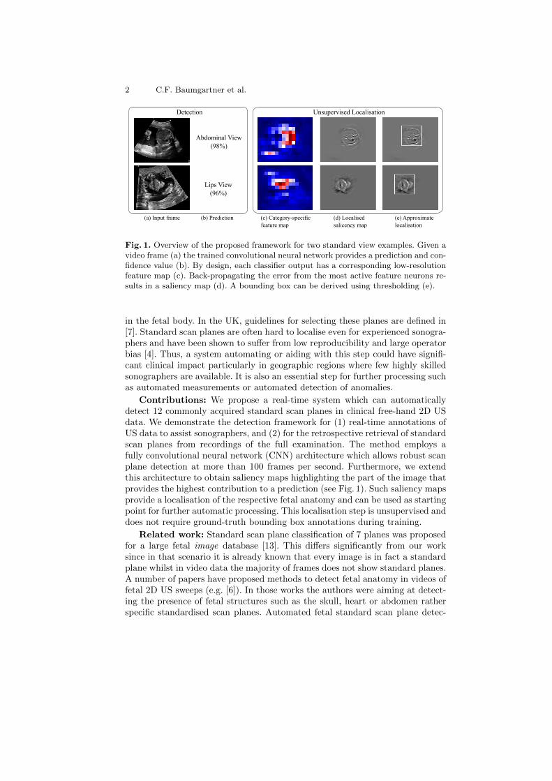
Abdominal View(98%)
Lips View(96%)
Detection Unsupervised Localisation
(a) Input frame (b) Prediction (c) Category-specific feature map
(d) Localised salicency map
(e) Approximatelocalisation
Fig. 1. Overview of the proposed framework for two standard view examples. Given avideo frame (a) the trained convolutional neural network provides a prediction and con-fidence value (b). By design, each classifier output has a corresponding low-resolutionfeature map (c). Back-propagating the error from the most active feature neurons re-sults in a saliency map (d). A bounding box can be derived using thresholding (e).
in the fetal body. In the UK, guidelines for selecting these planes are defined in[7]. Standard scan planes are often hard to localise even for experienced sonogra-phers and have been shown to suffer from low reproducibility and large operatorbias [4]. Thus, a system automating or aiding with this step could have signifi-cant clinical impact particularly in geographic regions where few highly skilledsonographers are available. It is also an essential step for further processing suchas automated measurements or automated detection of anomalies.
Contributions: We propose a real-time system which can automaticallydetect 12 commonly acquired standard scan planes in clinical free-hand 2D USdata. We demonstrate the detection framework for (1) real-time annotations ofUS data to assist sonographers, and (2) for the retrospective retrieval of standardscan planes from recordings of the full examination. The method employs afully convolutional neural network (CNN) architecture which allows robust scanplane detection at more than 100 frames per second. Furthermore, we extendthis architecture to obtain saliency maps highlighting the part of the image thatprovides the highest contribution to a prediction (see Fig. 1). Such saliency mapsprovide a localisation of the respective fetal anatomy and can be used as startingpoint for further automatic processing. This localisation step is unsupervised anddoes not require ground-truth bounding box annotations during training.
Related work: Standard scan plane classification of 7 planes was proposedfor a large fetal image database [13]. This differs significantly from our worksince in that scenario it is already known that every image is in fact a standardplane whilst in video data the majority of frames does not show standard planes.A number of papers have proposed methods to detect fetal anatomy in videos offetal 2D US sweeps (e.g. [6]). In those works the authors were aiming at detect-ing the presence of fetal structures such as the skull, heart or abdomen ratherspecific standardised scan planes. Automated fetal standard scan plane detec-

Real-time Standard Plane Detection and Localisation in Fetal Ultrasound 3
tion has been demonstrated for 1–3 standard planes in 2D fetal US sweeps [2,3, 8]. Notably, [2, 3] also employed CNNs. US sweeps are acquired by movingthe US probe from the cervix upwards in one continuous motion [3]. However,not all standard views required to determine the fetus’ health status are ade-quately visualised using a sweep protocol. For example, visualising the femuror the lips normally requires careful manual scan plane selection. Furthermore,data obtained using the sweep protocol are typically only 2–5 seconds long andconsist of fewer than 50 frames [3]. To the best of our knowledge, fetal stan-dard scan plane detection has never been performed on true free-hand US datawhich typically consist of 10,000+ frames. Moreover, none of related works weredemonstrated to run in real-time, typically requiring multiple seconds per frame.
2 Materials and Methods
Data and preprocessing: Our dataset consists of 1003 2D US scans of con-sented volunteers with gestational ages between 18–22 weeks which have beenacquired by a team of expert sonographers using GE Voluson E8 systems. Foreach scan a screen capture video of the entire procedure was recorded. Addition-ally, the sonographers saved “freeze frames” of a number of standard views foreach subject. A large fraction of these frames have been annotated allowing usto infer the correct ground-truth (GT) label. All video frames and images weredownsampled to a size of 225x273 pixels.
We considered 12 standard scan planes based on the guidelines in [7]. In par-ticular, we selected the following: two brain views at the level of the ventricles(Vt.) and the cerebellum (Cb.), the standard abdominal view, the transversekidney view, the coronal lip, the median profile, and the femur and spine views.We also included four commonly acquired cardiac views: the left and right ven-tricular outflow tracts (LVOT and RVOT), the three vessel view (3VV) and the4 chamber view (4CH)4. In addition to the labelled freeze frames, we sampled50 random frames from each video in order to model the background class, i.e.,the “not a standard scan plane” class.
Network architecture: The architecture of our proposed CNN is sum-marised in Fig. 2. Following recent advances in computer vision, we opted fora fully convolutional network architecture which replaces traditional fully con-nected layers with convolution layers using a 1x1 kernel [5, 9]. In the final convo-lutional layer (C6) the input is reduced to K 13x13 feature maps Fk, where K isthe number of classes. Each of these feature maps is then averaged to obtain theinput to the final Softmax layer. This architecture makes the network flexiblewith regard to the size of the input images. Larger images will simply result inlarger feature maps, which will nevertheless be mapped to a scalar for the finalnetwork output. We use this fact to train on cropped square images rather thanthe full field of view which is beneficial for data augmentation.
4 A detailed description of the considered standard planes is included in the sup-plementary material available at http://www.doc.ic.ac.uk/~cbaumgar/dwnlds/
miccai2016/.

4 C.F. Baumgartner et al.
...
Glo
bal A
vera
ge P
ooli
ng
Sof
tmax
...C1 (7x7/2) - MP C2 (5x5/2) - MP C3 (3x3/1) C4 (3x3/1) C5 (1x1/1) C6 (1x1/1)
225x225x1
55x55x32
13x13x64 13x13x128 13x13x128 13x13x64
13x13x
Fig. 2. Overview of the proposed network architecture. The size and stride of the con-volutional kernels are indicated at the top (notation: kernel size/stride). Max-poolingsteps are indicated by MP (2x2 bins, stride of 2). The activation functions of all con-volutions except C6 are rectified non-linear units (ReLUs). C6 is followed by a globalaverage pooling step. The sizes at the bottom of each image/feature map refer to thetraining phase and will be slightly larger during inference due to larger input images.
A key aspect of our proposed network architecture is that we enforce a one-to-one correspondence between each feature map Fk and the respective predictionyk. Since each neuron in the feature maps Fk has a receptive field in the originalimage, during training, the neurons will learn to activate only if an object of classk is in that field. This allows to interpret Fk as a spatially encoded confidencemap for class k [5]. In this paper, we take advantage of this fact to generatelocalised saliency maps as described below.
Training: We split our dataset into a test set containing 20% of the subjectsand a training set containing 80%. We use 10% of the training data as validationset to monitor the training progress. In total, we model 12 standard view planes,plus one background class resulting in K = 13 categories.
We train the model using mini-batch gradient descent and the categoricalcross-entropy cost function. In order to prevent overfitting we add 50% dropoutafter the C5 and C6 layers. To account for the significant class imbalance in-troduced by the background category, we create mini-batches with even class-sampling. Additionally, we augment each batch by a factor of 5 by taking 225x225square sub-images with a random horizontal translation and transforming themwith a small random rotation and flips along the vertical axis. Taking randomsquare sub-images allows to introduce more variation to the augmented batchesthan by training on the full field of view. We train the network for 50 epochsand choose the network parameters with the lowest error on the validation set.
Frame annotation and retrospective retrieval: After training we feedthe network with video frames containing the full field of view (225x273 pixels)of the input videos. This results in larger category-specific feature maps of 13x16.The prediction yk and confidence ck of each frame are given by the predictionwith the highest probability and the probability itself.
For retrospective frame retrieval, for each subject we calculate and record theconfidence for each class over the entire duration of an input video. Subsequently,we retrieve the frame with the highest confidence for each class.

Real-time Standard Plane Detection and Localisation in Fetal Ultrasound 5
Fig. 3. Saliency maps obtained from the input frame (LVOT class) shown on the left.The middle map was obtained using guided back-propagation from the average poollayer output [12]. The map on the right was obtained using our proposed method.
Saliency maps and unsupervised localisation: After obtaining the cat-egory yk of the current frame X from a forward pass through the network, wecan examine the feature map Fk (i.e. the output of the C6 layer) correspondingto the predicted category k. Two examples of feature maps are shown in Fig. 1c.The Fk could already be used to make an approximate estimate of the locationof the respective anatomy similar to [9].
Here, instead of using the feature maps directly, we present a novel methodto obtain localised saliency with the resolution of the original input images. For
each neuron F(p,q)k at the location p, q in the feature map it is possible calculate
how much each original input pixel X(i,j) contributed to the activation of thisneuron. This corresponds to calculating the partial derivatives
S(i,j)k =
∂F(p,q)k
∂X(i,j),
which can be solved efficiently using an additional backwards pass through thenetwork. [12] proposed a method for performing this back-propagation in a guidedmanner by allowing only error signals which contribute to an increase of theactivations in the higher layers (i.e. layers closer to the network output) to back-propagate. In particular, the error is only back-propagated through each neuron’sReLU unit if the input to the neuron x, as well as the error in the higher layerδ` are positive. That is, the back-propagated error δ`−1 of each neuron is givenby δ`−1 = δ`σ(x)σ(δ`), where σ(·) is the unit step function.
In contrast to [12] who back-propagated from the final output, in this work wetake advantage of the spatial encoding in the category specific feature maps andonly back-propagate the errors for the 10% most active feature map neurons, i.e.the spatial locations where the fetal anatomy is predicted. The resulting saliencymaps are significantly more localised compared to [12] (see Fig. 3).
These saliency maps can be used as starting point for various image analysistasks such as automated segmentation or measurements. Here, we demonstratehow they can be used for approximate localisation using basic image processing.We blur the absolute value image of a saliency map |Sk| using a 25x25 Gaussiankernel and apply a thresholding using Otsu’s method [10]. Finally, we computethe minimum bounding box of the components in the thresholded image.

6 C.F. Baumgartner et al.
Table 1. Precision pc = TP/(TP + FP ) and recall rc = TP/(TP + FN) for theclassification of the modelled scan planes. Background class: pc = 0.96, rc = 0.93.
view pc rc view pc rc view pc rc
Brain (Vt.) 0.96 0.90 Lips 0.85 0.88 LVOT 0.63 0.63Brain (Cb.) 0.92 0.94 Profile 0.71 0.82 RVOT 0.40 0.46Abdominal 0.85 0.80 Femur 0.79 0.93 3VV 0.46 0.60Kidneys 0.64 0.87 Spine 0.51 0.99 4CH 0.61 0.74
Table 2. % of correctly retrieved frames for each standard view for all 201 test subjects.
view % view % view %
Brain (Vt.) 0.95 Lips 0.77 LVOT 0.73Brain (Cb.) 0.89 Profile 0.76 RVOT 0.70Abdominal 0.79 Femur 0.75 3VV 0.66Kidneys 0.87 Spine 0.77 4CH 0.78
3 Experiments and Results
Frame annotation: We evaluated the ability of our method to detect standardframes by classifying the test data including the randomly sampled backgroundclass. We report the achieved precision (pc) and recall (rc) scores in Tab. 1. Thelowest scores were obtained for cardiac views, which are also the most difficultto scan for expert sonographers. This fact is reflected in the low detection ratesfor serious cardiac anomalies (e.g. only 35% in the UK).
[2] have recently reported pc/rc scores of 0.75/0.75 for the abdominal stan-dard view, and 0.77/0.61 for the 4CH view in US sweep data. We obtainedcomparable values for the 4CH view and considerably better values for the ab-dominal view. However, with 12 modelled standard planes and free-hand US dataour problem is significantly more complex. Using a Nvidia Tesla K80 graphicsprocessing unit (GPU) we were able to classify 113 frames per second (FPS) onaverage, which significantly exceeds the recording rate of the ultrasound machineof 25 FPS. We include an annotated video in the supplementary material.
Retrospective frame retrieval: We retrieved the standard views fromvideos of all test subjects and manually evaluated whether the retrieved framescorresponded to the annotated GT frames for each category. Several cases didnot have GTs for all views because they were not manually included by thesonographer in the original scan. For those cases we did not evaluate the retrievedframe. The results are summarised in Tab. 2. We show examples of the retrievedframes for two volunteers in Fig. 4. Note that in many cases the retrieved planesmatch the expert GT almost exactly. Moreover, some planes which were notannotated by the experts were nevertheless found correctly. As before, mostcardiac views achieved lower scores compared to other views.
Localisation: We show results for the approximate localisation of the re-spective fetal anatomy in the retrieved frames for one representative case inFig. 4b and in the supplemental video. We found that performing the localisa-tion reduced the frame rate to 39 FPS on average.

Real-time Standard Plane Detection and Localisation in Fetal Ultrasound 7
Fig. 4. Retrieved standard frames (RET ) and GT frames annotated and saved byexpert sonographers for two volunteers. Correctly retrieved and incorrectly retrievedframes are denoted with a green check mark or red cross, respectively. Frames with noGT annotation are indicated. The confidence is shown in the lower right of each image.The frames in (b) additionally contain the results of our proposed localisation (boxes).
4 Discussion and Conclusion
We have proposed a system for the automatic detection of twelve fetal standardscanplanes from real clinical fetal US scans. The employed fully CNN architec-ture allowed for robust real-time inference. Furthermore, we have proposed anovel method to obtain localised saliency maps by combining the information incategory-specific feature maps with a guided back-propagation step. To the bestof our knowledge, our approach is the first to model a large number of fetal stan-dard views from a substantial population of free-hand US scans. We have shown
8 C.F. Baumgartner et al.
that the method can be used to robustly annotate US data with classificationscores exceeding values reported in related work for some standard planes, butin a much more challenging scenario. A system based on our approach couldpotentially be used to assist or train inexperienced sonographers. We have alsoshown how the framework can be used to retrieve standard scan planes retro-spectively. In this manner, relevant key frames could be extracted from a videoacquired by an inexperienced operator and sent for further analysis to an ex-pert. We have also demonstrated how the proposed localised saliency maps canbe used to extract an approximate bounding box of the fetal anatomy. This isan important stepping stone for further, more specialised image processing.
Acknowledgments Supported by the Wellcome Trust IEH Award [102431].
References
1. Bull, C., et al.: Current and potential impact of fetal diagnosis on prevalenceand spectrum of serious congenital heart disease at term in the UK. The Lancet354(9186), 1242–1247 (1999)
2. Chen, H., Dou, Q., Ni, D., Cheng, J.Z., Qin, J., Li, S.and Heng, P.A.: Automaticfetal ultrasound standard plane detection using knowledge transferred recurrentneural networks. In: Proc MICCAI, pp. 507–514. Springer (2015)
3. Chen, H., Ni, D., Qin, J., Li, S., Yang, X., Wang, T., Heng, P.: Standard planelocalization in fetal ultrasound via domain transferred deep neural networks. IEEEJ Biomed Health Inform 19(5), 1627–1636 (2015)
4. Kurinczuk, J., Hollowell, J., Boyd, P., Oakley, L., Brocklehurst, P., Gray, R.: Thecontribution of congenital anomalies to infant mortality. National Perinatal Epi-demiology Unit, University of Oxford (2010)
5. Lin, M., Chen, Q., Yan, S.: Network in network. arXiv:1312.4400 (2013)6. Maraci, M., Napolitano, R., Papageorghiou, A., Noble, J.: Searching for structures
of interest in an ultrasound video sequence. In: Proc MLMI, pp. 133–140 (2014)7. NHS Screening Programmes: Fetal anomalie screen programme handbook pp. 28–
35 (2015)8. Ni, D., Yang, X., Chen, X., Chin, C.T., Chen, S., Heng, P.A., Li, S., Qin, J.,
Wang, T.: Standard plane localization in ultrasound by radial component modeland selective search. Ultrasound Med Biol 40(11), 2728–2742 (2014)
9. Oquab, M., Bottou, L., Laptev, I., Sivic, J.: Is object localization for free?-weakly-supervised learning with convolutional neural networks. In: IEEE Proc CVPR. pp.685–694 (2015)
10. Otsu, N.: A threshold selection method from gray-level histograms. Automatica11(285-296), 23–27 (1975)
11. Salomon, L., Alfirevic, Z., Berghella, V., Bilardo, C., Leung, K.Y., Malinger, G.,Munoz, H., et al.: Practice guidelines for performance of the routine mid-trimesterfetal ultrasound scan. Ultrasound Obst Gyn 37(1), 116–126 (2011)
12. Springenberg, J., Dosovitskiy, A., Brox, T., Riedmiller, M.: Striving for simplicity:The all convolutional net. arXiv:1412.6806 (2014)
13. Yaqub, M., Kelly, B., Papageorghiou, A., Noble, J.: Guided random forests foridentification of key fetal anatomy and image categorization in ultrasound scans.In: Proc MICCAI, pp. 687–694. Springer (2015)