incorporating differentials into every complete blood … · incorporating differentials into every...
TRANSCRIPT

Incorporating Differentials Into Every Complete Blood Count
Paige Flowers, LVT Dogwood Veterinary Internal Medicine

Complete Blood Count
Diagnostic performed to evaluate the quantity and morphology of the cellular components of blood. Automated cell count PCV/TS Manual Differential

Sample Collection and Handling
• Fill EDTA tubes until vacuum flow stops – Ensures appropriate blood volume to
anticoagulant ratio – Prevents overfilling
• If not analyzed within 2-3 hours, EDTA blood should be refrigerated – Minimal changes in refrigerator for up to 24 hours


Automated Cell Counters
Impedance Principle {aka: The Coultier Principle} Quantifies cells Identifies cells by size and density

Automated Cell Counters
• Platelet-erythrocyte histograms
– used to evaluate for mechanical errors – Overlap in the size of platelets and erythrocytes indicates the
machine cannot differentiate between the two

• A clear separation “valley” must be identified between erythrocyte and platelet peaks

Automated Cell Counters
• Laser Light Scatter – Laser detection system in a flow cytometer to
measure size and internal complexity of cells – Able to make distinctions that are undetectable
using impedance

Cytogram

PCV vs HCT – HCT
• (RBCs x MCV) 10 – PCV
• Centrifugation of blood to pack erythrocytes and determine a packed cell volume
The variation between HCT and PCV should not exceed 5%
• Suggests technical error

The Differential
• Complements automated cell counters • Often provides diagnostic information that an
automated cell counter cannot – Even without an automated cell count

The Blood Smear
• Prepare immediately • Monolayer
– Provides optimal morphologic detail
– Good distribution – Stains well – Cells lay flat

The Blood Smear
• Only cells with sufficient morphologic detail should be included in the differential cell count – A large percentage could be
cause for concern

LEUKOGRAM
• Total and differential leukocyte count
• Assessment of WBC morphologic features.

Neutrophils

Neutrophils
• Toxic neutrophils – Indicates more
severe disease, a worse prognosis and longer hospital stays
– Moderate to severe toxic changes can cause signs such as fever, vomiting, diarrhea or shock like symptoms


Eosinophils
• Orange - pink granules
• Shorter and less segmented than neutrophil nuclei • Cytoplasm, if visible, is pale blue.

Basophils
• Canine • ribbon-like nucleus • gray-lavender hue
• Feline
• small, slightly oval granules • pale lavender

Monocytes
• Variable in size and appearance
• Usually largest leukocyte
• Nucleus can be round to kidney-shaped to pseudo-lobulated

Lymphocytes
• small (mature) cells
• round nuclei
• smooth, dense chromatin
• small rim of light blue cytoplasm

Lymphocyte sizes
Lymphocyte size Nucleus size relative to neutrophil Small Nucleus of lymphocyte can fit
inside a neutrophil Intermediate Nucleus of lymphocyte is the
same size as a neutrophil Large Neutrophil can fit inside the
nucleus


Reactive Lymphocytes

Erythrogram
• Assessment of red blood cell numbers
• Assessment of
morphologic features

Erythropoesis

Spherocytes
• More spherical than normal disc shaped erythrocytes
• Do not lay flat - appear smaller
• Darker in color • Lack central palor

Autoagglutination
• Immune-induced aggregation of erythrocytes into clusters

Target Cell
• Resemble a bullseye target
• Regenerative anemia • Iron deficiency anemia

Acanthocytes
• Club shaped spicules protruding from erythrocyte

Howell-Jolly Body
• Blue in color • Perfectly round • Small fragments of
nonfunctional nucleus
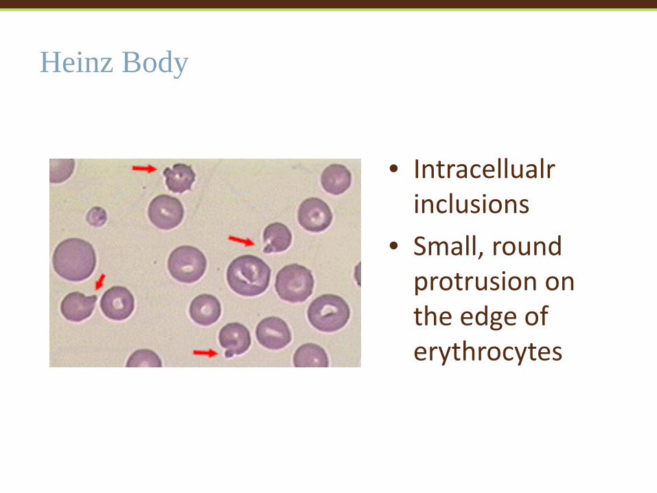
Heinz Body
• Intracellualr inclusions
• Small, round protrusion on the edge of erythrocytes

Mycoplasma
• Small, blue cocci or rod-like on the edges or across the faces of erythrocytes

Immature neutrophil vs monocyte vs reactive lymphocyte

Immature Erythrocyte vs Lymphocyte
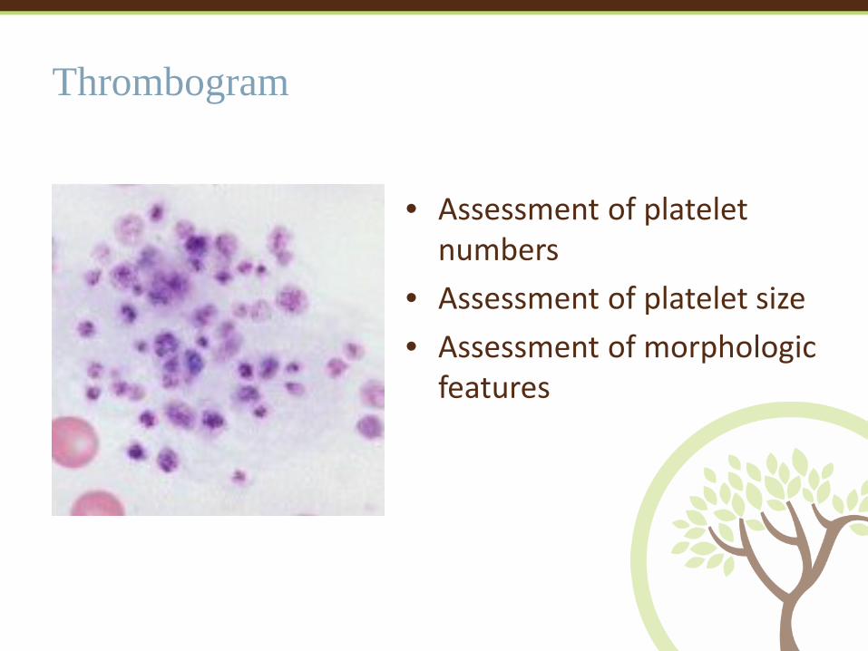
Thrombogram
• Assessment of platelet numbers
• Assessment of platelet size • Assessment of morphologic
features

Macroplatelets
• Large, immature platelet


Willard, DVM, MS, DACVIM, Michael, and Harold Tvedten, DVM, PhD, DACVP, DACVCP. "The Complete Blood Count, Bone Marrow Examination and Blood Banking." Small Animal Clinical Diagnosis by Laboratory Methods. Fifth ed. St. Louis: Elsevier, 2012. 12-29. Print. "Hemogram Basics." EClinpath. Cornell University College of Veterinary Medicine, 2013. Web. 16 Oct. 2015.