cytotune -ips 2.0 sendai reprogramming kit · sendai virus (sev) sendai virus is a respiratory...
TRANSCRIPT
USER GUIDE
CytoTune®-iPS 2.0 Sendai Reprogramming KitFor efficient, integration-free reprogramming of somatic cells into induced pluripotent stem cells (iPSC)
Catalog Numbers A16517, A16518
Publication Number MAN0009378
Revision 1.0
For Research Use Only. Not for use in diagnostic procedures.

Information in this document is subject to change without notice.
DISCLAIMER LIFE TECHNOLOGIES CORPORATION AND/OR ITS AFFILIATE(S) DISCLAIM ALL WARRANTIES WITH RESPECT TO THIS DOCUMENT, EXPRESSED OR IMPLIED, INCLUDING BUT NOT LIMITED TO THOSE OF MERCHANTABILITY, FITNESS FOR A PARTICULAR PURPOSE, OR NON-INFRINGEMENT. TO THE EXTENT ALLOWED BY LAW, IN NO EVENT SHALL LIFE TECHNOLOGIES AND/OR ITS AFFILIATE(S) BE LIABLE, WHETHER IN CONTRACT, TORT, WARRANTY, OR UNDER ANY STATUTE OR ON ANY OTHER BASIS FOR SPECIAL, INCIDENTAL, INDIRECT, PUNITIVE, MULTIPLE OR CONSEQUENTIAL DAMAGES IN CONNECTION WITH OR ARISING FROM THIS DOCUMENT, INCLUDING BUT NOT LIMITED TO THE USE THEREOF.
PURCHASER NOTIFICATION
CytoTune®-iPS 2.0 Sendai Reprogramming Kit This kit is a product of DNAVEC Corporation and supplied to the Life Technologies Corporation. The right to use the four Yamanaka factors for reprogramming in this kit has been granted by iPS Academia Japan to DNAVEC Corporation.
Important Licensing Information: These products may be covered by one or more Limited Use Label Licenses. By use of these products, you accept the terms and conditions of all applicable Limited Use Label Licenses.
TRADEMARKS The trademarks mentioned herein are the property of Life Technologies Corporation and/or its affiliate(s) or their respective owners. CytoTune is a registered trademark of DNAVEC Corporation. Parafilm is a registered trademark of Bemis Company, Inc. TaqMan is a registered trademark of Roche Molecular Systems, Inc, used under permission and license. Triton is a registered trademark of Union Carbide Corporation. TRIzol is a registered trademark of Molecular Research Center, Inc. © 2013 Life Technologies Corporation. All rights reserved.
1
Table of Contents Product Information ................................................................................................................... 2
Kit Contents and Storage ............................................................................................................................. 2
Description of the System ............................................................................................................................ 3
Safety Features of the System ..................................................................................................................... 6
Before You Begin ........................................................................................................................ 7 Guidelines for Generating iPSCs ................................................................................................................ 7
Reprogramming Fibroblasts ....................................................................................................... 9 Experiment Outline (Feeder-Dependent) ................................................................................................. 9
Reprogramming Fibroblasts (Feeder-Dependent) ................................................................................. 10
Experiment Outline (Feeder-Free) ........................................................................................................... 14
Reprogramming Fibroblasts (Feeder-Free) ............................................................................................. 15
Reprogramming PBMCs ........................................................................................................... 19 Experiment Outline .................................................................................................................................... 19
Reprogramming Peripheral Blood Mononuclear Cells (PBMCs) ........................................................ 20
Reprogramming CD34+ Cells .................................................................................................... 25 Experiment Outline .................................................................................................................................... 25
Reprogramming StemPro® CD34+ Cells .................................................................................................. 26
Identifying and Picking iPSC Colonies ....................................................................................... 31 Visual Identification ................................................................................................................................... 31
Live Staining ............................................................................................................................................... 32
Picking iPSC Colonies ................................................................................................................................ 34
Generating Vector-Free iPSCs .................................................................................................. 35 Generating Vector-Free iPSCs .................................................................................................................. 35
Troubleshooting ....................................................................................................................... 38 Appendix A: Recipes ................................................................................................................. 39
Preparing Media and Reagents ................................................................................................................ 39
Appendix B: Preparing Culture Vessels .................................................................................... 43 Preparing MEF Culture Dishes................................................................................................................. 43
Coating Culture Vessels with Vitronectin............................................................................................... 45
Coating Culture Vessels with Geltrex® Matrix ....................................................................................... 47
Appendix C: Support Protocols ................................................................................................. 48 CytoTune®-EmGFP Reporter Control Transduction ............................................................................. 48
Passaging iPSCs Using EDTA .................................................................................................................. 51
Cryopreserving iPSCs ................................................................................................................................ 52
Appendix D: Ordering Information ............................................................................................ 54 Accessory Products .................................................................................................................................... 54
Documentation and Support ..................................................................................................... 57 Obtaining Support ...................................................................................................................................... 57
References .................................................................................................................................................... 58
2
Product Information
Kit Contents and Storage
Kit contents The CytoTune®-iPS 2.0 Sendai Reprogramming Kit contains three CytoTune® 2.0
reprogramming vectors that are used for delivering and expressing key genetic factors necessary for reprogramming somatic cells into iPSCs.
The kit is available in two sizes: 1 pack (1 × 3 vials) and 3 packs (3 × 3 vials), with each vial containing 100 µL of one of the CytoTune® 2.0 reprogramming vector at a concentration of ≥ 8 × 10⁷ cell infectious units/mL (CIU/mL). Note: The titer of each CytoTune® 2.0 reprogramming vector is lot-dependent. For the specific titer of your vectors, refer to the Certificate of Analysis (CoA) available on our website. Go to www.lifetechnologies.com/cytotunegfp and search for the CoA by product lot number, which is printed on the vial.
Amount
Component Cap color A16517 A16518 CytoTune® 2.0 KOS clear 100 µL 3 × 100 µL
CytoTune® 2.0 hc-Myc white 100 µL 3 × 100 µL
CytoTune® 2.0 hKlf4 red 100 µL 3 × 100 µL
CytoTune® 2.0 reprogramming vectors are not compatible with the reprogramming vectors from the original CytoTune®-iPS Reprogramming Kits (Cat. nos. A13780-01, A13780-02). Do not mix or substitute CytoTune® 2.0 reprogramming vectors with the reprogramming vectors from the original kits.
Shipping and storage
x CytoTune®-iPS 2.0 Sendai Reprogramming Kit is shipped on dry ice.
x Immediately upon receipt, store each component at –80°C.
x Avoid repeated freezing and thawing of your reprogramming vectors. Viral titer is not guaranteed for kits that have been refrozen or thawed.
x Use the kit by the expiration date specified on the Certificate of Analysis (CoA).
Product use For Research Use Only. Not for use in diagnostic procedures.
This product must be used under Biosafety Level 2 (BL-2) containment with biological safety cabinet and laminar flow hood, and with appropriate personal safety equipment to prevent mucosal exposure/splash. For more information on BL-2 guidelines, see page 6.
3
Description of the System
Induced pluripotent stem cells (iPSC)
Induced pluripotent stem cells (iPSCs) are genetically reprogrammed adult cells which exhibit a pluripotent stem cell-like state similar to embryonic stem cells (Meissner et al., 2007; Park et al., 2008; Takahashi et al., 2007; Takahashi & Yamanaka, 2006; Wernig et al., 2007; Yu et al., 2007). While these artificially generated cells are not known to exist in the human body, they show qualities remarkably similar to those of embryonic stem cells (ESC); thus, they are an invaluable new source of pluripotent cells for drug discovery, cell therapy, and basic research.
There are multiple methods to generate iPSCs, including retrovirus-mediated gene transduction and chemical induction. While retroviral vectors require integration into host chromosomes to express reprogramming genes, DNA-based vectors such as adenovirus, adeno-associated virus, and plasmid vectors exist episomally and do not require integration; however, they may still be integrated into host chromosomes at certain frequencies. Unlike these vectors, the CytoTune® 2.0 reprogramming vectors do not integrate into the host genome or alter the genetic information of the host cell (Fusaki et al., 2009; Li et al., 2000; Seki et al., 2010).
CytoTune®-iPS 2.0 Reprogramming System
CytoTune®-iPS 2.0 Reprogramming System uses vectors based on a modified, non-transmissible form of Sendai virus (SeV) to safely and effectively deliver and express key genetic factors necessary for reprogramming somatic cells into iPSCs. In contrast to many available protocols, which rely on viral vectors that integrate into the genome of the host cell, the CytoTune®-iPS 2.0 Reprogramming System uses vectors that are non-integrating and remain in the cytoplasm (i.e., they are zero-footprint). In addition, the host cell can be cleared of the vectors and reprogramming factor genes by exploiting the cytoplasmic nature of SeV and the functional temperature sensitivity mutations introduced into the key viral proteins.
The CytoTune®-iPS 2.0 Sendai Reprogramming Kit contains three SeV-based reprogramming vectors, and are optimized for generating iPSCs from human somatic cells. The reprogramming vectors in this kit have been engineered to increase biological and environmental safety (see Safety Features of the System, page 6).
Continued on next page

4
Description of the System, continued
Sendai virus (SeV) Sendai virus is a respiratory virus of mouse and rat, classified as mouse
parainfluenza virus type I belonging to the Paramyxoviridae family. SeV was first isolated in Japan in the early 1950s (Kuroya et al., 1953) and is also called Hemagglutinating Virus of Japan (HVJ). SeV is an enveloped virus of 150–250 nm in diameter whose genome is a single chain RNA (15,384 bases) in the minus sense. Six genes coding for viral proteins are situated sequentially on the genome of the wild-type SeV in the following order (starting from the 3’ end):
x Nucleocapsid protein (NP) forms the core nucleocapsid complex with the genome RNA.
x Phosphoprotein (P) is the small subunit of the RNA polymerase.
x Matrix protein (M) supports the envelope structure from the inside.
x Fusion protein (F) fuses the viral envelope with cell membrane when the virus enters the cell. Note: The gene encoding the F protein is deleted from the CytoTune® 2.0 reprogramming vectors, rendering them incapable of producing infectious particles from infected cells (see page 6).
x Hemagglutinin-Neuraminidase (HN) recognizes the cell surface receptor, sialic acid.
x Large protein (L) is the large subunit of RNA polymerase.
Because SeV infects cells by attaching itself to the sialic acid receptor present on the surface of many different cells, it can infect a wide range of cell types of various animal species. Activation of F protein by a protease is required for the virus-cell fusion process to take place. After infection, the virus goes through genome replication and protein synthesis, and then daughter virus particles are assembled and released.
Figure 1 Comparison of the lifecycles of non-integrating SeV vectors and other, integrating vectors
Continued on next page

5
Description of the System, continued
CytoTune® 2.0 reprogramming vectors
The table below lists the CytoTune® 2.0 reprogramming vectors included in the CytoTune®-iPS 2.0 Sendai Reprogramming Kit. The reprogramming vectors include the four Yamanaka factors, Oct, Sox2, Klf4, and c-Myc, shown to be sufficient for efficient reprogramming (Takahashi et al., 2007).
CytoTune® Sendai vector Cap color Factor GenBank ID
CytoTune® 2.0 KOS clear Human Klf4 Human Oct3/4 Human Sox2
BC029923.1 NM_002701.4 NM_003106.2
CytoTune® 2.0 hc-Myc white Human c-Myc K02276.1
CytoTune® 2.0 hKlf4 red Human Klf4 BC029923.1
Advantages of CytoTune®-iPS 2.0 Sendai Reprogramming Kit
x No genotoxicity: CytoTune® 2.0 Sendai reprogramming vectors do not integrate into chromosomes of the target cells and potentially disrupt important genes.
x Wide range of targets: CytoTune® 2.0 Sendai reprogramming vectors are capable of transducing a wide range of cell types in proliferative and quiescent states.
x High transduction efficiency with low multiplicity of infection (MOI).
x Short contact time of virus with target cells is sufficient to establish transduction.
x High level of expression of the transgenes.
x Fast expression of the transgenes: expression is detectable as early as 6–10 hours after transduction, with maximum expression detected more than 24 hours after transduction.
x Zero footprint: the vectors and transgenes can be eliminated from the cells.
x No production of infectious particles by the transduced cells.
x Derived from a virus that is non-pathogenic to humans.
6
Safety Features of the System
Sendai virus (SeV) safety information
Host species: The host species for the Sendai virus (SeV) reported so far are mouse, rat, hamster, and guinea pigs, all of which have been described to be serologically positive.
Transmission: SeV is transmitted by aerosol and contact with respiratory secretions. The virus is highly contagious, but the infection does not persist in immunocompetent animals.
CytoTune® 2.0 Sendai reprogramming vectors: CytoTune® 2.0 Sendai reprogramming vectors in this kit are based on a modified, non-transmissible form of SeV, which has the Fusion protein (F) deleted, rendering the virus incapable of producing infectious particles from infected cells.
Inoculating animals with transduced cells: Although the CytoTune® 2.0 Sendai reprogramming vectors are non-transmissible, cells that have been exposed to the virus should be tested with PCR or antibody staining to ensure the absence of the virus before being inoculated into animals. Animals that have already been infected with wild type SeV may be able to make infectious CytoTune®-2.0 Sendai virus.
Non-transmissible CytoTune® 2.0 Sendai reprogramming vectors
SeV vectors used in this kit consist of viral proteins NP, P, M, F (activated), HN, and L, and the SeV genome RNA, from which the F gene is deleted. Because SeV infects cells by attaching itself to cell surface receptor sialic acid, present on the surface of many cell types of different species, the vectors are able to transduce a wide range of cells. However, they are no longer capable of producing infectious particles from infected cells, because the viral genome lacks the F-gene. In addition, the presence of functional mutations such as temperature sensitivity in the amino acid sequence of several SeV proteins (SeV/TSF, SeV/TS12F, and SeV/TS15F) renders the vectors easily removable from transduced cells. Note: SeV vectors used in this kit were developed by DNAVEC Corporation (http://www.dnavec.co.jp) and their rights for commercial use are the property of DNAVEC Corporation.
Biosafety Level 2
Although human is not the natural host for the SeV, and the virus is non-pathogenic to humans, appropriate care must be taken to prevent the potential mucosal exposure to the virus. This product must be used under Biosafety Level 2 (BL-2) containment with biological safety cabinet and laminar flow hood, and with appropriate personal safety equipment to prevent mucosal exposure/splash. In the event that the virus comes into contact with skin or eyes, decontaminate by flushing with plenty of water and consult a physician. For more information on BL-2 guidelines, refer to Biosafety in Microbiological and Biomedical Laboratories, 5th ed., published by the Centers for Disease Control, which is available for downloading at: www.cdc.gov/od/ohs/biosfty/bmbl5/bmbl5toc.htm.
7
Before You Begin
Guidelines for Generating iPSCs
Experimental guidelines
x To maintain sterile culture conditions, carry out all of the procedures in this manual using sterile laboratory practices in a laminar flow hood.
x You can use the CytoTune®-iPS 2.0 Sendai Reprogramming Kit to reprogram a wide range of cell types in proliferative and quiescent states. However, the reprogramming efficiency may vary between different cell types (~0.01%–1%).
x For successful reprogramming, transduce your cells using all three reprogramming vectors. Note: For successful reprogramming, all four Yamanaka factors (i.e., Oct4, Sox2, Klf4, and c-Myc) need to be expressed in your host cell.
x Cells that have already been infected with Sendai virus are refractive to further infection by Sendai virus. Therefore, you cannot transduce cells with CytoTune® 2.0 reprogramming vectors that have already been transduced with other Sendai vectors such as the CytoTune®-EmGFP Sendai Fluorescence Reporter or vice versa.
x One CytoTune®-iPS 2.0 Reprogramming Kit of three tubes supplies sufficient reagents to transduce a minimum of 1.5 × 10⁶ cells at MOI=5-5-3 (i.e., KOS MOI=5, hc-Myc MOI=5, hKlf4 MOI=3).
x The titer of each CytoTune® 2.0 Sendai reprogramming vector is lot-dependent. For the specific titer of your vectors, refer to the Certificate of Analysis (CoA) available on our website. Go to www.lifetechnologies.com/cytotunegfp and search for the CoA by product lot number, which is printed on the vial.
x Viral titers can decrease dramatically with each freeze/thaw cycle. Avoid repeated freezing and thawing of your reprogramming vectors. Viral titer is not guaranteed for kits that have been refrozen or thawed.
x Prior to starting, ensure that the media are equilibrated to 37°C and appropriately gassed.
CytoTune® 2.0 reprogramming vectors are not compatible with the reprogramming vectors from the original CytoTune®-iPS Reprogramming Kits (Cat. nos. A13780-01, A13780-02). Do not mix or substitute CytoTune® 2.0 reprogramming vectors with the reprogramming vectors from the original kits.
Positive control For positive control, we recommend performing a reprogramming experiment
with human neonatal foreskin fibroblast cells (strain BJ; ATCC no. CRL2522). Note that experimental conditions may vary among target cells and need to be optimized for each cell type. The example given in the following protocol does not guarantee the generation of iPSCs for all cell types.
Continued on next page
8
Guidelines for Generating iPSCs, continued
CytoTune®-EmGFP Sendai Fluorescence Reporter
The CytoTune®-EmGFP Sendai Fluorescence Reporter (Cat. no. A16519), available separately from Life Technologies, is a fluorescent control vector carrying the Emerald Green Fluorescent Protein (EmGFP) gene. The fluorescent control vector allows you to determine whether your cell line of interest is amenable or refractive to transduction by the Sendai reprogramming vectors, including the vectors from the original CytoTune®-iPS Sendai Reprogramming Kits. We recommend testing your cell lines of interest using the CytoTune®-EmGFP Sendai Fluorescence Reporter before starting your reprogramming experiments.
Note that you cannot transduce cells with CytoTune® reprogramming vectors that have already been transduced with the CytoTune®-EmGFP Sendai Fluorescence Reporter or vice versa. If you wish to use the CytoTune®-EmGFP Sendai Fluorescence Reporter during reprogramming, you must add it to the cells at the same time as the reprogramming vectors.
For detailed instructions on using the CytoTune®-EmGFP Sendai Fluorescence Reporter, see page 48.
9
Reprogramming Fibroblasts
Experiment Outline (Feeder-Dependent)
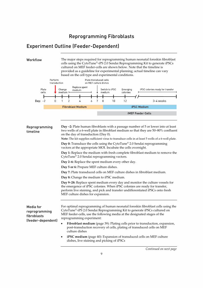
Workflow The major steps required for reprogramming human neonatal foreskin fibroblast
cells using the CytoTune®-iPS 2.0 Sendai Reprogramming Kit to generate iPSCs cultured on MEF feeder-cells are shown below. Note that the timeline is provided as a guideline for experimental planning; actual timeline can vary based on the cell type and experimental conditions.
Reprogramming timeline
Day –2: Plate human fibroblasts with a passage number of 5 or lower into at least two wells of a 6-well plate in fibroblast medium so that they are 50–80% confluent on the day of transduction (Day 0). Note: The kit supplies sufficient virus to transduce cells in at least 5 wells of a 6-well plate.
Day 0: Transduce the cells using the CytoTune® 2.0 Sendai reprogramming vectors at the appropriate MOI. Incubate the cells overnight.
Day 1: Replace the medium with fresh complete fibroblast medium to remove the CytoTune® 2.0 Sendai reprogramming vectors.
Day 2–6: Replace the spent medium every other day.
Day 5 or 6: Prepare MEF culture dishes.
Day 7: Plate transduced cells on MEF culture dishes in fibroblast medium.
Day 8: Change the medium to iPSC medium.
Day 9–28: Replace spent medium every day and monitor the culture vessels for the emergence of iPSC colonies. When iPSC colonies are ready for transfer, perform live staining, and pick and transfer undifferentiated iPSCs onto fresh MEF culture dishes for expansion.
Media for reprogramming fibroblasts (feeder-dependent)
For optimal reprogramming of human neonatal foreskin fibroblast cells using the CytoTune®-iPS 2.0 Sendai Reprogramming Kit to generate iPSCs cultured on MEF feeder-cells, use the following media at the designated stages of the reprogramming experiment:
x Fibroblast medium (page 39): Plating cells prior to transduction, expansion, post-transduction recovery of cells, plating of transduced cells on MEF culture dishes
x iPSC medium (page 40): Expansion of transduced cells on MEF culture dishes, live staining and picking of iPSCs
Continued on next page
10
Reprogramming Fibroblasts (Feeder-Dependent)
Materials needed Cells and vectors
x CytoTune® 2.0 Sendai reprogramming vectors Note: For successful reprogramming, you need all three tubes of reprogramming vectors.
x Human fibroblast cells to reprogram
x Optional: Human neonatal foreskin fibroblast cells (strain BJ; ATCC no. CRL2522) as a positive reprogramming control
x Gibco® Mouse Embryonic Fibroblasts (Irradiated) (Cat. no. S1520-100)
Media and reagents x DMEM with GlutaMAX™-I (high glucose) (Cat. no. 10569-010)
x KnockOut™ DMEM/F-12 (Cat. no. 12660-012)
x Fetal Bovine Serum (FBS), ES Cell-Qualified (Cat. no. 16141-079)
x KnockOut™ Serum Replacement (KSR) (Cat. no. 10828-028)
x MEM Non-essential Amino Acids (NEAA) (Cat. no. 11140-050)
x GlutaMAX™-I Supplement (Cat. no. 35050-061)
x Basic FGF, recombinant human (Cat. no. PHG0264)
x Ά-mercaptoethanol (Cat. no. 21985-023)
x Optional: Penicillin-Streptomycin, liquid (Cat. no. 15140-122)
x Attachment Factor (Cat. no. S-006-100)
x TrypLE™ Select Cell Dissociation Reagent (Cat. no. 12563) or 0.05% Trypsin/EDTA (Cat. no. 25300)
x Dulbecco’s PBS (DPBS) without Calcium and Magnesium (Cat. no. 14190)
CytoTune® 2.0 reprogramming vectors are not compatible with the reprogramming vectors from the original CytoTune®-iPS Reprogramming Kits (Cat. nos. A13780-01, A13780-02). Do not mix or substitute CytoTune® 2.0 reprogramming vectors with the reprogramming vectors from the original kits.
Continued on next page
11
Reprogramming Fibroblasts (Feeder-Dependent), continued
Reprogramming protocol
The following protocol has been optimized to transduce one well of human neonatal foreskin fibroblast cells (strain BJ; ATCC no. CRL2522), as a positive control. We recommend that you optimize the protocol for your cell type, and add an appropriate number of conditions/wells to utilize the entire volume of virus.
Day –2: Prepare the cells for transduction 1. Two days before transduction, plate human neonatal foreskin fibroblast cells
onto at least two wells of a 6-well plate at the appropriate density to achieve between 2 × 10⁵–3 × 10⁵ cells per well on the day of transduction (Day 0). One of the wells will be used to count cells for viral volume calculations. Note: Each CytoTune®-iPS 2.0 Sendai Reprogramming Kit supplies sufficient virus to transduce cells in at least 5 wells of a 6-well plate. We recommend using the entire volume of virus. Note: We recommend about 50–80% confluency on the day of transduction. Because overconfluency results in decreased transduction efficiency, we recommend replating your cells to achieve 50–80% confluency if your cells have become overconfluent during culturing.
2. Culture the cells for two more days, ensuring the cells have fully adhered and extended.
Day 0: Perform transduction 3. On the day of transduction, warm 1 mL of fibroblast medium in a water bath
(see page 39 for recipe) for each well to be transduced.
4. Harvest the cells from one well to perform a cell count. These cells will not be transduced, but will be used to estimate the cell number in the other well(s) plated in Step 1.
5. Remove the cells from this well using 0.5 mL of TrypLE™ Select reagent or 0.05% trypsin/EDTA following the procedure recommended by the manufacturer and incubating at room temperature. When the cells have rounded up (1–3 minutes later), add 1 mL of fibroblast medium into each well, and collect the cells in a 15-mL conical centrifuge tube.
6. Count the cells using the desired method (e.g., Countess® Automated Cell Counter), and calculate the volume of each virus needed to reach the target MOI using the live cell count and the titer information on the CoA.
Volume of virus (µL) = MOI (CIU/cell) ! number of cells
titer of virus (CIU/mL) ! 10–3 (µL/mL)
Note: We recommend initially performing the transductions with MOIs of 5, 5, and 3 (i.e., KOS MOI=5, hc-Myc MOI=5, hKlf4 MOI=3). These MOIs can be optimized for your application. Note: The titer of each CytoTune® 2.0 reprogramming vector is lot-dependent. For the specific titer of your vectors, go to www.lifetechnologies.com/cytotunegfp and search for the CoA by product lot number, which is printed on the vial. Avoid re-freezing and thawing of the reprogramming vectors since viral titers can decrease dramatically with each freeze/thaw cycle.
Continued on next page

12
Reprogramming Fibroblasts (Feeder-Dependent), continued
Reprogramming protocol, continued
7. Remove one set of CytoTune® 2.0 Sendai tubes from the –80°C storage. Thaw each tube one at a time by first immersing the bottom of the tube in a 37°C water bath for 5–10 seconds, and then removing the tube from the water bath and allowing it to thaw at room temperature. Once thawed, briefly centrifuge the tube and place it immediately on ice.
8. Add the calculated volumes of each of the three CytoTune® 2.0 Sendai tubes to 1 mL of fibroblast medium, pre-warmed to 37°C. Ensure that the solution is thoroughly mixed by pipetting the mixture gently up and down. Complete the next step within 5 minutes.
9. Aspirate the fibroblast medium from the cells, and add the reprogramming virus mixture prepared in Step 8 to the well containing the cells. Incubate the cells overnight in a 37°C incubator with a humidified atmosphere of 5% CO2.
Day 1: Replace medium and culture cells 10. 24 hours after transduction, replace the medium with fresh fibroblast
medium. Note: Depending on your cell type, you should expect to see some cytotoxicity 24–48 hours post-transduction, which can affect >50% of your cells. This is an indication of high uptake of the virus. We recommend that you continue culturing your cells and proceed with the protocol.
11. Culture the cells for 6 more days, changing the spent medium with fresh fibroblast medium every other day. Note: Depending on your cell type, you may observe high cell density before Day 5. We do not recommend passaging your cells onto MEF culture dishes before 7 days post-transduction. You may replace spent medium daily with fresh fibroblast medium if cultures become very dense.
Day 5 or 6: Prepare MEF culture dishes 12. One to two days before passaging the transduced fibroblasts onto MEF
feeder-cells, prepare 100-mm MEF culture dishes (see page 43).
Day 7: Plate transduced cells on MEF culture dishes 13. Seven days after transduction (Step 6, page 11), fibroblast cells are ready to
be harvested and plated on MEF culture dishes. Remove the medium from the fibroblasts, and wash cells once with D-PBS.
14. To remove the cells from the 6-well plate, use 0.5 mL of TrypLE™ Select reagent or 0.05% trypsin/EDTA following the procedure recommended by the manufacturer and incubate at room temperature. When the cells have rounded up (1–3 minutes later), add 2 mL of fibroblast medium into each well, and collect the cells in a 15-mL conical centrifuge tube. Note: Because the cells can be very sensitive to trypsin at this point, minimize trypsin exposure time and incubate the cells at room temperature.
15. Centrifuge the cells at 200 × g for 4 minutes, aspirate the medium, and re-suspend the cells in an appropriate amount of fibroblast medium.
Continued on next page

13
Reprogramming Fibroblasts (Feeder-Dependent), continued
Reprogramming protocol, continued
16. Count the cells using the desired method (e.g., Countess® Automated Cell Counter), and seed the MEF culture dishes with 5 × 10⁴–2 × 10⁵ cells per 100-mm dish and incubate overnight in a 37°C incubator with a humidified atmosphere of 5% CO2. Note: We recommend plating 5 × 10⁴, 1 × 10⁵, and 2 × 10⁵ cells per 100-mm dish. Depending on your cell type, you may need to plate most of your cells on the same plate to ensure sufficient numbers of colonies. Note: Set aside any remaining cells for RNA extraction to be used as a positive control in the RT-PCR detection of the SeV genome (see page 36).
Day 8 to 28: Feed and monitor the cells 17. 24 hours later, change the medium to iPSC medium (see page 40 for recipe),
and replace the spent medium every day thereafter.
18. Starting on Day 8, observe the plates every other day under a microscope for the emergence of cell clumps indicative of reprogrammed cells (see Visual identification, page 31). Note: For BJ fibroblasts, we normally observe colony formation on Day 12 post-transduction. However, depending on your cell type, you may need to culture for up to 4 weeks before seeing colonies.
19. Three to four weeks after transduction, colonies should have grown to an appropriate size for transfer. The day before transferring the colonies, prepare MEF culture plates using Attachment Factor-coated 12- or 24-well plates. Note: We typically harvest colonies closer to three weeks to avoid differentiation.
20. When colonies are ready for transfer, perform live staining using Tra1-60 or Tra1-81 for selecting reprogrammed colonies (see Live Staining, page 32).
21. Manually pick colonies and transfer them onto MEF plates (see Picking iPSC Colonies, page 34).

14
Experiment Outline (Feeder-Free)
Workflow The major steps required for reprogramming human neonatal foreskin fibroblast
cells using the CytoTune®-iPS 2.0 Sendai Reprogramming Kit to generate iPSCs cultured feeder-free on vitronectin-coated culture dishes are shown below. Note that the timeline is provided as a guideline for experimental planning; actual timeline can vary based on the cell type and experimental conditions.
Reprogramming timeline
Day –2: Plate human fibroblasts into at least two wells of a 6-well plate in fibroblast medium so that they are 50–80% confluent on the day of transduction (Day 0). Note: The kit supplies sufficient virus to transduce cells in at least 5 wells of a 6-well plate. Day 0: Transduce the cells using the CytoTune® 2.0 Sendai reprogramming vectors at the appropriate MOI. Incubate the cells overnight.
Day 1: Replace the medium with fresh complete fibroblast medium to remove the CytoTune® 2.0 Sendai reprogramming vectors.
Day 2–6: Replace the spent medium every other day.
Day 7: Plate transduced cells on vitronectin-coated culture dishes in fibroblast medium.
Day 8: Change the medium to complete Essential 8™ Medium.
Day 9–28: Replace spent medium every day and monitor the culture vessels for the emergence of iPSC colonies. When iPSC colonies are ready for transfer, perform live staining, and pick and transfer undifferentiated iPSCs onto fresh culture dishes for expansion.
Media for reprogramming fibroblasts (feeder-free)
For optimal reprogramming of human neonatal foreskin fibroblast cells using the CytoTune®-iPS 2.0 Sendai Reprogramming Kit to generate iPSCs cultured feeder free on vitronectin-coated culture dishes, use the following media at the designated stages of the reprogramming experiment:
x Fibroblast medium (page 39): Plating cells prior to transduction, expansion, post-transduction recovery of cells, plating of transduced cells on vitronectin-coated culture dishes
x Complete Essential 8™ Medium (page 40): Expansion of transduced cells on vitronectin-coated culture dishes, live staining and picking of iPSCs
Continued on next page
15
Reprogramming Fibroblasts (Feeder-Free)
Materials needed Cells and vectors
x CytoTune® 2.0 Sendai reprogramming vectors Note: For successful reprogramming, you need all three tubes of reprogramming vectors.
x Human fibroblast cells to reprogram
x Optional: Human neonatal foreskin fibroblast cells (strain BJ; ATCC no. CRL2522) as a positive reprogramming control
x Gibco® Mouse Embryonic Fibroblasts (Irradiated) (Cat. no. S1520-100)
Media and reagents x DMEM with GlutaMAX™-I (high glucose) (Cat. no. 10569-010)
x KnockOut™ DMEM/F-12 (Cat. no. 12660-012)
x Fetal Bovine Serum (FBS), ES Cell-Qualified (Cat. no. 16141-079)
x KnockOut™ Serum Replacement (KSR) (Cat. no. 10828-028)
x MEM Non-essential Amino Acids (NEAA) (Cat. no. 11140-050)
x GlutaMAX™-I Supplement (Cat. no. 35050-061)
x Basic FGF, recombinant human (Cat. no. PHG0264)
x Ά-mercaptoethanol, 55 mM (Cat. no. 21985-023)
x Optional: Penicillin-Streptomycin, liquid (Cat. no. 15140-122)
x Attachment Factor (Cat. no. S-006-100)
x TrypLE™ Select Cell Dissociation Reagent (Cat. no. 12563) or 0.05% Trypsin/EDTA (Cat. no. 25300)
x Dulbecco’s PBS (DPBS) without Calcium and Magnesium (Cat. no. 14190)
x Essential 8™ Medium (Cat. no. A1517001)
x Vitronectin, truncated recombinant human (VTN-N) (Cat. no. A14700)
CytoTune® 2.0 reprogramming vectors are not compatible with the reprogramming vectors from the original CytoTune®-iPS Reprogramming Kits (Cat. nos. A13780-01, A13780-02). Do not mix or substitute CytoTune® 2.0 reprogramming vectors with the reprogramming vectors from the original kits.
Continued on next page
16
Reprogramming Fibroblasts (Feeder-Free), continued
Reprogramming protocol
The following protocol has been optimized to transduce one well of human neonatal foreskin fibroblast cells (strain BJ; ATCC no. CRL2522), as a positive control. We recommend that you optimize the protocol for your cell type, and add an appropriate number of conditions/wells to utilize the entire volume of virus.
Day –2: Prepare the cells for transduction 1. Two days before transduction, plate human neonatal foreskin fibroblast cells
onto at least two wells of a 6-well plate at the appropriate density to achieve between 2 × 10⁵–3 × 10⁵ cells per well on the day of transduction (Day 0). One of the wells will be used to count cells for viral volume calculations. Note: Each CytoTune®-iPS 2.0 Sendai Reprogramming Kit supplies sufficient virus to transduce cells in at least 5 wells of a 6-well plate. We recommend using the entire volume of virus. Note: We recommend about 50–80% confluency on the day of transduction. Because overconfluency results in decreased transduction efficiency, we recommend replating your cells to achieve 50–80% confluency if your cells have become overconfluent during culturing.
2. Culture the cells for two more days, ensuring the cells have fully adhered and extended.
Day 0: Perform transduction 3. On the day of transduction, warm 1 mL of fibroblast medium in a water bath
(see page 39 for recipe) for each well to be transduced.
4. Harvest the cells from one well to perform a cell count. These cells will not be transduced, but will be used to estimate the cell number in the other well(s) plated in Step 1.
5. Remove the cells from the 6-well plate using 0.5 mL of TrypLE™ Select reagent or 0.05% trypsin/EDTA following the procedure recommended by the manufacturer and incubating at room temperature. When the cells have rounded up (1–3 minutes later), add 1 mL of fibroblast medium into each well, and collect the cells in a 15-mL conical centrifuge tube.
6. Count the cells using the desired method (e.g., Countess® Automated Cell Counter), and calculate the volume of each virus needed to reach the target MOI using the live cell count and the titer information on the CoA.
Volume of virus (µL) = MOI (CIU/cell) ! number of cells
titer of virus (CIU/mL) ! 10–3 (µL/mL)
Note: We recommend initially performing the transductions with MOIs of 5, 5, and 3 (i.e., KOS MOI=5, hc-Myc MOI=5, hKlf4 MOI=3). These MOIs can be optimized for your application. Note: The titer of each CytoTune® 2.0 reprogramming vector is lot-dependent. For the specific titer of your vectors, go to www.lifetechnologies.com/cytotunegfp and search for the CoA by product lot number, which is printed on the vial. Avoid re-freezing and thawing of the reprogramming vectors since viral titers can decrease dramatically with each freeze/thaw cycle.
Continued on next page
17
Reprogramming Fibroblasts (Feeder-Free), continued
Reprogramming protocol, continued
7. Remove one set of CytoTune® 2.0 Sendai tubes from the –80°C storage. Thaw each tube one at a time by first immersing the bottom of the tube in a 37°C water bath for 5–10 seconds, and then removing the tube from the water bath and allowing it to thaw at room temperature. Once thawed, briefly centrifuge the tube and place it immediately on ice.
8. Add the calculated volumes of each of the three CytoTune® 2.0 Sendai tubes to 1 mL of fibroblast medium, pre-warmed to 37°C. Ensure that the solution is thoroughly mixed by pipetting the mixture gently up and down. Complete the next step within 5 minutes.
9. Aspirate the fibroblast medium from the cells, and add the reprogramming virus mixture prepared in Step 8 to the well containing the cells. Incubate the cells overnight in a 37°C incubator with a humidified atmosphere of 5% CO2.
Day 1: Replace medium and culture cells 10. 24 hours after transduction, replace the medium with fresh fibroblast
medium. Note: Depending on your cell type, you should expect to see some cytotoxicity 24–48 hours post-transduction, which can affect >50% of your cells. This is an indication of high uptake of the virus. We recommend that you continue culturing your cells and proceed with the protocol.
11. Culture the cells for 6 more days, changing the spent medium with fresh fibroblast medium every other day. Note: Depending on your cell type, you may observe high cell density before Day 5. We do not recommend passaging your cells before 7 days post-transduction. You may replace spent medium daily with fresh fibroblast medium if cultures become very dense.
Day 7: Plate transduced cells on vitronectin-coated culture dishes 12. Coat a sufficient number of tissue culture dishes (e.g. 6-well, 60-mm, or
100-mm) with vitronectin (see page 45 for coating protocol). Note: Geltrex® Membrane Matrix can be substituted for vitronectin; see page 45 for coating protocol.
13. Seven days after transduction (Step 9, above), fibroblast cells are ready to be harvested and plated on vitronectin-coated culture dishes. Remove the medium from the fibroblasts, and wash cells once with D-PBS.
14. To remove the cells from the 6-well plate, use 0.5 mL of TrypLE™ Select reagent or 0.05% trypsin/EDTA following the procedure recommended by the manufacturer and incubate at room temperature. When the cells have rounded up (1–3 minutes later), add 2 mL of fibroblast medium into each well, and collect the cells in a 15-mL conical centrifuge tube. Note: Because the cells can be very sensitive to trypsin at this point, minimize trypsin exposure time and incubate the cells at room temperature.
15. Centrifuge the cells at 200 × g for 4 minutes, aspirate the medium, and re-suspend the cells in an appropriate amount of fibroblast medium.
Continued on next page

18
Reprogramming Fibroblasts (Feeder-Free), continued
Reprogramming protocol, continued
16. Count the cells using the desired method (e.g., Countess® Automated Cell Counter), and seed the vitronectin-coated culture dishes with 1 × 10⁵–5 × 10⁵ cells per 100-mm dish and incubate overnight in a 37°C incubator with a humidified atmosphere of 5% CO2. Note: Reprogramming efficiencies will typically be lower when using feeder-free conditions, so the number of cells plated should be increased accordingly. We recommend plating at least two different densities (e.g. 1 × 10⁵ and 5 × 10⁵ cells per 100-mm dish). Plating can also be scaled down to a 60-mm dish or 6-well plates. Depending on your cell type, you may need to plate most of your cells on the same plate to ensure sufficient numbers of colonies. Note: Set aside any remaining cells for RNA extraction to be used as a positive control in the RT-PCR detection of the SeV genome.
Day 8 to 28: Feed and monitor the cells 17. 24 hours later, change the medium to complete Essential 8™ Medium (see
page 40), and replace the spent medium every day thereafter.
18. Starting on Day 8, observe the plates every other day under a microscope for the emergence of cell clumps indicative of reprogrammed cells. Note: For BJ fibroblasts, we normally observe colony formation on Day 12 post-transduction. However, depending on your cell type, you may need to culture for up to 4 weeks before seeing colonies.
19. Three to four weeks after transduction, colonies should have grown to an appropriate size for transfer. When the colonies are ready for transfer, perform live staining using Tra1-60 or Tra1-81 for selecting reprogrammed colonies (see Live Staining, page 32). Note: We typically harvest colonies closer to three weeks to avoid differentiation.
20. Manually pick undifferentiated iPSC colonies (see Picking iPSC Colonies, page 34) and transfer them onto vitronectin-coated culture dishes for further expansion or analysis.

19
Reprogramming PBMCs
Experiment Outline
Workflow The major steps required for reprogramming peripheral blood mononuclear cells
(PBMCs) using the CytoTune®-iPS 2.0 Sendai Reprogramming Kit to generate iPSCs cultured on MEF feeder-cells are shown below. Note that the timeline is provided as a guideline for experimental planning; actual timeline can vary based on the cell type and experimental conditions.
Reprogramming timeline
Day –4: Plate peripheral blood mononuclear cells (PBMCs) at 5 × 10⁵ cells/mL to the middle section of a 24-well plate in complete PBMC medium.
Day –3 to –1: Replace half of the medium with 0.5 mL of fresh complete PBMC medium.
Day 0: Transduce the cells using the CytoTune® 2.0 Sendai reprogramming vectors at the appropriate MOI. Incubate the cells overnight.
Day 1: Replace the medium with fresh complete PBMC medium to remove the CytoTune® 2.0 Sendai reprogramming vectors. Prepare MEF culture dishes for use on Day 3.
Day 3: Plate the transduced cells on MEF culture dishes in complete StemPro®-34 medium without cytokines.
Day 4–6: Replace spent complete StemPro®-34 medium without cytokines every other day.
Day 7: Start transitioning into iPSC medium by replacing half of the StemPro®-34 medium without cytokines with complete iPSC medium.
Day 8: Replace the entire medium with complete iPSC medium to conclude the transitioning, and continue culturing cells on MEF culture dishes
Day 9–28: Replace spent medium with fresh complete iPSC medium every day and monitor the culture vessels for the emergence of iPSC colonies. When iPSC colonies are ready for transfer, perform live staining, and pick and transfer undifferentiated iPSCs onto fresh MEF culture dishes for expansion.
Continued on next page
20
Reprogramming Peripheral Blood Mononuclear Cells (PBMCs)
Media for reprogramming PBMCs (feeder-dependent)
For optimal reprogramming of PBMCs using the CytoTune®-iPS 2.0 Sendai Reprogramming Kit to generate iPSCs cultured on MEF feeder-cells, use the following media at the designated stages of the reprogramming experiment: x PBMC medium (page 39): Plating cells prior to transduction, expansion,
post-transduction recovery of cells
x StemPro®-34 medium without cytokines (page 41): Plating of transduced cells on MEF culture dishes
x iPSC medium (page 40): Expansion of transduced cells on MEF culture dishes, live staining and picking of iPSCs
Materials needed Cells and vectors
x CytoTune® 2.0 Sendai reprogramming vectors Note: For successful reprogramming, you need all three tubes of reprogramming vectors.
x Peripheral blood mononuclear cells (PBMCs) to reprogram Note: You can use PBMCs extracted from blood by a conventional method (i.e., Ficoll-Paque purification) or frozen PBMCs.
x Optional: Human neonatal foreskin fibroblast cells (strain BJ; ATCC no. CRL2522) as a positive reprogramming control Note: If you are using this as a control, follow the protocol for reprogramming fibroblasts within this manual (page 10).
x Gibco® Mouse Embryonic Fibroblasts (Irradiated) (Cat. no. S1520-100)
Media and reagents x StemPro®-34 SFM Medium (Cat. no. 10639-011)
x L-Glutamine (Cat. no. 25030)
x DMEM with GlutaMAX™-I (High Glucose) (Cat. no. 10569-010)
x KnockOut™ DMEM/F-12 (Cat. no. 12660-012)
x Fetal Bovine Serum (FBS), ES Cell-Qualified (Cat. no. 16141-079)
x KnockOut™ Serum Replacement (KSR) (Cat. no. 10828-028)
x MEM Non-Essential Amino Acids (NEAA) (Cat. no. 11140-050)
x GlutaMAX™-I Supplement (Cat. no. 35050-061
x Basic FGF, Recombinant Human (Cat. no. PHG0264)
x SCF (C-Kit Ligand), Recombinant Human (Cat. no. PHC2111)
x FLT-3 Ligand, Recombinant Human (Cat. no. PHC9414)
x IL-3, Recombinant Human (Cat. no. PHC 0034)
x IL-6, Recombinant Human (Cat. no. PHC0065)
x ȕ-Mercaptoethanol, 55 mM (Cat. no. 21985-023)
x Optional: Penicillin-Streptomycin, Liquid (Cat. no. 15140-122)
x Optional: Polybrene Hexadimethrine Bromide (Sigma, Cat. no. H9268)
x Attachment Factor (Cat. no. S-006-100)
x Dulbecco’s PBS (DPBS) without Calcium and Magnesium (Cat. no. 14190)
Continued on next page
21
Reprogramming PBMCs, continued
CytoTune® 2.0 reprogramming vectors are not compatible with the reprogramming vectors from the original CytoTune®-iPS Reprogramming Kits (Cat. nos. A13780-01, A13780-02). Do not mix or substitute CytoTune® 2.0 reprogramming vectors with the reprogramming vectors from the original kits.
Reprogramming protocol
The following protocol has been optimized for peripheral blood mononuclear cells (PBMCs) isolated through density gradient centrifugation via Ficoll-Paque and frozen in FBS and DMSO-containing medium. We recommend that you optimize the protocol for your cell type.
Day –4: Seed PBMCs 1. Four days before transduction, remove vial(s) of PBMCs from liquid nitrogen
storage. Thaw the vial quickly in 37°C water bath. When only a small ice crystal remains in the vial, remove it from the water bath. Spray the outside of the vial with 70% ethanol before placing it in the cell culture hood.
2. Gently transfer the PBMCs into a 15-mL conical tube. Slowly (drop-wise) add 5–10 mL pre-warmed complete PBMC medium (see page 41 for recipe) to the cell suspension. Remove an aliquot of cells to count and determine cell viability. Note: PBMC medium consists of complete StemPro®-34 medium containing the appropriate cytokines; aliquot the cytokines and add fresh daily.
3. Centrifuge the cell suspension at 200 × g for 10 minutes, discard the supernatant, and resuspend the cells in complete PBMC medium to 5 × 10⁵ cells/mL.
4. Add 1 mL per well to the middle section of a 24-well plate to prevent excessive evaporation of the medium during incubation.
5. Incubate the cells in a 37°C incubator with a humidified atmosphere of 5% CO2.
Day –3 to –1: Observe cells and add fresh medium 6. Count the cells daily, gently remove 0.5 mL of the medium from each well,
and replace it with 0.5 mL of fresh complete PBMC medium, trying not to disturb the cells. If cells are present in 0.5 mL removed from the wells, centrifuge the cell suspension at 200 × g for 10 minutes, discard the supernatant, and resuspend the cells in 0.5 mL fresh PBMC medium before adding them back to the plate. Note: Some cell death is generally observed the first day after the thaw. Some cells may adhere to the surface of the tissue culture plate. Proceed with the cells in suspension. Cells will not proliferate, but should maintain stable cell number for the first few days (PBMCs contain a variety of cells, and the current media system is only targeting a small population).
Continued on next page
22
Reprogramming PBMCs, continued
Reprogramming protocol, continued
Day 0: Count cells and perform transduction 7. Count the cells using the desired method (e.g., Countess® Automated Cell
Counter), and calculate the volume of each virus needed to reach the target MOI using the live cell count and the titer information on the CoA.
Volume of virus (µL) = MOI (CIU/cell) ! number of cells
titer of virus (CIU/mL) ! 10–3 (µL/mL)
Note: We recommend initially performing the transductions with MOIs of 5, 5, and 3 (i.e., KOS MOI=5, hc-Myc MOI=5, hKlf4 MOI=3). These MOIs can be optimized for your application. Note: The titer of each CytoTune® 2.0 reprogramming vector is lot-dependent. For the specific titer of your vectors, go to www.lifetechnologies.com/cytotunegfp and search for the CoA by product lot number, which is printed on the vial. Avoid re-freezing and thawing of the reprogramming vectors since viral titers can decrease dramatically with each freeze/thaw cycle.
8. Harvest the cells and seed the wells of a 12-well plate with 2.5 × 10⁵–5 × 10⁵ cells/well for transduction.
9. Remove CytoTune® 2.0 Sendai tubes from the –80°C storage. Thaw each tube one at a time by first immersing the bottom of the tube in a 37°C water bath for 5–10 seconds, and then removing the tube from the water bath and allowing it to thaw at room temperature. Once thawed, briefly centrifuge the tube and place it immediately on ice.
10. Add the calculated volumes of each of the three CytoTune™ 2.0 Sendai tubes to 1 mL of PBMC medium, pre-warmed to 37°C. Ensure that the solution is thoroughly mixed by pipetting the mixture gently up and down. Complete the next step within 5 minutes.
11. Seal the edges of the plate with Parafilm® laboratory film and centrifuge at 2250 rpm for 90 minutes at room temperature. Add an additional 1 mL of complete PBMC medium to each well and incubate the plate overnight at 37°C in a humidified atmosphere of 5% CO2. Note: If preferred, this centrifugation step can be performed in sterile, round-bottom culture tubes rather than in the 12-well plate. Transfer the cells and the medium containing the virus to a 12-well plate in a total volume of 2 mL for overnight incubation after centrifugation. Note: Although this centrifugation step is not required, it increases the transduction and reprogramming efficiencies. If the centrifugation step is omitted, transductions can be performed in a 24-well plate using 0.3 mL of total volume of cells, virus, and medium. Adding 4 µg/mL of Polybrene to the medium at the time of transduction may increase transduction efficiencies if the centrifugation step is not performed.
Day 1: Replace medium and culture cells 12. The next day, remove the cells and medium from the culture plate and
transfer to a 15-mL centrifuge tube. Rinse the well gently with 1 mL of medium to ensure most of the cells are harvested.
Continued on next page

23
Reprogramming PBMCs, continued
Reprogramming protocol, continued
13. Remove the CytoTune® 2.0 Sendai viruses by centrifuging the cell suspension at 200 × g for 10 minutes, aspirating the supernatant, and resuspending the cells in 0.5 mL of complete PBMC medium per well of a 24-well plate. Note: The cells may have drastic cell death (>60%); continue with the protocol using the live cell count. For the first 48 hours, observe the cells under the microscope for changes in cell morphology as a validation of transduction. Expect large, aggregated cells.
14. Culture the cells at 37°C in a humidified atmosphere of 5% CO2 for 2 days. Note: While the cells are incubating (1–2 days before plating the transduced cells), prepare MEF culture plates. You will need to have MEF feeder cells in at least two wells of a 6-well plate for each well of transduced cells (see page 43).
Day 3: Plate cells on MEF culture dishes 15. Count the cells using the desired method (e.g., Countess® Automated Cell
Counter) and seed the 6-well MEF culture plates with 10,000 and 50,000 live PBMCs per well in 2 mL of complete StemPro®-34 medium without the cytokines. Plate any excess cells in an additional MEF culture dish or harvest for extracting RNA to be used as a positive control in the RT-PCR detection of the SeV genome (see page 36).
16. Incubate the cells at 37°C in a humidified atmosphere of 5% CO2.
Day 4–6: Replace spent medium 17. Every other day, gently remove 1 mL (half) of the spent medium from the
cells and replace it with 1 mL of fresh complete StemPro®-34 medium without cytokines and without disturbing cells.
Day 7: Start transitioning cells to iPSC medium 18. Prepare 100 mL of complete iPSC medium as described on page 40.
19. Remove 1 mL (half) of StemPro®-34 medium from the cells and replace it with 1 mL of iPSC medium to start the adaptation of the cells to the new culture medium.
Day 8 to 28: Feed and monitor the cells 20. 24 hours later (day 8), change the full volume of the medium to iPSC medium,
and replace the spent medium every day thereafter.
21. Starting on day 8, observe the plates every other day under a microscope for the emergence of cell clumps indicative of reprogrammed cells (see Figure 2, page 24).
22. By day 15 to 21 after transduction, colonies should have grown to an appropriate size for transfer. The day before transferring the colonies, prepare MEF culture plates using Attachment Factor-coated 12- or 24-well plates (see page 43). Note: We typically harvest colonies closer to 3 weeks to avoid differentiation.
23. When colonies are ready for transfer, perform live staining using Tra1-60 or Tra1-81 for selecting reprogrammed colonies if desired (see Live Staining, page 32).
24. Manually pick colonies and transfer them onto prepared MEF plates (see Picking iPSC Colonies, page 34).
Continued on next page

24
Reprogramming PBMCs, continued
Expected results Figure 2 Colony formation for iPSC generated from PBMC. Cells are cultured in complete
PBMC medium (complete StemPro®-34 SFM + cytokines) for 4 days. On day 0 (panel A) cells are transduced overnight at an MOI of 5-5-3 (KOS MOI=5, hc-Myc MOI=5, hKlf4 MOI=3). At day 3 (panel B), the cells show morphological changes indicating reprogramming and are plated on MEF feeder layers. The cells are allowed to proliferate on MEF feeder layers and colony formation is observed from day 8 (panel C) to day 14 (panel D).
25
Reprogramming CD34+ Cells
Experiment Outline
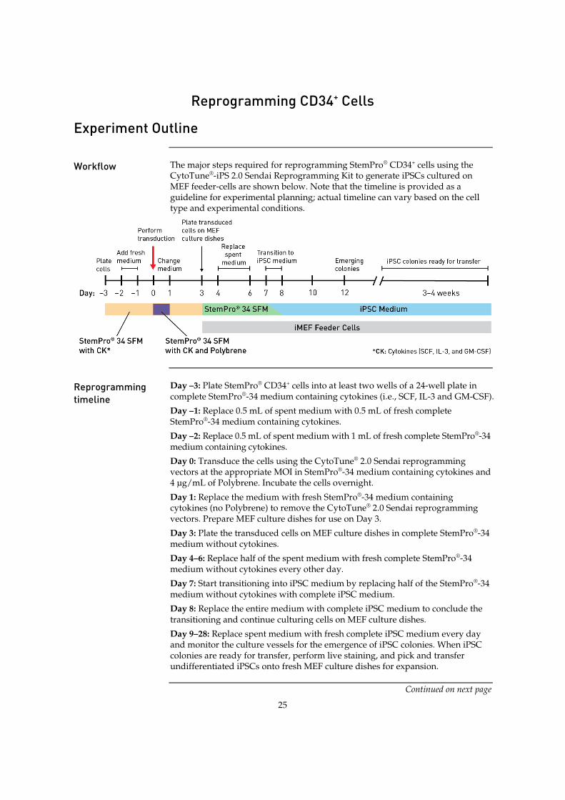
Workflow The major steps required for reprogramming StemPro® CD34+ cells using the
CytoTune®-iPS 2.0 Sendai Reprogramming Kit to generate iPSCs cultured on MEF feeder-cells are shown below. Note that the timeline is provided as a guideline for experimental planning; actual timeline can vary based on the cell type and experimental conditions.
Reprogramming timeline
Day –3: Plate StemPro® CD34+ cells into at least two wells of a 24-well plate in complete StemPro®-34 medium containing cytokines (i.e., SCF, IL-3 and GM-CSF).
Day –1: Replace 0.5 mL of spent medium with 0.5 mL of fresh complete StemPro®-34 medium containing cytokines.
Day –2: Replace 0.5 mL of spent medium with 1 mL of fresh complete StemPro®-34 medium containing cytokines.
Day 0: Transduce the cells using the CytoTune® 2.0 Sendai reprogramming vectors at the appropriate MOI in StemPro®-34 medium containing cytokines and 4 µg/mL of Polybrene. Incubate the cells overnight.
Day 1: Replace the medium with fresh StemPro®-34 medium containing cytokines (no Polybrene) to remove the CytoTune® 2.0 Sendai reprogramming vectors. Prepare MEF culture dishes for use on Day 3.
Day 3: Plate the transduced cells on MEF culture dishes in complete StemPro®-34 medium without cytokines.
Day 4–6: Replace half of the spent medium with fresh complete StemPro®-34 medium without cytokines every other day.
Day 7: Start transitioning into iPSC medium by replacing half of the StemPro®-34 medium without cytokines with complete iPSC medium.
Day 8: Replace the entire medium with complete iPSC medium to conclude the transitioning and continue culturing cells on MEF culture dishes.
Day 9–28: Replace spent medium with fresh complete iPSC medium every day and monitor the culture vessels for the emergence of iPSC colonies. When iPSC colonies are ready for transfer, perform live staining, and pick and transfer undifferentiated iPSCs onto fresh MEF culture dishes for expansion.
Continued on next page

26
Reprogramming StemPro® CD34+ Cells
Media for reprogramming StemPro® CD34+ cells (feeder-dependent)
For optimal reprogramming of CD34+ cells using the CytoTune®-iPS 2.0 Sendai Reprogramming Kit to generate iPSCs cultured on MEF feeder-cells, use the following media at the designated stages of the reprogramming experiment:
x StemPro®-34 medium containing cytokines (i.e., SCF, IL-3 and GM-CSF) (page 41): Plating cells prior to transduction, post-transduction recovery of cells
x StemPro®-34 medium containing cytokines + 4 µg/mL of Polybrene: Transduction
x StemPro®-34 medium without cytokines (page 41): Plating of transduced cells on MEF culture dishes
x iPSC medium (page 40): Expansion of transduced cells on MEF culture dishes, live staining and picking of iPSCs
Materials needed Cells and vectors
x CytoTune® 2.0 Sendai reprogramming vectors Note: For successful reprogramming, you need all three tubes of reprogramming vectors.
x StemPro® CD34+ cells to reprogram Note: StemPro® CD34+ cells are available as part of the StemPro®-34 Medium and CD34+ Cell Kit (Cat. no. A14059) from Life Technologies.
x Optional: Human neonatal foreskin fibroblast cells (strain BJ; ATCC no. CRL2522) as a positive reprogramming control Note: If you are using this as a control, follow the protocol for reprogramming fibroblasts within this manual (page 10).
x Gibco® Mouse Embryonic Fibroblasts (Irradiated) (Cat. no. S1520-100)
Media and reagents x StemPro®-34 Medium and CD34+ Cell Kit (Cat. no. A14059)
x Recombinant Human SCF Lyophilized (Cat. no. PHC2111)
x Recombinant Human IL-3 Lyophilized (Cat. no. PHC0031)
x Recombinant Human GM-CSF Lyophilized (Cat. no. PHC2011)
x Dulbecco’s Modified Eagle Medium (DMEM), High Glucose, with GlutaMAX™-I and Pyruvate (Cat. no. 10569-010)
x KnockOut™ DMEM/F-12 (Cat. no. 12660-012)
x Fetal Bovine Serum (FBS), ESC-Qualified, US Origin (Cat. no. 16141-079)
x KnockOut™ Serum Replacement (KSR) (Cat. no. 10828-028)
x MEM Non-Essential Amino Acids Solution, 10 mM (Cat. no. 11140-050)
x GlutaMAX™-I (100X) (Cat. no. 35050-061)
x Basic Fibroblast Growth Factor (bFGF) (Cat. no. PHG0264)
x ȕ-mercaptoethanol, 1000X (Cat. no. 21985-023)
Continued on next page

27
Reprogramming StemPro® CD34+ Cells, continued
Materials needed, continued
x Optional: Penicillin-Streptomycin, Liquid (Cat. no. 15140-122)
x Polybrene Hexadimethrine Bromide (Sigma Cat. no. H9268)
x Attachment Factor (Cat. no. S006100)
x TrypLE™ Select Cell Dissociation Reagent (Cat. no. 12563) or 0.05% Trypsin/EDTA (Cat. no. 25300)
x Dulbecco’s PBS (DPBS) without Calcium and Magnesium (Cat. no. 14190)
CytoTune® 2.0 reprogramming vectors are not compatible with the reprogramming vectors from the original CytoTune®-iPS Reprogramming Kits (Cat. nos. A13780-01, A13780-02). Do not mix or substitute CytoTune® 2.0 reprogramming vectors with the reprogramming vectors from the original kits.
Reprogramming protocol
The following protocol has been optimized for StemPro® CD34+ cells derived from the human umbilical cord blood of mixed donors. Note that experimental conditions may vary among target cells and need to be optimized for each cell type. The example given in the following protocol does not guarantee the generation of iPSCs for all cell types.
Day –3: Seed cells 1. Three days before transduction, remove one vial of StemPro® CD34+ cells
(0.5 × 10⁶ cells) from the liquid nitrogen storage tank.
2. Briefly roll the cryovial between hands to remove frost, and swirl it gently in a 37°C water bath to thaw the StemPro® CD34+ cells.
3. When only a small ice crystal remains in the vial, remove it from water bath. Spray the outside of the vial with 70% ethanol before placing it in the cell culture hood.
4. Pipet the thawed cells gently into a 15-mL conical tube.
5. Add 10 mL of pre-warmed complete StemPro®-34 medium (see page 41) drop-wise to the cells. Gently mix by pipetting up and down. Note: Adding the medium slowly helps the cells to avoid osmotic shock.
6. Centrifuge the cell suspension at 200 × g for 10 minutes.
7. Discard the supernatant and resuspend the cells in 1 mL of complete StemPro®-34 medium containing cytokines (i.e., SCF, IL-3 and GM-CSF) (see page 41).
8. Place 0.5 mL each of cell suspension into two wells of a 24-well plate and incubate at 37°C in a humidified atmosphere of 5% CO2. Note: We recommend using the wells in the middle section of the 24-well plate to prevent excessive evaporation of the medium during incubation.
Day –2: Observe cells and add fresh medium 9. Two days before transduction, add 0.5 mL of fresh complete StemPro®-34
medium containing cytokines without disturbing the cells.
Continued on next page

28
Reprogramming StemPro® CD34+ Cells, continued
Reprogramming protocol
Day –1: Observe cells and add fresh medium 10. One day before transduction, gently remove 0.5 mL of medium and add 1 mL
of fresh complete StemPro®-34 medium containing cytokines without disturbing the cells.
Figure 3 CD34+ cells grown in StemPro®-34 SFM.
Day 0: Count cells and perform transduction 11. Count the cells using the desired method (e.g., Countess® Automated Cell
Counter), and calculate the volume of each virus needed to reach the target MOI using the live cell count and the titer information on the CoA.
Volume of virus (µL) = MOI (CIU/cell) ! number of cells
titer of virus (CIU/mL) ! 10–3 (µL/mL)
Note: We recommend initially performing the transductions with MOIs of 5, 5, and 3 (i.e., KOS MOI=5, hc-Myc MOI=5, hKlf4 MOI=3). These MOIs can be optimized for your application. Note: The titer of each CytoTune® 2.0 reprogramming vector is lot-dependent. For the specific titer of your vectors, go to www.lifetechnologies.com/cytotunegfp and search for the CoA by product lot number, which is printed on the vial. Avoid re-freezing and thawing of the reprogramming vectors since viral titers can decrease dramatically with each freeze/thaw cycle.
12. Harvest the cells and seed the necessary number of wells of a 24-well plate in a minimal volume (~100 µL) with 1.0 × 10⁵ cells/well for transduction.
13. Remove one set of CytoTune® 2.0 Sendai tubes from the –80°C storage. Thaw each tube one at a time by first immersing the bottom of the tube in a 37°C water bath for 5–10 seconds, and then removing the tube from the water bath and allowing it to thaw at room temperature. Once thawed, briefly centrifuge the tube and place it immediately on ice.
Continued on next page

29
Reprogramming StemPro® CD34+ Cells, continued
Reprogramming protocol, continued
14. Add the calculated volumes of each of the three CytoTune® 2.0 Sendai viruses to 0.4 mL of pre-warmed StemPro®-34 medium containing cytokines and 4 µg/mL of Polybrene. Ensure that the solution is thoroughly mixed by pipetting the mixture gently up and down. Complete the next step within 5 minutes.
15. Add the reprogramming virus mixture (from Step 14) to the well(s) containing cells (from Step 12). Incubate the cells at 37°C in a humidified atmosphere of 5% CO2 overnight.
Day 1: Replace medium and culture cells 16. Remove the CytoTune® 2.0 Sendai viruses by centrifuging the cells at 400 × g
for 10 minutes. Aspirate and discard the supernatant.
17. Resuspend the cells in 0.5 mL of complete StemPro®-34 Medium containing cytokines (see page 41) in the 24-well plate.
18. Incubate the cells in at 37°C in a humidified atmosphere of 5% CO2 for two days. Note: While the cells are incubating (i.e., 1–2 days before passaging the transduced cells), prepare the necessary number of MEF culture dishes for each well containing transduced cells (see page 43).
Day 3: Plate cells on MEF dishes 19. Count the cells using the desired method (e.g., Countess® Automated Cell
Counter) and seed the MEF dishes with 5 × 10⁴ and 1 × 10⁵ CD34+ cells per 60-mm dish in 5 mL of complete StemPro®-34 Medium without cytokines. Plate any excess cells in an additional MEF culture dish or harvest for RNA extraction to be used as a positive control in the RT-PCR detection of the SeV genome (see page 36)
20. Incubate the cells at 37°C in a humidified atmosphere of 5% CO2 for three days.
21. Replace half of the spent medium every other day. Gently remove 2.5 mL of medium from the cells and replace with 2.5 mL of complete StemPro®-34 Medium without cytokines.
Day 7: Transition to iPSC medium 22. Remove 2.5 mL of medium from the cells and add 2.5 mL of iPSC medium
(see page 40) to transition the cells to the new culture medium.
23. Incubate the cells in a 37°C, 5% CO2 incubator overnight.
Continued on next page
30
Reprogramming StemPro® CD34+ Cells, continued
Reprogramming protocol, continued
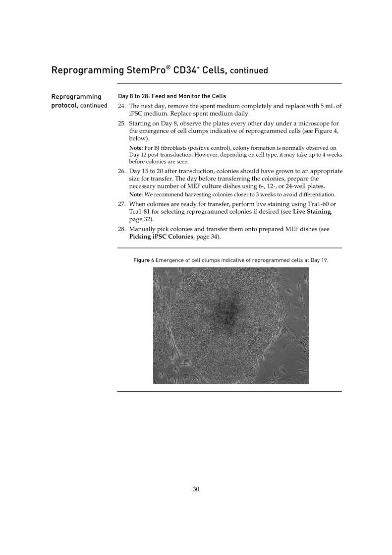
Day 8 to 28: Feed and Monitor the Cells 24. The next day, remove the spent medium completely and replace with 5 mL of
iPSC medium. Replace spent medium daily.
25. Starting on Day 8, observe the plates every other day under a microscope for the emergence of cell clumps indicative of reprogrammed cells (see Figure 4, below). Note: For BJ fibroblasts (positive control), colony formation is normally observed on Day 12 post-transduction. However, depending on cell type, it may take up to 4 weeks before colonies are seen.
26. Day 15 to 20 after transduction, colonies should have grown to an appropriate size for transfer. The day before transferring the colonies, prepare the necessary number of MEF culture dishes using 6-, 12-, or 24-well plates. Note: We recommend harvesting colonies closer to 3 weeks to avoid differentiation.
27. When colonies are ready for transfer, perform live staining using Tra1-60 or Tra1-81 for selecting reprogrammed colonies if desired (see Live Staining, page 32).
28. Manually pick colonies and transfer them onto prepared MEF dishes (see Picking iPSC Colonies, page 34).
Figure 4 Emergence of cell clumps indicative of reprogrammed cells at Day 19.
31
Identifying and Picking iPSC Colonies
Visual Identification
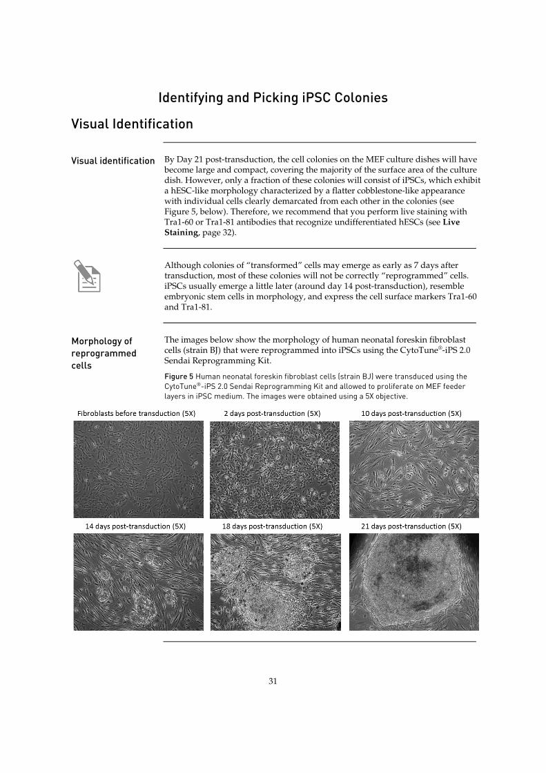
Visual identification By Day 21 post-transduction, the cell colonies on the MEF culture dishes will have
become large and compact, covering the majority of the surface area of the culture dish. However, only a fraction of these colonies will consist of iPSCs, which exhibit a hESC-like morphology characterized by a flatter cobblestone-like appearance with individual cells clearly demarcated from each other in the colonies (see Figure 5, below). Therefore, we recommend that you perform live staining with Tra1-60 or Tra1-81 antibodies that recognize undifferentiated hESCs (see Live Staining, page 32).
Although colonies of “transformed” cells may emerge as early as 7 days after transduction, most of these colonies will not be correctly “reprogrammed” cells. iPSCs usually emerge a little later (around day 14 post-transduction), resemble embryonic stem cells in morphology, and express the cell surface markers Tra1-60 and Tra1-81.
Morphology of reprogrammed cells
The images below show the morphology of human neonatal foreskin fibroblast cells (strain BJ) that were reprogrammed into iPSCs using the CytoTune®-iPS 2.0 Sendai Reprogramming Kit.
Figure 5 Human neonatal foreskin fibroblast cells (strain BJ) were transduced using the CytoTune®-iPS 2.0 Sendai Reprogramming Kit and allowed to proliferate on MEF feeder layers in iPSC medium. The images were obtained using a 5X objective.
32
Live Staining
Live staining with antibodies
One of the fastest and most reliable methods for identifying a reprogrammed colony is live staining with Tra1-60 or Tra1-81 antibodies that recognize undifferentiated iPSCs and enable the identification of reprogrammed cells from a variety of human cell types. Note: Other methods of identifying iPSCs (such as alkaline phosphatase staining) are also acceptable.
Required antibodies
Primary Antibodies You can use one or more of the following primary antibodies, diluted to the appropriate working concentration (see instructions provided with the antibody) in KnockOut™ DMEM/F-12.
x Mouse anti-Tra1-60 antibody (Cat. no. 41-1000)
x Mouse anti-Tra1-81 antibody (Cat. no. 41-1100)
x Mouse anti-SSEA4 (Cat. no. 41-4000)
Secondary Antibodies You can use one or more of the following secondary antibodies, diluted to the appropriate working concentration (see instructions provided with the antibody) in KnockOut™ DMEM/F-12.
x Alexa Fluor® 488 goat anti-mouse IgG (H+L) antibody (Cat. no. A11029)
x Alexa Fluor® 594 goat anti-mouse IgG (H+L) antibody (Cat. no. A11032)
Live staining protocol
If live-stained cells are to be used for further culture, be sure to use antibodies that are sterile (filter sterilize as necessary) and work aseptically. .
1. Aspirate the medium from the reprogramming dish.
2. Wash the cells once with 1X KnockOut™ DMEM/F-12.
3. Add the diluted primary antibody to the cells (2 mL per 60-mm dish, 6 mL per 100-mm dish).
4. Incubate the primary antibody and the cells at 37°C for 60 minutes.
5. Remove the primary antibody solution from the dish. Note: The primary antibody solution can be stored at 4°C for 1 week and re-used up to 2 times.
6. Wash cells three times with KnockOut™ DMEM/F-12.
7. Add the diluted secondary antibody to the cells (2 mL per 60-mm dish, 6 mL per 100-mm dish).
Continued on next page
33
Live Staining, continued
Live staining protocol, continued
8. Incubate the secondary antibody and the cells at 37°C for 60 minutes.
9. Remove the secondary antibody solution from the dish. Note: The secondary antibody solution can be stored at 4°C for 1 week and re-used up to 2 times.
10. Wash cells three times with KnockOut™ DMEM/F-12 and add fresh KnockOut™ DMEM/F-12 to cover the surface of the cells (2 mL per 60-mm dish, 6 mL per 100-mm dish).
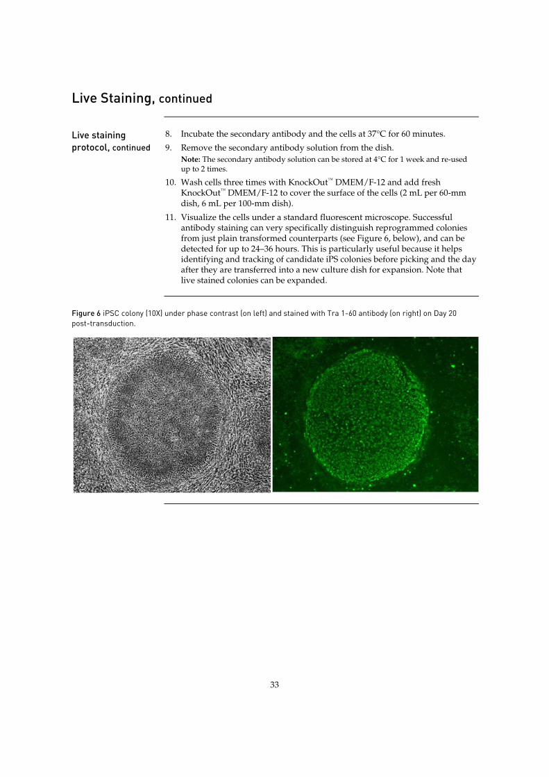
11. Visualize the cells under a standard fluorescent microscope. Successful antibody staining can very specifically distinguish reprogrammed colonies from just plain transformed counterparts (see Figure 6, below), and can be detected for up to 24–36 hours. This is particularly useful because it helps identifying and tracking of candidate iPS colonies before picking and the day after they are transferred into a new culture dish for expansion. Note that live stained colonies can be expanded.
Figure 6 iPSC colony (10X) under phase contrast (on left) and stained with Tra 1-60 antibody (on right) on Day 20 post-transduction.
34
Picking iPSC Colonies
Protocol for picking iPSC colonies (feeder-dependent)
1. Place the culture dish containing the reprogrammed cells under an inverted microscope and examine the colonies under 10X magnification.
2. Mark the colony to be picked on the bottom of the culture dish. Note: We recommend picking at least 10 distinct colonies by the end of each reprogramming experiment and expanding them in separate 24-well MEF culture plates (see below).
3. Transfer the culture dish to a sterile cell culture hood (i.e., biosafety cabinet) equipped with a stereomicroscope.
4. Using a 25 gauge 1½ inch needle, cut the colony to be picked into 5–6 pieces in a grid-like pattern.
5. Using a 200 µL pipette, transfer the cut pieces to a freshly prepared 24-well MEF culture plate (see page 43) containing iPSC medium (see page 40).
6. Incubate the MEF culture plate containing the picked colonies in a 37°C incubator with a humidified atmosphere of 5% CO2.
7. Allow the colonies to attach to the culture plate for 48 hours before replacing the spent medium with fresh iPSC medium. After that, change the medium every day.
8. Treat the reprogrammed colonies like normal human ESC colonies and passage, expand, and maintain them using standard culture procedures until you have frozen cells from two 60-mm plates (see Freezing iPSCs in iPSC freezing medium, page 52).
Protocol for picking iPSC colonies (feeder-free)
1. Pick the iPSCs as described on above, up to step 4.
2. Using a 200 μL pipette, transfer the cut pieces onto a vitronectin-coated culture plate (see page 45) containing complete Essential 8™ Medium (page 40).
3. Incubate the vitronectin-culture plate containing the picked colonies in a 37°C incubator with a humidified atmosphere of 5% CO2.
4. Allow the colonies to attach to the culture plate for 48 hours before replacing the spent medium with fresh complete Essential 8™ Medium. After that, change the medium every day.
5. When the colonies cover ~85% of the surface area of the culture vessel, they are ready for passaging. Passage the colonies using 0.5 mM EDTA prepared in Dulbecco's Phosphate-Buffered Saline (DPBS) without calcium or magnesium (see Passaging iPSCs Using EDTA, page 51). Note: Enzymes such as collagenase and dispase do not work well with cells cultured in Essential 8™ Medium on vitronectin-coated plates. Use of these enzymes for passaging cells results in compromised viability and attachment.
6. Continue to culture, expand, and maintain the reporgrammed colonies in complete Essential 8™ Medium until you have frozen cells from two 60-mm plates (see Freezing iPSCs in Essential 8™ Freezing Medium, page 53).
35
Generating Vector-Free iPSCs
Generating Vector-Free iPSCs
Guidelines for generating vector-free iPSCs
x The time needed to derive vector-free iPSCs may vary depending on culture and passage conditions. In the case of human neonatal foreskin fibroblast cells (strain BJ), it takes about 1–2 months after gene transduction to obtain iPSCs free of CytoTune® 2.0 Sendai reprogramming vectors.
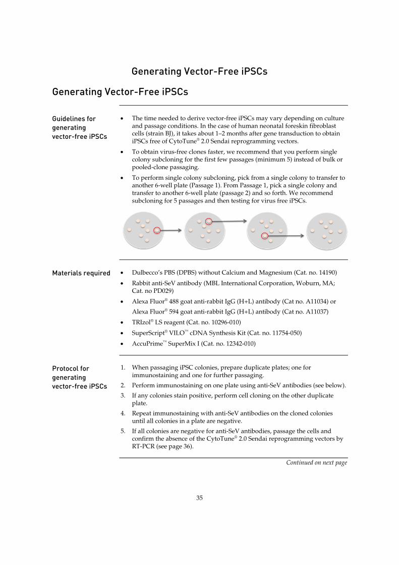
x To obtain virus-free clones faster, we recommend that you perform single colony subcloning for the first few passages (minimum 5) instead of bulk or pooled-clone passaging.
x To perform single colony subcloning, pick from a single colony to transfer to another 6-well plate (Passage 1). From Passage 1, pick a single colony and transfer to another 6-well plate (passage 2) and so forth. We recommend subcloning for 5 passages and then testing for virus free iPSCs.
Materials required x Dulbecco’s PBS (DPBS) without Calcium and Magnesium (Cat. no. 14190)
x Rabbit anti-SeV antibody (MBL International Corporation, Woburn, MA; Cat. no PD029)
x Alexa Fluor® 488 goat anti-rabbit IgG (H+L) antibody (Cat no. A11034) or
Alexa Fluor® 594 goat anti-rabbit IgG (H+L) antibody (Cat no. A11037)
x TRIzol® LS reagent (Cat. no. 10296-010)
x SuperScript® VILO™ cDNA Synthesis Kit (Cat. no. 11754-050)
x AccuPrime™ SuperMix I (Cat. no. 12342-010)
Protocol for generating vector-free iPSCs
1. When passaging iPSC colonies, prepare duplicate plates; one for immunostaining and one for further passaging.
2. Perform immunostaining on one plate using anti-SeV antibodies (see below).
3. If any colonies stain positive, perform cell cloning on the other duplicate plate.
4. Repeat immunostaining with anti-SeV antibodies on the cloned colonies until all colonies in a plate are negative.
5. If all colonies are negative for anti-SeV antibodies, passage the cells and confirm the absence of the CytoTune® 2.0 Sendai reprogramming vectors by RT-PCR (see page 36).
Continued on next page
36
Generating Vector-Free iPSCs, continued
Immunocyto-chemistry with anti-SeV antibodies
1. Wash cells once with D-PBS
2. Fix the cells in 4% paraformaldehyde for 5 minutes at room temperature.
3. Wash cells twice with D-PBS.
4. Add the anti-SeV antibody (MBL, Cat. no PD029) diluted in 0.1% Triton® X-100 in D-PBS to the cells and incubate for 1 hour at 37°C.
5. Remove the antibody solution. Wash the cells 3 times with D-PBS.
6. Add the secondary antibody diluted in 0.1% Triton® X-100 in D-PBS to the cells and incubate for 1 hour at 37°C.
7. Remove the secondary antibody solution from the dish. Wash the cells 3 times with D-PBS.
8. Visualize the cells under a fluorescence microscope.
RT-PCR protocol for detecting the SeV genome and transgenes
1. Extract the total RNA from 5 × 10⁶ iPSCs using the TRIzol® Reagent (Cat. no. 15596-026) following the instructions provided with the reagent. As a positive control, use cells set aside during the reprogramming procedure.
2. Carry out a reverse transcription reaction using 1 µg of RNA (from step 1) and the SuperScript® VILO™ cDNA Synthesis Kit (Cat. no. 11754-050) following the instructions provided with the kit. Note: Because the CytoTune® 2.0 Sendai reprogramming vectors are based on SeV, which is an RNA virus, reverse transcription is required for detecting the presence of the SeV genome in your reprogrammed cells.
3. Carry out the PCR using 10 µL of cDNA from the reverse transcription reaction (Step 2, above) and AccuPrime™ SuperMix I (Cat. no. 12342-010) with the parameters below. For the RT-PCR primer sequences and the expected product size, refer to the table on page 37.
Step Temperature Time Cycles Denaturation 95°C 30 seconds
30–35 Annealing 55°C 30 seconds Elongation 72°C 30 seconds
4. Analyze the PCR products using 2% agarose gel electrophoresis.
If you still detect CytoTune® 2.0 Sendai virus in your iPSC lines after more than 10 passages, and have performed RT-PCR to show that hKlf4 is absent from your cells (this vector does not have the temperature sensitive mutation), then you can perform a temperature shift to remove the c-Myc vector. CytoTune® 2.0 Sendai hc-Myc tends to persist in the cells longer than the other CytoTune® 2.0 Sendai reprogramming vectors. However, because this vector contains a temperature sensitivity mutation, you can enhance its removal and obtain complete absence of Sendai virus by incubating your cells at 38–39°C for 5 days.
Continued on next page
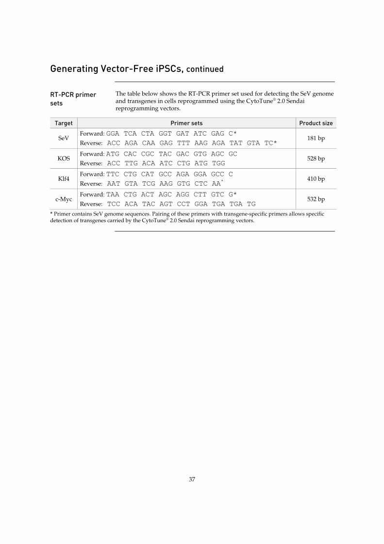
37
Generating Vector-Free iPSCs, continued
RT-PCR primer sets
The table below shows the RT-PCR primer set used for detecting the SeV genome and transgenes in cells reprogrammed using the CytoTune® 2.0 Sendai reprogramming vectors.
Target Primer sets Product size
SeV Forward: GGA TCA CTA GGT GAT ATC GAG C*
Reverse: ACC AGA CAA GAG TTT AAG AGA TAT GTA TC* 181 bp
KOS Forward: ATG CAC CGC TAC GAC GTG AGC GC
Reverse: ACC TTG ACA ATC CTG ATG TGG 528 bp
Klf4 Forward: TTC CTG CAT GCC AGA GGA GCC C
Reverse: AAT GTA TCG AAG GTG CTC AA* 410 bp
c-Myc Forward: TAA CTG ACT AGC AGG CTT GTC G*
Reverse: TCC ACA TAC AGT CCT GGA TGA TGA TG 532 bp
* Primer contains SeV genome sequences. Pairing of these primers with transgene-specific primers allows specific detection of transgenes carried by the CytoTune® 2.0 Sendai reprogramming vectors.
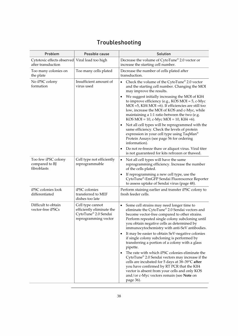
38
Troubleshooting
Problem Possible cause Solution Cytotoxic effects observed after transduction
Viral load too high Decrease the volume of CytoTune® 2.0 vector or increase the starting cell number.
Too many colonies on the plate
Too many cells plated Decrease the number of cells plated after transduction.
No iPSC colony formation
Insufficient amount of virus used
x Check the volume of the CytoTune® 2.0 vector and the starting cell number. Changing the MOI may improve the results.
x We suggest initially increasing the MOI of Klf4 to improve efficiency (e.g., KOS MOI = 5, c-Myc MOI =5, Klf4 MOI =6). If efficiencies are still too low, increase the MOI of KOS and c-Myc, while maintaining a 1:1 ratio between the two (e.g. KOS MOI = 10, c-Myc MOI = 10, Klf4 =6).
x Not all cell types will be reprogrammed with the same efficiency. Check the levels of protein expression in your cell type using TaqMan® Protein Assays (see page 56 for ordering information).
x Do not re-freeze thaw or aliquot virus. Viral titer is not guaranteed for kits refrozen or thawed.
Too few iPSC colony compared to BJ fibroblasts
Cell type not efficiently reprogrammable
x Not all cell types will have the same reprogramming efficiency. Increase the number of the cells plated.
x If reprogramming a new cell type, use the CytoTune®-EmGFP Sendai Fluorescence Reporter to assess uptake of Sendai virus (page 48).
iPSC colonies look differentiated
iPSC colonies transferred to MEF dishes too late
Perform staining earlier and transfer iPSC colony to fresh feeder cells.
Difficult to obtain vector-free iPSCs
Cell type cannot efficiently eliminate the CytoTune® 2.0 Sendai reprogramming vector
x Some cell strains may need longer time to eliminate the CytoTune® 2.0 Sendai vectors and become vector-free compared to other strains. Perform repeated single colony subcloning until you obtain negative cells as determined by immunocytochemistry with anti-SeV antibodies.
x It may be easier to obtain SeV-negative colonies if single colony subcloning is performed by transferring a portion of a colony with a glass pipette.
x The rate with which iPSC colonies eliminate the CytoTune® 2.0 Sendai vectors may increase if the cells are incubated for 5 days at 38–39°C after you have confirmed by RT PCR that the Klf4 vector is absent from your cells and only KOS and/or c-Myc vectors remain (see Note on page 36).
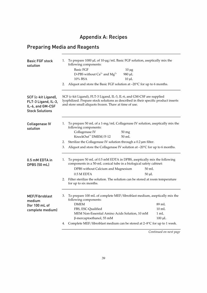
39
Appendix A: Recipes
Preparing Media and Reagents
Basic FGF stock solution
1. To prepare 1000 µL of 10-μg/mL Basic FGF solution, aseptically mix the following components:
Basic FGF 10 µg D-PBS without Ca2+ and Mg2+ 980 µL 10% BSA 10 µL
2. Aliquot and store the Basic FGF solution at –20°C for up to 6 months.
SCF (c-kit Ligand), FLT-3 Ligand, IL-3, IL-6, and GM-CSF Stock Solutions
SCF (c-kit Ligand), FLT-3 Ligand, IL-3, IL-6, and GM-CSF are supplied lyophilized. Prepare stock solutions as described in their specific product inserts and store small aliquots frozen. Thaw at time of use.
Collagenase IV solution
1. To prepare 50 mL of a 1-mg/mL Collagenase IV solution, aseptically mix the following components:
Collagenase IV 50 mg KnockOut™ DMEM/F-12 50 mL
2. Sterilize the Collagenase IV solution through a 0.2 μm filter.
3. Aliquot and store the Collagenase IV solution at –20°C for up to 6 months.
0.5 mM EDTA in DPBS (50 mL)
1. To prepare 50 mL of 0.5 mM EDTA in DPBS, aseptically mix the following components in a 50-mL conical tube in a biological safety cabinet:
DPBS without Calcium and Magnesium 50 mL
0.5 M EDTA 50 µL
2. Filter sterilize the solution. The solution can be stored at room temperature for up to six months.
MEF/Fibroblast medium (for 100 mL of complete medium)
3. To prepare 100 mL of complete MEF/fibroblast medium, aseptically mix the following components:
DMEM 89 mL FBS, ESC-Qualified 10 mL MEM Non-Essential Amino Acids Solution, 10 mM 1 mL ȕ-mercaptoethanol, 55 mM 100 µL
4. Complete MEF/fibroblast medium can be stored at 2–8°C for up to 1 week.
Continued on next page
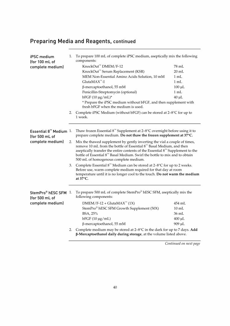
40
Preparing Media and Reagents, continued
iPSC medium (for 100 mL of complete medium)
1. To prepare 100 mL of complete iPSC medium, aseptically mix the following components:
KnockOut™ DMEM/F-12 78 mL KnockOut™ Serum Replacement (KSR) 20 mL MEM Non-Essential Amino Acids Solution, 10 mM 1 mL GlutaMAX™-I 1 mL ȕ-mercaptoethanol, 55 mM 100 µL Penicillin-Streptomycin (optional) 1 mL bFGF (10 µg/mL)* 40 µL * Prepare the iPSC medium without bFGF, and then supplement with fresh bFGF when the medium is used.
2. Complete iPSC Medium (without bFGF) can be stored at 2–8°C for up to 1 week.
Essential 8™ Medium (for 500 mL of complete medium)
1. Thaw frozen Essential 8™ Supplement at 2–8°C overnight before using it to prepare complete medium. Do not thaw the frozen supplement at 37°C.
2. Mix the thawed supplement by gently inverting the vial a couple of times, remove 10 mL from the bottle of Essential 8™ Basal Medium, and then aseptically transfer the entire contents of the Essential 8™ Supplement to the bottle of Essential 8™ Basal Medium. Swirl the bottle to mix and to obtain 500 mL of homogenous complete medium.
3. Complete Essential 8™ Medium can be stored at 2–8°C for up to 2 weeks. Before use, warm complete medium required for that day at room temperature until it is no longer cool to the touch. Do not warm the medium at 37°C.
StemPro® hESC SFM (for 500 mL of complete medium)
1. To prepare 500 mL of complete StemPro® hESC SFM, aseptically mix the following components:
DMEM/F-12 + GlutaMAX™ (1X) 454 mL StemPro® hESC SFM Growth Supplement (50X) 10 mL BSA, 25% 36 mL bFGF (10 µg/mL) 400 µL ȕ-mercaptoethanol, 55 mM 909 µL
2. Complete medium may be stored at 2–8°C in the dark for up to 7 days. Add ȕ-Mercaptoethanol daily during storage, at the volume listed above.
Continued on next page
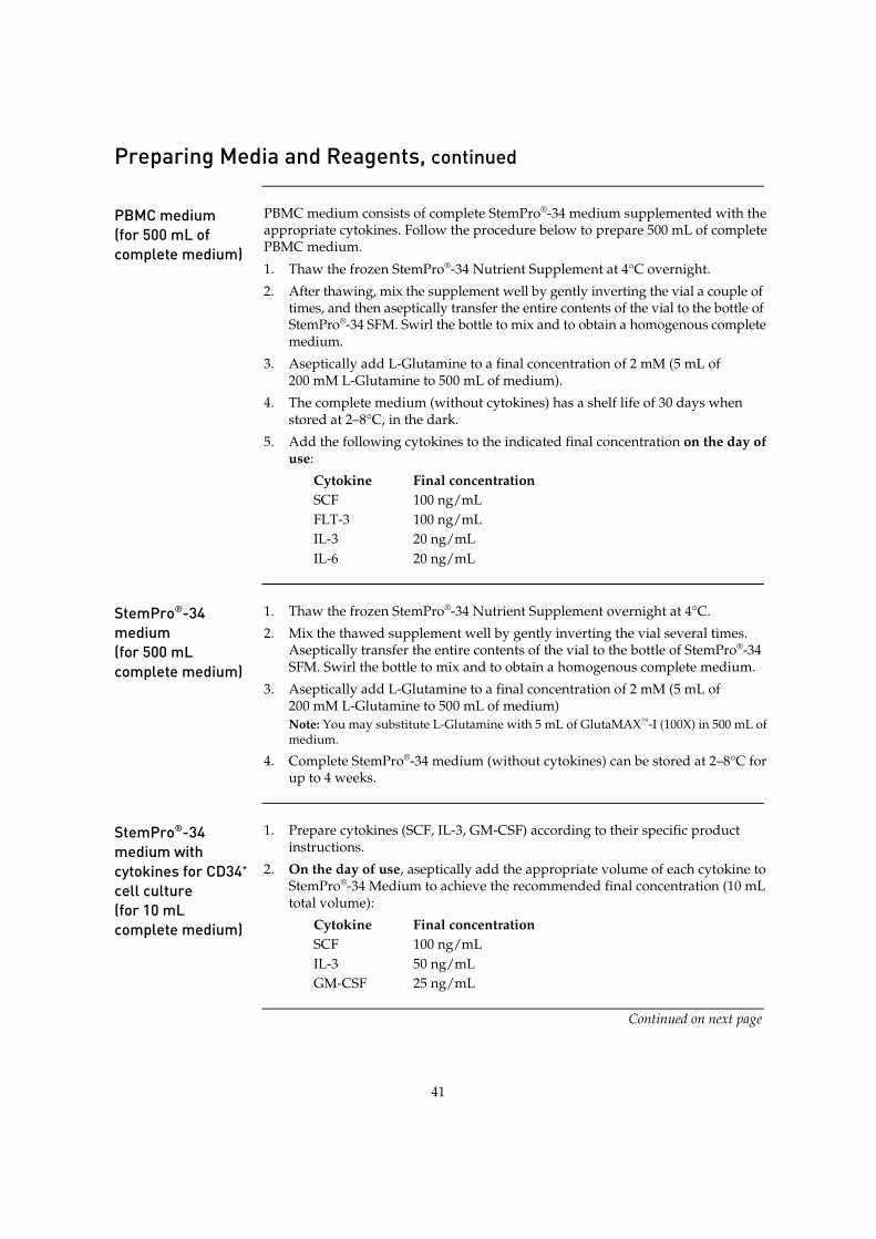
41
Preparing Media and Reagents, continued
PBMC medium (for 500 mL of complete medium)
PBMC medium consists of complete StemPro®-34 medium supplemented with the appropriate cytokines. Follow the procedure below to prepare 500 mL of complete PBMC medium.
1. Thaw the frozen StemPro®-34 Nutrient Supplement at 4°C overnight.
2. After thawing, mix the supplement well by gently inverting the vial a couple of times, and then aseptically transfer the entire contents of the vial to the bottle of StemPro®-34 SFM. Swirl the bottle to mix and to obtain a homogenous complete medium.
3. Aseptically add L-Glutamine to a final concentration of 2 mM (5 mL of 200 mM L-Glutamine to 500 mL of medium).
4. The complete medium (without cytokines) has a shelf life of 30 days when stored at 2–8°C, in the dark.
5. Add the following cytokines to the indicated final concentration on the day of use:
Cytokine Final concentration SCF 100 ng/mL FLT-3 100 ng/mL IL-3 20 ng/mL IL-6 20 ng/mL
StemPro®-34 medium (for 500 mL complete medium)
1. Thaw the frozen StemPro®-34 Nutrient Supplement overnight at 4°C.
2. Mix the thawed supplement well by gently inverting the vial several times. Aseptically transfer the entire contents of the vial to the bottle of StemPro®-34 SFM. Swirl the bottle to mix and to obtain a homogenous complete medium.
3. Aseptically add L-Glutamine to a final concentration of 2 mM (5 mL of 200 mM L-Glutamine to 500 mL of medium) Note: You may substitute L-Glutamine with 5 mL of GlutaMAX™-I (100X) in 500 mL of medium.
4. Complete StemPro®-34 medium (without cytokines) can be stored at 2–8°C for up to 4 weeks.
StemPro®-34 medium with cytokines for CD34+ cell culture (for 10 mL complete medium)
1. Prepare cytokines (SCF, IL-3, GM-CSF) according to their specific product instructions.
2. On the day of use, aseptically add the appropriate volume of each cytokine to StemPro®-34 Medium to achieve the recommended final concentration (10 mL total volume):
Cytokine Final concentration SCF 100 ng/mL IL-3 50 ng/mL GM-CSF 25 ng/mL
Continued on next page

42
Preparing Media and Reagents, continued
iPSC freezing medium
Prepare the Freezing Media A and B immediately before use.
1. In a sterile 15-mL tube, mix together the following reagents for every 1 mL of freezing medium A needed:
iPSC medium 0.5 mL KnockOut™ Serum Replacement 0.5 mL
2. In another sterile 15-mL tube, mix together the following reagents for every 1 mL of freezing medium B needed:
iPSC medium 0.8 mL DMSO 0.2 mL
3. Place the tube with freezing medium B on ice until use (you can keep freezing medium A at room temperature). Discard any remaining freezing medium after use.
Essential 8™ freezing medium
1. Pre-warm the required volume of Essential 8™ Medium at room temperature until it is no longer cool to the touch. Do not warm the medium in a 37°C water bath.
2. Prepare Essential 8™ Freezing Medium. For every 1 mL of freezing medium needed, aseptically combine the components listed below in a sterile 15-mL tube:
Complete Essential 8™ Medium 0.9 mL DMSO 0.1 mL
3. Place the tube with Essential 8™ Freezing Medium on ice until use. Discard any remaining freezing medium after use.
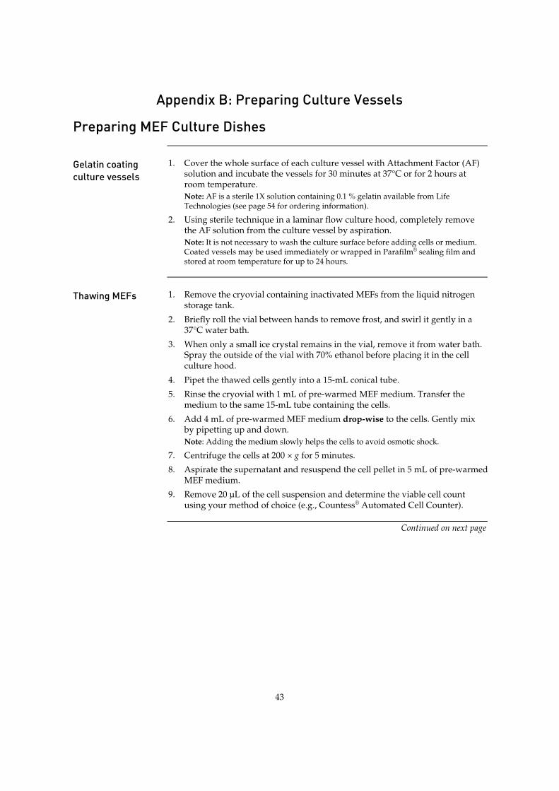
43
Appendix B: Preparing Culture Vessels
Preparing MEF Culture Dishes
Gelatin coating culture vessels
1. Cover the whole surface of each culture vessel with Attachment Factor (AF) solution and incubate the vessels for 30 minutes at 37°C or for 2 hours at room temperature. Note: AF is a sterile 1X solution containing 0.1 % gelatin available from Life Technologies (see page 54 for ordering information).
2. Using sterile technique in a laminar flow culture hood, completely remove the AF solution from the culture vessel by aspiration. Note: It is not necessary to wash the culture surface before adding cells or medium. Coated vessels may be used immediately or wrapped in Parafilm® sealing film and stored at room temperature for up to 24 hours.
Thawing MEFs 1. Remove the cryovial containing inactivated MEFs from the liquid nitrogen
storage tank.
2. Briefly roll the vial between hands to remove frost, and swirl it gently in a 37°C water bath.
3. When only a small ice crystal remains in the vial, remove it from water bath. Spray the outside of the vial with 70% ethanol before placing it in the cell culture hood.
4. Pipet the thawed cells gently into a 15-mL conical tube.
5. Rinse the cryovial with 1 mL of pre-warmed MEF medium. Transfer the medium to the same 15-mL tube containing the cells.
6. Add 4 mL of pre-warmed MEF medium drop-wise to the cells. Gently mix by pipetting up and down. Note: Adding the medium slowly helps the cells to avoid osmotic shock.
7. Centrifuge the cells at 200 × g for 5 minutes.
8. Aspirate the supernatant and resuspend the cell pellet in 5 mL of pre-warmed MEF medium.
9. Remove 20 µL of the cell suspension and determine the viable cell count using your method of choice (e.g., Countess® Automated Cell Counter).
Continued on next page

44
Preparing MEF Dishes, continued
Plating MEFs 1. Centrifuge the remaining cell suspension (step 9, page 43) at 200 × g for
5 minutes at room temperature.
2. Aspirate the supernatant. Resuspend the cell pellet in MEF medium to a density of 2.5 × 10⁵ cells/mL.
3. Aspirate the gelatin solution from the gelatin coated culture vessel.
4. Add the appropriate amount of MEF medium into each culture vessel (refer to the table below).
5. Into each of these culture vessels, add the appropriate amount of MEF suspension (refer to the table below). Note: The recommended plating density for Gibco® Mouse Embryonic Fibroblasts (Irradiated) is 2.5 × 10⁴ cells/cm2.
6. Move the culture vessels in several quick back-and-forth and side-to-side motions to disperse the cells across the surface of the vessels.
7. Incubate the cells in a 37°C incubator with a humidified atmosphere of 5% CO2.
8. Use the MEF culture vessels within 3–4 days after plating.
Culture vessel Surface area
Volume of media Number of MEFs Volume of MEF
suspension 96-well plate 10 cm2 per well 0.1 mL 1.0 × 10⁴/well 40 µL
24-well plate 4 cm2 per well 0.5 mL 5.0 × 10⁴/well 200 µL
12-well plate 2 cm2 per well 1 mL 1.0 × 10⁵/well 400 µL
6-well plate 10 cm2 2 mL 2.5 × 10⁵/well 1 mL
60-mm dish 20 cm2 5 mL 5.0 × 10⁵ 2 mL
100-mm dish 60 cm2 10 mL 1.5 × 10⁶ 6 mL
25-cm2 flask 25 cm2 5 mL 6.3 × 10⁵ 2.5 mL
75-cm2 flask 75 cm2 15 mL 1.9 × 10⁶ 7.5 mL
Figure 7 Mitotically inactivated Mouse Embryonic Fibroblasts (MEFs) plated on culture
vessels coated with Attachment Factor, shown here 4 days after plating.
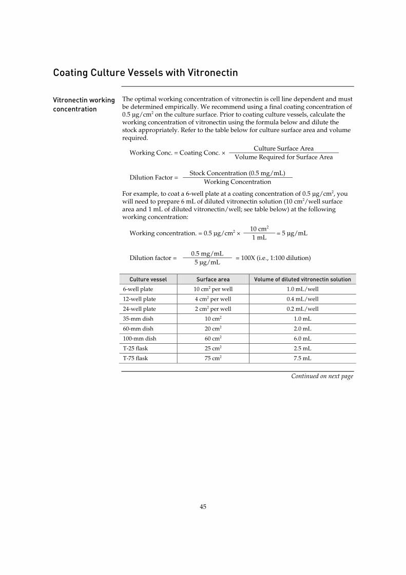
45
Coating Culture Vessels with Vitronectin
Vitronectin working concentration
The optimal working concentration of vitronectin is cell line dependent and must be determined empirically. We recommend using a final coating concentration of 0.5 µg/cm2 on the culture surface. Prior to coating culture vessels, calculate the working concentration of vitronectin using the formula below and dilute the stock appropriately. Refer to the table below for culture surface area and volume required.
Working Conc. = Coating Conc. × Culture Surface Area
Volume Required for Surface Area
Dilution Factor = Stock Concentration (0.5 mg/mL)
Working Concentration
For example, to coat a 6-well plate at a coating concentration of 0.5 µg/cm2, you will need to prepare 6 mL of diluted vitronectin solution (10 cm2/well surface area and 1 mL of diluted vitronectin/well; see table below) at the following working concentration:
Working concentration. = 0.5 µg/cm2 × 10 cm2
= 5 µg/mL 1 mL
Dilution factor = 0.5 mg/mL
= 100X (i.e., 1:100 dilution) 5 µg/mL
Culture vessel Surface area Volume of diluted vitronectin solution
6-well plate 10 cm2 per well 1.0 mL/well
12-well plate 4 cm2 per well 0.4 mL/well
24-well plate 2 cm2 per well 0.2 mL/well
35-mm dish 10 cm2 1.0 mL
60-mm dish 20 cm2 2.0 mL
100-mm dish 60 cm2 6.0 mL
T-25 flask 25 cm2 2.5 mL
T-75 flask 75 cm2 7.5 mL
Continued on next page
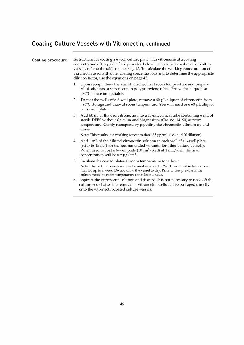
46
Coating Culture Vessels with Vitronectin, continued
Coating procedure Instructions for coating a 6-well culture plate with vitronectin at a coating
concentration of 0.5 µg/cm2 are provided below. For volumes used in other culture vessels, refer to the table on the page 45. To calculate the working concentration of vitronectin used with other coating concentrations and to determine the appropriate dilution factor, use the equations on page 45.
1. Upon receipt, thaw the vial of vitronectin at room temperature and prepare 60-µL aliquots of vitronectin in polypropylene tubes. Freeze the aliquots at –80°C or use immediately.
2. To coat the wells of a 6-well plate, remove a 60-µL aliquot of vitronectin from –80°C storage and thaw at room temperature. You will need one 60-µL aliquot per 6-well plate.
3. Add 60 µL of thawed vitronectin into a 15-mL conical tube containing 6 mL of sterile DPBS without Calcium and Magnesium (Cat. no. 14190) at room temperature. Gently resuspend by pipetting the vitronectin dilution up and down. Note: This results in a working concentration of 5 µg/mL (i.e., a 1:100 dilution).
4. Add 1 mL of the diluted vitronectin solution to each well of a 6-well plate (refer to Table 1 for the recommended volumes for other culture vessels). When used to coat a 6-well plate (10 cm2/well) at 1 mL/well, the final concentration will be 0.5 µg/cm2.
5. Incubate the coated plates at room temperature for 1 hour. Note: The culture vessel can now be used or stored at 2–8°C wrapped in laboratory film for up to a week. Do not allow the vessel to dry. Prior to use, pre-warm the culture vessel to room temperature for at least 1 hour.
6. Aspirate the vitronectin solution and discard. It is not necessary to rinse off the culture vessel after the removal of vitronectin. Cells can be passaged directly onto the vitronectin-coated culture vessels.
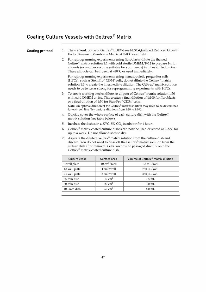
47
Coating Culture Vessels with Geltrex® Matrix
Coating protocol 1. Thaw a 5-mL bottle of Geltrex® LDEV-Free hESC-Qualified Reduced Growth
Factor Basement Membrane Matrix at 2–8°C overnight.
2. For reprogramming experiments using fibroblasts, dilute the thawed Geltrex® matrix solution 1:1 with cold sterile DMEM/F-12 to prepare 1-mL aliquots (or another volume suitable for your needs) in tubes chilled on ice. These aliquots can be frozen at –20°C or used immediately.
For reprogramming experiments using hematopoietic progenitor cells (HPCs), such as StemPro® CD34+ cells, do not dilute the Geltrex® matrix solution 1:1 to create the intermediate dilution. The Geltrex® matrix solution needs to be twice as strong for reprogramming experiments with HPCs.
3. To create working stocks, dilute an aliquot of Geltrex® matrix solution 1:50 with cold DMEM on ice. This creates a final dilution of 1:100 for fibroblasts or a final dilution of 1:50 for StemPro® CD34+ cells. Note: An optimal dilution of the Geltrex® matrix solution may need to be determined for each cell line. Try various dilutions from 1:30 to 1:100.
4. Quickly cover the whole surface of each culture dish with the Geltrex® matrix solution (see table below).
5. Incubate the dishes in a 37°C, 5% CO2 incubator for 1 hour.
6. Geltrex® matrix-coated culture dishes can now be used or stored at 2–8°C for up to a week. Do not allow dishes to dry.
7. Aspirate the diluted Geltrex® matrix solution from the culture dish and discard. You do not need to rinse off the Geltrex® matrix solution from the culture dish after removal. Cells can now be passaged directly onto the Geltrex® matrix-coated culture dish.
Culture vessel Surface area Volume of Geltrex® matrix dilution
6-well plate 10 cm2/well 1.5 mL/well
12-well plate 4 cm2/well 750 µL/well
24-well plate 2 cm2/well 350 µL/well
35-mm dish 10 cm2 1.5 mL
60-mm dish 20 cm2 3.0 mL
100-mm dish 60 cm2 6.0 mL
48
Appendix C: Support Protocols
CytoTune®-EmGFP Reporter Control Transduction
CytoTune®-EmGFP Sendai Fluorescence Reporter
The CytoTune®-EmGFP Sendai Fluorescence Reporter (Cat. no. A16519), available separately from Life Technologies, is a control vector carrying the Emerald Green Fluorescent Protein (EmGFP) gene. The fluorescent control vector allows you to determine whether your cells of interest are amenable or refractive to transduction by Sendai reprogramming vectors. We recommend testing your cell line interest using the CytoTune®-EmGFP Sendai Fluorescence Reporter before starting your reprogramming experiments.
Guidelines for using the CytoTune®-EmGFP Sendai Fluorescence Reporter
x Transducing your cell line of interest using the CytoTune®-EmGFP Sendai Fluorescence Reporter allows you to determine whether or not the cells can be transduced by the Sendai virus vectors; it does not indicate the cell line’s capability to be reprogrammed.
x Different cell types require different MOIs to express detectable levels of EmGFP. As such, cells should be transduced using a range of different MOIs. We suggest initially transducing your cells with at least 2–3 different MOIs (e.g. 1, 3, and 9).
x Expression of EmGFP should be detectable at 24 hours post-transduction by fluorescence microscopy, and reach maximal levels at 48–72 hours.
x The titer of the CytoTune® EmGFP vector is lot-dependent. For the specific titer of the vector, refer to the Certificate of Analysis (CoA) available on our website. Go to www.lifetechnologies.com/cytotunegfp and search for the CoA by product lot number, which is printed on the vial.
x Avoid re-freezing and thawing of the CytoTune®-EmGFP Sendai Fluorescence Reporter since viral titers can decrease dramatically with each freeze/thaw cycle.
CytoTune® 2.0 reprogramming vectors are not compatible with the reprogramming vectors from the original CytoTune®-iPS Reprogramming Kits (Cat. nos. A13780-01, A13780-02). Do not mix or substitute CytoTune® 2.0 reprogramming vectors with the reprogramming vectors from the original kits.
Control transduction protocol for adherent cells
Day –1 to –2: Prepare the cells for transduction 1. 1–2 days before transduction, plate the cells of interest onto the necessary
number of wells of a multi-well plate at the appropriate density to achieve 70–80% confluency on the day of transduction (Day 0). One extra well can be used to count cells for viral volume calculations.
2. Culture the cells for one to two more days, ensuring the cells have fully adhered and extended.
Continued on next page
49
CytoTune®-EmGFP Reporter Control Transduction, continued
Control transduction protocol for adherent cells, continued
Day 0: Perform transduction 3. On the day of transduction, warm an appropriate volume of cell culture
medium for each well to be transduced (e.g., 0.5 mL for each well of a 12-well plate) in a 37°C water bath.
4. Harvest cells from one well of the multi-well plate and perform a cell count. These cells will not be transduced, but will be used to estimate the cell number in the other well(s) plated in Step 1. Note: This step is optional and is performed to obtain more accurate MOI calculations. If exact MOIs are not needed, a rough estimate of the number of cells in the well (based on plating density and growth rates) will also suffice.
5. Count (or estimate) the cell number using the desired method (e.g., Countess® Automated Cell Counter), and calculate the volume of the virus needed to reach the target MOI(s). Titer information can be found on the CoA.
Volume of virus (µL) = MOI (CIU/cell) ! number of cells
titer of virus (CIU/mL) ! 10–3 (µL/mL)
6. Remove one tube of CytoTune®-EmGFP Sendai Fluorescence Reporter from the –80°C storage. Thaw the vector by first immersing the bottom of the tube in a 37°C water bath for 5–10 seconds, and then removing the tube from the water bath and allowing its contents to thaw at room temperature. Once thawed, briefly centrifuge the tube and place it immediately on ice.
7. Add the calculated volume of CytoTune®-EmGFP Sendai Fluorescence Reporter to the pre-warmed cell culture medium prepared in Step 3. Ensure that the solution is thoroughly mixed by pipetting the mixture gently up and down. Complete the next step within 5 minutes.
8. Aspirate the cell culture medium from the cells, and add the solution prepared in Step 7 to the well. Incubate the cells in a 37°C, 5% CO2 incubator overnight.
Day 1: Replace medium and culture cells 9. 24 hours after transduction, replace the medium with fresh cell culture
medium. Note: Depending on your cell type, you should expect to see some cytotoxicity 24–48 hours post-transduction, which can affect >50% of your cells. This is an indication of high uptake of the virus. We recommend that you continue culturing your cells and proceed with the protocol.
10. Visualize the cells on a fluorescence microscope using a standard FITC filter set. EmGFP expression should be visible in some cells (expression will reach maximum levels between 48–72 hours).
Day 2+: Replace medium and culture cells 11. 48 hours after transduction, replace the medium with fresh cell culture
medium.
12. Visualize the cells on a fluorescence microscope using a standard FITC filter set. EmGFP expression should be much brighter than Day 1, and should be visible in many cells (see Figure 8, page 50).
Continued on next page

50
CytoTune®-EmGFP Reporter Control Transduction, continued
Expected results Figure 8 BJ HDFn cells transduced with the CytoTune®-EmGFP Sendai Fluorescence
Reporter at the indicated MOI (1 or 3) and at the indicated time post-transduction (24 or 48 hours).

51
Passaging iPSCs Using EDTA
Passaging protocol 1. Pre-warm complete Essential 8™ Medium (see page 40) and vitronectin-coated
culture vessels (see page 45) to room temperature.
2. Aspirate the spent medium from the vessel containing PSCs and rinse the vessel twice with DPBS without Calcium and Magnesium (refer to the table below for the recommended volume).
3. Add 0.5 mM EDTA in DPBS to the vessel containing PSCs (refer to the table below). Swirl the vessel to coat the entire cell surface.
4. Incubate the vessel at room temperature for 5 to 8 minutes or at 37°C for 4 to 5 minutes. When the cells start to separate and round up, and the colonies appear to have holes in them when viewed under a microscope, they are ready to be removed from the vessel.
5. Aspirate the EDTA solution, and add pre-warmed complete Essential 8™ Medium to the vessel (refer to the table below).
6. Remove the cells from the well(s) by gently squirting medium and pipetting the colonies up. Avoid creating bubbles. Collect cells in a 15-mL conical tube. There may be obvious patches of cells that were not dislodged and left behind. Do not scrape the cells from the dish in an attempt to recover them. Note: Work with no more than 1 to 3 wells at a time, and work quickly to remove cells after adding Essential 8™ Medium to the well(s), which quickly neutralizes the initial effect of the EDTA. Some cell lines re-adhere very rapidly after medium addition, and must be removed 1 well at a time. Others are slower to re-attach, and may be removed 3 wells at a time.
7. Add an appropriate volume of pre-warmed complete Essential 8™ Medium to each well of a vitronectin-coated 6-well plate so that each well contains 2 mL of medium after the cell suspension has been added. Refer to the table below for the recommended volumes for other culture vessels.
8. Move the vessel in several quick figure eight motions to disperse the cells across the surface of the vessels. Place the vessel gently into the 37°C, 5% CO2 incubator and incubate the cells overnight.
9. Feed the PSC cells beginning the second day after splitting. Replace the spent medium daily. Note: It is normal to see cell debris and small colonies after passage.
Culture vessel Surface area DPBS 0.5 mM EDTA
in DPBS Complete Essential 8™
Medium 6-well plate 10 cm2/well 2 mL/well 1 mL/well 2 mL/well
12-well plate 4 cm2/well 1 mL/well 0.4 mL/well 1 mL/well
24-well plate 2 cm2/well 0.5 mL/well 0.2 mL/well 0.5 mL/well
35-mm dish 10 cm2 2 mL 1 mL 2 mL
60-mm dish 20 cm2 4 mL 2 mL 4 mL
100-mm dish 60 cm2 12 mL 6 mL 12 mL
52
Cryopreserving iPSCs
Freezing iPSCs in iPSC freezing medium
Follow the following protocol to cryopreserve iPSCs maintained on MEF-feeder cells by freezing them in iPSC freezing medium (see page 42 for recipe).
1. Prepare the required volume of fresh iPSC freezing medium and place it on ice (see page 42).
2. Aspirate the culture medium and rinse the dishes twice with D-PBS without Ca2+ and Mg2+ (2 mL per 35-mm or 4 mL per 60-mm dish).
3. Gently add Collagenase IV solution (page 39) to the culture dish (1 mL per 35-mm or 2 mL per 60-mm dish).
4. Incubate the dish with cells for 5–20 minutes in a 37°C incubator with a humidified atmosphere of 5% CO2. Note: Incubation times may vary among different batches of collagenase. Therefore, the appropriate incubation time should be optimized by examining the colonies periodically under microscope during incubation.
5. Stop the incubation when the edges of the colonies are starting to pull away from the plate.
6. Remove the culture dish from the incubator, aspirate the Collagenase IV solution, and gently rinse the dish with D-PBS without Ca2+ and Mg2+.
7. Add 2 mL of iPSC culture medium or DMEM/F-12 and gently dislodge the cells off the surface of the culture dish using a sterile pipette or a cell scraper. Transfer the cells to a sterile 15-mL centrifuge tube. Rinse the dish with additional iPSC medium or DMEM/F-12 to collect any leftover colonies.
8. Centrifuge the cells at 200 × g for 2–4 minutes at room temperature.
9. Discard the supernatant, gently tap the tube to dislodge the cell pellet from the tube bottom, and resuspend the cells in freezing medium A. After the cell clumps have been uniformly suspended, add an equal volume of freezing medium B to the cell suspension in a drop-wise manner while gently swirling the cell suspension to mix. Note: At this point, the cells are in contact with DMSO, and work must be performed efficiently with no or minimal delays. After the cells come into contact with DMSO, they should be aliquoted and frozen within 2–3 minutes.
10. Aliquot 1 mL of the cell suspension into each cryovial.
11. Quickly place the cryovials containing the cells in a cryo freezing container (e.g., Mr. Frosty) to freeze the cells at 1°C per minute and transfer them to –80°C overnight.
12. After overnight storage at –80°C, transfer the cells to a liquid nitrogen tank vapor phase for long term storage.

53
Cryopreserving iPSCs, continued
Freezing iPSCs in Essential 8™ freezing medium
Follow the following protocol to cryopreserve iPSCs maintained in Essential 8™ Medium on vitronectin-coated culture dishes by freezing them in Essential 8™ freezing medium (see page 42 for recipe).
1. Prepare the required volume of fresh Essential 8™ freezing medium and place it on ice until use. Note: Discard any remaining Essential 8™ freezing medium after use.
2. Aspirate the spent medium from the dish using a Pasteur pipette, and rinse the cells twice with DPBS without Calcium and Magnesium (refer to the table below).
3. Add 0.5 mM EDTA solution to the dish. To adjust the volume of EDTA for various dish sizes, refer to the table below. Swirl the dish to coat the entire cell surface.
4. Incubate the dish at room temperature for 5–8 minutes or at 37°C for 4–5 minutes. When the cells start to separate and round up, and the colonies appear to have holes in them when viewed under a microscope, they are ready to be removed from the vessel.
5. Aspirate the EDTA solution with a Pasteur pipette.
6. Add 1 mL of ice-cold Essential 8™ freezing medium to each well of a 6-well plate. To adjust the volume of Essential 8™ freezing medium for various dish sizes, refer to the table below.
7. Remove the cells by gently squirting the colonies from the well using a 5-mL glass pipette. Avoid creating bubbles. Collect the cells in a 15-mL conical tube and place on ice.
8. Resuspend the cells gently. Aliquot 1 mL of the cell suspension into each cryovial.
9. Quickly place the cryovials containing the cells in a cryo freezing container (e.g., Mr. Frosty) to freeze the cells at 1°C per minute and transfer them to –80°C overnight.
10. After overnight storage at –80°C, transfer the cells to a liquid nitrogen tank vapor phase for long term storage.
Culture vessel Surface area DPBS 0.5 mM EDTA
in DPBS Essential 8™
freezing medium
6-well plate 10 cm2/well 2 mL/well 1 mL/well 2 mL/well
12-well plate 4 cm2/well 1 mL/well 0.4 mL/well 1 mL/well
24-well plate 2 cm2/well 0.5 mL/well 0.2 mL/well 0.5 mL/well
35-mm dish 10 cm2 2 mL 1 mL 2 mL
60-mm dish 20 cm2 4 mL 2 mL 4 mL
100-mm dish 60 cm2 12 mL 6 mL 12 mL
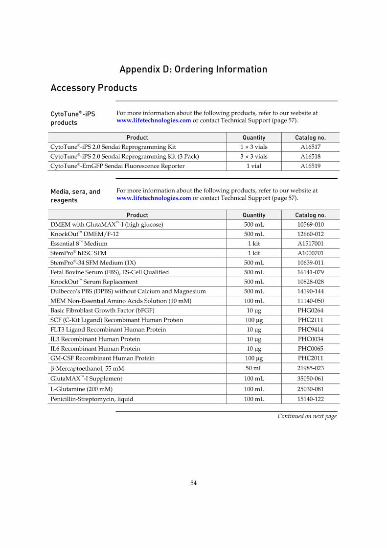
54
Appendix D: Ordering Information
Accessory Products
CytoTune®-iPS products
For more information about the following products, refer to our website at www.lifetechnologies.com or contact Technical Support (page 57).
Product Quantity Catalog no. CytoTune®-iPS 2.0 Sendai Reprogramming Kit 1 × 3 vials A16517 CytoTune®-iPS 2.0 Sendai Reprogramming Kit (3 Pack) 3 × 3 vials A16518 CytoTune®-EmGFP Sendai Fluorescence Reporter 1 vial A16519
Media, sera, and reagents
For more information about the following products, refer to our website at www.lifetechnologies.com or contact Technical Support (page 57).
Product Quantity Catalog no. DMEM with GlutaMAX™-I (high glucose) 500 mL 10569-010 KnockOut™ DMEM/F-12 500 mL 12660-012 Essential 8™ Medium 1 kit A1517001 StemPro® hESC SFM 1 kit A1000701 StemPro®-34 SFM Medium (1X) 500 mL 10639-011 Fetal Bovine Serum (FBS), ES-Cell Qualified 500 mL 16141-079 KnockOut™ Serum Replacement 500 mL 10828-028 Dulbecco’s PBS (DPBS) without Calcium and Magnesium 500 mL 14190-144 MEM Non-Essential Amino Acids Solution (10 mM) 100 mL 11140-050 Basic Fibroblast Growth Factor (bFGF) 10 μg PHG0264 SCF (C-Kit Ligand) Recombinant Human Protein 100 μg PHC2111 FLT3 Ligand Recombinant Human Protein 10 μg PHC9414 IL3 Recombinant Human Protein 10 μg PHC0034 IL6 Recombinant Human Protein 10 μg PHC0065 GM-CSF Recombinant Human Protein 100 μg PHC2011
Ά-Mercaptoethanol, 55 mM 50 mL 21985-023
GlutaMAX™-I Supplement 100 mL 35050-061
L-Glutamine (200 mM) 100 mL 25030-081
Penicillin-Streptomycin, liquid 100 mL 15140-122
Continued on next page
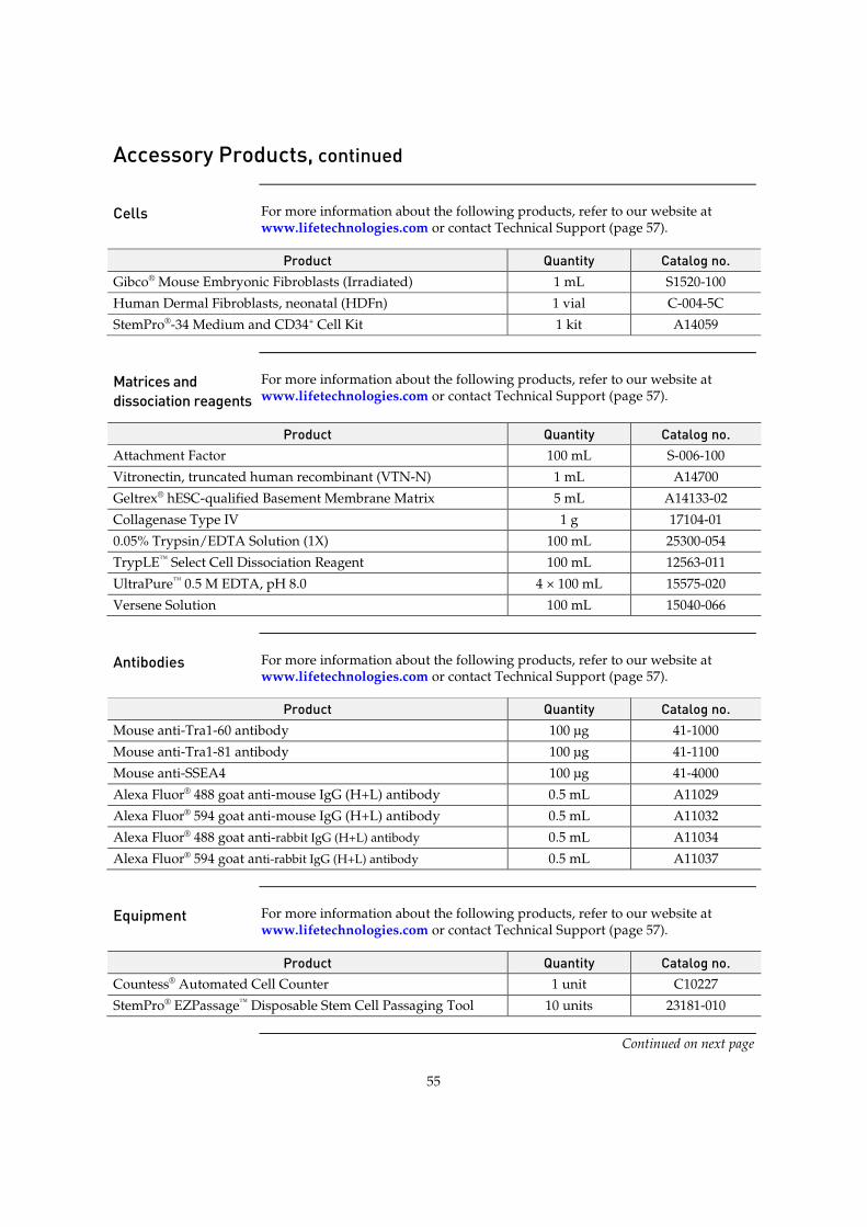
55
Accessory Products, continued
Cells For more information about the following products, refer to our website at
www.lifetechnologies.com or contact Technical Support (page 57).
Product Quantity Catalog no. Gibco® Mouse Embryonic Fibroblasts (Irradiated) 1 mL S1520-100 Human Dermal Fibroblasts, neonatal (HDFn) 1 vial C-004-5C StemPro®-34 Medium and CD34+ Cell Kit 1 kit A14059
Matrices and dissociation reagents
For more information about the following products, refer to our website at www.lifetechnologies.com or contact Technical Support (page 57).
Product Quantity Catalog no. Attachment Factor 100 mL S-006-100 Vitronectin, truncated human recombinant (VTN-N) 1 mL A14700 Geltrex® hESC-qualified Basement Membrane Matrix 5 mL A14133-02 Collagenase Type IV 1 g 17104-01 0.05% Trypsin/EDTA Solution (1X) 100 mL 25300-054 TrypLE™ Select Cell Dissociation Reagent 100 mL 12563-011 UltraPure™ 0.5 M EDTA, pH 8.0 4 × 100 mL 15575-020 Versene Solution 100 mL 15040-066
Antibodies For more information about the following products, refer to our website at
www.lifetechnologies.com or contact Technical Support (page 57).
Product Quantity Catalog no. Mouse anti-Tra1-60 antibody 100 μg 41-1000 Mouse anti-Tra1-81 antibody 100 μg 41-1100 Mouse anti-SSEA4 100 μg 41-4000 Alexa Fluor® 488 goat anti-mouse IgG (H+L) antibody 0.5 mL A11029 Alexa Fluor® 594 goat anti-mouse IgG (H+L) antibody 0.5 mL A11032 Alexa Fluor® 488 goat anti-rabbit IgG (H+L) antibody 0.5 mL A11034 Alexa Fluor® 594 goat anti-rabbit IgG (H+L) antibody 0.5 mL A11037
Equipment For more information about the following products, refer to our website at
www.lifetechnologies.com or contact Technical Support (page 57).
Product Quantity Catalog no. Countess® Automated Cell Counter 1 unit C10227 StemPro® EZPassage™ Disposable Stem Cell Passaging Tool 10 units 23181-010
Continued on next page
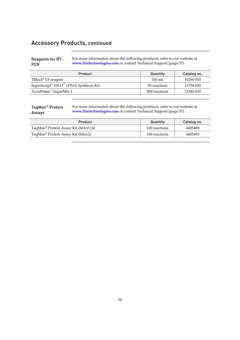
56
Accessory Products, continued
Reagents for RT-PCR
For more information about the following products, refer to our website at www.lifetechnologies.com or contact Technical Support (page 57).
Product Quantity Catalog no. TRIzol® LS reagent 100 mL 10296-010 SuperScript® VILO™ cDNA Synthesis Kit 50 reactions 11754-050 AccuPrime™ SuperMix I 200 reactions 12342-010
TaqMan® Protein Assays
For more information about the following products, refer to our website at www.lifetechnologies.com or contact Technical Support (page 57).
Product Quantity Catalog no. TaqMan® Protein Assay Kit (hOct3/4) 100 reactions 4405489
TaqMan® Protein Assay Kit (hSox2) 100 reactions 4405495
57
Documentation and Support
Obtaining Support
Technical Support For the latest services and support information for all locations, go to www.lifetechnologies.com.
At the website, you can:
x Access worldwide telephone and fax numbers to contact Technical Support and Sales facilities
x Search through frequently asked questions (FAQs)
x Submit a question directly to Technical Support ([email protected])
x Search for user documents, SDSs, vector maps and sequences, application notes, formulations, handbooks, certificates of analysis, citations, and other product support documents
x Obtain information about customer training
x Download software updates and patches
Safety Data Sheets (SDS)
Safety Data Sheets (SDSs) are available at www.lifetechnologies.com/sds.
Limited Product Warranty
Life Technologies Corporation and/or its affiliate(s) warrant their products as set forth in the Life Technologies’ General Terms and Conditions of Sale found on Life Technologies’ website at www.lifetechnologies.com/termsandconditions. If you have any questions, please contact Life Technologies at www.lifetechnologies.com/support.

58
References
Fusaki, N., Ban, H., Nishiyama, A., Saeki, K., and Hasegawa, M. (2009) Efficient induction of transgene-
free human pluripotent stem cells using a vector based on Sendai virus, an RNA virus that does not integrate into the host genome. Proc Jpn Acad Ser B Phys Biol Sci 85, 348-362
Kuroya, M., Ishida, N., and Shiratori, T. (1953) Newborn virus pneumonitis (type Sendai). II. The isolation of a new virus. Tohoku J Exp Med 58, 62
Li, H. O., Zhu, Y. F., Asakawa, M., Kuma, H., Hirata, T., Ueda, Y., Lee, Y. S., Fukumura, M., Iida, A., Kato, A., Nagai, Y., and Hasegawa, M. (2000) A cytoplasmic RNA vector derived from nontransmissible Sendai virus with efficient gene transfer and expression. J Virol 74, 6564-6569
Meissner, A., Wernig, M., and Jaenisch, R. (2007) Direct reprogramming of genetically unmodified fibroblasts into pluripotent stem cells. Nat Biotechnol 25, 1177-1181
Park, I. H., Lerou, P. H., Zhao, R., Huo, H., and Daley, G. Q. (2008) Generation of human-induced pluripotent stem cells. Nat Protoc 3, 1180-1186
Seki, T., Yuasa, S., Oda, M., Egashira, T., Yae, K., Kusumoto, D., Nakata, H., Tohyama, S., Hashimoto, H., Kodaira, M., Okada, Y., Seimiya, H., Fusaki, N., Hasegawa, M., and Fukuda, K. (2010) Generation of induced pluripotent stem cells from human terminally differentiated circulating T cells. Cell Stem Cell 7, 11-14
Takahashi, K., Tanabe, K., Ohnuki, M., Narita, M., Ichisaka, T., Tomoda, K., and Yamanaka, S. (2007) Induction of pluripotent stem cells from adult human fibroblasts by defined factors. Cell 131, 861-872
Takahashi, K., and Yamanaka, S. (2006) Induction of pluripotent stem cells from mouse embryonic and adult fibroblast cultures by defined factors. Cell 126, 663-676
Wernig, M., Meissner, A., Foreman, R., Brambrink, T., Ku, M., Hochedlinger, K., Bernstein, B. E., and Jaenisch, R. (2007) In vitro reprogramming of fibroblasts into a pluripotent ES-cell-like state. Nature 448, 318-324
Yu, J., Vodyanik, M. A., Smuga-Otto, K., Antosiewicz-Bourget, J., Frane, J. L., Tian, S., Nie, J., Jonsdottir, G. A., Ruotti, V., Stewart, R., Slukvin, II, and Thomson, J. A. (2007) Induced pluripotent stem cell lines derived from human somatic cells. Science 318, 1917-1920

Headquarters5791 Van Allen Way | Carlsbad, CA 92008 USA | Phone +1 760 603 7200 | Toll Free in USA 800 955 6288For support visit lifetechnologies.com/support or email [email protected]
lifetechnologies.com8 October 2013