detection of antibodies to sendai virus by … of antibodies to sendai virus by enzyme-linked...
TRANSCRIPT

Journal of lmmunological Melhods, 28 (1979) 163-176 © Elsevier/North-Holland Biomedical Press
163
DETECTION OF ANTIBODIES TO SENDAI VIRUS BY ENZYME-LINKED IMMUNOSORBENT ASSA Y (ELISA)
H.C.J. ERTL *, W. GERLICH and U.H. KOSZINOWSKI **
Hygiene-Inslilut der Universitiil Göttingen, Lehrstuhl Medizinische Mikrobiologie, Kreuzbergring 67, D·34 Göttingen, G.F.R.
(Received 6 November 1978, accepted 6 December 1978)
An enzyme-linked immunosorbent assay (ELISA) for the detection of antibodies to Sendai virus, a paramyxovirus, is described. The assay was found to be about 20-fold more sensitive than the hemagglutination inhibition assay. Differentiation between virus specific IgG and IgM is possible. The test appears to be especially useful in the study of early events in antibody formation in vivo as weil as in vitro.
INTRODUCTION
Viruses induce cellular and humoral immune responses in the host. This dichotomy is govemed by principles not yet fully understood. In cellular immune responses the major histocompatibility complex (MHC) is of importance at the level of T-cell differentiation (Bennink and Doherty, 1978) as well as of T-effector cell function (Doherty et al., 1976). The degree of MHC restricted antiviral responses has been shown to be under the control of immune response genes (Doherty et al., 1978). It is not yet known to what extent the antiviral antibody response is regulated by immune response genes. Since the induction of antibody formation against various protein antigens is under T-helper cell control (Roberts et al. , 1975) it is to be
* Present address: John Curtis School of Medical Research, Microbiology Department, The Australian National University, Post Office Box 4, Canberra, ACT 2600, Australia. ** Deutsches Krebsforschungszentrum, Institut für Immunologie und Genetik, Im Neuen· heimer Feld 280, D-69 Heidelberg, G.F.R. List of abbreviations: BSA, bovine serum albumin; CF, chorioallantoic fluid; ChHC, chicken host components; ELISA, enzyme-linked immunosorbent assay; FCS, fetal calf serum; HAU, hemagglutinating units; HAIU, hemagglutination inhibition units; HIA, hemagglutination inhibition assay; HRPO, horse radish peroxidase; i.n., intranasal; i.p., intraperitoneal; i.v., intravenous; M, molar; A, absorbance; PBS, phosphate buffered saline; RAMjlg, rabbit anti-mouse-Ig; RAM/IgG, rabbit anti-mouse-lgG; RAMjlgM, rabbit anti-mouse-lgM (Fe); RIA, radioimmunoassay; T-cells, thymus derived cells; TCIDso , 50% tissue culture infective doses; s.c., subcutaneous; SV, Sendai virus; wjv, weight per volume.

164
expect.ed that regulatory functi01ls of T <'ells govprn the production of antibodies as weil.
The analysis of antibody response to viral antigens requires sensitive tests. Virus neutralisation assay is very sensitive (Svenag and Mandel, 1964), but it is too laborious to use as a routine method. Complement binding is not sensitive enough. The hemagglutination inhibition assay is also not sufficiently sensitive and is furthermore restricted t r) antibodies to hemagglut.inin. Furthermore the conventional tests are not able to discriminate between different dasses of immunoglobulins and the antibody level is difficult to quantify.
This report describes different modifications of the enzyme-linked immunosorbent assay for the detectioll of antibodies to Sendai virus. The assays allow the demonstration of low levels of antibodies very early after sensitization and the differentiation hetween JgG and IgM. Small differences in antibody levels can be detected and the sensitivity of the assays allows the investigation of antibodies produced by cel1s in a primary in vitro induction phase.
MATERIALS AND METHODS
Mice Adult male and female C3HjTIF and DBAj2 inbrpd mice (Bornholdgard,
Ry, Denmark) were used at 8- -12 weeks of age.
Viruses Sendai virus was grown in the chorioallantoic fluid of 10-day-old em
bryonated hen eggs for 44--48 h at 37°C. Influenza virus A, straül Victoria, was grown in the chorioal1antoic fluid
of ll-day-old embryonated hen eggs for 40-44 h at 37°C. Hemagglutination titration was carried out by Salk's pattern method
(Salk, 1944) using 0.2% chicken red blood cells. Purification of Sendai virus was done by centrifugation of the virus-containing chorioallantoic fluid at 3000 rev./min for 30 min to remove ('eIl debris and chicken red blood cells. The supernatant was layered on 10 ml 60% sucrose (w jv) and centrifuged for 90 min at 20,000 X g (SW-27 rotor, Beckmann). The virus layer was collected and purified by a linear sucrose gradient from 10% to 60% (wjv) centrifugation for 90 min at 20,000 X g (SW-50.1 rotor, Beckmann). The visible virus layer was removed and dialysed against phosphate buffered saline overnight. Aliquots of purified 01' unpurified Sendai virus were stored at -70°C.
Protein content of the purified virus was determined by the method of Lowry et a1. (1951).
Inactivation of Sendai virus was carried out by treatment with ß-propiolactone for 18 h at a concentration of 0.025% (Koszinowski et a1., 1977b).
Vaccinia virus, strain WR, was propagated in Vel'o cells (monkey kidney cells) as de~wribed narlipl' (Ec""zinowskiflnrl Thomssen, 1975).

16fJ
Antisera Sendai virus (50---100 hemagglutinating units of llupurified, ur 500 ng uf
purified, inactivated virus) was injected intraperitoneally, intravenously, subcutaneously or intranasally. Booster injections were carried out in weekly intervals. Mice were bled by retro-orbital puncture. Immediately before infection, sera were taken fur controls.
Antisera to influenza virus were obtained by Lp. injection of 50--100 hemagglutinating units.
Titers of antisera to Sendai or influenza virus were determined in a hemagglutination inhibition assay as described earlier (Ertl and Koszinowski, 1977).
Vaccinia virus antisera were prepared by Lp. injection of 1 X 10⁵ TCIDso vaccinia virus. Titers were measured in a complement-dependent 51Cr-release assay, using vaccinia virus infected L-929 cells as targets (Koszinowski et al. , 1977a). The serum used in th:s report had a titer of 1 : 4048.
Antiserum to rubella virus was kindly provided by Prof. R. Thomssen. Antisera to chorioallantoic fluid were prepared by Lp. injection of 1 ml
chorioallantoic fluid of 12-day-old embryonated hen eggs. Antisera to mouse-y-globulin from rabbit (RAMjIg) were obtained from
Behringwerke, MarburgjLahn, G.F.R. Antisera to mouse-IgG (RAMjIgG) and mouse-IgM (RAMjJgM) from
rabbit were obtained from Nordic Immunological Laboratories, Tilburg, The N etherlands.
Purification of [gG One millilitre of antiserum was dialysed for 2 h against 0.02 M phosphate
buffer, pH 8.0, and then added to a chromatography column of 20 cm X 0.5 cm diethylaminoethylcellulose (DEAE-Sephacel, lot No. 1482, Phannacia, Fine Chemicals AB, Uppsala, Sweden). IgG was eluated with 0.02 M phosphate bu ffer , pH 8.0. Twelve to sixteen 1 ml fractions were collected, and the protein content was estimated by optical density at 280 nm. The four peak fractions were collected and used for further assays.
Preparation of conjugates Antisera and purified Sendai virus were labeled with horse radish peroxi
dase (HRPO, Type VI, Prod. No. p-8375, Sigma Chemieal Co., St. Louis) by a combination of the methods of Nakane (1975) and Avrameas and Ternynk (1971). Briefly, 20 mg HRPO were dissolved in 1 ml of 0.3 M sodium bicarbonate buffer, pH 8.4. Then 0.3 ml of 25% glutaraldehyde (Serva, Heidelberg) was added for 30 min at room temperature. To remove excess glutaraldehyde the solution was applied tu a 5.0 X 1.0 Sephadex G-25 M column (Column PD-10, Pharmacia, Fine Chemieals, Uppsala, Sweden) in 0.3 M sodium bicarbonate buffer, pH 8.0. The eluated enzyme was incubated with 0.5 ml of 0.1 M NaI04 for 30 min at room temperature. Then 0.1 ml of ethylene glycol was added for 10 min at room temperature. The solution was

166
applied to a Sephadex PD-10 column in 0.01 M sodium bicarbonate buffer, pH 9.5. The activated peroxidase was used for labeling proteins within the next four hours.
Labeling of Sendai virus or antisera: 10 mg of IgG in 1 ml buffer or 4 mg of Sendai virus in 5 ml of 0.01 M sodium bicarbonate buffer, pH 9.5, were added to 20 mg of activated peroxidase. The solution was stirred for 2 h at room temperature. After cooling to 4°C, 5 mg solid NaBH4 were added and the solution left at 4°C for 2 h. The solution was then dialysed at 4°C for 24 h.
Purification of labeled Sendai virus from free HRPO was by 2-fold linear sucrose gradient (10% to 60% w/v) centrifugation for 90 min at 20,000 X g (SW 50.1 rotor, Beckmann). To separate conjugated IgG from free HRPO, the sampIes were applied to a 100 cm X 1.6 cm Biogel P-300 column in 0.02 M Tris-HCl, 0.13 M NaCI buffer, pH 7.4. Fractions of 1 ml were collected and optical density was measured at 280 nm and 403 nm. RZ values (A403/A280) were calculated. Fractions with RZ values of 0.5 to 0.7 were collected and used in the further assays.
Enzyme linked immunosorbent assay (ELISA) WeHs of flexible polyvinyl microtiter plates (M 24, Cooke Laboratory)
were coated with 200 ng purified Sendai virus or with a 1 : 200 dilution of RAM/IgM suspended in a volume of 100 /-,tl, 0.02 M phosphate buffer, pH 7.4, for 18-24 h in a sealed humidified plastic box at 4°C. Control weHs were coated with chicken chorioaHantoic fluid 1 : 10 diluted in phosphate buffer or merely with buffer. After rinsing (3 X 2 min with 0.02 M phosphate buffer, pH 7.4) the weHs were filled with 150}11 normal guinea pig serum for 2 h. After another rinsing step, 100 }11 of test serum or negative control serum were added and the plates left for 20-30 min at room temperature. Control weHs were filled with buffer instead of serum. After a further 3-fold rinsing, 100 }11 of conjugate containing 50-200 ng of SV-HRPO or 100 ng of labeled anti-mouse--y-globulin IgG (RAM/Ig, RAM/IgG or RAM/IgM) diluted in buffer containing 2% BSA, were incubated for 20-30 min at room temperature. After 3-fold rinsing 100 /-11 of freshly prepared substrate (orthophenylenediamine 0.4 mg/mI + 0.006% H20 2 in 0.1 M citrate buffer, pH 5.0) was added and the plates incubated in the dark at room temperature. After 30 min, 50 /-11 2 M sulfuric acid were added to each weH to stop the enzyme reaction and the absorbance was measured at 493 nm. A solution of 100/-11 of substrate and 50 /-LI of sulfuric acid was taken as blank. Absorbance was measured in disposable cuvettes, 1 to 2.5 diluted in 5% Na2S04. The experimental schedule is shown in Fig. 1.
Primary induction of antibody formation in vitro 1 X 10⁸ viable spleen lymphocytes/ml of DBA/2 mice were cultured in
25 ml RPMI 1640 medium (Grand Island Biological Co., N.Y.) supplemented with 10% FCS (fetal calf serum), antibiotics, 2 X 10⁻⁵ M 2-mercaptoethanol

167
EXPERIMENTAL SCHEDULE
\y y y y I I.Coating
I I. Test serum
11. Conjugate
o Sendai virus Y RAM IlgM V test serum eSV-HRPO "RAM/lg-HRPO
Fig. 1. Principle of different ELISA techniques. I. Coating with SV or RAM/IgM (18-24 h). 11. After 3-fold rinsing, normal guinea pig serum was incubated in the weHs for 2 h and rinsed out. The test serum was then added and incubated at RT for 20-30 min. 111. After 3-fold rinsing SV-HRPO or enzyme-Iabeled RAM/Ig, RAM/IgG or RAM/IgM was added for 20-30 min. Orthiphenylenediamine and H2 0 2 were used as substrate.
and 1% glutamine with 100 ng ß-propiolactone inactivated Sendai virus/mI for 4 days at 37°C in a humidified atmosphere with 5% CO 2 • After 3-fold washings the cells were resuspended in aserum free medium for another 2 days. Control cells were cultured under the same conditions in a virus free medium. Supematants were coHected and freeze-dried.
RESULTS
Test requirements Coating conditions. The sensitivity of ELISA depends on the concentra
tion of Sendai virus or RAM/IgM used for coating of wells (Fig. 2). High concentrations of antigen and a 10-2 to 10-3 dilution of RAM/IgM were found to give best results. For further assays 200 ng SV/weH or a 1 : 200 dilution of RAM/IgM were used.
Coating procedure of SV or RAM/IgM is time-dependent. Maximal values of specific bin ding were obtained if the assay was done about 24 h after coating of weHs with SV or RAM/IgM. Coating for a time exceeding 48 h resulted in loss of activity probably due to antigen degradation in the case of virus or to a loss of binding of proteins to the plastic weHs.
Coating procedure is independent from temperature. WeHs coated with SV or RAM/lgM at 4°C, 22°C or 37°C showed nearly identical results. To

168
E B c: .., 01
o· 0
10 05
SV (l111/welI) dilution. 2.10"
Fig. 2. Coating concentration of SV or RAM/IgM. WeHs were coated with different concentrations of SV (A) or RAM/IgM (B). (e---e) serum of DBA/2 mice, 1 week after infection with 100 HAU SV (serum No. 13.1); (0---0) Pre-infection serum (serum No. 13.0). Sera were used at a dilution of 1 : 100. SV-HRPO was used as enzyme-labeled reagent.
prevent bacterial contamination of the weHs and to decrease the degradation of the temperature-sensitive viral antigens, weHs were incubated at 4°C. Within the range pH 5.0 to pH 9.0 there was no major difference in the coating capacity of SV or RAMjIgM. Results with weHs coated at neutral pH were slightly superior to those with weHs coated at basic or acid pH. Therefore weHs were usually coated in 0.02 M phosphate buffer, pH 7.4.
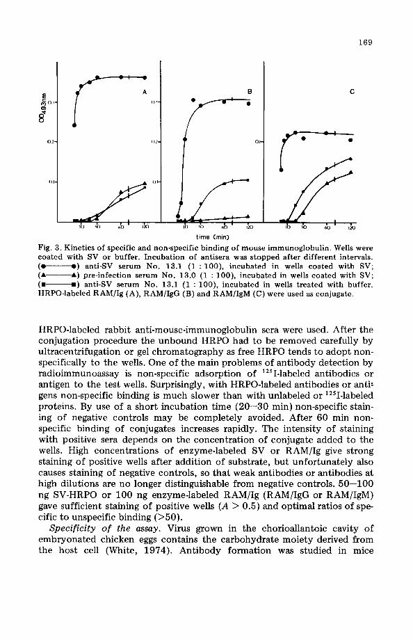
Antibody binding conditions. Immunoglobulins have a high affinity for plastic surfaces. Thus after the coating procedure weHs were incubated with a 20% solution of guinea pig serum, which did not react with SV, mouse immunoglobulins or anti-mouse-immunoglobulin sera. This step is necessary to block non-specific adsorption of antibodies to the weHs. Non-specific adsorption of antibodies to plastic weHs is much slower than specific binding to a solid phase reagent. Binding of SV-antibodies to untreated weHs is almost undetectable after 20 min, whereas specific binding to SV-coated weHs is complete after this time. The non-specific adsorption of mouseimmunoglobulin to SV-coated and untreated weHs in comparison to specific binding of SV-antibodies is shown in Fig. 3. Coating weHs with SV seems to inhibit the unspecific adsorption of IgG but not of IgM. The ratio of specific to unspecific binding is optimal after an incubation period of 20 min for the test serum.
Optimal conditions for specific binding of enzyme-labeled reagents. For the detection of Sendai virus antibodies of aH immunoglobulin classes, we used HRPO-labeled SV. To differentiate murine IgG from IgM antibodies
169
E A B C
~o' (H • • 0>
ff 0.2 0.2 o.
0.1
time (min)
Fig. 3. Kinetics of specific and non-specific binding of mouse immunoglobulin. WeHs were coated with SV or buffer. Incubation of antisera was stopped after different intervals. (e e) anti-SV serum No. 13.1 (1 : 100), incubated in weHs coated with SV; ( ... ---... ) pre-infection serum No. 13.0 (1 : 100), incubated in weHs coated with SV; (. .) anti-SV serum No. 13.1 (1 : 100), incubated in weHs treated with buffer. HRPO-Iabeled RAM/Ig (A), RAM/IgG (B) and RAM/IgM (e) were used as conjugate.
HRPO-Iabeled rabbit anti-mouse-immunoglobulin sera were used. After the conjugation procedure the unbound HRPO had to be removed carefully by ultracentrifugation or gel chromatography as free HRPO tends to adopt nonspecifically to the wells. One of the main problems of antibody detection by radioimmunoassay is non-specific adsorption of 12sI-Iabeled antibodies or antigen to the test weHs. Surprisingly, with HRPO-labeled antibodies or anti! gens non-specific bin ding is much slower than with unlabeled or 12sI-Iabeled proteins. By use of a short incubation time (20-30 min) non-specific staining of negative controls may be completely avoided. After 60 min nonspecific binding of conjugates increases rapidly. The intensity of staining with positive sera depends on the concentration of conjugate added to the wells. High concentrations of enzyme-labeled SV or RAMjIg give strong staining of positive wells after addition of substrate, but unfortunately also causes staining of negative controls, so that weak antibodies or antibodies at high dilutions are no longer distinguishable from negative controls. 50-100 ng SV-HRPO or 100 ng enzyme-Iabeled RAMjIg (RAMjIgG or RAMjIgM) gave sufficient staining of positive wells (A > 0.5) and optimal ratios of specific to unspecific binding (>50).
Specificity of the assay. Virus grown in the chorioallantoic cavity of embryonated chicken eggs contains the carbohydrate moiety derived from the host cell (White, 1974). Antibody formation was studied in mice

170
~ e g
03
02
0'
" , ,
--
" \
\ \
\
\~ \
\ \ \ '\ , , , , ..
A B
5< 5'! 5·~·< 5" ., serum dilution (x 10)
Fig. 4. Blocking of ChHC antibodies. WeHs were coated at solid phase with SV (A) or a 1 : 10 dilution of CF (B). Antisera were added for 30 min at room temperature. (+) SV antiserum No. 13.1; (e) Influenza virus antiserum; ("') preserum No. 13.0. (Pre-infection serum showed no activity and is omitted for the sake of clarity.) After 3-fold rinsing weHs were treated for 2 h with buffer (---), or CF (1 : 5) (- - - - - -). SV-HRPO was used as conjugate.
infected with egg-grown virus and the same virus was used, after purification, for coating or also for conjugation. Therefore this assay detects not only Sendai virus antibodies but also antibodies directed against chicken host components (ChHC). By this mechanis, high crossreactivity with sera directed against other egg-grown viruses such as influenza is found. Sera from virus-primed mice bind to wells coated either with Sendai virus or ChHC (Fig.4). By addition of excess chorioallantoic fluid, antibodies directed against ChHC are saturated and are no longer stained by SV-HRPO as is shown by negative results with wells coated with ChHC (Fig. 4B) while specific binding for viral antigen remains positive (Fig. 4A). The activity of Sendai virus antisera was reduced by 25% to 30%, indicating that 25% to 30% of the antibodies are directed against ChHC. A similar percentage is found after sensitizing mice to influenza virus A (Gerhard et al., 1975). The percentage of antibody directed against ChHC depends on the virus preparation used for sensitization and on the application route. Injection of large amounts of inactivated virus results in a higher fraction of ChHC antibodies than infection with a small amount of infective virus. Antisera produced by intranasal infection show no crossreactivity with ChHC (data not shown). The blocking of anti-ChHC specificity of bound antibodies with chorioallantoic fluid is only effective when SV-HRPO is used as the labeled reagent. In

0.5
e_ -
." --\ 1\ \ \ \
• \ \
\~ , ........
A
0.5 \ \ \ e
\ \ \
lll\ \ .
\ \ ,
e , "
B
58 5' 52 53 54 55 56 57 58 serum dilution (x2")
171
Fig. 5. Sensitivity of different test procedures for the measurement of SV antibodies. WeHs were coated with SV, with exception of procedure All, for which RAM/IgM was used. Dilutions of SV antiserum No. 13.1 (e) and corresponding pre-infection serum (e) were added for 30 min at room temperature. SV·HRPO was the labeled reagent for figure A. In figure B HRPO labeled antibodies against immunoglobulins were used (Ill RAM/Ig, IV RAM/IgG, V RAM/IgM).
assays where enzyme-Iabeled RAM/Ig preparations were used, test sera were routinely preabsorbed three times for 15 min at room temperature on weHs coated with chorioallantoic fluid.
Crossreactivities with sera directed to other viruses such as rubella or vaccinia were not observed (data not shown).
Sensitivity of the assay. A SV-hyperimmune serum was titrated by several modifications of ELISA as shown in Fig. 5. For demonstration of the total amount of SV antibodies two methods were used. Curve I shows the results with wells coated with SV and SV-HRPO as enzyme-Iabeled reagent, while in curve 111 enzyme-Iabeled RAM/Ig was used. Both methods are equivalent in sensitivity, detecting SV antibodies (serum No. 13.2) to a dilution of ab out 1 : 75.000. The serum has a titer of 1 : 2560 in HIA.
Two methods were used to demonstrate IgM antibodies. In the first, wells were coated with RAM/IgM (Fig. 5, curve 11), total IgM was then bound to the solid phase and SV specific IgM was detected by SV-HRPO. In the second procedure (Fig. 5, curve V) SV was used as solid phase reagent, SV antibodies were bound irrespective of Ig cIass and demonstration of IgM was by enzyme-Iabeled RAM/Igl\ll. The first procedure was superior in sensitivity to the second. Selective determination of anti-SV IgG was done by enzymelabeled RAM/IgG (Fig. 5, curve IV).

172
High concentrations of preinfection serum were non-specifically stained especially if RAM/IgG was used as enzyme reagent.
In our experiment (Fig. 5) we used an antiserum from DBA/2 mice, which was produced by three injections of 100 HAU of SV at weekly intervals. Mice were bled one week after the last injection.
Application of the assay Early antibody synthesis. Infection of mice with 100 HAU of Sendai virus
using different application routes (intraperitoneal, intravenous, intranasal or subcutaneous) resulted in all cases in good antibody production after 2 to 4 days (Fig. 6). Infection by Lp., Ln. or s.c. injection gave comparable amounts and time courses of antibody production, while after i.v. infection antibodies increased earlier and reached, especially in the case of IgM, higher levels. HIA gave low, if any, demonstrable titers during the first week.
Propagation of SV in mice is not necessary to provide powerful stimulus for antibody production. Lp. injection of a high dose of ß-propiolactone
SVs.c. SVi.n.
05
'00 SV inactivated
0.5 ::J
~ 100
14
SVi.p. SV · ,1\, t.V. I ,
I \
./~~, '00 '/ ~
(l· I I
1/
'. / \ '"
0.5 05
100
0246814 02468 14
days after infection days after infection
Fig. 6. Influence of application route on antibody formation: Sera, which were pooled from three mice, were obtained at different intervals after Lp., Lv., s.c. and Ln. injection of SV and tested in a dilution of 1 : 100. Total SV antibody (e e) was demonstrated by procedure I (Fig. 5), SV-IgM (e- - - - - -e) by procedure 11 and SV-IgG by pro-cedure IV (e---e). Titers in HIA were also determined (e e).
Fig. 7. Antibody formation after Lp. injection of inactivated SV: See legend of Fig. 6. Serum was obtained after Lp. injection of 500 ng of ß-propiolactone inactivated SV.

173
inactivated virus (0.5 j.1g/mouse) caused antibody production on a similar scale to that after injection of infective virus (Fig. 7). Production of specific IgM was if anything slightly increased.
Antibody production in vitro. In the Sendai virus system it is possible to stimulate cytolytic T-Iymphocytes in a primary in vitro induction phase (Koszinowski, unpublished results). It was not known whether specific antibodies are also produced in these cultures. Freeze-dried supernatants (25 ml) of primary induction phase cultures were collected and diluted in 1 ml 0.02 M phosphate buffer, pH 7.4. Total Sendai virus antibodies and IgM antibodies against SV were determined. As seen in Fig. 8, spleen lymphocytes cultured in the presence of SV produced antibodies to SV. In comparison with the in vivo production of Sendai virus antibodies by DBA/2 mice in one week (Fig. 8B), antibody content of the in vitro cultures was rather low. This may be due to the formation of immune complexes with the large amount of Sendai virus (2.5 j.1g) per culture or to the short duration of antibody production (2 days after rem oval of medium, containing fetal calf serum).
E c M Cl)
" o o
03
02
01
A 20
10
...... e\ ., '. "., e~
e ___ • ' -e_e _,
5-' 5-- 5- \ 5- 4 5-' 5-6 5-' 5-8
dilution Fig. 8. A: titration of SY-antibodies produced in vitro. Total SY-antibody (---) or SY-IgM (- - -) were determined in supernatants of DBA/2lymphocytes previously cultured in the presence (.) or absence (e) of SV. B: titration of SY-antibodies in the serum of DBA/2 mice one week after infection with SY (.) and test results for the corresponding pre-infection serum (e).

174
DISCUSSION
This report describes an enzyme-linked immunosorbent assay (ELISA) for the demonstration of Sendai virus antibodies. The main advantages of ELISA are greater sensitivity in comparison with standard assays and the possibility of differentiating between immunoglobulins of the IgG and IgM c1ass (Leinikki and Passila, 1977). In routine procedures antibodies to paramyxoviruses are determined by hemagglutination inhibition assay or by complement fixation. Titers demonstrable by these methods rarely exceed 1 : 200-1 : 500 and often change from one test to the next. Ta prove a primary infection IgG and IgM have to be separated by ultracentrifugation.
The enzyme-linked immunosorbent assay is about 20-fold more sensitive than the hemagglutination inhibition assay; it detects other (and more) antigenic determinants, and it is not influenced by non-specific inhibitors wh ich often confuse the results of HIA. Virus neutralisation tests are very sensitive and detect low levels of antibodies at an early stage of infection (Svenag and Mandel, 1964) but are too laborious for routine work, while results are often non-specific because of the influence of interferon. The sensitivity of ELISA is comparable with that of RIA (Ertl, unpublished results). ELISA has several advantages over RIA. The chloramine T method genetically used (Hunter and Greenwood, 1962) causes chemical damage to the antigens of the Sendai virus; for example, hemagglutinin activity is totally destroyed. Enzyme-Iabeling of the virus is more gentle and hemagglutinin activity is only slightly reduced. Also, enzyme conjugates are more stable than iodinated proteins. Other advantages c1aimed for such forms of non-radioactive labeled tracers are the absence of health hazard associated with the labeling procedure and work with radioactive products, and the low cost of both the enzyme required for labeling and the colorimetric equipment used for detection. Estimates of antibody titer can be even made visually. For labeling of antigen and anti-mouse immunoglobulins, horseradish peroxidase was used, because it has the advantages of being easily detectable with high sensitivity by various substrates and also of being very stable.
Peroxidase may be coupled to proteins by bifunctional reagents such as glutaraldehyde. This is simple to perfarm but requires availability of amino and/or hydroxyl groups on both the peroxidase and the protein. In many peroxidase preparations a majority of these groups is blocked by allythiocyanate (Ornstein, 1966) so that only a low proportion of reaction sites is available for coupling.
An alternative method of coupling peroxidase to protein is oxidation of the glycoside portion of the enzyme by periodate to form aldehyde groups able to bind to the amino groups of the pro tein (Nakane, 1975). Unfortunately some peroxidase preparations contain only low amounts of carbohydrates which are necessary for this coupling procedure. In this report a combination of both labeling procedures is used which was found to be highly reliable and reproducible. Final treatment with sodium borohydride

175
stabilizes the linkages of glutaraldehyde and glycoside-derived aldehydes to the amino groups.
Five ways of assessing antibodies to Sendai virus are described. The total amount of SV antibodies bound to surface coated SV antigens is demonstrable either by using enzyme-Iabeled SV or altematively by the application of enzyme-Iabeled rabbit anti-mouse Ig. The use of SV-HRPO has the advantage of being applicable for testing SV-antibodies from different species. The use of enzyme-Iabeled anti-mouse Ig is restricted to one animal species, but one HRPO-Iabeled reagent can be used for detection of antibodies to various viruses on weHs coated with the corresponding antigens. This method has a certain risk of increased non-specific activity but this can be avoided by use of short incubation times.
Differentiation of immunoglobulin classes is possible by the demonstration of SV specific antibodies bound to SV coated weHs with an Ig class specific conjugate. However, for the measurement of virus specific IgM the coating of weHs with RAMjIgM and quantification with enzyme-Iabeled SV was found to be superior. The amount of IgM in the serum is much sm aller than that of IgG, but the percentage of specific IgM is higher than that of IgG at the early phase of immune reaction. Much of the IgM fixed by antiIgM serum to the solid phase should therefore bind SV-HRPO. On the other hand, if SV is coated, binding of specific IgM to SV is competitively inhibited by specific IgG so that the amount of IgM detectable by enzyme-Iabeled anti-IgM is reduced.
In the ELISA test it is desirable to have a highly purified antigen and a system whereby specific binding of the test antibody can be distinguished from non-specific adherence phenomena. Since paramyxoviruses grown in embryonated eggs contain chicken host components even after purification it is particularly important to determine antibodies with specificities for ehHe. Using wells coated with ehHe we were able to distinguish between antiviral and anti-eh He activity of the sera. Anti-ehHe activity, which amounted to about 30% of total antibody activity, could be easily removed by eompetition with chorioallantoic fluid or by preadsorption of test sera on wells eoated with ehorioallantoic fluid. Although it is possible to use viral antigens grown in cell cultures in this test (data not shown) the requirement for speeifieity ean be easily fulfilled by egg grown virus. With little effort the assay system ean be modified for investigation of antibodies to a particular protein of the virus using purified viral pro teins, for example a purified hemagglutinin or a fusion faetor, which could be used as coating material or as the peroxidase labeled conjugate.
The main advantage of the assay is its ability to detect low levels of specific IgG or IgM antibodies. After infection of mice by different routes, the kinetics of IgG and IgM antibody formation were studied. Antibody production after inoculation of virus by the i.p., s.c. or i.n. routes showed similar kinetics, while infection by the i.v. route caused earlier on set of antibody formation and higher IgM levels. Sensitization with ß-propiolactone inacti-

176
vated SV showed similar results with enhanced IgM production. It is not yet dear which mechanisms are responsible for these differences. Data obtained with the HIA indicate that this test is too insensitive to study early events in antibody formation.
We were able to demonstrate specific antibody formation in the supernatants of cell cultures after 6 days of primary sensitization of lymphocytes with viral antigens in vitro. These antibodies were not demonstrable by HIA at all. In a similar way the assay should help in selecting viral antibody producing hybrid omas at an early stage. Use of this assay for analysis of cellular and viral requirements for induction and regulation of antibody formation is in progress.
ACKNOWLEDGEMENTS
We thank Prof. R. Thomssen for helpful discussion and Mrs. 1. Dreyer for her valuable help in the preparation of the manuscript.
REFERENCES
Avrameas, S. and T. Ternynek, 1971, Immunoehemistry 8,1175. Bennink, J.R. and P.C. Doherty, 1978, J. Exp. Med. 148, 128. Doherty, P.C., RV. Blanden and RM. Zinkernagel, 1976, Transplant. Rev. 29, 89. Doherty, P.C., W.E. Biddison, J.R Bennink and B. B. Knowles, 1978, J. Exp. Med. 148,
534. Ertl, H. and U. Koszinowski, 1976, Immunobiology 152, 128. Gerhard, W., T.J. Braciale and N.L. Klinman, 1975, Eur. J. Immunol. 5,720. Hunter, W.M. and F.C. Greenwood, 1962, Nature 194, 495. Koszinowski, U. and R Thomssen, 1975, Eur. J. Immunol. 5,245. Koszinowski, U., H. Ertl, H. Wekerle and R. Thomssen, 1977a, Cold Spr. Harb. Symp.
Quant. Biol. 41, 529. Koszinowski, U., M.J. Gething and M. Waterfield, 1977b, Nature 267,160. Leinikki, P.O. and S. Passila, 1977, J. Infeet. Dis. 136, 294. Lowry, O.H., N.J. Rosenbrough and P.J. RandalI, 1951, J. Biol. Chem. 193,265. Nakane, R.P., 1975, Ann. N.Y. Aead. Sei. 254, 203. Ornstein, L., 1966, J. Histoehem. Cytoehem. 14,790. Roberts, I.M., C.C. Bernard, G.N. Vyas and I.R. Maekay, 1975, Nature 254, 606. Salk, J.E., 1944, J. Immunol. 49,87. Svenag, S.E. and B. Mandel, 1964, J. Exp. Med. 119,1. White, D.O., 1974, Curr. Top. Mierobiol. Immunol. 63, 1.