reactive lymphadenitis “acute &...
TRANSCRIPT
بسم هللا الرحمن الرحيم
Reactive Lymphadenitis
“Acute & Chronic”
presented by:
Mohammed Burhan
Mohammed Kaader
Mazin Saeed

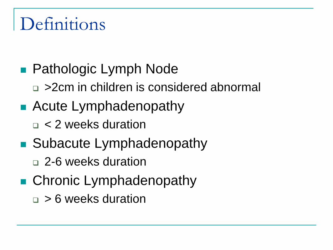
Definitions
Pathologic Lymph Node
>2cm in children is considered abnormal
Acute Lymphadenopathy
< 2 weeks duration
Subacute Lymphadenopathy
2-6 weeks duration
Chronic Lymphadenopathy
> 6 weeks duration

Etiology of Lymphadenopathy
Acute Infectious
Subacute
Chronic Infectious
Acute Infectious
Lymphadenopathy
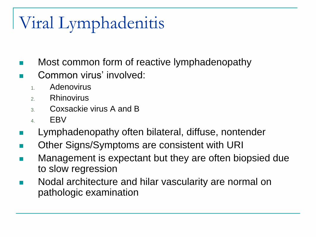
Viral Lymphadenitis
Most common form of reactive lymphadenopathy
Common virus’ involved:1. Adenovirus
2. Rhinovirus
3. Coxsackie virus A and B
4. EBV
Lymphadenopathy often bilateral, diffuse, nontender
Other Signs/Symptoms are consistent with URI
Management is expectant but they are often biopsied due to slow regression
Nodal architecture and hilar vascularity are normal on pathologic examination
Suppurative Bacterial Lymphadenitis
Staphylococcus aureus and Group A Streptococcus
Common history reveals recent URI
Earache
Sore Throat/Toothache
Skin Lesions
Management is initially with oral or IV antibiotics depending on severity of infection
If not resolving or getting worse CT with contrast and/or Ultrasound to evaluate for
phlegmon/abscess/infiltrate
FNA vs Surgical I&D vs Surgical Excision if abscess is identified
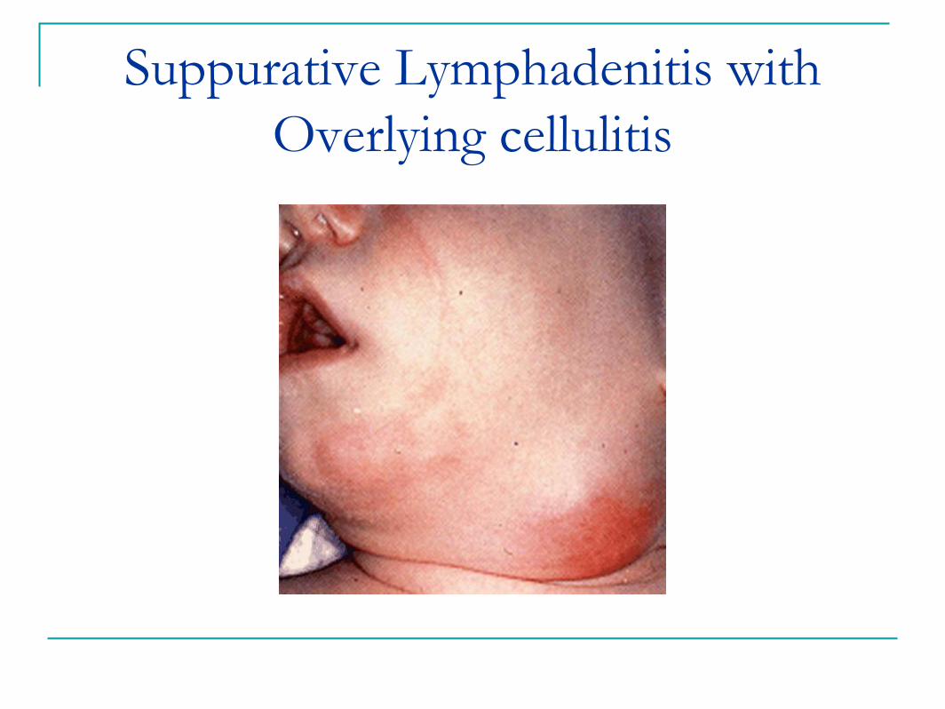
Suppurative Lymphadenitis with
Overlying cellulitis
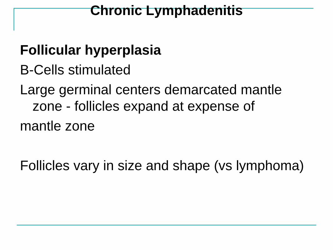
Chronic Lymphadenitis
Follicular hyperplasia
B-Cells stimulated
Large germinal centers demarcated mantle
zone - follicles expand at expense of
mantle zone
Follicles vary in size and shape (vs lymphoma)

Follicular hyperplasia could be non-specific
Or due to specific causes;
Toxoplasmosis, rheumatoid arithritis, SLE,
AIDS
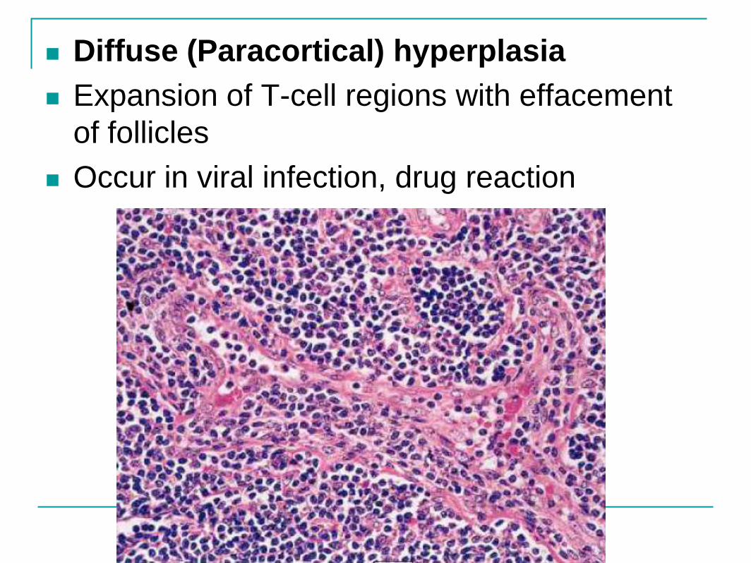
Diffuse (Paracortical) hyperplasia
Expansion of T-cell regions with effacement
of follicles
Occur in viral infection, drug reaction

Sinus Pattern of Hyperplasia
Seen in nodes draining cancer
Prominence of sinusoids - distended with
histiocytes

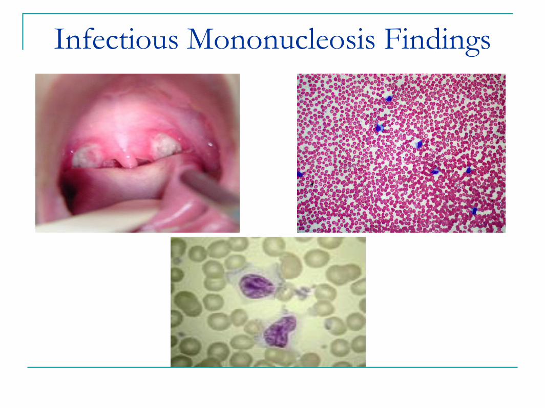
Infectious Mononucleosis
Caused by Epstein Barr Virus
Epidemiology 50% seropositive by age 5
90% seropositive by age 25
Signs/Symptoms Fever
Exudative pharyngitis
Painless generalized lymphadenopathy
Axillary LAD and Splenic enlargement increase likelihood
50% lymphocytosis with >10% Atypical lymphocytes on peripheral smear is suggestive
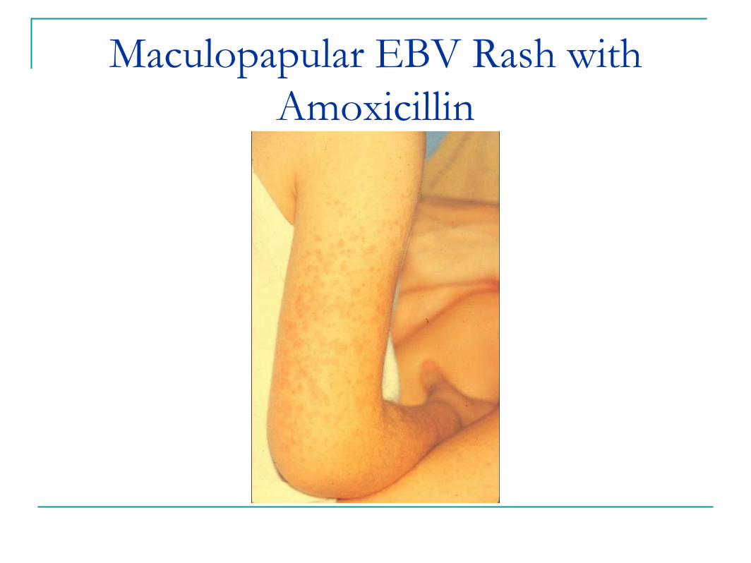
Maculopapular EBV Rash with
Amoxicillin