quantitative analysis of the relationship between
TRANSCRIPT
Citation: Vaishnavi D, Harshitha V and Kishore K. Quantitative Analysis of the Relationship between Maxillary Incisors and the Incisive Canal by Cone- Beam Computed Tomography in an Adult Population of Mangaluru. Austin J Dent. 2021; 8(2): 1156.
Austin J Dent - Volume 8 Issue 2 - 2021ISSN : 2381-9189 | www.austinpublishinggroup.com Vaishnavi et al. © All rights are reserved
Austin Journal of DentistryOpen Access
Abstract
Background: Maxillary anterior teeth play a crucial role in aesthetics, phonetics, and mastication. For successful orthodontic treatment evaluating the morphology of the alveolar bone and incisive canal would help in avoiding root resorption, dehiscence, and fenestration. This study is aimed to research the configurational relationships among maxillary incisors, alveolar bone, and incisive canal through Cone Beam Computerated Tomography (CBCT).
Methods: CBCT images of 35 orthodontic patients were evaluated for length of the canal (L); angles between the palatal plane and the maxillary alveolar border (θ1),the incisive canal (θ2), and maxillary incisor (θ3); distance from the right maxillary incisor to the incisive canal (D). All the measurements were performed on sagittal plane with the exception of (D) which was made on axial plane. Statistical analysis was performed on the above parameters using two sample test and Pearson’s correlation analysis.
Results: There was no statistically significant difference between males and females for all the variables although there were large interindividual variation.There was a positive moderate correlation between θ1 and θ2 (0.480), θ1 and θ3 (0.487), θ2 and θ3 (0.345). The mean value for L and D were 10.38mm and 4.14mm respectively.
Conclusion: There exists a large interindividual variability for incisive canal, proximity of incisors with that of incisive canal which could not be precisely predicted by the conventional cephalograms. The results of the study could be helpful clinically in planning orthodontic treatment for significant intrusion and retraction of maxillary incisors.
IntroductionWhile not specifically in the Hippocratic Oath, primum non
nocere is believed to be derived from it and means first, do no harm or above all else, do no harm. In other words, before you do anything to a patient, make sure that you are not making matters worse. What good does it do to straighten teeth if in doing so you cause the patient to lose them? [1].With regard to maxillary incisor movement, Ackerman et al. presented the concept of “envelope of discrepancy” [2], which describes the limitations of the range of orthodontic movement of the maxillary incisor [3]. Contact with hard tissue structures, such as the labial, palatal, or incisive canal cortical plates, is a risk factor for apical root resorption in the maxillary incisor [4-6], and it is one of the iatrogenic complications of orthodontic treatment. The incisive canal is an anatomic structure that runs posterior and more close to the roots of the central incisors in the median plane of the palatine process of the maxilla, surrounded by thick cortical bone [5,6]. It connects the floor of the nasal cavity with the palate and opens into the oral cavity as incisive foramen. It runs parallel to the maxillary central incisors and transmits nasopalatine vessels and nerves, branches of the maxillary division of the trigeminal nerve,
Research Article
Quantitative Analysis of the Relationship between Maxillary Incisors and the Incisive Canal by Cone- Beam Computed Tomography in an Adult Population of MangaluruVaishnavi D*, Harshitha V and Kishore KDepartment of Orthodontics, A J Institute of Dental Sciences, Mangaluru, India
*Corresponding author: D Vaishnavi, A J Institute of Dental Sciences, Mangaluru, A J Resident hostel, kuntikana, Mangaluru PC: 575004, India
Received: June 04, 2021; Accepted: July 09, 2021; Published: July 16, 2021
and the maxillary artery [7]. Because of proximity of incisive canal to the maxillary incisors, the surgical invasion and its complications of the incisive canal during dental procedures in maxillary incisor region can cause nonosseointegration of dental implants or sensory dysfunction [8,9]. The precise location of incisive canal in reference to the maxillary incisors isn’t well documented within the orthodontic literature due to the difficulties in detecting incisive canal morphology using conventional twodimensional radiographs.
The objective of this study was to evaluate the morphologic features and the relationship between roots of maxillary incisors and the incisive canal using Cone Beam Computed Tomography (CBCT).
Materials and MethodsSubjects
The study was carried out on the patients visiting the out-patient section of the Department of Orthodontics and Dentofacial Orthopaedics, AJ Institute of dental sciences, Mangaluru. CBCT scans were taken for those who required CBCT for diagnosis and treatment planning. The inclusion criteria include patients having lateral cephalogram, normal anteroposterior skeletal relationship
Austin J Dent 8(2): id1156 (2021) - Page - 02
Vaishnavi D Austin Publishing Group
Submit your Manuscript | www.austinpublishinggroup.com
(ANB of 0°- 4°), normal overjet and overbite with Class I molar relationship. The exclusion criteria were history of orthodontic treatment, missing or supernumerary maxillary incisors, prosthesis in relation to maxillary incisors, history of trauma to maxillary incisors, and congenital anomalies like cleft lip and palate. Based on the inclusion and exclusion criteria, 35 subjects were selected.
MethodsThe data was obtained using NewTom GiANO NNT Scanner
with the patient in upright position and head positioned along the Frankfort horizontal plane, running parallel to the floor. All the scans were taken using the same machine by the same operator. The operating parameters were set at 3mA and 90kV, dose of 80- 100 µSv and the scan time of 9 seconds. All CBCT images were taken using a large dentoalveolar field of view. It was determined that the sagittal plane was perpendicular to the axial plane and parallel to the plane passing through anterior nasal spine and posterior nasal spine.
CBCT images of 35 orthodontic patients were evaluated for the following measurements
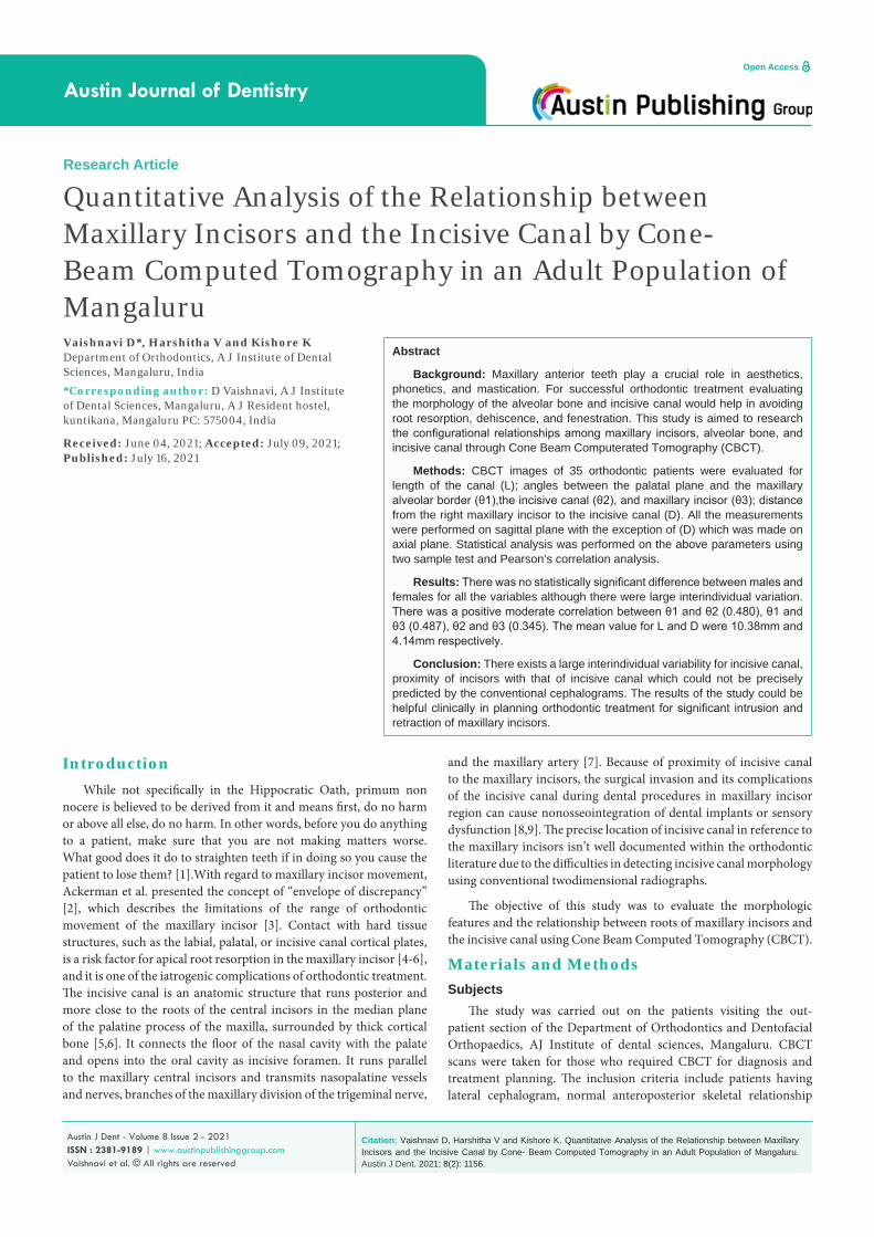
• Length of the canal (L).
• Angles between the palatal plane and the maxillary alveolar border (θ1) the incisive canal (θ2) the maxillary incisor (θ3).
• Distance from the right maxillary incisor to the incisive canal (D) (Figure 1).
ResultsMean values of the variables (descriptive statistics)
See Table 1.
Pearson’s correlation test:
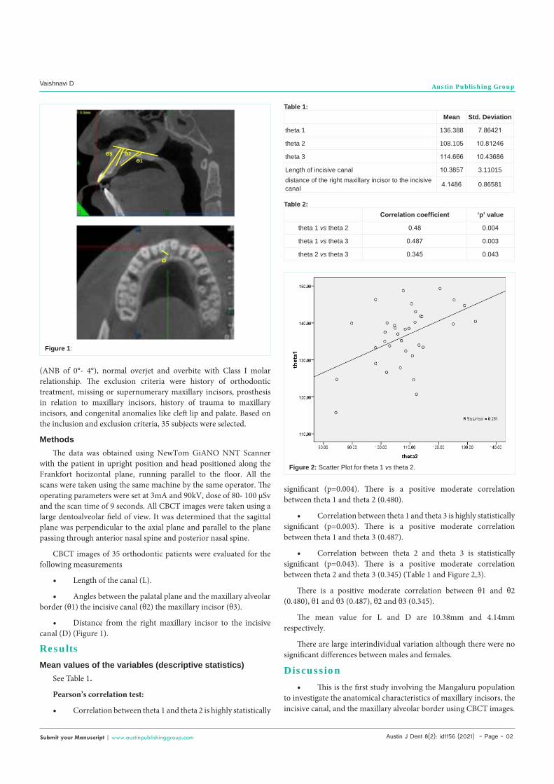
• Correlation between theta 1 and theta 2 is highly statistically
significant (p=0.004). There is a positive moderate correlation between theta 1 and theta 2 (0.480).
• Correlation between theta 1 and theta 3 is highly statistically significant (p=0.003). There is a positive moderate correlation between theta 1 and theta 3 (0.487).
• Correlation between theta 2 and theta 3 is statistically significant (p=0.043). There is a positive moderate correlation between theta 2 and theta 3 (0.345) (Table 1 and Figure 2,3).
There is a positive moderate correlation between θ1 and θ2 (0.480), θ1 and θ3 (0.487), θ2 and θ3 (0.345).
The mean value for L and D are 10.38mm and 4.14mm respectively.
There are large interindividual variation although there were no significant differences between males and females.
Discussion• This is the first study involving the Mangaluru population
to investigate the anatomical characteristics of maxillary incisors, the incisive canal, and the maxillary alveolar border using CBCT images.
Figure 1:
Mean Std. Deviation
theta 1 136.388 7.86421
theta 2 108.105 10.81246
theta 3 114.666 10.43686
Length of incisive canal 10.3857 3.11015distance of the right maxillary incisor to the incisive canal 4.1486 0.86581
Table 1:
Correlation coefficient ‘p’ value
theta 1 vs theta 2 0.48 0.004
theta 1 vs theta 3 0.487 0.003
theta 2 vs theta 3 0.345 0.043
Table 2:
Figure 2: Scatter Plot for theta 1 vs theta 2.

Austin J Dent 8(2): id1156 (2021) - Page - 03
Vaishnavi D Austin Publishing Group
Submit your Manuscript | www.austinpublishinggroup.com
• Inclination of maxillary incisors is significantly correlated with those of the maxillary alveolar border and axis of the incisive canal.
• First, in the present study, the length of the incisive canal in male patients is similar to that in female patients, which is discordant with the findings of a previous study [10].
• The FOV of CBCT images in the present study is larger in comparison with those reported in previous studies [12].
• The average anterior-posterior distance between the maxillary central incisor roots and the incisive canal measured is 4.14 which is lesser than previous studies [11] . This 4.14mm distance does not necessarily imply the “safety zone” for retraction because individuals with relatively large interroot distances are not at risk of canal invasion or contact even following maximum retraction. However, a large diversity in anatomy, morphology and size of the incisive canal and incisive foramen in different people are frequently reported with three-dimensional evaluation. The incisive canal, in many circumstances, is deviated toward the right central incisor [6,13-15].
• Because of the proximity of maxillary incisors to incisive canal, the possibility of sensory dysfunction in the anterior region and failure of osseointegration has been reported in cases of contact of the incisive canal through surgical interventions such as dental implant placement [16,17].
Conclusion• This study has the proximity between incisive canal and
maxillary central measured at 4.14 which is quite less indicating the closeness of incisor root to the canal which would aid us in carrying out the orthodontic procedures effectively.
• This study demonstrated that the incisive canal had large inter-individual variability, and the proximity between the incisive canal and the incisal root could not be precisely predicted by the conventional cephalogram.
Figure 3: Scatter Plot for theta 2 vs theta 3.
References1. Joseph Morneau.How to prevent root resorption using the golden rule of
orthodontics.Dentistry iq. 2006.
2. Arnett GW, Bergman RT. Facial keys to orthodontic diagnosis and treatment planning. Part I. Am J Orthod. 1993;103: 299–312.
3. Kokich V. Esthetics and anterior tooth position: an orthodontic perspective part III: mediolateral relationships. J Esthet Restor Dent. 1993;5: 200-207.
4. Chung CJ, Choi YJ, Kim KH. Approximation and contact of the maxillary central incisor roots with the incisive canal after maximum retraction with temporary anchorage devices: report of two cases. Am J Orthod Dentofac Orthop. 2015; 148: 493-502.
5. Proffit W, Fields H, Sarver D. Contemporary orthodontics. 5th ed. St Louis: Elsevier. 2013.
6. Liang X, Jacobs R, Martens W, Hu Y, Adriaensens P, Quirynen M, et al. Macro- and micro- anatomical, histological and computed tomography scan characterization of the nasopalatine canal. J Clin Periodontol 2009; 36: 598-603.
7. Song WC, Jo DI, Lee JY, Kim JN, Hur MS, Hu KS, et al. Microanatomy of the incisive canal using three-dimensional reconstruction of microCT images: an ex vivo study. Oral Surg Oral Med Oral Pathol Oral Radiol Endod. 2009; 108: 583-590.
8. Kraut RA, Boyden DK. Location of incisive canal in relation to central incisor implants. Implant Dent. 1998; 7: 221-225.
9. Kan JY, Rungcharassaeng K, Roe P, Mesquida J, Chatriyamuyoke P, Caruso JM. Maxillary centeral incisor-incisive canal relationship: a cone beam computed tomography study. Am J Esthet Dent. 2012; 2: 180-187.
10. Güncü GN, Yıldırım YD, Yılmaz HG, Galindo-Moreno P, Velasco-Torres M, Al-Hezaimi K, et al. Is there a gender difference in anatomic features of incisive canal and maxillary environmental bone? Clin Oral Implants Res. 2013; 24: 1023-1026.
11. Matsumura T, Ishida Y, Kawabe A, Ono T. Quantitative analysis of the relationship between maxillary incisors and the incisive canal by cone-beam computed tomography in an adult Japanese population. Progress in Orthodontics. 2017; 18: 24.
12. Khurana S, Parasher P, Mukherjee P, Mupparapu M, Lotlikar PP, Creanga AG. Cone beam computed tomographic–Based retrospective study on newark population for the assessment of distance between incisive canal and maxillary central incisors: Clinical implications. Indian J Dent Res. 2020; 31: 175-179.
13. Song WC, Jo DI, Lee JY, Kim JN, Hur MS, Hu KS, et al. Microanatomy of the incisive canal using three-dimensional reconstruction of microCT images: an ex vivo study. Oral Surg Oral Med Oral Pathol Oral Radiol Endod. 2009; 108: 583-590.
14. Kim SJ, Lim SH. Anatomic study of the incisive canal in relation to midpalatal placement of mini- implant. Korean J Orthod. 2009; 39:146-158.
15. Asaumi R, Kawai T, Sato I, Yoshida S, Yosue T. Three-dimensional observations of the incisive canal and the surrounding bone using cone-beam computed tomography. Oral Radiol. 2010; 26: 20-28.
16. Artzi Z, Nemcovsky CE, Bitlitum I, Segal P. Displacement of the incisive foramen in conjunction with implant placement in the anterior maxilla without jeopardizing vitality of nasopalatine nerve and vessels: a novel surgical approach. Clin Oral Implants Res. 2000; 11: 505-510.
17. Kan JY, Rungcharassaeng K, Roe P, Mesquida J, Chatriyamuyoke P, Caruso JM. Maxillary central incisor-incisive canal relationship: a cone beam computed tomography study. Am J Esthet Dent. 2012; 2: 180-187.