pysm an integrated data management and analysis platform for single molecule experimentation

1
examined using fluorescence polarization microscopy. We characterized the domain organization of the FG nups, which are required for transport of cargo through the NPC. This approach revealed both structured and unstructured do- mains: the tips of the FG domains are disordered, whereas the NPC-anchored domains are ordered. This technique allows the collection of structural informa- tion in vivo with the ability to probe the organization of protein domains within the NPC. This has particular relevance for the FG domain nups, which are im- plicated in the mechanism of cargo transport. 755-Pos Board B555 The Structure of the Nuclear Pore Complex Studied by Fluorescence Anisotropy Claire E. Atkinson, Martin Kampmann, Alexa L. Mattheyses, Sanford M. Simon. A major challenge in determining structures of large macromolecular com- plexes is the integration of high-resolution crystal structures of individual proteins with low-resolution structures of the entire assembly. The nuclear pore complex (NPC), which has a molecular mass of greater than 50 MDa, is one example of this. It is composed of multiple copies of ~30 different proteins, termed nucleoporins. While EM structures of the complex as a whole are available, it is unclear how individual proteins are arranged in the complex. Polarized fluorescence microscopy can be used to deter- mine the orientation of immobile, isotropically ordered fluorophores. We present a theoretical framework for determining the orientation of GFP within the NPC and relating this to the orientation of structured nucleopor- ins using polarized fluorescence microscopy in combination with structural information from crystal structures. This framework can be used to deter- mine nucleoporin orientation within both yeast and mammalian NPCs and has the potential to complement X-ray crystallography and electron mi- croscopy to generate a high-resolution map of the entire NPC. This method could be adapted to yield unique insights into the molecular mechanisms of other biologically important processes, such as co-translational protein translocation. 756-Pos Board B556 Homo-FRET Imaging as a Tool to Quantify Protein and Lipid Clustering Hans Gerritsen, Arjen Bader, Erik Hofman, Jarno Voortman, Paul van Bergen, en Henegouwen, Gerrit van Meer. Fluorescence anisotropy based Homo-FRET methods have the potential to be- come valuable tools in molecular cell biology. Here, use is being made of the rapid change in fluorescenc anisotropy due to homo energy transfer. Homo- FRET measurements can be employed to determine the distance between fluo- rophores but also for quantifying the size of the clusters, as well as distributions of cluster sizes. The interpretation of homo-FRET signals is complicated by the fact that both the mutual orientations of the fluorophores and the number of fluorophores per cluster affect the fluorescence anisotropy in a similar way. The properties of the fluorescence probes are of great important and taking these aspects into account is crucial for the cor- rect interpretation of homo-FRET signals in protein and lipid clustering studies. In this study cluster sizes are quan- tified using reference values obtained from controlled di- merization and oligomirization experiments. Homo-FRET is applied in a study on the clustering of lipid raft markers GPI (see Fig.) and K-ras, as well as for EGF receptor clus- tering in the plasma membrane. The results on K-ras are compared with an TEM study on gold labeled K-ras. 757-Pos Board B557 No Need to FRET Josef Lazar, Alexey Bondar, Stuart Firestein. Membrane proteins are a large, diverse group of proteins, serving a multi- tude of cellular functions. They are difficult to study, due to their require- ment of a lipid membrane for function. Our mathematical model shows that polarization fluorescence microscopy using fluorescent proteins can take ad- vantage of the cell membrane requirement to yield insights into membrane protein structure and function, in living cells and organisms. We have now experimentally demonstrated that polarization microscopy can be used for imaging of G-protein activation, changes in intracellular calcium concentra- tion, and other cellular processes, in living cells, with sensitivity compara- ble to, or even exceeding that of current FRET probes. Crucially, in contrast to FRET, polarization fluorescence microscopy only requires pres- ence of a single fluorescent protein. Therefore, as both our theoretical and experimental work shows, many existing constructs can be used as optical probes of molecular processes involving membrane proteins. Apart from utilizing existing constructs, polarization microscopy offers a clear path towards development of new genetically encoded optical probes of mem- brane protein function, including a usable genetically encoded optical sen- sor of cell membrane voltage. Our results indicate that in many biological applications, FRET is likely to be complemented or even replaced by po- larization microscopy. 758-Pos Board B558 PySM an Integrated Data Management and Analysis Platform for Single Molecule Experimentation Jacob C. Bolewski, Jose M. Moran-Mirabal, Larry Walker. Application of single molecule tracking (SMT) and super-resolution (SR) mi- croscopy methods is expanding with easier to use tools and off the shelf com- mercial hardware from major vendors. Key to these methods application to biologically relevant questions is non-trivial image and data analysis. Although the progression of hardware has broadened SPT and SR microscopy among re- searchers, the development and dissemination of easy to use software tools has lagged behind. PySM (PythonSingleMolecule) is an effort to develop an open source user friendly GUI which allows users from non-technical backgrounds to analyze and visualize SPT and SR microscopy data. Some of the features in- clude a persistent database for storing images, analyzed data, and metadata, built in algorithms for single particle detection, tracking and stage registration, an internal python analysis/scripting environment, and the ability to parallelize data analysis run across multiple cores or workstations. The aim is to provide a built solution for researchers more interested in analyzing data as opposed to writing code while enabling developers to incorporate additional algorithms through python bindings to external libraries or by running scripts with the built in IPython interpreter. The application of PySM is demonstrated for the various encoded routines with sample data where single molecules are detected over a series of 600 frames, the stage positioning is tracked through fiducial marks included in the sample, and tracks are generated by linking the particles be- tween frames. Additional data analysis is performed by calculating the anisot- ropy of the generated tracks to discriminate between different types of motion for the molecules tracked. 759-Pos Board B559 Accurate FRET Measurements and Testing of the Theory for Multimeric Complexes Using Reference Fluorescence Standards Suparna Patowary, Luca F. Pisterzi, Michael R. Stoneman, Vyacheslav Strogolov, Julie A. Oliver, James W. Wells, Valerica ˘ Raicu. Fo ¨ rster Resonance Energy Transfer (FRET) is a process in which a donor (D) in the excited state transfers its energy nonradiatively to an acceptor (A) in the ground state. The underlying theory has been confirmed countless times, partic- ularly with regard to the dependence of the FRET efficiency on the sixth power of the distance between D and A. In contrast, a complete FRET theory for mul- tiple donors and acceptors in oligomeric complexes has been developed only recently (Raicu, 2007, J. Biol. Phys. 33:109-127), in parallel with technology of sufficient accuracy for tests in living cells (Raicu et al., 2009, Nature Photon. 3:107-113). This novel approach now has been applied to linked fluorescent proteins located in the cytoplasm and at the plasma membrane. The cytoplas- mic probes were fused combinations of a donor (Cerulean, C), an acceptor (Ve- nus, V), and a chromophore-deficient, Venus-like molecule that cannot absorb or transfer energy (Amber, A) (Koushik et al., 2009, PLoS ONE 4(11):e8031): namely, ACVA, ACAV, VCAA, and VCVV. The membrane-bound probes were fused dimers and trimers of eGFP2 (G2) and eYFP (Y): namely, G2Y, YG2, G2YG2, and YG2Y. According to the theory (Raicu, 2007), the FRET efficiency of a tetramer such as VCVV can be predicted from that of analogues that contain a single acceptor (e.g., ACVA, ACAV, VCAA); also, the apparent FRET efficiency of a trimer such as G2YG2 or YG2Y can be predicted from the pair-wise efficiency that corresponds to that of dimers such as G2Y and YG2. These predictions have been confirmed for FRET efficiencies measured by means of two-photon microspectroscopy (Raicu et al., 2009), in accord with the theory and underlying assumptions for FRET within multimers. 760-Pos Board B560 In Vivo Monitoring of Agonist-Induced Relative Movements Between G Protein Coupled Receptor Segments in Oligomeric Complexes Using Spectrally Resolved FRET Michael R. Stoneman, Suparna Patowary, Michael Roesch, Madhusudan Dey, Valerica Raicu. Fo ¨rster Resonance Energy Transfer (FRET) from an electronically excited do- nor to an acceptor molecule is used to quantify the extent of interactions be- tween molecules. For example, FRET can be utilized to determine how proteins form complexes by tagging them differentially with donor and accep- tor fluorophores. Recent advances in the FRET theory combined with a novel spectrally resolved two-photon microscope have strengthened the effectiveness of the FRET technique and have enabled us to determine the size and structure 140a Sunday, March 6, 2011

Upload: larry

Post on 24-Nov-2016

212 views

Category:

Documents


0 download

TRANSCRIPT

140a Sunday, March 6, 2011

examined using fluorescence polarization microscopy. We characterized thedomain organization of the FG nups, which are required for transport of cargothrough the NPC. This approach revealed both structured and unstructured do-mains: the tips of the FG domains are disordered, whereas the NPC-anchoreddomains are ordered. This technique allows the collection of structural informa-tion in vivo with the ability to probe the organization of protein domains withinthe NPC. This has particular relevance for the FG domain nups, which are im-plicated in the mechanism of cargo transport.

755-Pos Board B555The Structure of the Nuclear Pore Complex Studied by FluorescenceAnisotropyClaire E. Atkinson, Martin Kampmann, Alexa L. Mattheyses,Sanford M. Simon.A major challenge in determining structures of large macromolecular com-plexes is the integration of high-resolution crystal structures of individualproteins with low-resolution structures of the entire assembly. The nuclearpore complex (NPC), which has a molecular mass of greater than 50 MDa,is one example of this. It is composed of multiple copies of ~30 differentproteins, termed nucleoporins. While EM structures of the complex asa whole are available, it is unclear how individual proteins are arrangedin the complex. Polarized fluorescence microscopy can be used to deter-mine the orientation of immobile, isotropically ordered fluorophores. Wepresent a theoretical framework for determining the orientation of GFPwithin the NPC and relating this to the orientation of structured nucleopor-ins using polarized fluorescence microscopy in combination with structuralinformation from crystal structures. This framework can be used to deter-mine nucleoporin orientation within both yeast and mammalian NPCsand has the potential to complement X-ray crystallography and electron mi-croscopy to generate a high-resolution map of the entire NPC. This methodcould be adapted to yield unique insights into the molecular mechanisms ofother biologically important processes, such as co-translational proteintranslocation.

756-Pos Board B556Homo-FRET Imaging as a Tool to Quantify Protein and Lipid ClusteringHans Gerritsen, Arjen Bader, Erik Hofman, Jarno Voortman, Paul vanBergen, en Henegouwen, Gerrit van Meer.Fluorescence anisotropy based Homo-FRET methods have the potential to be-come valuable tools in molecular cell biology. Here, use is being made of therapid change in fluorescenc anisotropy due to homo energy transfer. Homo-FRET measurements can be employed to determine the distance between fluo-rophores but also for quantifying the size of the clusters, as well as distributions

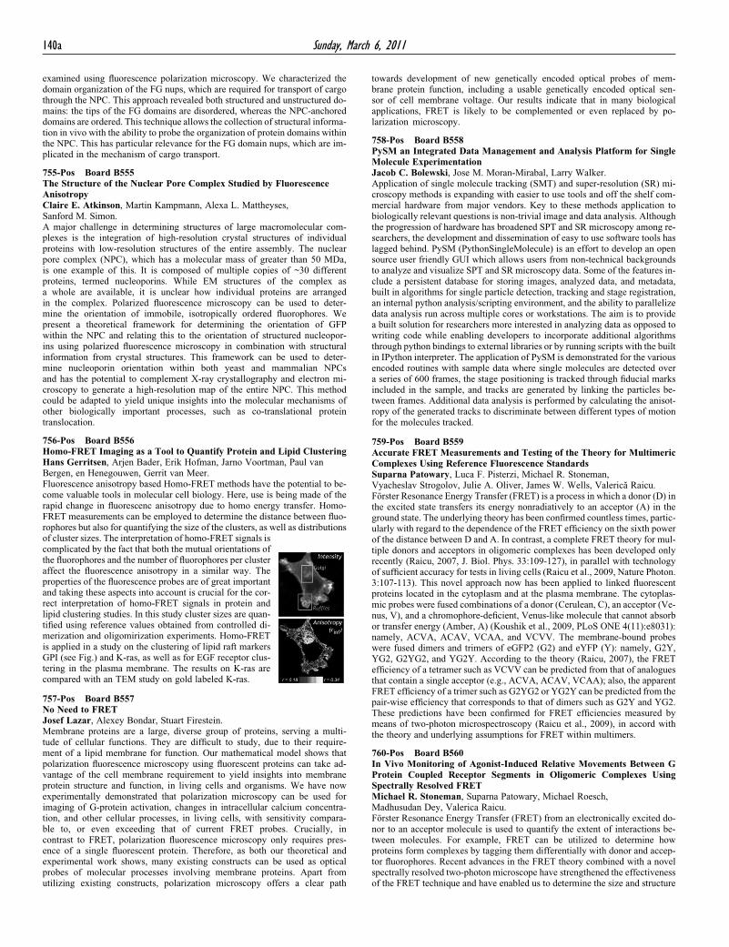
of cluster sizes. The interpretation of homo-FRET signals iscomplicated by the fact that both the mutual orientations ofthe fluorophores and the number of fluorophores per clusteraffect the fluorescence anisotropy in a similar way. Theproperties of the fluorescence probes are of great importantand taking these aspects into account is crucial for the cor-rect interpretation of homo-FRET signals in protein andlipid clustering studies. In this study cluster sizes are quan-tified using reference values obtained from controlled di-merization and oligomirization experiments. Homo-FRETis applied in a study on the clustering of lipid raft markersGPI (see Fig.) and K-ras, as well as for EGF receptor clus-tering in the plasma membrane. The results on K-ras arecompared with an TEM study on gold labeled K-ras.

757-Pos Board B557

No Need to FRETJosef Lazar, Alexey Bondar, Stuart Firestein.Membrane proteins are a large, diverse group of proteins, serving a multi-tude of cellular functions. They are difficult to study, due to their require-ment of a lipid membrane for function. Our mathematical model shows thatpolarization fluorescence microscopy using fluorescent proteins can take ad-vantage of the cell membrane requirement to yield insights into membraneprotein structure and function, in living cells and organisms. We have nowexperimentally demonstrated that polarization microscopy can be used forimaging of G-protein activation, changes in intracellular calcium concentra-tion, and other cellular processes, in living cells, with sensitivity compara-ble to, or even exceeding that of current FRET probes. Crucially, incontrast to FRET, polarization fluorescence microscopy only requires pres-ence of a single fluorescent protein. Therefore, as both our theoretical andexperimental work shows, many existing constructs can be used as opticalprobes of molecular processes involving membrane proteins. Apart fromutilizing existing constructs, polarization microscopy offers a clear path

towards development of new genetically encoded optical probes of mem-brane protein function, including a usable genetically encoded optical sen-sor of cell membrane voltage. Our results indicate that in many biologicalapplications, FRET is likely to be complemented or even replaced by po-larization microscopy.

758-Pos Board B558PySM an Integrated Data Management and Analysis Platform for SingleMolecule ExperimentationJacob C. Bolewski, Jose M. Moran-Mirabal, Larry Walker.Application of single molecule tracking (SMT) and super-resolution (SR) mi-croscopy methods is expanding with easier to use tools and off the shelf com-mercial hardware from major vendors. Key to these methods application tobiologically relevant questions is non-trivial image and data analysis. Althoughthe progression of hardware has broadened SPT and SR microscopy among re-searchers, the development and dissemination of easy to use software tools haslagged behind. PySM (PythonSingleMolecule) is an effort to develop an opensource user friendly GUI which allows users from non-technical backgroundsto analyze and visualize SPT and SR microscopy data. Some of the features in-clude a persistent database for storing images, analyzed data, and metadata,built in algorithms for single particle detection, tracking and stage registration,an internal python analysis/scripting environment, and the ability to parallelizedata analysis run across multiple cores or workstations. The aim is to providea built solution for researchers more interested in analyzing data as opposed towriting code while enabling developers to incorporate additional algorithmsthrough python bindings to external libraries or by running scripts with the builtin IPython interpreter. The application of PySM is demonstrated for the variousencoded routines with sample data where single molecules are detected overa series of 600 frames, the stage positioning is tracked through fiducial marksincluded in the sample, and tracks are generated by linking the particles be-tween frames. Additional data analysis is performed by calculating the anisot-ropy of the generated tracks to discriminate between different types of motionfor the molecules tracked.

759-Pos Board B559Accurate FRET Measurements and Testing of the Theory for MultimericComplexes Using Reference Fluorescence StandardsSuparna Patowary, Luca F. Pisterzi, Michael R. Stoneman,Vyacheslav Strogolov, Julie A. Oliver, James W. Wells, Valerica Raicu.Forster Resonance Energy Transfer (FRET) is a process in which a donor (D) inthe excited state transfers its energy nonradiatively to an acceptor (A) in theground state. The underlying theory has been confirmed countless times, partic-ularly with regard to the dependence of the FRET efficiency on the sixth powerof the distance between D and A. In contrast, a complete FRET theory for mul-tiple donors and acceptors in oligomeric complexes has been developed onlyrecently (Raicu, 2007, J. Biol. Phys. 33:109-127), in parallel with technologyof sufficient accuracy for tests in living cells (Raicu et al., 2009, Nature Photon.3:107-113). This novel approach now has been applied to linked fluorescentproteins located in the cytoplasm and at the plasma membrane. The cytoplas-mic probes were fused combinations of a donor (Cerulean, C), an acceptor (Ve-nus, V), and a chromophore-deficient, Venus-like molecule that cannot absorbor transfer energy (Amber, A) (Koushik et al., 2009, PLoS ONE 4(11):e8031):namely, ACVA, ACAV, VCAA, and VCVV. The membrane-bound probeswere fused dimers and trimers of eGFP2 (G2) and eYFP (Y): namely, G2Y,YG2, G2YG2, and YG2Y. According to the theory (Raicu, 2007), the FRETefficiency of a tetramer such as VCVV can be predicted from that of analoguesthat contain a single acceptor (e.g., ACVA, ACAV, VCAA); also, the apparentFRET efficiency of a trimer such as G2YG2 or YG2Y can be predicted from thepair-wise efficiency that corresponds to that of dimers such as G2Y and YG2.These predictions have been confirmed for FRET efficiencies measured bymeans of two-photon microspectroscopy (Raicu et al., 2009), in accord withthe theory and underlying assumptions for FRET within multimers.

760-Pos Board B560In Vivo Monitoring of Agonist-Induced Relative Movements Between GProtein Coupled Receptor Segments in Oligomeric Complexes UsingSpectrally Resolved FRETMichael R. Stoneman, Suparna Patowary, Michael Roesch,Madhusudan Dey, Valerica Raicu.Forster Resonance Energy Transfer (FRET) from an electronically excited do-nor to an acceptor molecule is used to quantify the extent of interactions be-tween molecules. For example, FRET can be utilized to determine howproteins form complexes by tagging them differentially with donor and accep-tor fluorophores. Recent advances in the FRET theory combined with a novelspectrally resolved two-photon microscope have strengthened the effectivenessof the FRET technique and have enabled us to determine the size and structure