publicar con estndares web mediante herramientas de coste
TRANSCRIPT
Lesions from patients with sporadic cerebralcavernous malformations harbor somatic mutationsin the CCM genes: evidence for a commonbiochemical pathway for CCM pathogenesis
David A. McDonald1,{, Changbin Shi2, Robert Shenkar2, Carol J. Gallione1, Amy L. Akers1,3,
Stephanie Li1, Nicholas De Castro1, Michel J. Berg4, David L. Corcoran5, Issam A. Awad2
and Douglas A. Marchuk1,∗
1Molecular Genetics and Microbiology Department, Duke University Medical Center, Durham, NC 27710, USA, 2Section
of Neurosurgery, Biological Sciences Division, University of Chicago, Chicago, IL 60637, USA, 3Angioma Alliance,
Norfolk, VA 23517, USA, 4School of Medicine and Dentistry, University of Rochester Medical Center, Rochester, NY
14642, USA and 5Institute for Genome Sciences and Policy, Duke University, Durham, NC 27710, USA
Received December 4, 2013; Revised and Accepted March 31, 2014
Cerebral cavernous malformations (CCMs) are vascular lesions affecting the central nervous system. CCMoccurs either sporadically or in an inherited, autosomal dominant manner. Constitutional (germline) mutationsin any of three genes, KRIT1, CCM2 and PDCD10, can cause the inherited form. Analysis of CCM lesions frominherited cases revealed biallelicsomatic mutations, indicating that CCM follows a Knudsonian two-hit mutationmechanism. It is still unknown, however, if the sporadic cases of CCM also follow this genetic mechanism.We extracted DNA from 11 surgically excised lesions from sporadic CCM patients, and sequenced the threeCCM genes in each specimen using anext-generation sequencing approach. Four sporadic CCM lesion samples(36%) were found to contain novel somatic mutations. Three of the lesions contained a single somatic mutation,and one lesion contained two biallelic somatic mutations. Herein, we also describe evidence of somatic mosai-cism in a patient presenting with over 130 CCM lesions localized to one hemisphere of the brain. Finally, in alesion regrowth sample, we found that the regrown CCM lesion contained the same somatic mutation as the ori-ginal lesion. Together, thesedatabolster the idea thatall formsofCCMhaveagenetic underpinning of the two-hitmutation mechanism in the known CCM genes. Recent studies have found aberrant Rho kinase activation ininherited CCM pathogenesis, and we present evidence that this pathway is activated in sporadic CCM patients.These results suggest that all CCM patients, including those with the more common sporadic form, are poten-tially amenable to the same therapy.
INTRODUCTION
Cerebral cavernous malformations (CCMs, OMIM #116860,603284, 603285) are vascular lesions affecting the centralnervous system. CCM lesions consist of grossly dilated, capillary-like structures lined by endothelium and lacking mature vascularwall structure. Lesions can be found in �0.5% of the generalpopulation (1–4) and affected individuals have a lifetime risk of
focal neurological deficits and hemorrhagic stroke. Sporadiclesions are typically solitary, whereas familial CCM is character-ized by multifocal cerebral lesions in the setting of autosomaldominant mutations in one of three genes, KRIT1 (CCM1),CCM2 and PDCD10 (CCM3) (5–9).
A Knudson two-hit mutation mechanism has been suggestedin the familial forms of CCM, where both copies of a particulargene must be mutated in order for disease to occur (10). Lesions
†Present address: Center for Science, Math and Technology Education, North Carolina Central University, Durham, NC 27707, USA.
∗To whom correspondence should be addressed at: Duke University Medical Center, PO Box 3175, Durham, NC 27710, USA. Tel: +1 9196841945;Fax: +1 9196842790; Email: [email protected]
# The Author 2014. Published by Oxford University Press. All rights reserved.For Permissions, please email: [email protected]
Human Molecular Genetics, 2014, Vol. 23, No. 16 4357–4370doi:10.1093/hmg/ddu153Advance Access published on April 3, 2014
by guest on September 30, 2014
http://hmg.oxfordjournals.org/
Dow
nloaded from
in familial cases would result from an inherited, constitutional(germline) mutation and biallelic somatic mutations. This mech-anism would also suggest that lesions in sporadic cases mayresult from two independent, biallelic, somatic mutations inCCM genes occurring in the same cell.
Our laboratory and others have identified biallelic somaticmutations in lesions from familial cases (11–13), with one mu-tation being the inherited constitutional mutation, and the otheroccurring somatically in the wild-type (WT) copy of the corre-sponding gene in the vascular lesional cells. These data demon-strated that the familial forms can be caused by a Knudsonian,two-hit mutation mechanism. However, although these studiesfocused on the inherited form of CCM, the majority of CCMpatients are sporadic cases (14). Based on the two-hit geneticmechanism, we hypothesize that sporadic CCM lesions arecaused by two independent, biallelic, somatic mutations inone of the known CCM genes. We investigated this hypothesisthrough next-generation sequencing (NGS) of the three CCMgenes using genomic DNA isolated from the lesions of sporadicCCM patients.
Previous studies on somatic mutations in CCM lesionsfocused on single nucleotide changes or relatively small indels(11–13). Mutant alleles in these samples were present at fre-quencies between 5 and 20%. If a single mutant cell became aCCM lesion by clonal expansion (such as that seen in canceroustumors), then the frequency of mutant alleles within the sampleswould be expected to approach 50%. The lower than expectedvalues observed for somatic mutations in CCM lesions are inpart because of the cellular heterogeneity of the cerebrovascularlesion. However, the somatic mutation frequency remains wellbelow 50% even in laser capture-microdissected lesion endothe-lial cells (11–13), suggesting that the CCM lesions do notdevelop from a purely clonal expansion of a mutant cell. Thelow frequencies of mutations within the lesions also demonstratethe need for using sensitive techniques to identify the somaticmutation.
In examining sporadic CCM lesions for two somatic muta-tions, both of which are likely to be found at low frequencies, aNGS strategy was used. Multiple sporadic CCM lesions werefound to contain somatic mutations, including one lesion withtwo biallelic, somatic mutations. These findings from sporadiclesion samples, along with a somatic mosaic patient and a caseof CCM lesion regrowth, support a somatic mutation mechanisminvolving the known CCM genes, suggesting a common pathwayfor CCM lesion pathogenesis.
RESULTS
For our NGS strategy, the coding exons (with .10 bp of flankingintronic sequence) of all three CCM genes were amplified bypolymerase chain reaction (PCR). All coding exons were suc-cessfully amplified from each sample, ruling out constitutionalor high-frequency trans deletions in the samples examined.PCR products were pooled and sequencing on a Roche 454GS-FLX Titanium platform. Fourteen lesion samples were ana-lyzed in this manner. A total of 379 394 reads aligned with thehuman reference sequence, with an average of 596 reads peramplicon, though this value ranged from 0 to 8292 reads and astandard deviation of 776 reads. Since the vast majority of
constitutional CCM mutations that have been identified severelyalter protein function (premature stop codons, splice-site muta-tions, insertions/deletions leading to frameshifts, etc.), thesequences from these sporadic CCM lesions were analyzed forthese same classes of somatic mutations. Thus, we may havemissed bona fide somatic missense mutations, but our approachwas designed to generate few false-positive mutant calls. Theresults are summarized in Table 1.
CCM lesions with constitutional mutations
Of the 14 CCM samples analyzed, 13 were from patients exhibit-ing a single CCM lesion, a common feature of sporadic CCM.The remaining CCM lesion was a sample from an inheritedcase previously analyzed by our laboratory (13) and served asa positive control for our sequencing strategy. Sample 2049was resected from a family member harboring the common His-panic constitutional mutation (13) in KRIT1 (c.1363C.T,p.Q455X). In the original study, we utilized Sanger sequencingof individual cloned exonic amplicons from the lesion DNA toidentify the trans somatic mutation in KRIT1 (c.1271_1274delTATAfs∗12) in 4.6% of the clones. Using NGS for the samesample, the constitutional mutation was identified in 39 of 94reads (42% frequency) and the somatic mutation was identifiedin 4 of 94 reads (4.3% frequency). Since both mutations occur inthe same exon, it was evident that the mutations are indeed intrans as no read contained both mutations. This positive controlsample served as a proof of principle that NGS was sensitiveenough to detect one of our previously reported somatic muta-tions, even though it can only be found at low frequency in thebulk lesion sample.
In nine of the CCM lesion samples, 10 heterozygous single nu-cleotide polymorphisms (SNPs) were identified (SupplementaryMaterial, Table S1), each with an associated reference number.For SNPs with coverage of at least 50 reads, the minor allelesranged in frequency from 45.0 to 58.4%. From these data, wecan surmise that heterozygous variants would be found atsimilar frequencies and somatic variants would be found atlower frequencies.
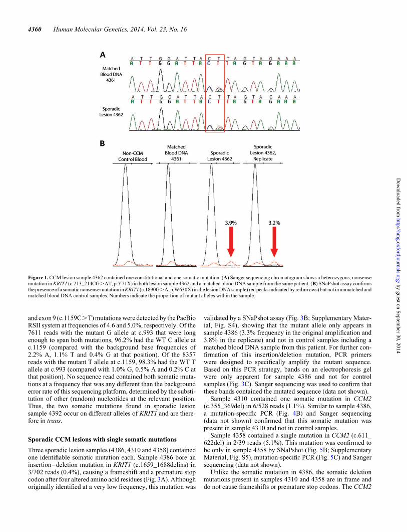
Surprisingly, analysis of the sequencing data revealed that 2 ofthe 13 CCM lesion samples (15%) from patients harboring asingle lesion instead contained high-frequency mutations inone of the CCM genes. In lesion 4362, a mutation was identifiedin KRIT1 (c.213_214CG.AT, p.Y71X) in 46% of the reads(321 of 703) and confirmed by Sanger sequencing (Fig. 1A).Analysis of a matched blood DNA sample from this patientrevealed the presence of the c.213_214CG.AT mutation,confirming that this high-frequency variant is a constitutional,heterozygous mutation. In the lesion sample 4362, a somatic mu-tation was also identified in KRIT1 (c.1890G.A, p.W630X) at afrequency of 2.5% (53 of 2097 reads).
A single-base extension assay (SNaPshot, Applied Biosys-tems) was used to validate these mutations. First, primers aredesigned to anneal adjacent to the base of interest, and then anextension reaction adds a single nucleotide to the primer. Theresulting fragments are separated by size, and different allelesof the same amplicon appear as adjacent peaks. A diagramdepicting SNaPshot results is shown in Supplementary Material,Figure S1, and it should be noted that only the data from the rele-vant bases are shown in the remaining figures. One of the major
4358 Human Molecular Genetics, 2014, Vol. 23, No. 16
by guest on September 30, 2014
http://hmg.oxfordjournals.org/
Dow
nloaded from
benefits of this assay is that it can be used to quantify the fre-quency of alleles at a particular DNA base.
As shown in Figure 1B and Supplementary Material, Figure S2,the c.1890G.A mutation in KRIT1 was confirmed by SNaPshotin sample 4362. The mutant allele appeared only in DNA fromthe lesion sample (two replicates, 3.0 and 2.8% frequencies ofthe mutant allele). Only the WT allele was present in DNAfrom this patient’s blood (sample 4361) and in a non-CCMcontrol DNA sample.
A high-frequency mutation was also found in lesion 4384, asplice-site mutation in CCM2 (c.472+1G.T, splice donorsite) occurring in 52% of the reads (43/82). This heterozygousmutation was verified by Sanger sequencing (data not shown).No obvious somatic mutation was identified in this sample.
Although thesesamples were obtained fromCCM patientswithno apparent family history and who exhibited a single lesion onmagnetic resonance imaging (MRI), both hallmarks of sporadicCCM disease, these patients did harbor high-frequency CCMgene mutations, indicating that they were either cryptic familialcases with constitutional mutations or possibly cases of somaticmosaicism. For patient sample 4362, we were able to confirmthe presence of the constitutional mutation in a matched bloodsample (Fig. 1A), so this patient is likely to be a cryptic familialcase of CCM. Since most of the samples were obtained throughan anonymous tissue bank, we were unable to follow up to deter-mine whether MRI analysis of the entire family might identifyhidden cases of CCM or whether the individuals instead representde novo constitutional mutations. Regardless, our sequence
analysis identified another case of biallelic somatic and constitu-tional mutations in an inherited case of CCM.
Sporadic CCM lesion containing two biallelic, somaticmutations
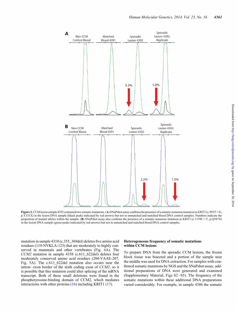
Of the 11 remaining sporadic CCM lesion samples analyzed byNGS, 5 somatic mutations were identified in 4 samples. Twosomatic mutations in KRIT1 were identified in sample 4392.The first somatic mutation was a premature stop codon incoding exon 8 (c.993T.G, p.Y331X) occurring in 44/610reads (7.2% frequency). The second somatic mutation was a dif-ferent premature stop codon in coding exon 9 (c.1159C.T,p.Q387X) occurring in 35 of 578 reads (6.1% frequency).
By SNaPshot, the exon 8 and exon 9 mutant alleles appeared inthe original lesion DNA sample (4392) at frequencies of 5.3 and2.2%, respectively (Fig. 2; Supplementary Material, Fig. S3).A second, separate PCR amplification also replicated theseresults with the exon 8 mutation measured at 8.5% frequencyand the exon 9 mutation measured at 1.5% frequency. Mutantalleles were not apparent in a matched blood DNA sample(4391) or a control DNA sample (non-CCM case).
To test whether these two somatic mutations were in trans, a1 kb region surrounding the two mutations was amplifiedusing the high-fidelity (15) PfuTurbo polymerase (Agilent)and sequenced using the Pacific Biosystems RS platform. ThePacBio RS NGS system was selected because it can producelonger reads than other platforms. The exon 8 (c.993T.G)
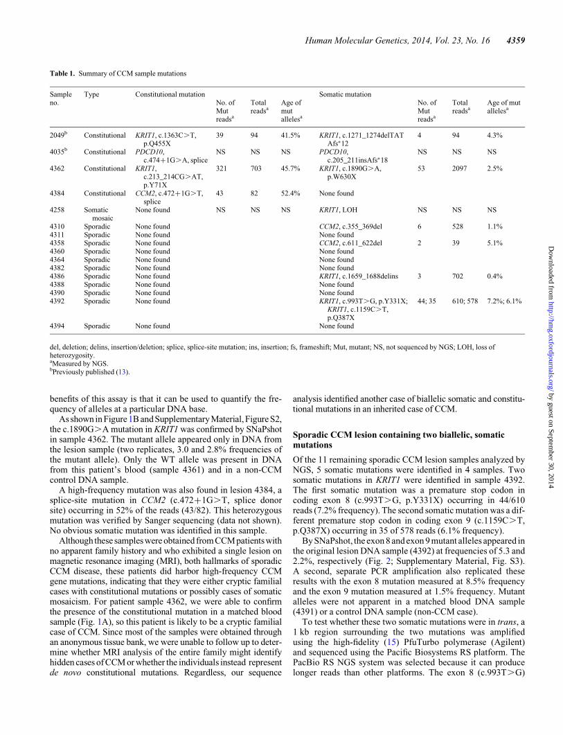
Table 1. Summary of CCM sample mutations
Sampleno.
Type Constitutional mutation Somatic mutationNo. ofMutreadsa
Totalreadsa
Age ofmutallelesa
No. ofMutreadsa
Totalreadsa
Age of mutallelesa
2049b Constitutional KRIT1, c.1363C.T,p.Q455X
39 94 41.5% KRIT1, c.1271_1274delTATAfs∗12
4 94 4.3%
4035b Constitutional PDCD10,c.474+1G.A, splice
NS NS NS PDCD10,c.205_211insAfs∗18
NS NS NS
4362 Constitutional KRIT1,c.213_214CG.AT,p.Y71X
321 703 45.7% KRIT1, c.1890G.A,p.W630X
53 2097 2.5%
4384 Constitutional CCM2, c.472+1G.T,splice
43 82 52.4% None found
4258 Somaticmosaic
None found NS NS NS KRIT1, LOH NS NS NS
4310 Sporadic None found CCM2, c.355_369del 6 528 1.1%4311 Sporadic None found None found4358 Sporadic None found CCM2, c.611_622del 2 39 5.1%4360 Sporadic None found None found4364 Sporadic None found None found4382 Sporadic None found None found4386 Sporadic None found KRIT1, c.1659_1688delins 3 702 0.4%4388 Sporadic None found None found4390 Sporadic None found None found4392 Sporadic None found KRIT1, c.993T.G, p.Y331X;
KRIT1, c.1159C.T,p.Q387X
44; 35 610; 578 7.2%; 6.1%
4394 Sporadic None found None found
del, deletion; delins, insertion/deletion; splice, splice-site mutation; ins, insertion; fs, frameshift; Mut, mutant; NS, not sequenced by NGS; LOH, loss ofheterozygosity.aMeasured by NGS.bPreviously published (13).
Human Molecular Genetics, 2014, Vol. 23, No. 16 4359
by guest on September 30, 2014
http://hmg.oxfordjournals.org/
Dow
nloaded from
and exon 9 (c.1159C.T) mutations were detected by the PacBioRSII system at frequencies of 4.6 and 5.0%, respectively. Of the7611 reads with the mutant G allele at c.993 that were longenough to span both mutations, 96.2% had the WT C allele atc.1159 (compared with the background base frequencies of2.2% A, 1.1% T and 0.4% G at that position). Of the 8357reads with the mutant T allele at c.1159, 98.3% had the WT Tallele at c.993 (compared with 1.0% G, 0.5% A and 0.2% C atthat position). No sequence read contained both somatic muta-tions at a frequency that was any different than the backgrounderror rate of this sequencing platform, determined by the substi-tution of other (random) nucleotides at the relevant position.Thus, the two somatic mutations found in sporadic lesionsample 4392 occur on different alleles of KRIT1 and are there-fore in trans.
Sporadic CCM lesions with single somatic mutations
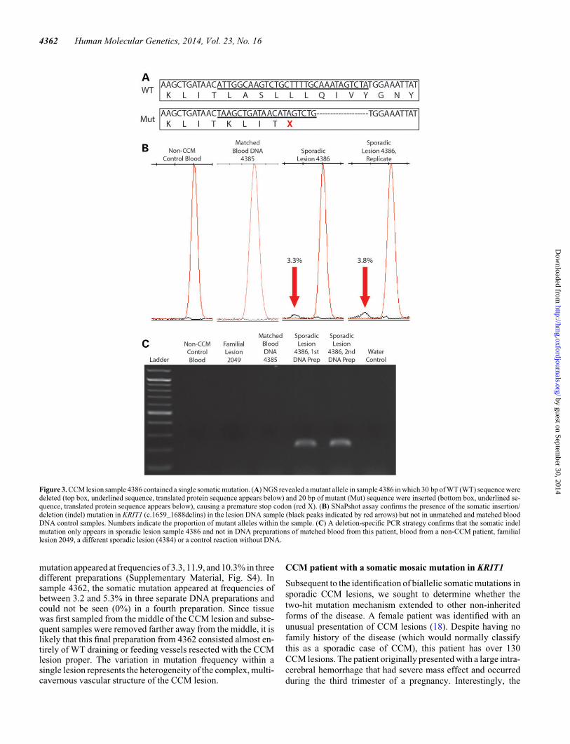
Three sporadic lesion samples (4386, 4310 and 4358) containedone identifiable somatic mutation each. Sample 4386 bore aninsertion–deletion mutation in KRIT1 (c.1659_1688delins) in3/702 reads (0.4%), causing a frameshift and a premature stopcodon after four altered amino acid residues (Fig. 3A). Althoughoriginally identified at a very low frequency, this mutation was
validated by a SNaPshot assay (Fig. 3B; Supplementary Mater-ial, Fig. S4), showing that the mutant allele only appears insample 4386 (3.3% frequency in the original amplification and3.8% in the replicate) and not in control samples including amatched blood DNA sample from this patient. For further con-firmation of this insertion/deletion mutation, PCR primerswere designed to specifically amplify the mutant sequence.Based on this PCR strategy, bands on an electrophoresis gelwere only apparent for sample 4386 and not for controlsamples (Fig. 3C). Sanger sequencing was used to confirm thatthese bands contained the mutated sequence (data not shown).
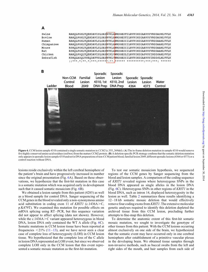
Sample 4310 contained one somatic mutation in CCM2(c.355_369del) in 6/528 reads (1.1%). Similar to sample 4386,a mutation-specific PCR (Fig. 4B) and Sanger sequencing(data not shown) confirmed that this somatic mutation waspresent in sample 4310 and not in control samples.
Sample 4358 contained a single mutation in CCM2 (c.611_622del) in 2/39 reads (5.1%). This mutation was confirmed tobe only in sample 4358 by SNaPshot (Fig. 5B; SupplementaryMaterial, Fig. S5), mutation-specific PCR (Fig. 5C) and Sangersequencing (data not shown).
Unlike the somatic mutation in 4386, the somatic deletionmutations present in samples 4310 and 4358 are in frame anddo not cause frameshifts or premature stop codons. The CCM2
Figure 1. CCM lesion sample 4362 contained one constitutional and one somatic mutation. (A) Sanger sequencing chromatogram shows a heterozygous, nonsensemutation in KRIT1 (c.213_214CG.AT, p.Y71X) in both lesion sample 4362 and a matched blood DNA sample from the same patient. (B) SNaPshot assay confirmsthe presence of a somatic nonsense mutation in KRIT1 (c.1890G.A, p.W630X) in the lesion DNA sample (red peaks indicatedby red arrows) but not in unmatched andmatched blood DNA control samples. Numbers indicate the proportion of mutant alleles within the sample.
4360 Human Molecular Genetics, 2014, Vol. 23, No. 16
by guest on September 30, 2014
http://hmg.oxfordjournals.org/
Dow
nloaded from
mutation in sample 4310 (c.355_369del) deletes five amino acidresidues (119-NVKLA-123) that are moderately to highly con-served in mammals and other vertebrates (Fig. 4A). TheCCM2 mutation in sample 4358 (c.611_622del) deletes fourmoderately conserved amino acid residues (204-VAAE-207,Fig. 5A). The c.611_622del mutation also occurs near theintron–exon border of the sixth coding exon of CCM2, so itis possible that this mutation could alter splicing of the mRNAtranscript. Both of these small deletions were found in thephosphotyrosine-binding domain of CCM2, which mediatesinteractions with other proteins (16) including KRIT1 (17).
Heterogeneous frequency of somatic mutationswithin CCM lesions
To prepare DNA from the sporadic CCM lesions, the frozen
block tissue was bisected and a portion of the sample near
the middle was used for DNA extraction. For samples with con-
firmed somatic mutations by NGS and the SNaPshot assay, add-
itional preparations of DNA were generated and examined
(Supplementary Material, Figs S2–S5). The frequency of the
somatic mutations within these additional DNA preparations
varied considerably. For example, in sample 4386 the somatic
Figure2. CCM lesion sample4392 contained two somaticmutations. (A) SNaPshot assayconfirms the presence of a somaticnonsense mutation in KRIT1 (c.993T.G,p.Y331X) in the lesion DNA sample (black peaks indicated by red arrows) but not in unmatched and matched blood DNA control samples. Numbers indicate theproportion of mutant alleles within the sample. (B) SNaPshot assay also confirms the presence of a somatic nonsense mutation in KRIT1 (c.1159C.T, p.Q387X)in the lesion DNA sample (green peaks indicated by red arrows) but not in unmatched and matched blood DNA control samples.
Human Molecular Genetics, 2014, Vol. 23, No. 16 4361
by guest on September 30, 2014
http://hmg.oxfordjournals.org/
Dow
nloaded from
mutation appeared at frequencies of 3.3, 11.9, and 10.3% in threedifferent preparations (Supplementary Material, Fig. S4). Insample 4362, the somatic mutation appeared at frequencies ofbetween 3.2 and 5.3% in three separate DNA preparations andcould not be seen (0%) in a fourth preparation. Since tissuewas first sampled from the middle of the CCM lesion and subse-quent samples were removed farther away from the middle, it islikely that this final preparation from 4362 consisted almost en-tirely of WT draining or feeding vessels resected with the CCMlesion proper. The variation in mutation frequency within asingle lesion represents the heterogeneity of the complex, multi-cavernous vascular structure of the CCM lesion.
CCM patient with a somatic mosaic mutation in KRIT1
Subsequent to the identification of biallelic somatic mutations in
sporadic CCM lesions, we sought to determine whether the
two-hit mutation mechanism extended to other non-inherited
forms of the disease. A female patient was identified with an
unusual presentation of CCM lesions (18). Despite having nofamily history of the disease (which would normally classify
this as a sporadic case of CCM), this patient has over 130
CCM lesions. The patient originally presented with a large intra-
cerebral hemorrhage that had severe mass effect and occurred
during the third trimester of a pregnancy. Interestingly, the
Figure 3. CCM lesion sample 4386 contained a single somatic mutation. (A) NGS revealed a mutant allele in sample 4386 in which 30 bp of WT (WT) sequence weredeleted (top box, underlined sequence, translated protein sequence appears below) and 20 bp of mutant (Mut) sequence were inserted (bottom box, underlined se-quence, translated protein sequence appears below), causing a premature stop codon (red X). (B) SNaPshot assay confirms the presence of the somatic insertion/deletion (indel) mutation in KRIT1 (c.1659_1688delins) in the lesion DNA sample (black peaks indicated by red arrows) but not in unmatched and matched bloodDNA control samples. Numbers indicate the proportion of mutant alleles within the sample. (C) A deletion-specific PCR strategy confirms that the somatic indelmutation only appears in sporadic lesion sample 4386 and not in DNA preparations of matched blood from this patient, blood from a non-CCM patient, familiallesion 2049, a different sporadic lesion (4384) or a control reaction without DNA.
4362 Human Molecular Genetics, 2014, Vol. 23, No. 16
by guest on September 30, 2014
http://hmg.oxfordjournals.org/
Dow
nloaded from
lesions reside exclusively within the left cerebral hemisphere ofthe patient’s brain and have progressively increased in numbersince the original presentation (Fig. 6A). Based on these obser-vations, we hypothesize that the first-hit mutation in this caseis a somatic mutation which was acquired early in developmentsuch that it caused somatic mosaicism (Fig. 6B).
We obtained a lesion sample from this patient (4285) as wellas a blood sample for control DNA. Sanger sequencing of theCCM genes in the blood revealed only a non-synonymous aminoacid substitution in coding exon 11 of KRIT1 (c.1436A.C,p.K479T). We examined this mutation for possible effects onmRNA splicing using RT–PCR, but this sequence variationdid not appear to affect splicing (data not shown). However,while the c.1436A.C variant appeared heterozygous in bloodDNA, lesion DNA only contained the WT A allele (Fig. 6C).Somatic mutations in CCM lesion tissue have been reported atfrequencies ,21% (11–13), and we have never seen a clearcase of complete loss of heterozygosity (LOH) in CCM lesiontissue. We hypothesized that the complete loss of the C allelein lesion DNA represented an LOH event, but since we observedcomplete LOH only in the CCM lesion that this event repre-sented a somatic mosaic mutation as the first-hit mutation.
To test our somatic mosaicism hypothesis, we sequencedregions of the CCM genes by Sanger sequencing from theblood and lesion samples. A comparison of the coding sequenceof KRIT1 revealed regions where heterozygous SNPs in theblood DNA appeared as single alleles in the lesion DNA(Fig. 6C). Heterozygous SNPs in other regions of KRIT1 in theblood DNA, such as intron 14, displayed heterozygosity in thelesion as well. Table 2 summarizes these results identifying a12–18 kb somatic mosaic deletion that would effectivelyremove four coding exons from KRIT1. The extensive moleculargenetic analyses required to identify this deletion depleted thearchived tissue from this CCM lesion, precluding furtherattempts to fine-map this deletion.
To determine the anatomic extent of this first-hit somaticmosaic mutation, we sought to investigate the genotype ofother tissues from this patient. With the CCM lesions occurringalmost exclusively on one side of the brain, we hypothesizedthat the somatic event may have occurred only in one cerebralhemisphere after establishment of a primitive vascular plexusin the developing brain. We obtained tissue samples throughnon-invasive methods, such as buccal swabs from the left andright sides of the mouth, and hair samples from each side of
Figure 4. CCM lesion sample 4310 contained a single somatic mutation in CCM2 (c.355_369del). (A) The in-frame deletion mutation in sample 4310 would removefive highly conserved amino acid residues (red box) from the mature CCM2 protein. (B) A deletion-specific PCR strategy confirms that the somatic deletion mutationonly appears in sporadic lesion sample 4310 and not in DNA preparations of non-CCMpatient blood, familial lesion2049, different sporadic lesions (4364 or 4373) or acontrol reaction without DNA.
Human Molecular Genetics, 2014, Vol. 23, No. 16 4363
by guest on September 30, 2014
http://hmg.oxfordjournals.org/
Dow
nloaded from
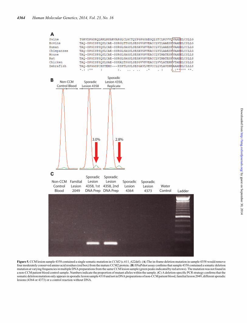
Figure 5. CCM lesion sample 4358 contained a single somatic mutation in CCM2 (c.611_622del). (A) The in-frame deletion mutation in sample 4358 would removefour moderately conserved amino acid residues (red box) from the mature CCM2 protein. (B) SNaPshot assay confirms that sample 4358 contained a somatic deletionmutation at varying frequencies in multiple DNA preparations from the same CCM lesion sample (green peaks indicated by red arrows). The mutation was not found ina non-CCM patient blood control sample. Numbers indicate the proportion of mutant alleles within the sample. (C) A deletion-specific PCR strategy confirms that thesomaticdeletion mutationonly appears in sporadic lesion sample 4310 and not in DNA preparations of non-CCMpatient blood, familial lesion 2049, different sporadiclesions (4364 or 4373) or a control reaction without DNA.
4364 Human Molecular Genetics, 2014, Vol. 23, No. 16
by guest on September 30, 2014
http://hmg.oxfordjournals.org/
Dow
nloaded from
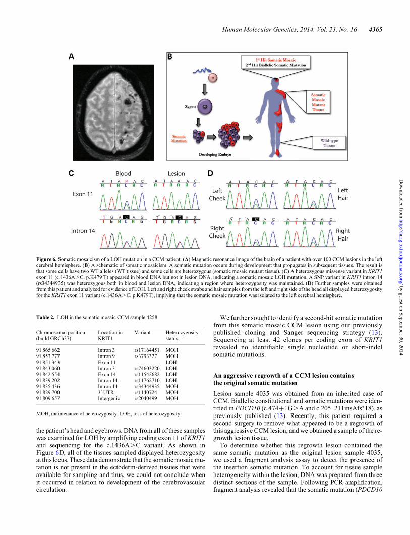
the patient’s head and eyebrows. DNA from all of these sampleswas examined for LOH by amplifying coding exon 11 of KRIT1and sequencing for the c.1436A.C variant. As shown inFigure 6D, all of the tissues sampled displayed heterozygosityat this locus. These data demonstrate that the somatic mosaic mu-tation is not present in the ectoderm-derived tissues that wereavailable for sampling and thus, we could not conclude whenit occurred in relation to development of the cerebrovascularcirculation.
We further sought to identify a second-hit somatic mutationfrom this somatic mosaic CCM lesion using our previouslypublished cloning and Sanger sequencing strategy (13).Sequencing at least 42 clones per coding exon of KRIT1revealed no identifiable single nucleotide or short-indelsomatic mutations.
An aggressive regrowth of a CCM lesion containsthe original somatic mutation
Lesion sample 4035 was obtained from an inherited case ofCCM. Biallelic constitutional and somatic mutations were iden-tified in PDCD10 (c.474+1G.A and c.205_211insAfs∗18), aspreviously published (13). Recently, this patient required asecond surgery to remove what appeared to be a regrowth ofthis aggressive CCM lesion, and we obtained a sample of the re-growth lesion tissue.
To determine whether this regrowth lesion contained thesame somatic mutation as the original lesion sample 4035,we used a fragment analysis assay to detect the presence ofthe insertion somatic mutation. To account for tissue sampleheterogeneity within the lesion, DNA was prepared from threedistinct sections of the sample. Following PCR amplification,fragment analysis revealed that the somatic mutation (PDCD10
Figure 6. Somatic mosaicism of a LOH mutation in a CCM patient. (A) Magnetic resonance image of the brain of a patient with over 100 CCM lesions in the leftcerebral hemisphere. (B) A schematic of somatic mosaicism. A somatic mutation occurs during development that propagates in subsequent tissues. The result isthat some cells have two WT alleles (WT tissue) and some cells are heterozygous (somatic mosaic mutant tissue). (C) A heterozygous missense variant in KRIT1exon 11 (c.1436A.C, p.K479 T) appeared in blood DNA but not in lesion DNA, indicating a somatic mosaic LOH mutation. A SNP variant in KRIT1 intron 14(rs34344935) was heterozygous both in blood and lesion DNA, indicating a region where heterozygosity was maintained. (D) Further samples were obtainedfrom this patient and analyzed for evidence of LOH. Left and right cheek swabs and hair samples from the left and right side of the head all displayed heterozygosityfor the KRIT1 exon 11 variant (c.1436A.C, p.K479T), implying that the somatic mosaic mutation was isolated to the left cerebral hemisphere.
Table 2. LOH in the somatic mosaic CCM sample 4258
Chromosomal position(build GRCh37)
Location inKRIT1
Variant Heterozygositystatus
91 865 662 Intron 3 rs17164451 MOH91 853 777 Intron 9 rs3793327 MOH91 851 343 Exon 11 LOH91 843 060 Intron 3 rs74603220 LOH91 842 554 Exon 14 rs11542682 LOH91 839 202 Intron 14 rs11762710 LOH91 835 436 Intron 14 rs34344935 MOH91 829 700 3′ UTR rs1140724 MOH91 809 657 Intergenic rs2040499 MOH
MOH, maintenance of heterozygosity; LOH, loss of heterozygosity.
Human Molecular Genetics, 2014, Vol. 23, No. 16 4365
by guest on September 30, 2014
http://hmg.oxfordjournals.org/
Dow
nloaded from
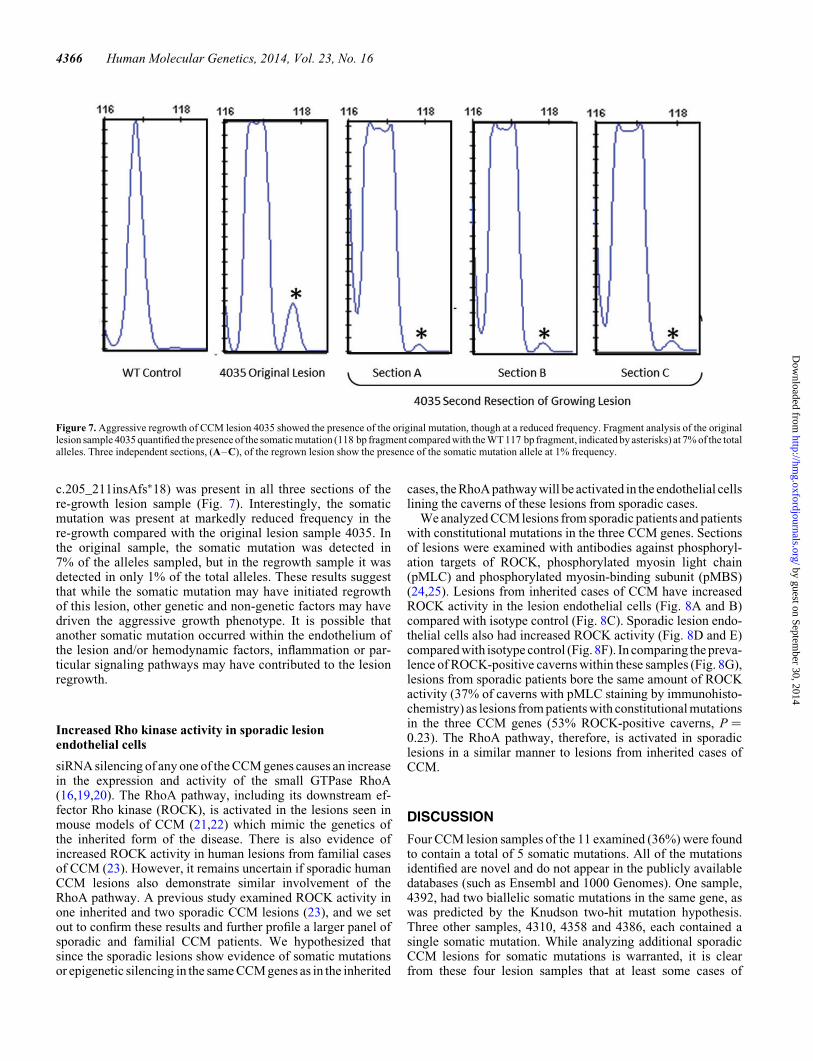
c.205_211insAfs∗18) was present in all three sections of there-growth lesion sample (Fig. 7). Interestingly, the somaticmutation was present at markedly reduced frequency in there-growth compared with the original lesion sample 4035. Inthe original sample, the somatic mutation was detected in7% of the alleles sampled, but in the regrowth sample it wasdetected in only 1% of the total alleles. These results suggestthat while the somatic mutation may have initiated regrowthof this lesion, other genetic and non-genetic factors may havedriven the aggressive growth phenotype. It is possible thatanother somatic mutation occurred within the endothelium ofthe lesion and/or hemodynamic factors, inflammation or par-ticular signaling pathways may have contributed to the lesionregrowth.
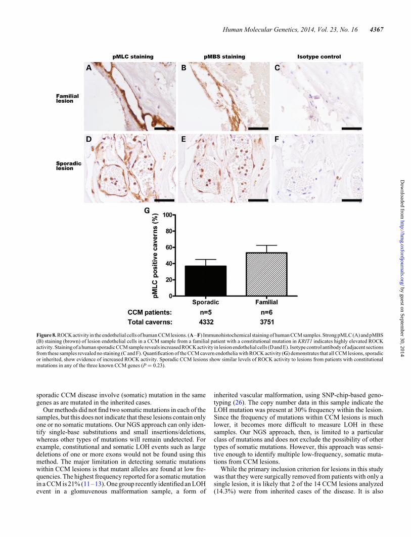
Increased Rho kinase activity in sporadic lesionendothelial cells
siRNA silencing of any one of the CCM genes causes an increasein the expression and activity of the small GTPase RhoA(16,19,20). The RhoA pathway, including its downstream ef-fector Rho kinase (ROCK), is activated in the lesions seen inmouse models of CCM (21,22) which mimic the genetics ofthe inherited form of the disease. There is also evidence ofincreased ROCK activity in human lesions from familial casesof CCM (23). However, it remains uncertain if sporadic humanCCM lesions also demonstrate similar involvement of theRhoA pathway. A previous study examined ROCK activity inone inherited and two sporadic CCM lesions (23), and we setout to confirm these results and further profile a larger panel ofsporadic and familial CCM patients. We hypothesized thatsince the sporadic lesions show evidence of somatic mutationsor epigenetic silencing in the same CCM genes as in the inherited
cases, the RhoA pathway will be activated in the endothelial cellslining the caverns of these lesions from sporadic cases.
We analyzed CCM lesions from sporadic patients and patientswith constitutional mutations in the three CCM genes. Sectionsof lesions were examined with antibodies against phosphoryl-ation targets of ROCK, phosphorylated myosin light chain(pMLC) and phosphorylated myosin-binding subunit (pMBS)(24,25). Lesions from inherited cases of CCM have increasedROCK activity in the lesion endothelial cells (Fig. 8A and B)compared with isotype control (Fig. 8C). Sporadic lesion endo-thelial cells also had increased ROCK activity (Fig. 8D and E)compared with isotype control (Fig. 8F). In comparing the preva-lence of ROCK-positive caverns within these samples (Fig. 8G),lesions from sporadic patients bore the same amount of ROCKactivity (37% of caverns with pMLC staining by immunohisto-chemistry) as lesions from patients with constitutional mutationsin the three CCM genes (53% ROCK-positive caverns, P ¼0.23). The RhoA pathway, therefore, is activated in sporadiclesions in a similar manner to lesions from inherited cases ofCCM.
DISCUSSION
Four CCM lesion samples of the 11 examined (36%) were foundto contain a total of 5 somatic mutations. All of the mutationsidentified are novel and do not appear in the publicly availabledatabases (such as Ensembl and 1000 Genomes). One sample,4392, had two biallelic somatic mutations in the same gene, aswas predicted by the Knudson two-hit mutation hypothesis.Three other samples, 4310, 4358 and 4386, each contained asingle somatic mutation. While analyzing additional sporadicCCM lesions for somatic mutations is warranted, it is clearfrom these four lesion samples that at least some cases of
Figure 7. Aggressive regrowth of CCM lesion 4035 showed the presence of the original mutation, though at a reduced frequency. Fragment analysis of the originallesion sample 4035 quantified the presence of the somatic mutation (118 bp fragment compared with the WT 117 bp fragment, indicated by asterisks) at 7% of the totalalleles. Three independent sections, (A–C), of the regrown lesion show the presence of the somatic mutation allele at 1% frequency.
4366 Human Molecular Genetics, 2014, Vol. 23, No. 16
by guest on September 30, 2014
http://hmg.oxfordjournals.org/
Dow
nloaded from
sporadic CCM disease involve (somatic) mutation in the samegenes as are mutated in the inherited cases.
Our methods did not find two somatic mutations in each of thesamples, but this does not indicate that these lesions contain onlyone or no somatic mutations. Our NGS approach can only iden-tify single-base substitutions and small insertions/deletions,whereas other types of mutations will remain undetected. Forexample, constitutional and somatic LOH events such as largedeletions of one or more exons would not be found using thismethod. The major limitation in detecting somatic mutationswithin CCM lesions is that mutant alleles are found at low fre-quencies. The highest frequency reported for a somatic mutationin a CCM is 21% (11–13). One group recently identified an LOHevent in a glomuvenous malformation sample, a form of
inherited vascular malformation, using SNP-chip-based geno-typing (26). The copy number data in this sample indicate theLOH mutation was present at 30% frequency within the lesion.Since the frequency of mutations within CCM lesions is muchlower, it becomes more difficult to measure LOH in thesesamples. Our NGS approach, then, is limited to a particularclass of mutations and does not exclude the possibility of othertypes of somatic mutations. However, this approach was sensi-tive enough to identify multiple low-frequency, somatic muta-tions from CCM lesions.
While the primary inclusion criterion for lesions in this studywas that they were surgically removed from patients with only asingle lesion, it is likely that 2 of the 14 CCM lesions analyzed(14.3%) were from inherited cases of the disease. It is also
Figure 8. ROCK activity in the endothelial cells of human CCM lesions. (A–F) Immunohistochemical staining of human CCM samples. Strong pMLC (A) and pMBS(B) staining (brown) of lesion endothelial cells in a CCM sample from a familial patient with a constitutional mutation in KRIT1 indicates highly elevated ROCKactivity. Stainingof a human sporadic CCM sample reveals increased ROCK activity in lesion endothelial cells (D and E). Isotype control antibody of adjacent sectionsfrom these samples revealed no staining (C and F). Quantification of the CCM cavern endothelia with ROCK activity (G) demonstrates that all CCM lesions, sporadicor inherited, show evidence of increased ROCK activity. Sporadic CCM lesions show similar levels of ROCK activity to lesions from patients with constitutionalmutations in any of the three known CCM genes (P ¼ 0.23).
Human Molecular Genetics, 2014, Vol. 23, No. 16 4367
by guest on September 30, 2014
http://hmg.oxfordjournals.org/
Dow
nloaded from
possible that these patients had de novo constitutional mutationsor somatic mosaic mutations early in development, but that is notpossible to determine without DNA samples from the patients’parents. Most inherited cases of CCM present with multipleCCM lesions, but some inherited cases show lower penetrance(27), owing either to the stochastic nature of this disease or tomutation- or environment-specific factors affecting lesiongrowth. As such, it would not be unexpected to find inheritedcases of CCM among these biobank samples.
In this study, NGS was a reliable method for identifying muta-tions in both sporadic and inherited cases of CCM. Indeed, NGSidentified somatic mutations at frequencies below those previ-ously observed with our cloning and sequencing strategy (13).It should be noted, however, that the frequencies of the somaticmutations reported here varied between different lesions andeven within the same lesion. Different somatic mutation fre-quencies were found in multiple DNA preparations from thesame lesion, reflecting the heterogeneous and complex structureof the lesions. Different assays (NGS, SNaPshot) using the sameDNA sample also found the somatic mutations at varying fre-quencies. While it is difficult to determine which of these assaysmost closely approximates the ‘true’ mutation frequency, NGSwas able to qualitatively identify the presence or absence ofthese somatic mutations.
In addition to the sporadic CCM patients with single lesionsand no apparent family history, we also examined the geneticmechanism underlying a patient with over 100 CCM lesions ex-clusively in one hemisphere of the brain (18). Though no second-hit mutation was identified, it was apparent that the first genetichit was a somatic mosaic LOH event, deleting 12–18 kb ofKRIT1. The presence of a somatic mosaic mutation potentiallyexplains why this patient presented with CCM lesions localizedto one side of the brain: endothelial cells in the affected region ofthe brain would require only one further somatic mutation tocause CCM genesis, whereas the unaffected portion of thebrain was still effectively WT, requiring two, independentsomatic mutations for lesions to form.
Lesion sample 4035 was previously reported in a study ofsomatic mutations in familial cases of CCM (13). After publica-tion of these results, this patient underwent further surgeries toremove apparent regrowth of a CCM lesion in the same stereo-tactic location of the brain. Samples of this regrowth lesionretained the somatic mutation signature of the original resectedlesion. We hypothesize that cells harboring the originalsomatic mutation evaded surgical resection and subsequentlyinitiated the regrowth of the CCM lesion. This is the firstreport of a CCM lesion demonstrating regrowth potential similarto a tumor. Further investigation of this phenomenon may revealthat more aggressive treatments are necessary in some cases toprevent regrowth and hemorrhage of CCM lesions.
Multiple groups have previously found evidence of a two-hitmutation mechanism in inherited cases of CCM (11–13,28).From these experiments, we have provided evidence that atleast some sporadic CCM lesions harbor somatic mutations inthe same genes, with one case showing biallelic somatic muta-tions. Since our approach can only identify certain classes ofmutations, we suggest that many sporadic CCMs may also becaused by a two-hit mutational mechanism. Furthermore, wehave presented other cases of somatic mosaicism and lesion re-growth that, while less typical, nonetheless reinforce that the
distinct presentations of CCM are all caused by a Knudsoniantwo-hit mechanism in the known CCM genes.
Examination of CCM lesions from humans (23) and CCMmouse models (21–23) has revealed activation of the RhoApathway through its effector ROCK. Through these data, wehave demonstrated that CCM lesions from both sporadic andfamilial cases of the disease contain endothelial cells with in-creased levels of ROCK activity. The ROCK activity in sporadiclesions closely resembled that in the lesions from familialpatients. Importantly, increased ROCK activity was heteroge-neous within the CCM lesions, with �40% of the cavernsshowing phosphorylation of MLC or MBS. The loss of CCMproteins due to biallelic mutations and aberrant ROCK activity,then, appear to be cell-autonomous events in CCM pathogenesis.
Novel therapeutic strategies are currently being tested inanimal models of CCM based on the genetics of the inheritedform of the disease (22). Previously, it was unknown if sporadicCCM patients, who represent the majority of CCM cases (29),would benefit from the pharmacological inhibition treatmentscurrently under investigation. Since these data demonstratethat sporadic form of CCM also follows an underlying geneticmechanism and shows activation of the RhoA pathway similarto inherited cases of CCM, it is likely that these novel therapieswill also be effective for the larger population with sporadicCCM.
MATERIALS AND METHODS
CCM samples
All CCM samples were obtained from the Angioma AllianceTissue Bank in accordance with Institutional Review Board stan-dards. Lesions were bisected and tissue samples were removedfrom the center of the lesion. DNA was isolated using the Pure-gene tissue protocol (Gentra).
Next-generation sequencing
PCR primers (IDT) were designed to amplify the coding exons(along with .10 bp of flanking intronic sequence) of all threeCCM genes. During primer design, 5′ overhangs were createdin accordance to 454 sequencing protocols (Roche), includingthe addition of a 4 bp identification sequence used for multiplex-ing samples (four samples per run). Regions of interest wereamplified by PCR and a small aliquot was checked for qualitycontrol by gel electrophoresis. Where necessary, PCR productsof interest were analyzed by gel electrophoresis and isolatedusing the GeneClean Turbo kit (MP Biomedicals).
PCR products were pooled and submitted to the Duke GenomeSequencing and Analysis Core for quality control and sequen-cing on a Roche 454 GS-FLX Titanium platform. The GS DataAnalysis Software Package (Roche) was used to split the multi-plexed sample into the four independent datasets. TheSWAP454 toolkit (30) was then used to generate a coveragemap and SNP calls from the amplified sections of the humanCCM genes. Briefly, SWAP454 aligned the individual reads tothe sequences of the amplified regions recording only theunique alignments. A coverage map was then constructed fromthe aligned reads after filtering based upon their neighborhood-quality score method (MIN_QUAL ¼ 20; NQ ¼ 15). Individual
4368 Human Molecular Genetics, 2014, Vol. 23, No. 16
by guest on September 30, 2014
http://hmg.oxfordjournals.org/
Dow
nloaded from
SNPs were then called that had a coverage of at least two reads,with at least one read mapping to both the forward and reversestrand, and a minimum variant/reference ratio of 0.02. A separateanalysis of these data was also performed using Geneious soft-ware (Biomatters) (31).
Foranalysis ofphase of somatic mutations in thesamesporadicCCM sample, PCR products were first amplified using the high-fidelity polymerase PfuTurbo (Agilent), and then purified by gelextraction (Geneclean Turbo, Qbiogene). Samples were run onthe Pacific Biosystems (PacBio) RSII platform. PacBio readswere first trimmed to remove adapter sequences and low-quality(≤0.75) bases from the ends of the reads. Reads that were at least50 nt in length were aligned to the KRIT1 sequence using the blasrtool from the SMRT Analysis Toolkit (Pacific Biosciences;default parameters). The number of reference and variant callsfor each of the two somatic SNP locations was then counted.Reads that had a base call at both locations were then used to iden-tify the monoallelic or biallelic nature of the variants.
SNaPshot analysis
PCR products with potential mutations were analyzed by SNaP-shot (Applied Biosystems). Primers were designed adjacent tothe base of interest, and SNaPshot was performed according toApplied Biosystems protocols. Results were visualized andquantified using GeneMapper software (Applied Biosystems).Allele frequencies were calculated by dividing the area underthe peak of a particular allele by the total area under both allelepeaks.
Sanger sequencing
Samples were analyzed by Sanger sequencing using the BigDyereaction kit (Applied Biosystems) on the 3130 Genetic Analyzer(Applied Biosystems). Sequences were examined using Sequen-cher software (Gene Codes Corporation).
ROCK activity assay
The ROCK activity was assessed by the expression of pMLC andpMBS, which are ROCK substrates. Excised CCM lesions fromfive sporadic patients (obtained from the University of Chicago)and six familial patients (including two patients with constitu-tional mutations in KRIT1, two patients with constitutionalmutations in CCM2, and two patients with constitutional muta-tions in PDCD10) were embedded in paraffin and cut into serial5 mm sections. After antigen retrieval, sections were blockedusing PBS supplemented with 0.5% fish skin gelatin (Sigma–Aldrich), 5% goat serum (Invitrogen) and an avidin/biotin block-ing kit (Vector Laboratories). Slides were probed with eitherrabbit polyclonal anti-pMLC (Thr18/Ser19) antibody (Cell Sig-naling Technology, 1:250) or rabbit polyclonal anti-pMBS(MYPT1, Thr853) antibody (MyBioSource, 1:2000 for sporadicsamples and 1:5000 for familial samples) overnight at 48C, fol-lowed by biotinylated goat anti-rabbit antibody (Vector Labora-tories, 1:400) for 2 h, as previously described (21–23). Isotypecontrols were used as negative controls and run simultaneouslywith all specimens. Quantification of pMLC expression was per-formed in each cavern of CCM lesions from a total of 11 patients.A negative cavern was defined as complete absence of pMLC
staining, whereas a positive cavern was defined as definite butdiffuse, intense and confluent staining. Student’s t-test was usedto compare the prevalence of positive pMLC caverns of CCMlesions between sporadic patient and familial patient samples.
Fragment analysis
The presence of the single-base insertion somatic mutation insample 4035 was validated by fragment analysis assay. PCR pro-ducts for exon 6 of PDCD10 were amplified using one fluores-cently labeled primer, 5′-6-FAM-CTCACACAAGACATCATTATG, and one unlabeled primer, 5′-CCATACGAAGAAGGGACTCC or 5′-AAACAAGGTTCTTCTGTCCGTTA. Thehigh-fidelity DNA polymerase Phusion (Thermo Scientific)was used for these reactions. The resulting PCR products wereresuspended in formamide (Applied Biosystems) and werecharacterized on an Applied Biosystems 3130 sequencer. Subse-quent analysis of fragment seizes was performed using Gene-Mapper software (Applied Biosystems). Positive and negativecontrols were colony PCR products from clones with sequenceshowing either WT (negative control) or somatic mutation (posi-tive control) sequences.
SUPPLEMENTARY MATERIAL
Supplementary Material is available at HMG online.
ACKNOWLEDGEMENTS
We thank St Joseph’s Hospital and Medical Center’s HumanSpecimen Procurement Service as well as Angioma Alliance’sDNA/Tissue Bank (www.angioma.org/DNA) for providing theCCM lesion samples used in this study.
Conflict of Interest statement. None declared.
FUNDING
This work was supported by the National Institutes of Health(R01-NS060748 and R01-NS077957 to D.A.M. and I.A.W.,F31-NS077702 to D.A.M. and F31-NS061468 to A.L.A.).
REFERENCES
1. Rigamonti, D., Hadley, M.N., Drayer, B.P., Johnson, P.C., Hoenig-Rigamonti,K., Knight, J.T. and Spetzler, R.F. (1988) Cerebral cavernous malformations.Incidence and familial occurrence. N. Engl. J. Med., 319, 343–347.
2. Otten, P., Pizzolato, G.P., Rilliet, B. and Berney, J. (1989) 131 cases ofcavernous angioma (cavernomas) of the CNS, discovered by retrospectiveanalysis of 24,535 autopsies. Neurochirurgie, 35, 82–83.
3. Del Curling, O. Jr, Kelly, D.L. Jr, Elster, A.D. and Craven, T.E. (1991) Ananalysis of the natural history of cavernous angiomas. J. Neurosurg., 75,702–708.
4. Robinson, J.R., Awad, I.A. and Little, J.R. (1991) Natural history of thecavernous angioma. J. Neurosurg., 75, 709–714.
5. Laberge-le Couteulx, S., Jung, H.H., Labauge, P., Houtteville, J.P., Lescoat,C., Cecillon, M., Marechal, E., Joutel, A., Bach, J.F. and Tournier-Lasserve,E. (1999) Truncating mutations in CCM1, encoding KRIT1, causehereditary cavernous angiomas. Nat. Genet., 23, 189–193.
6. Sahoo, T., Johnson, E.W., Thomas, J.W., Kuehl, P.M., Jones, T.L., Dokken,C.G., Touchman, J.W., Gallione, C.J., Lee-Lin, S.Q., Kosofsky, B. et al.(1999) Mutations in the gene encoding KRIT1, a Krev-1/rap1a binding
Human Molecular Genetics, 2014, Vol. 23, No. 16 4369
by guest on September 30, 2014
http://hmg.oxfordjournals.org/
Dow
nloaded from
protein, cause cerebral cavernous malformations (CCM1). Hum. Mol.Genet., 8, 2325–2333.
7. Liquori, C.L., Berg, M.J., Siegel, A.M., Huang, E., Zawistowski, J.S.,Stoffer, T., Verlaan, D., Balogun, F., Hughes, L., Leedom, T.P. et al. (2003)Mutations in a gene encoding a novel protein containing aphosphotyrosine-binding domain cause type 2 cerebral cavernousmalformations. Am. J. Hum. Genet., 73, 1459–1464.
8. Denier, C., Goutagny, S., Labauge, P., Krivosic, V., Arnoult, M., Cousin, A.,Benabid, A.L., Comoy, J., Frerebeau, P., Gilbert, B. et al. (2004) Mutationswithin the MGC4607 gene cause cerebral cavernous malformations.Am. J. Hum. Genet., 74, 326–337.
9. Bergametti, F., Denier, C., Labauge, P., Arnoult, M., Boetto, S., Clanet, M.,Coubes, P., Echenne, B., Ibrahim, R., Irthum, B. et al. (2005) Mutationswithin the programmed cell death 10 gene cause cerebral cavernousmalformations. Am. J. Hum. Genet., 76, 42–51.
10. Knudson, A.G. Jr. (1971) Mutation and cancer: statistical study ofretinoblastoma. Proc. Natl. Acad. Sci. USA, 68, 820–823.
11. Gault, J., Awad, I.A., Recksiek, P., Shenkar, R., Breeze, R., Handler, M. andKleinschmidt-DeMasters, B.K. (2009) Cerebral cavernous malformations:somatic mutations in vascular endothelial cells. Neurosurgery, 65, 138–144(discussion 144–135).
12. Gault, J., Shenkar, R., Recksiek, P. and Awad, I.A. (2005) Biallelic somaticand germ line CCM1 truncating mutations in a cerebral cavernousmalformation lesion. Stroke, 36, 872–874.
13. Akers, A.L., Johnson, E., Steinberg, G.K., Zabramski, J.M. and Marchuk,D.A. (2009) Biallelic somatic and germline mutations in cerebral cavernousmalformations (CCMs): evidence for a two-hit mechanism of CCMpathogenesis. Hum. Mol. Genet., 18, 919–930.
14. Pozzati, E., Acciarri, N., Tognetti, F., Marliani, F. and Giangaspero, F.(1996) Growth, subsequent bleeding, and de novo appearance of cerebralcavernous angiomas. Neurosurgery, 38, 662–669 (discussion 669–670).
15. Shafikhani, S. (2002) Factors affecting PCR-mediated recombination.Environ. Microbiol., 4, 482–486.
16. Crose, L.E., Hilder, T.L., Sciaky, N. and Johnson, G.L. (2009) Cerebralcavernous malformation 2 protein promotes smad ubiquitin regulatoryfactor 1-mediated RhoA degradation in endothelial cells. J. Biol. Chem.,284, 13301–13305.
17. Zawistowski, J.S., Stalheim, L., Uhlik, M.T., Abell, A.N., Ancrile, B.B.,Johnson, G.L. and Marchuk, D.A. (2005) CCM1 and CCM2 proteininteractions in cell signaling: implications for cerebral cavernousmalformations pathogenesis. Hum. Mol. Genet., 14, 2521–2531.
18. Reid, P.J., Campbell, S.S., Vates, G.E. and Allende, R. (2008) Extreme denovo appearance of cerebral cavernous malformations: case report.Neurosurgery, 62, E969–E970 (discussion E970).
19. Borikova, A.L., Dibble, C.F., Sciaky, N., Welch, C.M., Abell, A.N.,Bencharit, S. and Johnson, G.L. (2010) Rho kinase inhibition rescues theendothelial cell cerebral cavernous malformation phenotype. J. Biol. Chem.,285, 11760–11764.
20. Whitehead, K.J., Chan, A.C., Navankasattusas, S., Koh, W., London, N.R.,Ling, J., Mayo, A.H., Drakos, S.G., Marchuk, D.A., Davis, G.E. et al. (2009)The cerebral cavernous malformation signaling pathway promotes vascularintegrity via Rho GTPases. Nat. Med., 15, 177–184.
21. McDonald, D.A., Shenkar, R., Shi, C., Stockton, R.A., Akers, A.L.,Kucherlapati, M.H., Kucherlapati, R., Brainer, J., Ginsberg, M.H., Awad,I.A. et al. (2011) A novel mouse model of cerebral cavernous malformationsbased on the two-hit mutation hypothesis recapitulates the human disease.Hum. Mol. Genet., 20, 211–222.
22. McDonald, D.A., Shi, C., Shenkar, R., Stockton, R.A., Liu, F., Ginsberg,M.H., Marchuk, D.A. and Awad, I.A. (2012) Fasudil decreases lesion burdenin a murine model of cerebral cavernous malformation disease. Stroke, 43,571–574.
23. Stockton, R.A., Shenkar, R., Awad, I.A. and Ginsberg, M.H. (2010) Cerebralcavernous malformations proteins inhibit Rho kinase to stabilize vascularintegrity. J. Exp. Med., 207, 881–896.
24. Grassie, M.E., Moffat, L.D., Walsh, M.P. and MacDonald, J.A. (2011) Themyosin phosphatase targeting protein (MYPT) family: a regulatedmechanism for achieving substrate specificity of the catalytic subunit ofprotein phosphatase type 1delta. Arch. Biochem. Biophys., 510, 147–159.
25. Hudson, C.A., Heesom, K.J. and Lopez Bernal, A. (2012) Phasiccontractions of isolated human myometrium are associated with Rho-kinase(ROCK)-dependent phosphorylation of myosin phosphatase-targetingsubunit (MYPT1). Mol. Hum. Reprod., 18, 265–279.
26. Amyere, M., Aerts, V., Brouillard, P., McIntyre, B.A., Duhoux, F.P.,Wassef, M., Enjolras, O., Mulliken, J.B., Devuyst, O., Antoine-Poirel, H.et al. (2013) Somatic uniparental isodisomy explains multifocality ofglomuvenous malformations. Am. J. Hum. Genet., 92, 188–196.
27. Denier, C., Labauge, P., Bergametti, F., Marchelli, F., Riant, F., Arnoult, M.,Maciazek, J., Vicaut, E., Brunereau, L. and Tournier-Lasserve, E. (2006)Genotype–phenotype correlations in cerebral cavernous malformationspatients. Ann. Neurol., 60, 550–556.
28. Pagenstecher, A., Stahl, S., Sure, U. and Felbor, U. (2009) A two-hitmechanism causes cerebral cavernousmalformations: complete inactivationof CCM1, CCM2 or CCM3 in affected endothelial cells. Hum. Mol. Genet.,18, 911–918.
29. Aboian, M.S., Daniels, D.J., Rammos, S.K., Pozzati, E. and Lanzino, G.(2009) The putative role of the venous system in the genesis of vascularmalformations. Neurosurg. Focus, 27, E9.
30. Brockman, W., Alvarez, P., Young, S., Garber, M., Giannoukos, G., Lee,W.L., Russ, C., Lander, E.S., Nusbaum, C. and Jaffe, D.B. (2008) Qualityscores and SNP detection in sequencing-by-synthesis systems. Genome Res.,18, 763–770.
31. Drummond, A.J.A.B., Buxton, S., Cheung, M., Cooper, A., Heled, J., Kearse,M., Moir, R., Stones-Havas, S., Sturrock, S., Thierer, T. and Wilson, A. (2010)Geneious v5.3. Available from http://www.geneious.com. (date last accessed,9 April 2014).
4370 Human Molecular Genetics, 2014, Vol. 23, No. 16
by guest on September 30, 2014
http://hmg.oxfordjournals.org/
Dow
nloaded from