pseudo-meigs syndrome: a case report · heterogeneous pelvic tumor, ... on ultrasound of abdomen, a...
TRANSCRIPT

CASE REPORT PEER REVIEWED | OPEN ACCESS
www.edoriumjournals.com
International Journal of Case Reports and Images (IJCRI)International Journal of Case Reports and Images (IJCRI) is an international, peer reviewed, monthly, open access, online journal, publishing high-quality, articles in all areas of basic medical sciences and clinical specialties.
Aim of IJCRI is to encourage the publication of new information by providing a platform for reporting of unique, unusual and rare cases which enhance understanding of disease process, its diagnosis, management and clinico-pathologic correlations.
IJCRI publishes Review Articles, Case Series, Case Reports, Case in Images, Clinical Images and Letters to Editor.
Website: www.ijcasereportsandimages.com
Pseudo-Meigs syndrome: A case report
Divya Kallarackal, Dharampal Singh
ABSTRACT
Introduction: Meigs syndrome and pseudo-Meigs syndrome both presents with hydrothorax and ascites. Meigs syndrome is characteristically associated with ovarian fibroma whereas pseudo-Meigs syndrome is associated with any ovarian or pelvic tumors, other than ovarian fibroma. Case Report: A 48-year-old perimenopausal woman presented with a long history of 8–10 years of abdominal distension. Her examination revealed a right pleural effusion, massive ascites and large heterogeneous pelvic tumor, measuring 42x31 cm. After a preoperative ascitic tapping, the patient underwent an exploratory laparotomy with excision of the tumor, uterus and the right ovary. The tumor was diagnosed histologically as an ovarian mucinous cystadenoma. The postoperative resolution of hydrothorax and ascites confirmed the diagnosis of pseudo-Meigs syndrome. The patient remains in good condition 12 months after surgery. Conclusion: Pseudo-Meigs syndrome being a rare syndrome, with a good prognosis should be included in differential diagnosis in women presenting with unexplained hydrothorax and ascites.
(This page in not part of the published article.)

International Journal of Case Reports and Images, Vol. 8 No. 5, May 2017. ISSN – [0976-3198]
Int J Case Rep Images 2017;8(5):331–334. www.ijcasereportsandimages.com
Kallarackal et al. 331
CASE REPORT PEER REVIEWED | OPEN ACCESS
Pseudo-Meigs syndrome: A case report
Divya Kallarackal, Dharampal Singh
ABSTRACT
Introduction: Meigs syndrome and pseudo-Meigs syndrome both presents with hydrothorax and ascites. Meigs syndrome is characteristically associated with ovarian fibroma whereas pseudo-Meigs syndrome is associated with any ovarian or pelvic tumors, other than ovarian fibroma. Case Report: A 48-year-old perimenopausal woman presented with a long history of 8–10 years of abdominal distension. Her examination revealed a right pleural effusion, massive ascites and large heterogeneous pelvic tumor, measuring 42x31 cm. After a preoperative ascitic tapping, the patient underwent an exploratory laparotomy with excision of the tumor, uterus and the right ovary. The tumor was diagnosed histologically as an ovarian mucinous cystadenoma. The postoperative resolution of hydrothorax and ascites confirmed the diagnosis of pseudo-Meigs syndrome. The patient remains in good condition 12 months after surgery. Conclusion: Pseudo-Meigs syndrome being a rare syndrome, with a good prognosis should be included in differential diagnosis in women presenting with unexplained hydrothorax and ascites.
Divya Kallarackal1, Dharampal Singh2
Affiliations: 1MS OBGY, Consultant Gynaecologist, Depart-ment of Obstetrics and Gynaecology, Lethsolathebe II Me-morial Hospital, Maun, Botswana; 2MD Anaesthesiology, Consultant Anaesthesiologist, Department of Anaesthesiol-ogy, Lethsolathebe II Memorial, Hospital, Maun, Botswana.Corresponding Author: Divya Dayanandan Kallarackal, P.O. BOX, 12, Lethsolathebe II Memorial Hospital, Maun, Bot-swana; Email: [email protected]
Received: 07 January 2017Accepted: 25 February 2017Published: 01 May 2017
Keywords: Ascites, Hydrothorax, Ovarian tumor, Pseudo-Meigs syndrome
How to cite this article
Kallarackal D, Singh D. Pseudo-Meigs syndrome: A case report. Int J Case Rep Images 2017;8(5):331–334.
Article ID: Z01201705CR10796DK
*********
doi:10.5348/ijcri-201757-CR-10796
INTRODUCTION
Meigs syndrome is a rare condition, defined as the co-existence of benign ovarian fibroma, pleural effusion and ascites. While, pseudo-Meigs syndrome is characterized by the co-existence of pleural effusion, ascites and other ovarian or pelvic tumors. It was Meigs and Cass who brought out the significance of pleural effusion and ascites in ovarian fibroma. These syndromes should be considered in otherwise healthy postmenopausal women, who present with either hydrothorax or ascites. For both these syndromes, surgical resection of the tumor is the only therapeutic choice, resulting in resolution of fluid accumulations [1].
CASE REPORT
A 48-year-old perimenopausal woman came with history of abdominal distension since last 8–10 years, difficulty in breathing with increasing intensity over the past few months. She became very uncomfortable in supine position. She had no medical or surgical history of note. She is para 3 with uneventful vaginal deliveries. She took no regular medication and had no family medical

International Journal of Case Reports and Images, Vol. 8 No. 5, May 2017. ISSN – [0976-3198]
Int J Case Rep Images 2017;8(5):331–334. www.ijcasereportsandimages.com
Kallarackal et al. 332
history of note (Figure 1). Auscultation revealed absence of breath sounds at the right lower hemithorax and normal heart sounds. On abdominal examination massive ascites was noted. The mass was not palpable because of the tense ascites. Chest X-ray revealed mild right sided pleural effusion (Figure 2). Electrocardiography was within normal limits. On ultrasound of abdomen, a massive multi septate cystic mass with suspected ovarian origin, with massive ascites was noted. Computed tomography scan revealed a huge multiseptate mass with solid and cystic components measuring 42x31 cm arising from the pelvis. Left side ovary was not visualized and uterus was normal sized. Massive ascites was noted. No obvious lymphadenopathy was seen. Her serum CA 125 was 49 U/ml (normal <35U/ml). AFP was within normal limits while b-hCG was not detectable. Her serum proteins were slightly below normal. Routine blood investigations, including LFT’s and RFT’s were within normal limits. Ascitic tap fluid cytology revealed low cellular fluid comprising of lymphocytes and mesothelial cells. No evidence of malignancy was noted.
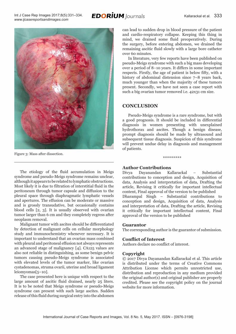
A preoperative diagnosis of left ovarian tumor was made and nearly 6 L of ascitic fluid was drained in two settings in ward three days before surgery and the day before surgery. In operation theatre, under epidural anesthesia, a wide bore silicone catheter was inserted and nearly 18 L of ascitic fluid was tapped slowly over a period of 60 minutes in lateral position. Then in supine position, through a midline incision from pubic symphysis to 2 cm above umbilicus, a mass measuring 42x31 cm, weighing 9 kg was removed, originating from left ovary.
There was no any evidence of metastasis or lymphadenopathy. Omental and peritoneal biopsy were taken. Hysterectomy with bilateral salpingo-oophorectomy was done. Grossly uterus with cervix measured 9x6 cm, right ovary measured 2.5x1.3 cm, both unremarkable (Figure 3).
On histopathology report, the mass was diagnosed as an ovarian mucinous cystadenoma. The pleural effusion resolved by postoperative day-10. The patient remains in good condition 12 months after surgery.
DISCUSSION
Meigs syndrome is defined as a triad of benign ovarian tumor (ovarian fibroma), ascites and pleural effusion. Though the association of pleural effusion with benign pelvic tumor was described by Salmon in 1934, it was Meigs and Cass whom brought out the significance of pleural effusion and ascites in ovarian fibroma. It is to be noted that the ascites and effusion resolves completely after resection of tumor. Meigs syndrome is a benign disease with a good prognosis.
Pseudo-Meigs syndrome on the other hand consists of ascites and pleural effusion associated with any pelvic tumor other than fibroma. It is clinically important as it may resemble metastatic pelvic cancer. Cytological
Figure 1: Preoperative picture of patient (written informed consent taken for this picture).
Figure 2: Preoperative chest X-ray of patient.
examination of the body cavity effusions is essential to differentiate between reactive process and metastatic tumor spread.
International Journal of Case Reports and Images, Vol. 8 No. 5, May 2017. ISSN – [0976-3198]
Int J Case Rep Images 2017;8(5):331–334. www.ijcasereportsandimages.com
Kallarackal et al. 333
The etiology of the fluid accumulation in Meigs syndrome and pseudo-Meigs syndrome remains unclear, although it appears to be related to lymphatic obstructions. Most likely it is due to filtration of interstitial fluid in the peritoneum through tumor capsule and diffusion to the pleural space through diaphragmatic lymphatic vessels and apertures. The effusion can be moderate or massive and is grossly transudative, but occasionally contains blood cells [2, 3]. It is usually observed with ovarian tumor larger than 6 cm and they completely regress after neoplasm removal.
Malignant tumor with ascites should be differentiated by detection of malignant cells on cellular morphology study and immunochemistry whenever necessary. It is important to understand that an ovarian mass combined with pleural and peritoneal effusion not always represents an advanced stage of malignancy [4]. CA125 values are also not reliable in distinguishing, as some benign pelvic tumors causing pseudo-Meigs syndrome is associated with elevated levels of the tumor marker, like ovarian cystadenomas, struma ovarii, uterine and broad ligament leiomyomas[5–10].
The case presented here is unique with respect to the large amount of ascitic fluid drained, nearly 25 liters. It is to be noted that Meigs syndrome or pseudo-Meigs syndrome can present with such large ascites. Sudden release of this fluid during surgical entry into the abdomen
can lead to sudden drop in blood pressure of the patient and cardio-respiratory collapse. Keeping this thing in mind, we drained some fluid preoperatively. During the surgery, before entering abdomen, we drained the remaining ascitic fluid slowly with a large bore catheter over 60 minutes.
In literature, very few reports have been published on pseudo-Meigs syndrome with such a big mass developing over a period of 8–10 years. It differs in some important respects. Firstly, the age of patient is below fifty, with a history of abdominal distension since 7–8 years back, much younger than when the majority of these tumors present. Secondly, we have not seen a case report with such a big ovarian tumor removed i.e. 42x31 cm size.
CONCLUSION
Pseudo-Meigs syndrome is a rare syndrome, but with a good prognosis. It should be included in differential diagnosis in women presenting with unexplained hydrothorax and ascites. Though a benign disease, prompt diagnosis should be made by ultrasound and subsequent tissue diagnosis. Suspicion of this syndrome will prevent undue delay in diagnosis and management of patients.
*********
Author ContributionsDivya Dayanandan Kallarackal – Substantial contributions to conception and design, Acquisition of data, Analysis and interpretation of data, Drafting the article, Revising it critically for important intellectual content, Final approval of the version to be publishedDharampal Singh – Substantial contributions to conception and design, Acquisition of data, Analysis and interpretation of data, Drafting the article, Revising it critically for important intellectual content, Final approval of the version to be published
GuarantorThe corresponding author is the guarantor of submission.
Conflict of InterestAuthors declare no conflict of interest.
Copyright© 2017 Divya Dayanandan Kallarackal et al. This article is distributed under the terms of Creative Commons Attribution License which permits unrestricted use, distribution and reproduction in any medium provided the original author(s) and original publisher are properly credited. Please see the copyright policy on the journal website for more information.
Figure 3: Mass after dissection.

International Journal of Case Reports and Images, Vol. 8 No. 5, May 2017. ISSN – [0976-3198]
Int J Case Rep Images 2017;8(5):331–334. www.ijcasereportsandimages.com
Kallarackal et al. 334
REFERENCES
1. Kazanov L, Ander DS, Enriquez E, Jaggi FM. Pseudo-Meigs’ Syndrome. Am J Emerg Med 1998 Jul;16(4):404–5.
2. Santopaolo O, Rotondo A, Alfè M, Canciello P, Rito Marcone G, Cusati B. Meigs syndrome with bilateral hydrothorax. [Article in Italian]. Minerva Ginecol 1993 May;45(5):263–6.
3. Amant F, Gabriel C, Timmerman D, Vergote I. Pseudo-Meigs’ syndrome caused by a hydropic degenerating uterine leiomyoma with elevated CA 125. Gynecol Oncol 2001 Oct;83(1):153–7.
4. Wiatrowska B, Krajci P, Berner A. Pseudo-Meigs’ syndrome. [Article in Norwegian]. Tidsskr Nor Laegeforen 2000 Jan 30;120(3):364–6.
5. Santangelo M, Battaglia M, Vescio G, et al. Meigs’ syndrome: Its clinical picture and treatment.
[Article in Italian] Ann Ital Chir 2000 Jan–Feb;71(1):115–9.
6. Domingo P, Montiel JA, Monill JM, Prat J. Pseudo-Meigs syndrome with elevated CA 125 levels. Arch Intern Med 1998 Jun 22;158(12):1378–9.
7. Huh JJ, Montz FJ, Bristow RE. Struma ovarii associated with pseudo-Meigs’ syndrome and elevated serum CA 125. Gynecol Oncol 2002 Aug;86(2):231–4.
8. Long CY, Chen YH, Chen SC, Lee JN, Su JH, Hsu SC. Pseudo-Meigs syndrome and elevated levels of tumor markers associated with benign ovarian tumors--two case reports. Kaohsiung J Med Sci 2001 Nov;17(11):582–5.
9. Kebapci M, Aslan O, Kaya T, Yalcin OT, Ozalp S. Pedunculated uterine leiomyoma associated with pseudo-Meigs’ syndrome and elevated CA-125 level: CT features. Eur Radiol 2002 Dec;12 Suppl 3:S127–9.
10. Migishima F, Jobo T, Hata H, et al. Uterine leiomyoma causing massive ascites and left pleural effusion with elevated CA 125: A case report. J Obstet Gynaecol Res 2000 Aug;26(4):283–7.
Access full text article onother devices
Access PDF of article onother devices

EDORIUM JOURNALS AN INTRODUCTION
Edorium Journals: On Web
About Edorium JournalsEdorium Journals is a publisher of high-quality, open ac-cess, international scholarly journals covering subjects in basic sciences and clinical specialties and subspecialties.
Edorium Journals www.edoriumjournals.com
Edorium Journals et al.
Edorium Journals: An introduction
Edorium Journals Team
But why should you publish with Edorium Journals?In less than 10 words - we give you what no one does.
Vision of being the bestWe have the vision of making our journals the best and the most authoritative journals in their respective special-ties. We are working towards this goal every day of every week of every month of every year.
Exceptional servicesWe care for you, your work and your time. Our efficient, personalized and courteous services are a testimony to this.
Editorial ReviewAll manuscripts submitted to Edorium Journals undergo pre-processing review, first editorial review, peer review, second editorial review and finally third editorial review.
Peer ReviewAll manuscripts submitted to Edorium Journals undergo anonymous, double-blind, external peer review.
Early View versionEarly View version of your manuscript will be published in the journal within 72 hours of final acceptance.
Manuscript statusFrom submission to publication of your article you will get regular updates (minimum six times) about status of your manuscripts directly in your email.
Our Commitment
Favored Author programOne email is all it takes to become our favored author. You will not only get fee waivers but also get information and insights about scholarly publishing.
Institutional Membership programJoin our Institutional Memberships program and help scholars from your institute make their research accessi-ble to all and save thousands of dollars in fees make their research accessible to all.
Our presenceWe have some of the best designed publication formats. Our websites are very user friendly and enable you to do your work very easily with no hassle.
Something more...We request you to have a look at our website to know more about us and our services.
We welcome you to interact with us, share with us, join us and of course publish with us.
Browse Journals
CONNECT WITH US
Invitation for article submissionWe sincerely invite you to submit your valuable research for publication to Edorium Journals.
Six weeksYou will get first decision on your manuscript within six weeks (42 days) of submission. If we fail to honor this by even one day, we will publish your manuscript free of charge.*
Four weeksAfter we receive page proofs, your manuscript will be published in the journal within four weeks (31 days). If we fail to honor this by even one day, we will pub-lish your manuscript free of charge and refund you the full article publication charges you paid for your manuscript.*
This page is not a part of the published article. This page is an introduction to Edorium Journals and the publication services.
* Terms and condition apply. Please see Edorium Journals website for more information.