psectag2 a, b, and c - thermo fisher...
TRANSCRIPT

pSecTag2 A, B, and C Catalog number V900-20
Revision date 19 January 2012
Publication Part number 28-0159
MAN0000651
For Research Use Only. Not for diagnostic procedures.
user guide

ii

iii
Table of Contents
Kit Contents and Storage ..................................................................................................................................... iv
Introduction ................................................................................................................... 1 Product Overview .................................................................................................................................................. 1
Methods ......................................................................................................................... 2 Cloning into pSecTag2 ........................................................................................................................................... 2
Transformation and Transfection ......................................................................................................................... 6
Expression and Purification .................................................................................................................................. 8
Appendix ........................................................................................................................ 9 pSecTag2 Vector ..................................................................................................................................................... 9
pSecTag2/PSA ...................................................................................................................................................... 11
Zeocin™ .................................................................................................................................................................. 12
Recipes ................................................................................................................................................................... 13
Accessory Products .............................................................................................................................................. 14
Technical Support ................................................................................................................................................. 15
Purchaser Notification ......................................................................................................................................... 16
References .............................................................................................................................................................. 17

iv
Kit Contents and Storage
Shipping and Storage
pSecTag2 vectors are shipped on wet ice. Upon receipt, store vectors at –20°C.
Kit Contents All vectors are supplied as detailed below. Store the vectors at –20°C.
Item Composition Amount
pSecTag2 A, B, and C 40 μL of 0.5 μg/μL vector in 10 mM Tris-HCl, 1 mM EDTA, pH 8.0
20 μg
pSecTag2/PSA 40 μL of 0.5 μg/μL vector in 10 mM Tris-HCl, 1 mM EDTA, pH 8.0
20 μg
Intended Use
For research use only. Not intended for any human or animal diagnostic or therapeutic uses.

1
Introduction
Product Overview
Description of the System
pSecTag2 A, B, and C are 5.2 kb expression vectors designed for high-level stable and transient expression in mammalian hosts. The pSecTag2 vectors represent an improvement over the pSecTag vectors. Potential stop codons have been removed from the multiple cloning sites to facilitate cloning in frame with the N-terminal and/or the C-terminal peptide sequence.
Proteins expressed from pSecTag2 are fused at the N-terminus to the murine Igκ chain leader sequence for protein secretion and at the C-terminus to a peptide containing the c-myc epitope and six tandem histidine residues for detection and purification. The Zeocin™ resistance gene allows selection in both E. coli and mammalian cells in the presence of the antibiotic Zeocin™ (for general and purchasing information on Zeocin™, refer to page 12). The pSecTag2 vector is supplied in three different versions (A, B, and C) to facilitate correct in-frame fusion with the Igκ-chain leader sequence.
To get started with cloning into pSecTag2, see page 2.
For more information on the pSecTag2 vector, see page 9.
Experimental Outline
Use the following outline to clone and express your gene of interest in pSecTag2:
1. Consult the multiple cloning sites described on pages 3-5 to determine which vector (A, B, or C) should be used to clone your gene in frame with the C-terminal myc epitope and the polyhistidine tag.
2. Ligate your insert into the appropriate vector and transform into E. coli. Select transformants on 50–100 μg/mL ampicillin or 25–50 μg/mL Zeocin™ in Low Salt LB. For more information, see page 13.
3. Analyze your transformants for the presence of insert by restriction digestion.
4. Select a transformant with the correct restriction pattern and use sequencing to confirm that your gene is cloned in-frame with the C-terminal peptide.
5. Transfect your construct into the cell line of choice using your own method of transfection. Generate a stable cell line, if desired.
6. Test for expression of your recombinant gene by western blot analysis or functional assay. For antibodies to the myc epitope or the C-terminal polyhistidine tag, see page 14.
7. To purify your recombinant protein, you may use metal-chelating resin such as ProBond™. ProBond™ resin is available separately (see page 14 for ordering information).

2
Methods
Cloning into pSecTag2
General Molecular Biology Techniques
For help with DNA ligations, E. coli transformations, restriction enzyme analysis, purification of single-stranded DNA, DNA sequencing, and DNA biochemistry, see Molecular Cloning: A Laboratory Manual (Sambrook et al., 1989), or Current Protocols in Molecular Biology (Ausubel et al., 1994).
E. coli Strain Many E. coli strains are suitable for the growth of this vector including TOP10F and DH10B™. We recommend propagating vectors containing inserts in E. coli strains that are recombination deficient (recA) and endonuclease A deficient (endA) For your convenience, TOP10F’ is available from Life Technologies as chemically competent or electrocompetent cells (see page 14). Note that any E. coli strain that contains the complete Tn5 transposable element (i.e. DH5αF′IQ, SURE, SURE2) encodes the ble (bleomycin) resistance gene. These strains will be resistant to Zeocin™. For the most efficient selection it is highly recommended that you choose an E. coli strain that does not contain the Tn5 gene.
Maintaining pSecTag2
To propagate and maintain pSecTag2 A, B, and C, we recommend that you transform the plasmids into E. coli and prepare glycerol stocks for long-term storage.
To transform plasmids into E. coli, use a small amount of the supplied 0.5 μg/μL stock solution in TE, pH 8.0 to transform a recA, endA E. coli strain like TOP10F , DH10B™ or equivalent. Note: Do not use strains that contain the Tn5 transposon (i.e., DH5 F IQ, SURE, SURE2). These strains contain the bleomycin resistance gene (ble) that confers resistance to Zeocin™. Transformants are selected on LB plates containing 50–100 μg/mL ampicillin or on Low Salt LB plates containing 25 μg/mL Zeocin™ (see page 13 for the recipe for Low Salt LB medium containing Zeocin™, or page 14 for ordering information). Analyze transformants for the appropriate plasmid and prepare glycerol stocks by mixing 0.85 mL of an overnight culture with 0.15 mL of sterile glycerol. Transfer the resulting solution to a cryovial and store at –80°C.
Cloning into the pSecTag2 Vectors
pSecTag2 A, B, and C vectors are fusion vectors requiring that you clone your gene of interest in frame with the initiation ATG of the N-terminal Igκ-chain leader sequence and/or the C-terminal myc epitope/polyhistidine tag. Three versions of this vector are provided to facilitate cloning. For proper expression, first determine which restriction sites are appropriate for ligation and then which vector will preserve the reading frame at BOTH the 5′ and the 3′ ends. It may be necessary to PCR your gene product to create a fragment with the appropriate restriction sites to clone in frame at both ends. Carefully inspect your gene and the multiple cloning site of each vector before cloning your gene of interest. See pages 3–5 for details of the multiple cloning sites.
Continued on next page

3
Cloning into pSecTag2, continued
Note If you wish to express and secrete your protein without the C-terminal tag, include a termination codon in your gene of interest. Note that you will be unable to detect the fusion protein with the Anti-myc Antibody or Anti-His (C-term) Antibody or purify it using nickel-chelating resin (i.e., ProBond™).
Multiple Cloning Site of pSecTag2 A
Below is the multiple cloning site for pSecTag2 A. Restriction sites are labeled to indicate the cleavage site. The variable region is the boxed region located after the Igκ-chain leader sequence. The multiple cloning site has been confirmed by sequencing and functional testing. The vector sequence of pSecTag2 A is available for downloading from our website (www.lifetechnologies.com) or from Technical Support(see page 15).
Continued on next page
������������������������������������������������������������������
���������������������������������������
�������� ������� �������������� �
�����������������������������������������������������������������
������������ ������������
���� ����
����� ������
����������������������
������� �!"
�� �� ��#����������
���
�#�$ ����������%��� �
�� �
����������������
���&'(�
����������
)��� ����������#
�&��*�����*�+�����#����
����������*��#����
�����������������������������������������������������������������
�����������������������������������������������������������������
����������������������������������������������������������������
��������������������������������������������������������������
������������������������������������������������������������������
�����������������������������������������������������������������
�����������������������������������������������������������������
���������������� ������ ������� ��� �������
��������������������������������������������������������������������� ��������������������� ��������������������������������������
���
��
��
���
���
���
�
����
����
���
����
����
���,�
���� ���," ������ ���� ����
� ��������
�����
-�#��� �����#�����
�.���� ��� �����/�������������� ��������0�1

4
Cloning into pSecTag2, continued
Multiple Cloning Site of pSecTag2 B
Below is the multiple cloning site for pSecTag2 B. Restriction sites are labeled to indicate the cleavage site. The variable region is the boxed region located after the Ig κ-chain leader sequence. The multiple cloning site has been confirmed by sequencing and functional testing. The vector sequence of pSecTag2 B is available for downloading from our website (www.lifetechnologies.com) or from Technical Support (see page 15).
Continued on next page
�������������������������������������������������������������������
��������������������������������������������������
�������� ������� �������������� �
�����������������������������������������������������������������
������������ ������������
���� ����
�����
������
���,�
���,"
����������������������
������� �!"
�� �� ��#����������
������������
���
�#�$ ����������%��� �
�����
� ����
�������������
���&'(�
����
���������� )��� ����������#
�&��*�����*�+�����#����
����������*��#����
�����������������������������������������������������������������
�����������������������������������������������������������������
����������������������������������������������������������������
��������������������������������������������������������������
�����������������������������������������������������������������
��������������������������������������������������������������
�����������������������������������������������������������������
���������������� ������ ������� ��� �������
�������������������������������������������������������������������� ��������������������� ������������������������������
�������
���
�
��
���
���
���
�
����
���
��
����
������
�����
-�#��� �����#�����
�.���� ��� �����/�������������� ��������0�1

5
Cloning into pSecTag2, continued
Multiple Cloning Site of pSecTag2 C
Below is the multiple cloning site for pSecTag2 C. Restriction sites are labeled to indicate the cleavage site. The variable region is the boxed region located after the Ig κ-chain leader sequence. The multiple cloning site has been confirmed by sequencing and functional testing. The vector sequence of pSecTag2 C is available for downloading from our website (www.lifetechnologies.com) or from Technical Support (see page 15).
�#�$ ����������%��� �
���������������������������������������������������������������
�������������������������������������������������������������
�������� ������� �������������� �
�����������������������������������������������������������������
������������ ������������
���� ����
�����
����������������������
������� �!"
�� �� ��#����������
���
�� �
� ���� ���&'(�
���������� )��� ����������#
�&��*�����*�+�����#����
����������*��#����
�����������������������������������������������������������������
�����������������������������������������������������������������
����������������������������������������������������������������
��������������������������������������������������������������
�����������������������������������������������������������������
�����������������������������������������������������������
�����������������������������������������������������������������
���������������� ������ ������� ��� �������
���������������������������������������������������������������������������� ��������������������� ����������������������
���������������
���
��
��
���
���
���
�
�� �
����
���
���
������ ���,� ���� ���," ������ ���� ���� ����
�����
�����
-�#��� �����#�����
�.���� ��� �����/�������������� ��������0�1

6
Transformation and Transfection
Introduction At this point you should have ligation mixtures that are ready to be transformed into competent E. coli. The following guidelines and recommendations are provided for your convenience. If you need more details about the techniques discussed, refer to the general molecular biology references (Ausubel et al., 1994; Sambrook et al., 1989)
E. coli Transformation
Transform your ligation mixtures into a competent recA, endA E. coli strain (e.g. TOP10F′, DH10B™) and select on LB plates containing 50–100 μg/mL ampicillin or Low Salt LB plates containing 25 μg/mL Zeocin™ (see page 13). Select 10–20 clones and analyze for the presence and orientation of your insert.
We recommend that you sequence your construct with the T7 forward and BGH reverse primers to confirm that your gene is correctly fused to the Igκ-chain leader sequence at the N-terminal and the C-terminal tag. For ordering primers, see page 14.
Plasmid Preparation
Once you have confirmed that your gene is in the correct reading frame, prepare plasmid DNA for transfection. Plasmid DNA for transfection into eukaryotic cells must be very clean and free from phenol and sodium chloride. Contaminants will kill the cells and salt will interfere with lipid complexing, decreasing transfection efficiency. We recommend isolating plasmid DNA using the PureLink® HiPure Miniprep Kit or the PureLink® HiPure Midiprep Kit (see page 14 for ordering information).
Methods of Transfection
For established cell lines (e.g. HeLa), consult original references or the supplier of your cell line for the optimal method of transfection. It is recommended that you follow exactly the protocol for your cell line. Pay particular attention to medium requirements, when to pass the cells, and at what dilution to split the cells. Further information is provided in Current Protocols in Molecular Biology (Ausubel et al., 1994).
Methods for transfection include calcium phosphate (Chen and Okayama, 1987; Wigler et al., 1977), lipid-mediated (Felgner et al., 1989; Felgner and Ringold, 1989) and electroporation (Chu et al., 1987; Shigekawa and Dower, 1988). Life Technologies offers the Lipofectamine® 2000 Reagent for mammalian transfection (page 14). For more information, see our website (www.lifetechnologies.com) or contact Technical Support (page 15).
����
�����
���

7
Creating Stable Cell Lines
Handling Zeocin™ • High salt and acidity or basicity inactivates Zeocin™. Therefore, we recommend that you reduce the salt in bacterial medium and adjust the pH to 7.5 to keep the drug active (see page 13).
Note: The salt concentration should not be adjusted for mammalian cells. Changes to the salt concentration are detrimental to cells.
• Store Zeocin™ at –20°C and thaw on ice before use.
• Zeocin™ is light sensitive. Store drug, plates, and medium containing drug in the dark.
• Wear gloves, a laboratory coat, and safety glasses or goggles when handling solutions containing Zeocin™.
• Zeocin™ is toxic. Do not ingest or inhale solutions containing the drug.
Determining Zeocin™ Sensitivity
To obtain a stable integrant, you must first determine if the cell line in question can grow as an isolated colony. You may already know this for your cell line. If you do not, seed ~100 cells in a 60 mm plate and feed every 4 days for 10–12 days. Count the number of colonies. Growing in soft agar can help cells to grow when they are diluted; however, some cell lines (e.g. NIH3T3) require plating at a certain density in order to grow properly (Ausubel et al., 1994).
Next, determine the minimal concentration of Zeocin™ required to prevent growth of the parental cell line using the protocol below:
1. Plate or split a confluent plate so there are approximately 2.5 × 10⁵ cells per 60–100 mm dish. Prepare 7 plates and add varying concentrations of Zeocin™
(0, 50, 125, 250, 500, 750, and 1000 μg/mL) to each plate.
2. Replenish the selective media every 3–4 days, and observe the percentage of surviving cells.
3. Count the number of viable cells at regular intervals to determine the appropriate concentration of Zeocin™ that prevents growth.
Selecting Stable Integrants
Once you have determined the appropriate Zeocin™ concentration to use, you can generate a stable cell line with your construct.
1. Transfect cells with your construct using the desired protocol and plate. Remember to include a plate of untransformed cells as a negative control.
2. 24 hours after transfection, wash the cells and add fresh medium to the cells.
3. 48 hours after transfection, split the cells into fresh medium containing Zeocin™ at the pre-determined concentration required for your cell line. Split the cells such that the cells are no more than 25% confluent.
4. Feed the cells with selective medium every 3–4 days until foci can be identified.
5. Pick and expand 20–40 foci to test for expression of your recombinant protein.

8
Expression and Purification
Introduction Expression of your recombinant protein can be detected using an antibody to the c-myc epitope encoded in the C-terminal fusion peptide. In addition, the metal binding domain allows simple, one-step purification of your recombinant protein by Immobilized Metal Affinity Chromatography (IMAC) using a nickel-chelating resin.(i.e., ProBond™, see page 14 for ordering).
Detecting Secreted Protein from Medium
The medium in which transfected cells are grown can be analyzed for secreted, recombinant protein by functional assay or western blot analysis. It may be necessary to perform a time course analysis to determine the optimal time for expression of your recombinant protein. If you do not have antibody to your particular protein, you can use the Anti-myc or Anti-His(C-term) antibodies to detect your protein.
A positive control vector, pSecTag2/PSA, is included to test for expression and secretion in your particular cell line. Prostate-specific antigen (PSA) is fused to the c-myc epitope and the polyhistidine tag. The resulting fusion protein is ~33 kDa which includes the N-terminal secretion signal. The signal adds 2.4 kDa to the fusion protein. Note: There are glycosylation sites in PSA. The secreted fusion protein expressed in human breast carcinoma cells migrates at ~45 kDa. The PSA protein is processed via the normal secretory pathway, but is glycosylated during this process.
Purifying Secreted Recombinant Protein
To purify secreted, recombinant protein from the medium, follow the manufacturer's instructions for the nickel-chelating resin that you are using. Start with about 3–5 mL of medium and load onto 1–2 mL of resin. Scale up or down depending on the level of expression.
Analyzing Cells for Recombinant Protein
If you do not detect any secreted protein in the medium, use the procedure below to check cells for production of recombinant protein. You will need 5 × 10⁶ to 1 × 10⁷ cells for purification on a 2 mL ProBond™ column (see ProBond™ Purification System manual).
1. Seed cells in five T-75 flasks or two to three T-175 flasks.
2. Grow the cells in selective medium until they are 80–90% confluent.
3. Harvest the cells by treating with trypsin-EDTA for 2–5 minutes or by scraping the cells in PBS.
4. Inactivate the trypsin by diluting with medium and transfer the cells to a sterile conical tube.
5. Centrifuge the cells at 240 × g for 5 minutes. You may lyse the cells immediately or freeze in liquid nitrogen and store at –80°C until needed.
Lysis of Cells If you are using ProBond™ resin, refer to the ProBond™ Purification System manual for details about sample preparation for chromatography.
If you are using other resin, refer to the manufacturer's instruction for recommendations on sample preparation.
9
Appendix
pSecTag2 Vector
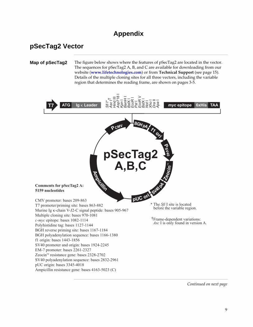
Map of pSecTag2 The figure below shows where the features of pSecTag2 are located in the vector. The sequences for pSecTag2 A, B, and C are available for downloading from our website (www.lifetechnologies.com) or from Technical Support (see page 15). Details of the multiple cloning sites for all three vectors, including the variable region that determines the reading frame, are shown on pages 3-5.
Continued on next page
����
�����
�����
�����
��
���
��
��� �
��
���
���
���
���
���
�����
��
���
���
���
�����
����
����
��
����������� ��� �
����
���������
����
��2
��� �������
�
�����
�� ���
����
����� !" #�#�
�� $!���%� &���'
����������� �����������������������
�!"��*���3+����456$(7��&��*���8��*��#����3+����(7�$((4!�����#�$ ���"$94$���#����������3+����65:$67&!������� �����#����3+����6&5$'5('������������3+����'5(4$''';)��� ����������#3+����''4&$'';;����������*��#����3+����''7&$''(;��������������������%��� �3+����''77$'�(5�'��#��3+����';;�$'(:7-";5��*��������#��3+����'64;$44;:<!$&��*���3+����447'$4�4&=�� ��2������� �#���3+����4�4($4&54-";5�����������������%��� �3+����4(�4$467'�>���#��3+������;:$;5'(�*�� ������������ �#���3+����;'7�$:54����
� ��� ��������� ����+����� �����+���#���1
�
?@�*�$������������������3������������������������1
10
pSecTag2 Vector, continued
Features of pSecTag2
pSecTag2 A (5159 bp), pSecTag2 B (5163 bp), and pSecTag2 C (5167 bp) contain the following elements. All features have been functionally tested.
Feature Benefit
Human cytomegalovirus (CMV) immediate-early promoter/enhancer
Permits efficient, high-level expression of your recombinant protein (Andersson et al., 1989; Boshart et al., 1985; Nelson et al., 1987).
T7 promoter/priming site Allows for in vitro transcription in the sense orientation and sequencing through the insert.
ATG initiation codon Permits initiation of translation of the pSecTag2 fusion protein.
Murine Ig κ-chain leader sequence Allows secretion of the fusion protein (Coloma et al., 1992).
Multiple cloning site Allows insertion of your gene and facilitates cloning.
c-myc epitope (Glu-Gln-Lys-Leu-Ile-Ser-Glu-Glu-Asp-Leu)
Allows detection of pSecTag2 fusion protein with the Anti-myc antibodies (Evan et al., 1985) (see page 14 for ordering).
Polyhistidine tag For high affinity binding to Ni2+-chelating resin (i.e. ProBond™) and easy purification. In addition, it allows detection of pSecTag2 fusion proteins with the Anti-His(C-term) antibodies (Lindner et al., 1997) (see page 14 for ordering).
BGH reverse priming site Permits sequencing through the insert.
Bovine growth hormone (BGH) polyadenylation signal
Efficient transcription termination and polyadenylation of mRNA (Goodwin and Rottman, 1992).
f1 origin Allows rescue of single-strand DNA.
SV40 early promoter and origin Allows efficient, high-level expression of the Zeocin™ resistance gene and episomal replication in cells expressing the SV40 large T antigen.
Zeocin™ resistance gene (Sh ble) For selection of transformants in E. coli and stable transfectants in mammalian cells (Drocourt, et al., 1990; Mulsant, et al., 1988).
SV40 polyadenylation signal Permits efficient transcription termination and polyadenylation of mRNA.
pUC origin Allows high-copy number replication and growth in E. coli.
Ampicillin resistance gene (β-lactamase)
Permits selection in E. coli.

11
pSecTag2/PSA
Map of pSecTag2/PSA
The figure below shows the features of pSecTag2/PSA. The vector sequence for pSecTag2/PSA is available for downloading from our website (www.lifetechnologies.com) or from Technical Support (see page 15).
� ��� ��������� ����+����� �����+���#���1
�
����
�����
����
�����
��
���
��
��
����������� ��� �
����
���������
����
��2
��� �������
�
�����
�� ������
����� !"(�� )*+�,-
�� $!���%� &���' ��
��
��
����������� �������� �����������������
�!"��*���3+����456$(7��&��*���8��*��#����3+����(7�$((4!�����#�$ ���"$94$���#����������3+����65:$67&)-�#���3+����'5;4$'&�4������������3+����'&;:$'&&&)��� ����������#3+����'&65$'(5&����������*��#����3+����'(�5$'(;&��������������������%��� �3+����'(46$45;��'��#��3+����'6:;$4�7&-";5��*��������#��3+����4:(&$465(<!$&��*���3+����464;$4665=�� ��2������� �#���3+����466'$��7:-";5�����������������%��� �3+�����;6:$�74;�>���#��3+����;55($;7('�*�� ������������ �#���3+����;(47$:7(7���

12
Zeocin™
Zeocin™ Zeocin™ is a member of the bleomycin/phleomycin family of antibiotics isolated
from Streptomyces. Antibiotics in this family are broad spectrum antibiotics that act as strong anti-bacterial and anti-tumor drugs. They show strong toxicity against bacteria, fungi (including yeast), plants, and mammalian cells.
The Zeocin™ resistance protein has been isolated and characterized (Calmels et al., 1991; Drocourt et al., 1990). This protein, the product of the Sh ble gene (Streptoalloteichus hindustanus bleomycin gene), is a 13.7 kDa protein that binds Zeocin™ in a stoichiometric manner to inhibit its DNA strand cleavage activity. Expression of this protein in eukaryotic and prokaryotic hosts confers resistance to Zeocin™.
Molecular Weight, Formula and Structure
The formula for Zeocin™ is C55H86O21N20S2Cu-HCl and the molecular weight is 1,527.5 daltons. Zeocin™ is an HCl salt. The diagram below shows the structure of Zeocin™.
Applications of Zeocin™
Zeocin™ is used for selection in mammalian cells (Mulsant et al., 1988); plants (Perez et al., 1989); yeast (Baron et al., 1992); and prokaryotes (Drocourt et al., 1990). Suggested concentrations of Zeocin™ for selection in mammalian cell lines and E. coli are listed below:
Organism Zeocin™ Concentration and Selective Medium
E. coli 25–50 μg/mL in Low Salt LB medium* (see page 13 for recipe)
Mammalian Cells 50–1,000 μg/mL (varies with cell line) *Efficient selection requires that the concentration of NaCl be no more than 5 g/liter (< 90 mM).
���
��
�
��� ��
� �
�
���
�
������
��
�
�
���
������
�
���� ��
��
��
��
�
��
�����
��
����
���
� �
�
�
�
���
�
�
�
�
�
�� �
�
��
��
�� �

13
Recipe
Low Salt LB Medium with Zeocin™
For Zeocin™ to be active, the salt concentration of the medium must remain low (< 90 mM) and the pH must be 7.5. Prepare LB broth and plates using the following recipe. Note the lower salt content of this medium.
Failure to lower the salt content of your LB medium will result in non-selection because of inactivation of the drug.
Low Salt LB Medium:
10 g Tryptone 5 g NaCl 5 g Yeast Extract
1. Combine the dry reagents above and add deionized, distilled water to 950 mL. Adjust pH to 7.5 with 1 N NaOH. Bring the volume up to 1 liter. For plates, add 15 g/L agar before autoclaving.
2. Autoclave on liquid cycle at 15 lbs./sq. in. and 121°C for 20 minutes.
3. Allow the medium to cool to at least 55°C before adding the Zeocin™ to 25 μg/mL final concentration.
4. Store plates at 4°C in the dark. Plates containing Zeocin™ are stable for 1–2 weeks.

14
Accessory Products
Introduction The following products may be used with the pSecTag2 vectors. For details, visit www.lifetechnologies.com or contact Technical Support (page 15).
Item Amount Catalog no.
ProBond™ Purification System 6 purifications K850-01
ProBond™ Resin 50 mL R801-01
150 mL R801-15
Electrocomp™ TOP10F´ 5 × 80 μL C665-55
Max Efficiency® DH10B™ (chemically competent cells)
5 × 0.2 mL 18297-010
PureLink® HiPure Plasmid Miniprep Kit 100 preps K2100-03
PureLink® HiPure Plasmid Midiprep Kit 25 preps K2100-04
Lipofectamine® 2000 Reagent 0.75 mL 11668-027
Zeocin™ 1 gram R250-01
5 grams R250-05
imMedia™ Zeo Liquid 200 mL Q620-20
imMedia™ Zeo Agar 20 each Q621-20
Antibodies If you do not have an antibody specific to your protein, Life Technologies offers
the Anti-myc, or Anti-His(C-term) antibodies to detect your recombinant fusion protein. Horseradish peroxidase (HRP)- and alkaline phosphatase (AP)-conjugated antibodies are available for convenient one-step detection.
Antibody Epitope Catalog no.
Anti-myc Detects a 10 amino acid epitope derived from c-myc (Evan et al., 1985): EQKLISEEDL
R950-25
Anti-myc-HRP R951-25
Anti-myc-AP R952-25
Anti-His(C-term) Detects the C-terminal polyhistidine tag (requires the free carboxyl group for detection) (Lindner et al., 1997): HHHHHH-COOH
R930-25
Anti-His(C-term)-HRP R931-25
Anti-His(C-term)-AP R932-25
Primers For your convenience, Life Technologies offers a custom primer synthesis
service. Visit www.lifetechnologies.com for more details.

15
Technical Support
Obtaining support For the latest services and support information for all locations, go to www.lifetechnologies.com. At the website, you can:
• Access worldwide telephone and fax numbers to contact Technical Support and Sales facilities
• Search through frequently asked questions (FAQs) • Submit a question directly to Technical Support
([email protected]) • Search for user documents, SDSs, vector maps and sequences, application
notes, formulations, handbooks, certificates of analysis, citations, and other product support documents
• Obtain information about customer training • Download software updates and patches
Safety Sata Sheets (SDS)
Safety Data Sheets (SDSs) are available at www.lifetechnologies.com/sds.
Certificate of Analysis
The Certificate of Analysis provides detailed quality control and product qualification information for each product. Certificates of Analysis are available on our website. Go to www.lifetechnologies.com/support and search for the Certificate of Analysis by product lot number, which is printed on the box.
Limited warranty Life Technologies and/or its affiliate(s) warrant their products as set forth in the
Life Technologies General Terms and Conditions of Sale found on the Life Technologies web site at www.lifetechnologies.com/termsandconditions. If you have any questions, please contact Life Technologies. Life Technologies and/or its affiliate(s) disclaim all warranties with respect to this document, expressed or implied, including but not limited to those of merchantability or fitness for a particular purpose. In no event shall Life Technologies and/or its affiliate(s) be liable, whether in contract, tort, warranty, or under any statute or on any other basis for special, incidental, indirect, punitive, multiple or consequential damages in connection with or arising from this document, including but not limited to the use thereof.

16
Purchaser Notification Limited Use Label License No. 358: Research Use Only
The purchase of this product conveys to the purchaser the limited, non-transferable right to use the purchased amount of the product only to perform internal research for the sole benefit of the purchaser. No right to resell this product or any of its components is conveyed expressly, by implication, or by estoppel. This product is for internal research purposes only and is not for use in commercial applications of any kind, including, without limitation, quality control and commercial services such as reporting the results of purchaser’s activities for a fee or other form of consideration. For information on obtaining additional rights, please contact [email protected] or Out Licensing, Life Technologies, 5791 Van Allen Way, Carlsbad, California 92008.

17
References
Andersson, S., Davis, D. L., Dahlbäck, H., Jörnvall, H., and Russell, D. W. (1989). Cloning, Structure, and Expression of the Mitochondrial Cytochrome P-450 Sterol 26-Hydroxylase, a Bile Acid Biosynthetic Enzyme. J. Biol. Chem. 264, 8222-8229.
Ausubel, F. M., Brent, R., Kingston, R. E., Moore, D. D., Seidman, J. G., Smith, J. A., and Struhl, K. (1994). Current Protocols in Molecular Biology (New York: Greene Publishing Associates and Wiley-Interscience).
Baron, M., Reynes, J. P., Stassi, D., and Tiraby, G. (1992). A Selectable Bifunctional -Galactosidase: Phleomycin-resistance Fusion Protein as a Potential Marker for Eukaryotic Cells. Gene 114, 239-243.
Berdy, J. (1980) Bleomycin-Type Antibiotics. In Amino Acid and Peptide Antibiotics, J. Berdy, ed. (Boca Raton, FL: CRC Press), pp. 459-497.
Boshart, M., Weber, F., Jahn, G., Dorsch-Häsler, K., Fleckenstein, B., and Schaffner, W. (1985). A Very Strong Enhancer is Located Upstream of an Immediate Early Gene of Human Cytomegalovirus. Cell 41, 521-530.
Chen, C., and Okayama, H. (1987). High-Efficiency Transformation of Mammalian Cells by Plasmid DNA. Mol. Cell. Biol. 7, 2745-2752.
Chu, G., Hayakawa, H., and Berg, P. (1987). Electroporation for the Efficient Transfection of Mammalian Cells with DNA. Nuc. Acids Res. 15, 1311-1326.
Coloma, M. J., Hastings, A., Wims, L. A., and Morrison, S. L. (1992). Novel Vectors for the Expression of Antibody Molecules Using Variable Regions Generated by Polymerase Chain Reaction. J. Imm. Methods 152, 89-104.
Drocourt, D., Calmels, T. P. G., Reynes, J. P., Baron, M., and Tiraby, G. (1990). Cassettes of the Streptoalloteichus hindustanus ble Gene for Transformation of Lower and Higher Eukaryotes to Phleomycin Resistance. Nuc. Acids Res. 18, 4009.
Evan, G. I., Lewis, G. K., Ramsay, G., and Bishop, V. M. (1985). Isolation of Monoclonal Antibodies Specific for c-myc Proto-oncogene Product. Mol. Cell. Biol. 5, 3610-3616.
Felgner, P. L., Holm, M., and Chan, H. (1989). Cationic Liposome Mediated Transfection. Proc. West. Pharmacol. Soc. 32, 115-121.
Felgner, P. L., and Ringold, G. M. (1989). Cationic Liposome-Mediated Transfection. Nature 337, 387-388.
Goodwin, E. C., and Rottman, F. M. (1992). The 3 -Flanking Sequence of the Bovine Growth Hormone Gene Contains Novel Elements Required for Efficient and Accurate Polyadenylation. J. Biol. Chem. 267, 16330-16334.
Lindner, P., Bauer, K., Krebber, A., Nieba, L., Kremmer, E., Krebber, C., Honegger, A., Klinger, B., Mocikat, R., and Pluckthun, A. (1997). Specific Detection of His-tagged Proteins With Recombinant Anti-His Tag scFv-Phosphatase or scFv-Phage Fusions. BioTechniques 22, 140-149.
Mulsant, P., Tiraby, G., Kallerhoff, J., and Perret, J. (1988). Phleomycin Resistance as a Dominant Selectable Marker in CHO Cells. Somat. Cell Mol. Genet. 14, 243-252.
Nelson, J. A., Reynolds-Kohler, C., and Smith, B. A. (1987). Negative and Positive Regulation by a Short Segment in the 5 -Flanking Region of the Human Cytomegalovirus Major Immediate-Early Gene. Mol. Cell. Biol. 7, 4125-4129.
Perez, P., Tiraby, G., Kallerhoff, J., and Perret, J. (1989). Phleomycin Resistance as a Dominant Selectable Marker for Plant Cell Transformation. Plant Mol. Biol. 13, 365-373.
Continued on next page

18
References, continued
Sambrook, J., Fritsch, E. F., and Maniatis, T. (1989). Molecular Cloning: A Laboratory Manual, Second Edition (Plainview, New York: Cold Spring Harbor Laboratory Press).
Shigekawa, K., and Dower, W. J. (1988). Electroporation of Eukaryotes and Prokaryotes: A General Approach to the Introduction of Macromolecules into Cells. BioTechniques 6, 742-751.
Wigler, M., Silverstein, S., Lee, L.-S., Pellicer, A., Cheng, Y.-C., and Axel, R. (1977). Transfer of Purified Herpes Virus Thymidine Kinase Gene to Cultured Mouse Cells. Cell 11, 223-232.
©2012 Life Technologies Corporation. All rights reserved. The trademarks mentioned herein are the property of Life Technologies Corporation or their respective owners. Zeocin is a trademark of Cayla.

Headquarters5791 Van Allen Way | Carlsbad, CA 92008 USA | Phone +1 760 603 7200 | Toll Free in USA 800 955 6288For support visit lifetechnologies.com/support or email [email protected]
lifetechnologies.com