projections of the nucleus of the basal optic root in ...dwylie/wylie et al vis neurosci...
TRANSCRIPT

Projections of the nucleus of the basal optic root in pigeons~Columba livia!: A comparison of the morphology anddistribution of neurons with different efferent projections
DOUGLAS R.W. WYLIE,1,2 JANELLE M.P. PAKAN,2 CAMERON A. ELLIOTT,2
DAVID J. GRAHAM,2 and ANDREW N. IWANIUK1
1Department of Psychology, University of Alberta, Edmonton, Alberta, Canada2University Centre for Neuroscience, University of Alberta, Edmonton, Alberta, Canada
(Received April 18, 2007; Accepted July 10, 2007!
Abstract
The avian nucleus of the basal optic root ~nBOR! is a visual structure involved in the optokinetic response. nBORconsists of several morphologically distinct cell types, and in the present study, we sought to determine if thesedifferent cell types had differential projections. Using retrograde tracers, we examined the morphology anddistribution of nBOR neurons projecting to the vestibulocerebellum ~VbC!, inferior olive ~IO!, dorsal thalamus, thepretectal nucleus lentiformis mesencephali ~LM!, the contralateral nBOR, the oculomotor complex ~OMC! and agroup of structures along the midline of the mesencephalon. The retrogradely labeled neurons fell into two broadcategories: large neurons, most of which were multipolar rather than fusiform and small neurons, which were eitherfusiform or multipolar. From injections into the IO, LM, contralateral nBOR, and structures along themidline-mesencephalon small nBOR neurons were labeled. Although there were no differences with respect to thesize of the labeled neurons from these injections, there were some differences with the respect to the distribution oflabeled neurons and the proportion of multipolar vs. fusiform neurons. From injections into the VbC, the largemultipolar cells were labeled throughout nBOR. The only other cases in which these large neurons were labeledwere contralateral OMC injections. To investigate if single neurons project to multiple targets we used pairedinjections of red and green fluorescent retrograde tracers into different targets. Double-labeled neurons were neverobserved indicating that nBOR neurons do not project to multiple targets. We conclude that individual nBORneurons have unique projections, which may have differential roles in processing optic flow and controlling theoptokinetic response.
Keywords: Optokinetic, Oculomotor, Accessory optic system, Pretectum, Vestibulocerebellum, Inferior olive
Introduction
Self-motion through the environment results in patterns of “opticflow” across the entire retina ~Gibson, 1954!. Together, nuclei inthe pretectum and the accessory optic system ~AOS! are involvedin the analysis of optic flow ~Simpson, 1984; Simpson et al.,1988b; Grasse & Cynader, 1990!. The pretectum and AOS arehighly conserved in vertebrates: the mammalian pretectal nucleusof the optic tract ~NOT! is homologous to the nucleus lentiformismesencephali ~LM! in birds, and the avian nucleus of the basaloptic root ~nBOR! of the AOS is homologous to the medial andlateral terminal nuclei ~MTN, LTN! of the mammalian AOS~Simpson, 1984; Fite, 1985; McKenna & Wallman, 1985; Weber,1985; Simpson et al., 1988b; Gamlin, 2005; Giolli et al., 2005!.
Previous reports have shown that neither the LM nor the nBORcan be regarded as homogeneous nuclei, but rather consist ofseveral morphologically distinct cell types ~Brecha et al., 1980;Gottlieb & McKenna, 1986; Gamlin & Cohen, 1988; Tang &Wang, 2002; Zayats et al., 2002, 2003!. As the analysis of opticflow subserves many functions ~Lee & Lishman, 1977; Simpson,1984; Lappe & Rauschecker, 1994!, it is possible that these distinctcell types are specialized with respect to their efferent projections,as well as their function. For example, Prochnow et al. ~2007!showed that different cell types in the rat NOT project to thesuperior colliculus and inferior olive. They also found that thesecell types have different physiological properties and are involvedin saccades and the slow phase of optokinetic nystagmus, respec-tively. Recently, we ~Pakan et al., 2006! showed that the differentefferent projections of LM originate from morphologically distincttypes of neurons, and we speculated that they are involved indifferent components of the optokinetic response or other visualbehaviors. Given that LM and nBOR are quite similar with re-spect to function, response properties, and efferent projections
Address correspondence and reprint requests to: Douglas Wong-WyliePhD, University of Alberta, Department of Psychology, Edmonton, Al-berta, Canada T6G 2E9. E-mail: [email protected]
Visual Neuroscience ~2007!, 24, 691–707. Printed in the USA.Copyright © 2007 Cambridge University Press 0952-5238007 $25.00DOI: 10.10170S0952523807070599
691

~McKenna & Wallman, 1985!, one might expect that the differentcell types in nBOR also have differential projections.
Electrophysiological studies have shown that nBOR neuronshave large receptive fields in the contralateral visual field andexhibit direction-selectivity to large-field moving visual stimuli~Burns & Wallman, 1981; Morgan & Frost, 1981; Wylie &Frost, 1990!. The nBOR receives primary input from the dis-placed ganglion cells in the contralateral retina ~Karten et al.,1977; Reiner et al., 1979; Fite et al., 1981! and projects toseveral structures. These include bilateral projections to the me-dial column of the inferior olive ~mcIO!, folium IXcd of thevestibulocerebellum ~VbC! and the oculomotor complex ~OMC!,ipsilateral projections to LM and parts of the anterior dorsalthalamus, and a projection to the contralateral nBOR. In addi-tion, the nBOR projects bilaterally to several structures alongthe midline in the mesencephalon, including the interstitial nu-cleus of Cajal ~IS!, nucleus Darkshewitsch ~D!, the central grey~CtG!, and the ventral tegmental area ~VTA; Brecha et al., 1980;Wild, 1989; Wylie & Linkenhoker, 1996; Wylie et al., 1997,1998b; Wylie, 2001; Pakan et al., 2006!.
nBOR consists of several morphologically distinct cell types~Brecha et al., 1980! with different immunochemical properties~Zayats et al., 2002!. However, it is not known if these differentneuronal subtypes are associated with different efferent projec-tions. In the present study, using retrograde tracing techniques, weexamined differences in the morphology and distribution of nBORneurons that project to several different targets: VbC, mcIO, LM,OMC, the contralateral nBOR, dorsal thalamus, and structuresalong the midline of the mesencephalon.
Materials and methods
The methods reported herein conformed to the guidelines estab-lished by the Canadian Council on Animal Care and were ap-proved by the Biosciences Animal Care and Policy Committee atthe University of Alberta. Silver King and homing pigeons, ob-tained from various suppliers, were anesthetized with an intramus-cular injection of a ketamine ~65 mg0kg!0xylazine ~9.4 mg0kg!cocktail, and were given supplemental doses as needed to maintainanesthesia. The animals were placed in a stereotaxic device withpigeon ear bars and beak adapter so that the orientation of the skullconformed to the atlas of Karten and Hodos ~1967!. Sufficientskull and dura were removed to expose the brain surface and allowaccess to one of the following: the dorsal thalamus, structuresalong the midline-mesencephalon, VbC, mcIO, OMC, LM, ornBOR. All target sites were localized using a stereotaxic atlas~Karten & Hodos, 1967!. For injections into the VbC, mcIO, LM,and nBOR we also relied on single-unit recordings made withglass micropipettes ~tip diameters 4–5 mm! filled with 2 M NaCl,which were advanced using an hydraulic microdrive to record theresponses of neurons in these structures to optic flow stimuli~e.g., Wylie & Frost, 1990, 1996; Winship & Wylie, 2001; Winshipet al., 2005!.
Studies using cholera toxin subunit B (CTB)as a retrograde tracer
For injections into the VbC, mcIO, LM, and nBOR, after recordingfrom optic flow sensitive cells, the recording electrode was re-placed with a micropipette ~tip diameter 20 mm! filled with CTB~low-salt version, Sigma, St. Louis, MO; 1% in 0.1 M phosphate-
buffered saline ~PBS, pH 7.4!! and the nucleus was located againby isolating cells responsive to large field visual stimuli. For allother target sites ~dorsal thalamus and midline structures!, injec-tions were made according to the stereotaxic coordinates. Inall cases, the CTB was injected iontophoretically for 10–15 min~�4 µamps, 7 s on, 7 s off !. Following the injection, the micro-pipette was left in place for 5 min then removed and the exposureswere closed. Once the animal regained consciousness, buprenor-phine ~2 mg0kg, i.m.! was administered as an analgesic.
After a survival time of 3–5 days, the animals were adminis-tered an overdose of sodium pentobarbital ~100 mg0kg!, andperfused with 0.9% saline followed by 4% paraformaldehyde in0.1 M phosphate buffer ~PB!. The brain was extracted from theskull, embedded in gelatin, and placed in 30% sucrose in 0.1 M PBfor cryoprotection. Using a microtome, frozen sections in thecoronal plane ~40 mm thick! were collected, and sections wereprocessed for CTB based on the protocol outlined by Wild et al.~1993; see also Veenman et al., 1992!. Sections were initiallyrinsed in 0.1 M PBS. They were then washed in a 25% methanol,0.9% hydrogen peroxide solution for 30 min to decrease endog-enous peroxidase activity. Sections were rinsed several times inPBS then placed in 4% rabbit serum with 0.4% Triton X-100 inPBS for 30 min. Tissue was subsequently incubated for 20 h in aprimary antibody for CTB, goat anti-Choleragenoid ~1:20,000;List Biological Laboratories, Campbell, CA! with 0.4% TritonX-100 in PBS. Sections were then rinsed in PBS ~several times!and incubated for 60 min in biotinylated rabbit anti-goat antiserum~1:600; Vector Laboratories, Burlingame, CA! with 0.4% TritonX-100 in PBS. Tissue was rinsed several times with PBS andincubated for 90 min in ExtrAvidin ~1:1,000; Sigma, St. Louis,MO! with 0.4% Triton X-100 in PBS. Subsequent to a few washeswith PBS, the tissue was incubated for 12 min in filtered 0.025%diaminobenzidine ~DAB! and 0.006% cobalt chloride in PBS.0.005% hydrogen peroxide was added to the DAB solution, andthe sections were reacted for up to 6 min. The sections were thenrinsed several times with PBS, mounted onto gelatin coated slides,lightly counterstained with Neutral Red and cover slipped withPermount.
Double-labeling fluorescent studies
We also performed double-labeling experiments using green andred fluorescent latex microspheres ~LumaFluor Corp, Naples, FL!as retrograde tracers. These were pressure injected through a glassmicropipette ~tip diameter 20 mm!, into the mcIO, LM, OMC,dorsal thalamus, VbC, and nBOR, using a Picospritzer II ~GeneralValve Corporation!. Viewing through a surgical microscope, wemonitored the movement of the meniscus to inject from 0.05 to0.2 µl. With larger injection volumes, typically used for injectioninto the cerebellum, the resultant injection sites were about 1 mmin diameter. As with the CTB injections, for injections into LM,VbC, nBOR and mcIO, the nuclei were first localized by recordingthe responses of neurons to optic flow stimuli. After a recoveryperiod of 2–5 days, the animals were deeply anesthetized withsodium pentobarbital ~100 mg0kg! and immediately perfused withheparinized phosphate buffered saline ~0.9% NaCl, 1 ml0100 mlheparin, 0.1 M phosphate buffer!. The brains were extracted, thenflash-frozen in 2-methylbutane and stored at �808C until sec-tioned. Brains were embedded in optimal cutting temperaturemedium and 40 mm coronal sections were cut through the brain-stem and cerebellum with a cryostat and mounted on electrostaticslides.
692 D.R.W. Wylie et al.
Microscopy
Sections were viewed with a compound light microscope ~LeicaDMRE! equipped with the appropriate fluorescence filters. Thered and green latex microspheres fluoresce under rhodamine andFITC filters, respectively. The CTB-reacted tissue was examinedusing standard light microscopy and drawings were made with theaid of a drawing tube. Images were obtained using the OPENLABImaging system ~Improvision, Lexington, MA! and Adobe Photo-shop software was used to compensate for brightness and contrast.OPENLAB was also used to measure the size ~area! of CTB-labeled neurons.
Nomenclature
Brecha et al. ~1980! defined the nBOR complex as consisting ofthree parts: nBOR proper ~nBORp!, which comprises the bulk ofthe nucleus; nBOR dorsalis ~nBORd! which is a cap that surroundsthe dorsal and caudal margins of nBORp; and nBOR lateralis,which sits atop the stratum opticum ~SOp! lateral to the nucleus.McKenna and Wallman ~1981! found that nBOR lateralis is con-tinuous with LM, and represents LM’s caudo-medial tail. More-over, they showed that with respect to direction-preference forvisual stimuli, lateralis resembles LM rather than nBOR. In dozensof species of birds, Iwaniuk and Wylie ~2007! observed in Nisslstained coronal sections that lateralis is continuous with LM. Also,the unique connections of nBOR, the contralateral nBOR and theOMC, do not involve lateralis. For these reasons, we considernBOR lateralis as part of LM rather than the nBOR complex.
Results
Data are described from experiments performed on 33 pigeons~Table 1!. Twenty-one pigeons were used for the retrograde studies
using CTB injected into a single target site. The CTB cases in-cluded unilateral injections in the VbC ~n � 5! IO ~n � 3!, contra-lateral nBOR ~n � 3!, dorsal thalamus ~n � 3!, LM ~n � 3!, andOMC ~n � 1!. In addition, there were three cases, two unilateral,and one bilateral that were grouped together as “midline-mesencephalon.” The other 12 birds received injections of red orgreen LumaFluors in different efferent targets of the nBOR. Fromboth the CTB and LumaFluor injections, we observed differencesin the size, morphology, and distribution of neurons associated withthe different projection sites. In our descriptions of morphology andquantification of neuron size, we relied only on the CTB cases, asthis tracer results in uniform and complete labeling of the entiresoma and proximal dendrites. Photomicrographs of representativeretrograde labeling from the CTB experiments are shown in Figs. 1and 2, and from the LumaFluor experiments in Fig. 3. Drawings ofcoronal sections through the nBOR illustrating the distribution oflabeled cells from various injections are shown in Figs. 6 and 7.
VBC-projecting nBOR neurons
Previous studies using anterograde techniques have shown that theprojection from nBOR to the VbC is bilateral and terminates in thegranule layer as mossy fiber rosettes ~Brecha et al., 1980!. Wedirected our injections to folium IXcd. The complex spike activityof Purkinje cells in folium IXcd responds to patterns of optic flowresulting from either self-translation or self-rotation ~Wylie &Frost, 1991, 1993, 1999b; Wylie et al., 1993, 1998a!. Once re-sponsive neurons were found, we made the injection in the adja-cent granule cell layer. Spread of the injection site to other foliawas not a concern because folium X does not receive direct inputfrom nBOR ~Brecha et al., 1980; Wylie & Linkenhoker, 1996;Wylie et al., 1997!, although the nBOR does project to foliaVI-IXab, retrograde studies show that the magnitude of this pro-
Table 1. Retrograde tracer injection sites and fluorescent cell count
CTB Injections
Case Target Case Target Case Target
VbC#1 VbC IO#1 IO DTHAL#1 dorsal thalamusVbC#2 VbC IO#2 IO DTHAL#2 dorsal thalamusVbC#3 VbC IO#3 IO DTHAL#3 dorsal thalamusVBC#4 VbC c-nBOR#1 nBOR LM#1 LMVBC#5 VbC c-nBOR#2 nBOR LM#2 LMOMC OMC c-nBOR#3 nBOR LM#3 LMMID-MES#1 Midline-Mesencephalon MID-MES#2 Midline-Mesencephalon MID-MES#3 Midline-Mesencephalon
LumaFluor Injections
Case Target-green # green cells Target-Red # red cells #double-labeled
DTHAL-IO dorsal thalamus 73 ipsi-IO 10 0DTHAL-LM dorsal thalamus 223 LM 888 0IO-LM#1 ipsi-IO 60 LM 224 0IO-LM#2 LM 370 contra-IO 168 0nBOR-DTHAL nBOR 186 dorsal thalamus 202 0nBOR-IO#1 nBOR 184 contra-IO 78 0nBOR-IO#2 nBOR 592 ipsi-IO 136 0nBOR-LM nBOR 370 LM 624 0nBOR-VbC nBOR 756 VbC 432 0OMC-VbC OMC 363 VbC 80 0VbC-IO#1 VbC 73 IO 380 0VbC-IO#2 VbC 738 IO 269 0
Projections of the Pigeon nBOR 693
jection is very small compared to the projection to IXcd ~Brechaet al., 1980; Pakan & Wylie, 2006!.
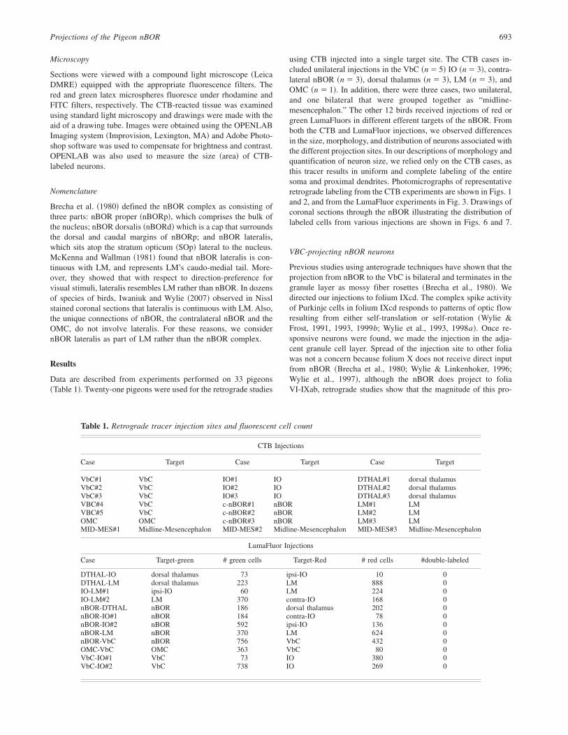
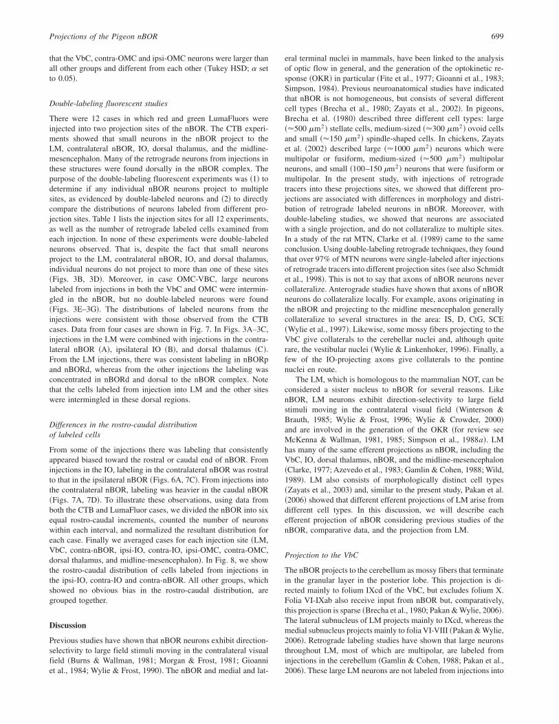
There were nine cases in which the VbC was injected unilat-erally; five CTB cases, and four LumaFluor cases. From theseinjections, large neurons ~330.2616.9 mm2; mean6 s.e.m.! wereinvariably labeled ~Figs. 1E, 1F, 3F!. The majority ~85%! of theseneurons was multipolar ~Fig. 4! and they were distributed uni-formly throughout nBOR ~Figs. 1E, 6B!.
IO-projecting nBOR neurons
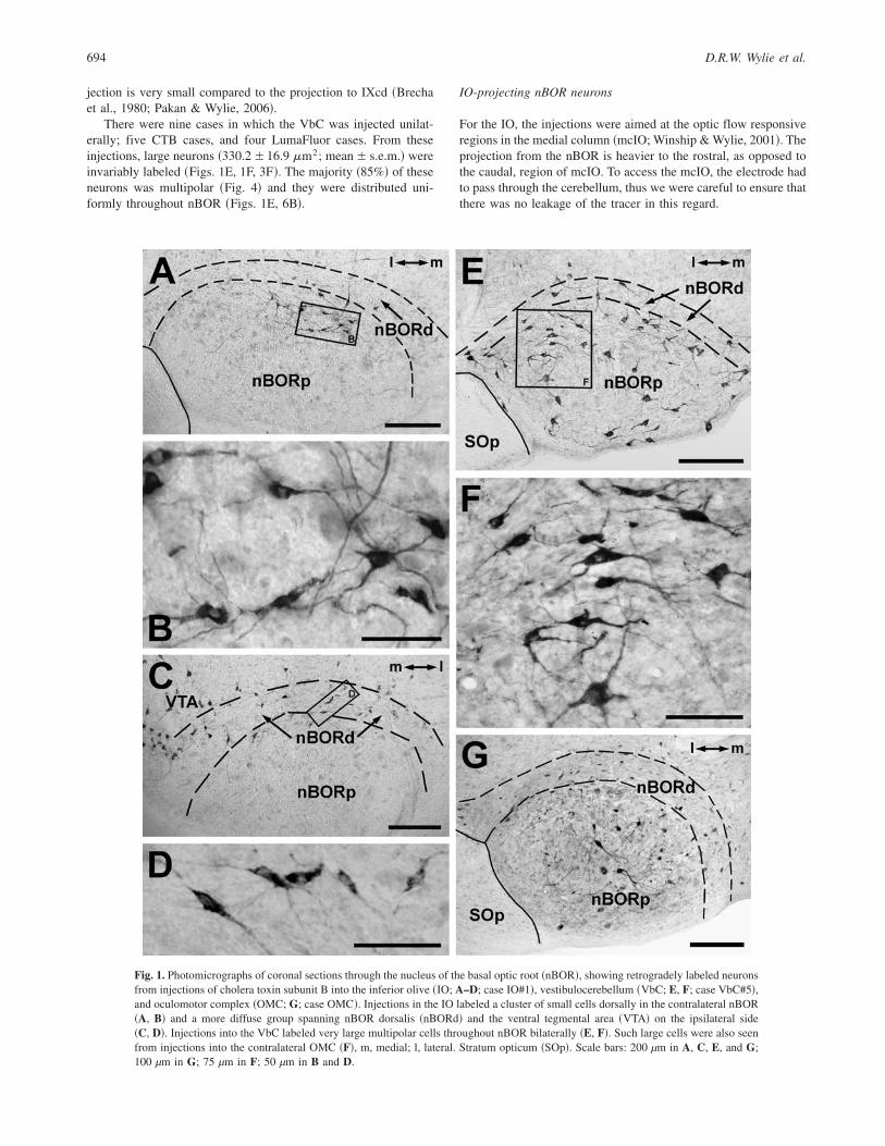
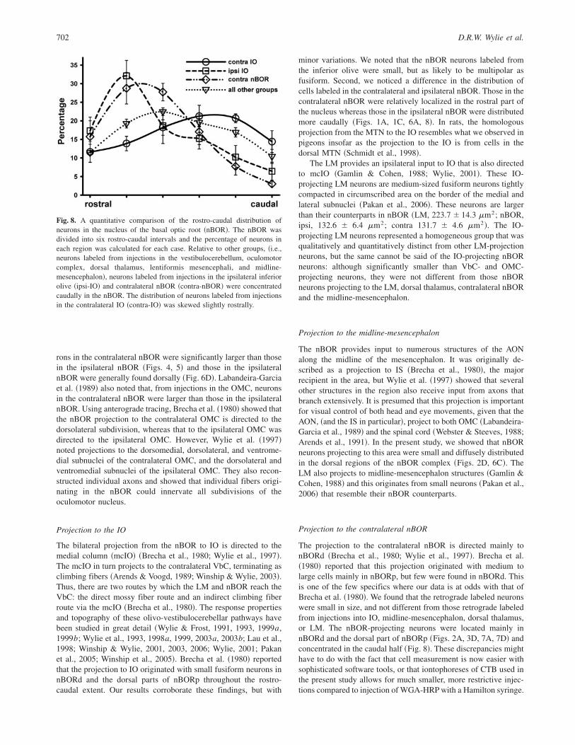
For the IO, the injections were aimed at the optic flow responsiveregions in the medial column ~mcIO; Winship & Wylie, 2001!. Theprojection from the nBOR is heavier to the rostral, as opposed tothe caudal, region of mcIO. To access the mcIO, the electrode hadto pass through the cerebellum, thus we were careful to ensure thatthere was no leakage of the tracer in this regard.
Fig. 1. Photomicrographs of coronal sections through the nucleus of the basal optic root ~nBOR!, showing retrogradely labeled neuronsfrom injections of cholera toxin subunit B into the inferior olive ~IO; A–D; case IO#1!, vestibulocerebellum ~VbC; E, F; case VbC#5!,and oculomotor complex ~OMC; G; case OMC!. Injections in the IO labeled a cluster of small cells dorsally in the contralateral nBOR~A, B! and a more diffuse group spanning nBOR dorsalis ~nBORd! and the ventral tegmental area ~VTA! on the ipsilateral side~C, D!. Injections into the VbC labeled very large multipolar cells throughout nBOR bilaterally ~E, F!. Such large cells were also seenfrom injections into the contralateral OMC ~F!, m, medial; l, lateral. Stratum opticum ~SOp!. Scale bars: 200 µm in A, C, E, and G;100 µm in G; 75 µm in F; 50 µm in B and D.
694 D.R.W. Wylie et al.

The IO was injected in three of the CTB cases and sevenof the LumaFluor cases. Small neurons were labelled bilaterallyin the nBOR ~ipsi, 132.6 6 6.4 mm2; contra 131.7 6 4.6 mm2;mean6 s.e.m.!. Neurons that were multipolar and fusiform0ovoidwere equally represented ~Figs. 1B, 1D, 4!. In the contralateralnBOR, the neurons were found in a circumscribed region in thedorsal part of nBORp and encroaching on the adjacent nBORd~Figs. 1A, 6A!. In the ipsilateral nBOR, the neurons were morewidely distributed and found mainly in nBORd and adjacentregions of the nBORp and VTA ~Figs. 1C, 6A!. The IO-projectingneurons in the ipsilateral nBOR were located more caudally thanthose in the contralateral nBOR ~Fig. 6A; see below!.
LM-Projecting nBOR neurons
The heaviest projection of the nBOR is to the ipsilateral LM. Thisprojection spans the rostro-caudal and dorso-ventral extent of the
medial and lateral subnuclei of LM ~Wylie et al., 1997!. The LMwas injected in three of the CTB cases and four of the LumaFluorcases. Critically, none of these injections spread into the dorsalthalamus. Small cells were labeled from these injections ~106.066.2 mm2; mean 6 s.e.m.!, most of which were fusiform0ovoid~85%! as opposed to multipolar ~Figs. 2E, 3C, 4!. Neurons werefound in both nBORp and nBORd and also dorsal to the complexitself ~Figs. 3C, 3D, 7A–7C!.
Dorsal thalamus-projecting nBOR neurons
Wylie et al. ~1998b! showed that the projection from the nBOR tothe thalamus is directed mainly to the lateral subnucleus of theipsilateral anterior dorsolateral thalamus ~DLL!. This is one com-ponent of the principal optic nucleus of the thalamus and is con-sidered the homolog of the mammalian dorsal lateral geniculatenucleus ~dLGN; Karten et al., 1973; Shimizu & Karten, 1991!. For
Fig. 2. Photomicrographs of coronal sections through the nucleus of the basal optic root ~nBOR! showing small neurons labeled withcholera toxin subunit B. A–C show cells labeled from injections in the contra nBOR ~case C-NBOR#1!. The rectangles in A indicatethe areas shown in B and C. Most cells were found in nBOR dorsalis ~nBORd! and the dorsal part of nBOR proper ~nBORp! ~B!, andsome were dorsal to the nBOR complex in the medial mesencephalic reticular formation ~FRM! ~C!. D, E, and F, respectively showcells retrogradely labeled from injections in the midline-mesencephalon ~case MID-MES#3!, lentiformis mesencephali ~LM; caseLM#3!, and the dorsal thalamus ~case DTHAL#1!. m, medial; l, lateral. Stratum opticum ~SOp!. Scale bars: 200 µm in A; 100 mm inD; 50 µm in B, C, E; 10 µm in F.
Projections of the Pigeon nBOR 695

Fig. 3. Photomicrographs of coronal sections through the nucleus of the basal optic root ~nBOR! showing cells labeled with fluorescentmicrospheres. ~A! Shows a cell labeled from an injection in the inferior olive ~IO; case LMIO2!. ~B and C! Show cells labeled fromcase DTHAL-LM in which green LumaFluor was injected in the dorsal thalamus and red was injected in the pretectal nucleuslentiformis mesencephali ~LM!. ~D) Shows labeled cells from case nBOR-LM, in which red was injected in the ipsilateral LM andgreen was injected in the contralateral nBOR. ~E! Shows a cell labeled from an injection in the vestibulocerebellum ~VbC; caseVbC-IO#1! and ~F and G! Show cells labeled from case OMC-VbC in which green was injected in the oculomotor complex ~OMC!and red was injected in the VbC. The cells labeled from the LM injections were found in nBOR proper ~nBORp! and dorsally ~nBORd!whereas cells labeled from contra-nBOR and dorsal thalamus injections were found mostly in nBORd. Note the large cells labeled fromthe VbC and OMC injections ~E, F!. Scale bars: 25 mm in A, C, E, F; 100 mm in B, D, G.
696 D.R.W. Wylie et al.

these injections, we were careful to ensure that they did not en-croach ventrolaterally on LM.
The dorsal thalamus was injected in three CTB cases and threeof the LumaFluor cases. Like those labeled from injections in theLM, the dorsal thalamic-projecting nBOR neurons were small~123.46 7.8 mm2; mean6 s.e.m.!, and the majority were fusiform0ovoid ~85%! as opposed to multipolar ~Figs. 2F, 3B, 4!. Theseneurons were distributed mainly in nBORd and dorsal to theborder of the nBOR complex ~Figs. 7B, 7D!.
Contralateral nBOR-projecting nBOR neurons
The nBOR projects to the contralateral nBOR and anterogradestudies have shown that the projection is mainly to the dorsal partsof the nBOR complex ~Brecha et al., 1980; Wylie et al., 1997!. The
nBOR was injected in three of the CTB cases and five of theLumaFluor cases. From these injections, small neurons ~124.1 67.3 mm2; mean 6 s.e.m.! were labeled in the contralateral nBOR~Figs. 2B, 2C!. Multipolar and fusiform0ovoid cells were found inequal proportions, and most labeled cells were located in nBORd,dorsal nBORp, and some were located dorsal to the nBOR com-plex in VTA and medial mesencephalic reticular formation ~FRM;Figs. 2A, 3D, 7A, 7D!.
nBOR neurons projecting to the midline-mesencephalon
Previous studies have shown that nBOR projects heavily to theaccessory optic nuclei ~AON!, a group of structures lying on themidline of the mesencephalon that includes IS and D ~Brecha et al.,1980!. In an anterograde study, Wylie et al. ~1997! showed thatfibers exit the nBOR medially, travel dorsally to the AON andcollateralize heavily to adjacent structures including VTA, the rednucleus ~Ru!, the stratum cellulare externum0internum and CtG.Injecting these small individual nuclei in this region is extremelydifficult, and because individual fibers collateralize throughout thisregion, we considered this area as a group. There were three casesin which CTB was injected along the midline-mesencephalon. Incase MID-MES#1, the injection was unilateral, and included IS, D,the medial Ru, VTA, SCE and the caudal part of Campi Foreli~CF!. In case MID-MES#2, the injection was centered on the mid-line and was largely confined to the VTA bilaterally, with somespread to Ru, IS, CF, and SCE. In case MID-MES#3, the injectionwas unilateral and centered on the lateral Ru with spread to theadjacent FRM, IS, CF, and SCE. The morphology, size, and distri-bution of the retrograde labeled cells in the nBOR were not appre-ciably different from these three cases. Cells were found bilaterallyin nBOR and were small ~132.96 8.3 mm2; mean6 s.e.m.!. Mostwere fusiform0ovoid ~70%! as opposed to multipolar ~30%!~Fig. 2D!. With respect to distribution, the labeling was found dor-sally in the nBORp, nBORd, and dorsal to the nBOR complex~Figs. 2D, 6C!.
OMC-projecting nBOR neurons
We had two OMC cases: one CTB injection ~case OMC!, and oneLumaFluor injection ~case OMC-VbC!. For the CTB case, the in-jection was largely unilateral, and included the dorsomedial, dor-solateral, and ventromedial subdivisions of the oculomotor nucleus,as well as the Edinger-Westphal nucleus. There was spread to theadjacent CtG, the medial longitudinal fasciculus, the vestibular-mesencephalic tract, the brachium conjuctivum, and diffuse spreadrostrally into midline-mesencephalon structures, including IS, D,and Ru. Retrograde labeled cells were observed in the lateralsubnucleus of LM, consistent with an injection in the midline-mesencephalon ~Pakan et al., 2006!. As the LM does not project tothe OMC, we must conclude that the injection spread to the midline-mesencephalon. Although the injection appeared to be unilateral,there were some labeled fibers in the contralateral oculomotor nerve.Most of these fibers were located medially and although these arelikely innervating the contralateral superior rectus, it is possiblethat some of this labeling was due to spread across the midline.Also there were retrograde labeled neurons in the contralateral LMand PPC, indicative of spread across the midline. Thus, it appearsthat although the injection was largely unilateral, there was somespread across the midline. In the LumaFluor case ~OMC-VbC!, theinjection was bilateral, and centered on the ventromedial sub-division of the oculomotor nucleus. The injection spread into the
Fig. 4. A comparison of the morphology and size of neurons in the nucleusof the basal optic root ~nBOR! that project to the vestibulocerebellum~VbC!, the ipsi- and contralateral oculomotor complex ~ipsi-, contra-OMC!, the nucleus lentiformis mesencephali ~LM!, the dorsal thalamus,the ipsi- and contralateral inferior olive ~ipsi-, contra-IO!, the contralteralnBOR ~contra-nBOR! and the midline-mesencephalon ~mid-mesen!. Sil-houettes of CTB-labeled neurons have been drawn to the same scale. TheVbC- and OMC-projecting neurons were much larger than the others andthey tend to be multipolar rather than fusiform. Scale bar � 50 mm.
Projections of the Pigeon nBOR 697

dorsomedial and dorsolateral subnuclei, but the Edinger-Westphalnucleus was largely spared. There was spread ventrally into theparts to the brachium conjuctivum, and rostrally into the midline-mesencephalic structures of the AON including IS, D, CtG, SCE,and Ru. As with the CTB case, there was retrograde labeling in theLM and PPC. As such, both injections included the OMC and themidline-mesencephalon. The retrograde labeled cells in nBOR fromthese injections were quite variable in size ~65–805 mm2 as mea-sured from the CTB case! and included small and large neurons~Figs. 1G, 3E, 3F!. Those in the contralateral nBOR were largeron average than those in the ipsilateral nBOR ~contra, 258.7 612.9mm2; ipsi, 180.569.2mm2, mean6 s.e.m.!.The large neuronsfound in the contralateral nBOR were as large as those retrogradelabeled from the VbC ~Figs. 3F, 5!. Neurons were found in nBORdand nBORp, with a dorsal emphasis, as expected from the spread ofthe injections to the midline-mesencephalon.
A Comparison of the size of nBOR neurons
Fig. 4 shows the silhouettes of nBOR neurons projecting toVbC, the contra- and ipsilateral OMC, LM, dorsal thalamus, the
contra- and ipsilateral IO, contralateral nBOR, and the midline-mesencephalon all drawn to the same scale. The OMC-projectingneurons, drawn from the CTB case represent midline-mesencephalonprojecting cells as well. Nonetheless, we purposefully selectedcells from nBORp as opposed to nBORd from this case, becauselabeling from the midline-mesencephalon was heavily biased tothe nBORd. Thus, the cells we selected are more likely to representOMC-projecting neurons. Clearly, the VbC-projecting neuronswere much larger than the others. From among the other groups,cells of this size were only seen from the contra-OMC projectingcells. The cells projecting to the contralateral nBOR, dorsal thal-amus, IO, LM, and midline-mesencephalon were all very similar insize. The only noticeable difference was that cells with a fusiform0ovoid as opposed to a multipolar profile were more prevalentamong the LM-, dorsal thalamic- and midline-mesencephalic-projecting neurons. To quantitatively examine the size of neurons,we used a one-way ANOVA with injection site as a grouping factorand compared all groups with Tukey’s HSD tests. We subjected thedata to an ln-transform to normalize the distributions. Fig. 5 showsbox plots of these data. There was a significant effect of group~F~8,577!� 48.9; p � 0.0001! and planned comparisons revealed
Fig. 5. A comparison of the sizes of neurons in nucleus of the basal optic root ~nBOR!. Box plots show the cross-sectional areasof nBOR cells retrograde labeled from injections into the nucleus lentiformis mesencephali ~LM!, dorsal thalamus, midline-mesencephalon ~mid-mesen!, contralateral nBOR, contra- and ipsilateral inferior olive ~IO!, contra- and ipsilateral oculomotor complex~OMC! and the vestibulocerebellum ~VbC!. Note that the data has been subjected to an ln transform. The asterisks ~*! indicate theipsi-OMC, contra-OMC and VbC groups were statistically different from all other groups and each other ~Tukey HSD, p � .05!.
698 D.R.W. Wylie et al.
that the VbC, contra-OMC and ipsi-OMC neurons were larger thanall other groups and different from each other ~Tukey HSD; a setto 0.05!.
Double-labeling fluorescent studies
There were 12 cases in which red and green LumaFluors wereinjected into two projection sites of the nBOR. The CTB experi-ments showed that small neurons in the nBOR project to theLM, contralateral nBOR, IO, dorsal thalamus, and the midline-mesencephalon. Many of the retrograde neurons from injections inthese structures were found dorsally in the nBOR complex. Thepurpose of the double-labeling fluorescent experiments was ~1! todetermine if any individual nBOR neurons project to multiplesites, as evidenced by double-labeled neurons and ~2! to directlycompare the distributions of neurons labeled from different pro-jection sites. Table 1 lists the injection sites for all 12 experiments,as well as the number of retrograde labeled cells examined fromeach injection. In none of these experiments were double-labeledneurons observed. That is, despite the fact that small neuronsproject to the LM, contralateral nBOR, IO, and dorsal thalamus,individual neurons do not project to more than one of these sites~Figs. 3B, 3D!. Moreover, in case OMC-VBC, large neuronslabeled from injections in both the VbC and OMC were intermin-gled in the nBOR, but no double-labeled neurons were found~Figs. 3E–3G!. The distributions of labeled neurons from theinjections were consistent with those observed from the CTBcases. Data from four cases are shown in Fig. 7. In Figs. 3A–3C,injections in the LM were combined with injections in the contra-lateral nBOR ~A!, ipsilateral IO ~B!, and dorsal thalamus ~C!.From the LM injections, there was consistent labeling in nBORpand nBORd, whereas from the other injections the labeling wasconcentrated in nBORd and dorsal to the nBOR complex. Notethat the cells labeled from injection into LM and the other siteswere intermingled in these dorsal regions.
Differences in the rostro-caudal distributionof labeled cells
From some of the injections there was labeling that consistentlyappeared biased toward the rostral or caudal end of nBOR. Frominjections in the IO, labeling in the contralateral nBOR was rostralto that in the ipsilateral nBOR ~Figs. 6A, 7C!. From injections intothe contralateral nBOR, labeling was heavier in the caudal nBOR~Figs. 7A, 7D!. To illustrate these observations, using data fromboth the CTB and LumaFluor cases, we divided the nBOR into sixequal rostro-caudal increments, counted the number of neuronswithin each interval, and normalized the resultant distribution foreach case. Finally we averaged cases for each injection site ~LM,VbC, contra-nBOR, ipsi-IO, contra-IO, ipsi-OMC, contra-OMC,dorsal thalamus, and midline-mesencephalon!. In Fig. 8, we showthe rostro-caudal distribution of cells labeled from injections inthe ipsi-IO, contra-IO and contra-nBOR. All other groups, whichshowed no obvious bias in the rostro-caudal distribution, aregrouped together.
Discussion
Previous studies have shown that nBOR neurons exhibit direction-selectivity to large field stimuli moving in the contralateral visualfield ~Burns & Wallman, 1981; Morgan & Frost, 1981; Gioanniet al., 1984; Wylie & Frost, 1990!. The nBOR and medial and lat-
eral terminal nuclei in mammals, have been linked to the analysisof optic flow in general, and the generation of the optokinetic re-sponse ~OKR! in particular ~Fite et al., 1977; Gioanni et al., 1983;Simpson, 1984!. Previous neuroanatomical studies have indicatedthat nBOR is not homogeneous, but consists of several differentcell types ~Brecha et al., 1980; Zayats et al., 2002!. In pigeons,Brecha et al. ~1980! described three different cell types: large~�500 mm2! stellate cells, medium-sized ~�300 mm2! ovoid cellsand small ~�150 mm2! spindle-shaped cells. In chickens, Zayatset al. ~2002! described large ~�1000 mm2! neurons which weremultipolar or fusiform, medium-sized ~�500 mm2! multipolarneurons, and small ~100–150 µm2! neurons that were fusiform ormultipolar. In the present study, with injections of retrogradetracers into these projections sites, we showed that different pro-jections are associated with differences in morphology and distri-bution of retrograde labeled neurons in nBOR. Moreover, withdouble-labeling studies, we showed that neurons are associatedwith a single projection, and do not collateralize to multiple sites.In a study of the rat MTN, Clarke et al. ~1989! came to the sameconclusion. Using double-labeling retrograde techniques, they foundthat over 97% of MTN neurons were single-labeled after injectionsof retrograde tracers into different projection sites ~see also Schmidtet al., 1998!. This is not to say that axons of nBOR neurons nevercollateralize. Anterograde studies have shown that axons of nBORneurons do collateralize locally. For example, axons originating inthe nBOR and projecting to the midline mesencephalon generallycollateralize to several structures in the area: IS, D, CtG, SCE~Wylie et al., 1997!. Likewise, some mossy fibers projecting to theVbC give collaterals to the cerebellar nuclei and, although quiterare, the vestibular nuclei ~Wylie & Linkenhoker, 1996!. Finally, afew of the IO-projecting axons give collaterals to the pontinenuclei en route.
The LM, which is homologous to the mammalian NOT, can beconsidered a sister nucleus to nBOR for several reasons. LikenBOR, LM neurons exhibit direction-selectivity to large fieldstimuli moving in the contralateral visual field ~Winterson &Brauth, 1985; Wylie & Frost, 1996; Wylie & Crowder, 2000!and are involved in the generation of the OKR ~for review seeMcKenna & Wallman, 1981, 1985; Simpson et al., 1988a!. LMhas many of the same efferent projections as nBOR, including theVbC, IO, dorsal thalamus, nBOR, and the midline-mesencephalon~Clarke, 1977; Azevedo et al., 1983; Gamlin & Cohen, 1988; Wild,1989!. LM also consists of morphologically distinct cell types~Zayats et al., 2003! and, similar to the present study, Pakan et al.~2006! showed that different efferent projections of LM arise fromdifferent cell types. In this discussion, we will describe eachefferent projection of nBOR considering previous studies of thenBOR, comparative data, and the projection from LM.
Projection to the VbC
The nBOR projects to the cerebellum as mossy fibers that terminatein the granular layer in the posterior lobe. This projection is di-rected mainly to folium IXcd of the VbC, but excludes folium X.Folia VI-IXab also receive input from nBOR but, comparatively,this projection is sparse ~Brecha et al., 1980; Pakan & Wylie, 2006!.The lateral subnucleus of LM projects mainly to IXcd, whereas themedial subnucleus projects mainly to folia VI-VIII ~Pakan & Wylie,2006!. Retrograde labeling studies have shown that large neuronsthroughout LM, most of which are multipolar, are labeled frominjections in the cerebellum ~Gamlin & Cohen, 1988; Pakan et al.,2006!. These large LM neurons are not labeled from injections into
Projections of the Pigeon nBOR 699

other targets of LM ~Pakan et al., 2006!. In the present study, wefound that large, generally mulitpolar neurons throughout nBORwere labeled from cerebellar injections. These likely correspond tothe large and medium sized cells described by Zayats et al. ~2002!.Our findings are consistent with Brecha et al. ~1980! who reportedthat large and medium-sized nBOR cells project to the cerebellum.With respect to morphology and size, the VbC-projecting LM cellsare quite similar to the VbC-projecting nBOR cells.
These mossy fiber projections are not found in all vertebrates.In turtles, an nBOR-cerebellar pathway has also been reported,
arising from medium and large neurons, but not small neurons~Reiner & Karten, 1978!. In fish, mossy fiber pathways to thecerebellum, originating in the homologs of nBOR and LM, havealso been described ~Finger & Karten, 1978!. However, thesepathways are not found in frogs ~Montgomery et al., 1981!.Finally, projections from the lateral and medial terminal nuclei~LTN, MTN! of the AOS have been found in some mammalianspecies ~chinchilla: Winfield et al., 1978; tree shrew: Haines &Sowa, 1985! but not others ~cats: Kawasaki & Sato, 1980; rats andrabbits: Giolli et al., 1984, 1985, 1988!.
Fig. 6. Distribution of retrograde labeling in the nucleus of the basal optic root ~nBOR! from injections of cholera toxin subunit B intothe inferior olive ~IO; A!, vestibuolocerebllum ~VbC; B!, the midline mesencephalon ~C!, and the oculomotor complex ~OMC; D!. Sixcoronal sections, at about 200–250 mm intervals, through the nBOR are shown from caudal ~top! to rostral. The injection sites areshown in the bottom row. Stratum opticum ~SOp!, ventral tegmental area ~VTA!, oculomotor nerve ~nIII!, nucleus of the hypoglossalnerve ~nXII!, nucleus of the glossopharyngeal nerve ~nIX!, medial longitudinal fasciculus ~FLM!. Scale bars � 1 mm.
700 D.R.W. Wylie et al.

Projection to the OMC
The only other injection target that resulted in labeling of largenBOR neurons was the OMC. Large neurons were clearly labeledin the contralateral nBOR and these resembled those labeled fromVbC injections in both size and morphology. The distribution ofthe VbC-projecting neurons was somewhat broader than the OMC-projecting neurons ~Fig. 6B versus 6D!. In the double-labelingcase ~OMC-VbC! no double-labeled neurons were found in nBOR.
Thus, the large nBOR neurons projecting to the VbC do not alsoproject to the OMC. Our statistical analysis suggested that theVbC-projecting neurons are larger than the contra-OMC projectingneurons ~Fig. 5!, however, the OMC injection spread to themidline-mesencephalon, which invariably resulted in the labelingof small cells in nBOR. Brecha et al. ~1980! reported that theprojection to the ipsilateral OMC originated in small neurons innBORd. Again noting the caution that our injections spread to themidline-mesencephalon, generally we confirmed these results: neu-
Fig. 7. Distribution of retrograde labeling in the nucleus of the basal optic root ~nBOR! from injections of red and green LumaFluorsinto the pretectal nucleus lentiformis mesencephali ~LM; A, B, C!, contralateral nBOR ~A, D!, dorsal thalamus ~B, D! and the inferiorolive ~IO; C!. Six coronal sections, at about 200–250 mm intervals, through the nBOR are shown from caudal ~top! to rostral. Theinjection sites are shown in the two bottom rows. Stratum opticum ~SOp!, ventral tegmental area ~VTA!, oculomotor nerve ~nIII!,nucleus of the hypoglossal nerve ~nXII!, ventral lamella of the inferior olive ~vl!, medial longitudinal fasciculus ~FLM!, tractus opticus~TrO!, nucleus laminaris precommissuralis ~LPC!, nucleus rotundus ~Rt!, ventral leaflet of the lateral geniculate nucleus ~GLv!, optictectum ~TeO!, nucleus lentiformis mesencephali ~medial0lateral subnucleus! ~LM~m0l!!, anterior dorsolateral thalamus, medialsubnucleus ~DLM!, anterior dorsolateral thalamus, lateral subnucleus ~DLL!, lateral prosencephalic fasiculus ~FPL!, tractus septom-esencephalicus ~TSM!, tractus quintofrontalis ~QF!, nucleus laminaris ~La!, nucleus triangularis ~T!, nucleus ovoidalis ~OV!,tectothalamic tract ~TT!. Scale bars � 1 mm.
Projections of the Pigeon nBOR 701
rons in the contralateral nBOR were significantly larger than thosein the ipsilateral nBOR ~Figs. 4, 5! and those in the ipsilateralnBOR were generally found dorsally ~Fig. 6D!. Labandeira-Garciaet al. ~1989! also noted that, from injections in the OMC, neuronsin the contralateral nBOR were larger than those in the ipsilateralnBOR. Using anterograde tracing, Brecha et al. ~1980! showed thatthe nBOR projection to the contralateral OMC is directed to thedorsolateral subdivision, whereas that to the ipsilateral OMC wasdirected to the ipsilateral OMC. However, Wylie et al. ~1997!noted projections to the dorsomedial, dorsolateral, and ventrome-dial subnuclei of the contralateral OMC, and the dorsolateral andventromedial subnuclei of the ipsilateral OMC. They also recon-structed individual axons and showed that individual fibers origi-nating in the nBOR could innervate all subdivisions of theoculomotor nucleus.
Projection to the IO
The bilateral projection from the nBOR to IO is directed to themedial column ~mcIO! ~Brecha et al., 1980; Wylie et al., 1997!.The mcIO in turn projects to the contralateral VbC, terminating asclimbing fibers ~Arends & Voogd, 1989; Winship & Wylie, 2003!.Thus, there are two routes by which the LM and nBOR reach theVbC: the direct mossy fiber route and an indirect climbing fiberroute via the mcIO ~Brecha et al., 1980!. The response propertiesand topography of these olivo-vestibulocerebellar pathways havebeen studied in great detail ~Wylie & Frost, 1991, 1993, 1999a,1999b; Wylie et al., 1993, 1998a, 1999, 2003a, 2003b; Lau et al.,1998; Winship & Wylie, 2001, 2003, 2006; Wylie, 2001; Pakanet al., 2005; Winship et al., 2005!. Brecha et al. ~1980! reportedthat the projection to IO originated with small fusiform neurons innBORd and the dorsal parts of nBORp throughout the rostro-caudal extent. Our results corroborate these findings, but with
minor variations. We noted that the nBOR neurons labeled fromthe inferior olive were small, but as likely to be multipolar asfusiform. Second, we noticed a difference in the distribution ofcells labeled in the contralateral and ipsilateral nBOR. Those in thecontralateral nBOR were relatively localized in the rostral part ofthe nucleus whereas those in the ipsilateral nBOR were distributedmore caudally ~Figs. 1A, 1C, 6A, 8!. In rats, the homologousprojection from the MTN to the IO resembles what we observed inpigeons insofar as the projection to the IO is from cells in thedorsal MTN ~Schmidt et al., 1998!.
The LM provides an ipsilateral input to IO that is also directedto mcIO ~Gamlin & Cohen, 1988; Wylie, 2001!. These IO-projecting LM neurons are medium-sized fusiform neurons tightlycompacted in circumscribed area on the border of the medial andlateral subnuclei ~Pakan et al., 2006!. These neurons are largerthan their counterparts in nBOR ~LM, 223.76 14.3 mm2; nBOR,ipsi, 132.6 6 6.4 mm2; contra 131.7 6 4.6 mm2!. The IO-projecting LM neurons represented a homogeneous group that wasqualitatively and quantitatively distinct from other LM-projectionneurons, but the same cannot be said of the IO-projecting nBORneurons: although significantly smaller than VbC- and OMC-projecting neurons, they were not different from those nBORneurons projecting to the LM, dorsal thalamus, contralateral nBORand the midline-mesencephalon.
Projection to the midline-mesencephalon
The nBOR provides input to numerous structures of the AONalong the midline of the mesencephalon. It was originally de-scribed as a projection to IS ~Brecha et al., 1980!, the majorrecipient in the area, but Wylie et al. ~1997! showed that severalother structures in the region also receive input from axons thatbranch extensively. It is presumed that this projection is importantfor visual control of both head and eye movements, given that theAON, ~and the IS in particular!, project to both OMC ~Labandeira-Garcia et al., 1989! and the spinal cord ~Webster & Steeves, 1988;Arends et al., 1991!. In the present study, we showed that nBORneurons projecting to this area were small and diffusely distributedin the dorsal regions of the nBOR complex ~Figs. 2D, 6C!. TheLM also projects to midline-mesencephalon structures ~Gamlin &Cohen, 1988! and this originates from small neurons ~Pakan et al.,2006! that resemble their nBOR counterparts.
Projection to the contralateral nBOR
The projection to the contralateral nBOR is directed mainly tonBORd ~Brecha et al., 1980; Wylie et al., 1997!. Brecha et al.~1980! reported that this projection originated with medium tolarge cells mainly in nBORp, but few were found in nBORd. Thisis one of the few specifics where our data is at odds with that ofBrecha et al. ~1980!. We found that the retrograde labeled neuronswere small in size, and not different from those retrograde labeledfrom injections into IO, midline-mesencephalon, dorsal thalamus,or LM. The nBOR-projecting neurons were located mainly innBORd and the dorsal part of nBORp ~Figs. 2A, 3D, 7A, 7D! andconcentrated in the caudal half ~Fig. 8!. These discrepancies mighthave to do with the fact that cell measurement is now easier withsophisticated software tools, or that iontophoreses of CTB used inthe present study allows for much smaller, more restrictive injec-tions compared to injection of WGA-HRP with a Hamilton syringe.
Fig. 8. A quantitative comparison of the rostro-caudal distribution ofneurons in the nucleus of the basal optic root ~nBOR!. The nBOR wasdivided into six rostro-caudal intervals and the percentage of neurons ineach region was calculated for each case. Relative to other groups, ~i.e.,neurons labeled from injections in the vestibulocerebellum, oculomotorcomplex, dorsal thalamus, lentiformis mesencephali, and midline-mesencephalon!, neurons labeled from injections in the ipsilateral inferiorolive ~ipsi-IO! and contralateral nBOR ~contra-nBOR! were concentratedcaudally in the nBOR. The distribution of neurons labeled from injectionsin the contralateral IO ~contra-IO! was skewed slightly rostrally.
702 D.R.W. Wylie et al.

Projection to the LM
There is a heavy reciprocal connection between LM and nBOR~Clarke, 1977; Brecha et al., 1980; Gamlin & Cohen, 1988; Wylieet al., 1997!. From injections into LM, small cells were labeledthroughout nBOR, especially in the dorsal two-thirds of nBORpand nBORd. Considering the homologous projection in rats, afterinjections in the NOT, retrograde labeled cells are found through-out MTN ~Schmidt et al., 1998!. Our double-labeling studiesshowed that, although the distribution of the LM-projecting neu-rons was ventral to that of the contra-nBOR-, dorsal thalamus-, andIO-projecting neurons, there was considerable overlap in nBORd~Figs. 3B, 3D, 7A–7C!. Brecha et al. ~1980! reported that large andmedium sized cells were labeled in the nBORp and nBORd afterinjections in LM. Although we noted a similar distribution ofretrograde labeled cells, once again our data are at odds with thisas we found that small nBOR neurons were labeled after injectionsin LM ~Figs. 2E, 3B, 4, 5!.
Projection to the dorsal thalamus
Wylie et al. ~1998b! described the projection as originating mainlyin nBORd, and terminating in several subnuclei in the dorsalthalamus, but mainly the lateral nucleus of the anterior dorslateralthalamus ~DLL!. DLL is part of the principal optic nucleus of thethalamus and considered to be equivalent to the lateral geniculatenucleus of the mammalian thalamus ~e.g., Karten et al., 1973!. Inthe present study, we confirmed that the cells projecting to thedorsal thalamus were located mainly in nBORd, and found thatthese were small neurons, not significantly different in size fromthose projecting to IO, LM, contralateral nBOR, and the midline-mesencephalon. In double-labeling studies, pairing injections inthe dorsal thalamus with injections in either IO, contralateralnBOR, or LM, no double labeled neurons were observed. Inparticular, the absence of double-labeling from the case involvingthe LM was surprising. The axons that travel to the dorsal thalamuscourse in a large bundle through the LM where the majorityterminates. We ~Wylie et al., 1997, 1998b! had suspected thatmany of the axons traveling to the dorsal thalamus would givecollaterals that terminate in LM.
Proposed functions of the different types of nBOR neurons
The results of the present study, along with previous studies ~e.g.,Brecha et al., 1980; Zayats et al., 2002!, emphasize that the nBORshould not be regarded as a homogeneous structure, but ratherconsists of morphologically distinct subtypes of neurons withparticular projections. The question remains, do different nBORcell populations have different physiological properties and differ-ential functional correlates? A few studies are beginning to addressthe various roles of the different cell types in visual informationprocessing. For example, using double-labeling retrograde tech-niques combined with recordings in vitro, Prochnow et al. ~2007!found that neurons in the rat NOT that project to both the contra-lateral NOT and the ipsilateral superior colliculus have differentelectrophysiological properties from those that project to IO. Theyproposed that the former are involved in monitoring visual activityduring saccades whereas the latter are involved in the optokineticresponse to reduce retinal slip. Below, we offer an assessment ofthe function of the various cell types in nBOR, much as Pakanet al. ~2006! did so for the LM. One of the key observations thatwe consider is that Zayats et al. ~2002! noted that small neurons in
the nBOR are GABAergic. Although they considered these to beinterneurons, based on functional and comparative considerations,we propose that some of these may give rise to various projections.
Function of nBOR neurons projecting to the inferiorolive and vestibulocerebellumThere are two routes from the nBOR to the VbC: large multi-
polar neurons project directly to the VbC as mossy fibers, andsmall neurons project to the medial column of the IO, whichprojects to the VbC as climbing fibers. The olivo-cerebellar path-way projecting to the VbC and originating in the pretectum andaccessory optic systems has been studied in detail in numerousspecies ~Simpson, 1984; Simpson et al., 1988a; Giolli et al., 2005!.The small IO-projecting neurons in nBOR are likely not GABAer-gic. In rats, Schmidt et al. ~1998! unequivocally showed that MTNneurons projecting to IO are not GABAergic. It is not known whatneurotransmitter is involved with this projection, although it is notcatecholaminergic in pigeons ~Winship et al., 2006! or rats ~Fallonet al., 1984!.
For LM, Pakan et al. ~2006! proposed a differential role of thelarge neurons projecting to the VbC as opposed to the medium-sized neurons projecting to the IO. We propose the same argumentfor their nBOR counterparts. The complex spike activity ~CSA! ofVbC Purkinje cells ~which reflects CF input: Eccles et al., 1966!,responds best to particular patterns of optic flow resulting fromeither self-translation or self-rotation ~birds: Wylie & Frost, 1991,1993, 1999a, 1999b; Wylie et al., 1993, 1998a; Graf et al., 1988!,and these neurons are critical for mediating the optokinetic re-sponse ~e.g., Robinson, 1976; Zee et al., 1981; Ito et al., 1982;Nagao, 1983; Waespe et al., 1983; Lisberger et al., 1984!. CSA ofVbC Purkinje cells responds to slow speed of stimulus motion,whereas visually driven mossy fiber activity in the VbC respondsto either slow or fast speeds ~Winship et al., 2005!. Crowder andWylie ~2001! recorded from nBOR neurons and noted that mostresponded to slowly moving stimuli, but some responded to fastspeeds. It follows that the small nBOR neurons projecting to theIO are responsive to slow speed, whereas the large multipolarneurons are responsive to slow or fast stimuli. Following thearguments of Ibbotson et al. ~1994! with respect to the role of fastversus slow pretectal neurons, both the small nBOR neuronsprojecting to IO and the large multipolar VbC-projecting nBORneurons are involved in processing slow speeds for charging thevelocity storage mechanism when retinal slip velocities are low.The large multipolar neurons responding to fast stimuli would beinvolved when retinal slip velocities are high, such as the latentperiod at the onset of optokinetic stimulation ~see also Wylie &Crowder, 2000!.
Functions of the small nBOR neuronsIn the present study, we found that small nBOR neurons project
to the contralateral nBOR, the midline-mesencephalon, LM andthe dorsal thalamus. Zayats et al. ~2002! found that small neuronsare GABAergic. It follows then that the projections to the abovementioned structures might be, at least in part, GABAergic. Basedon comparative and experimental studies, there is support that theprojections to the dorsal thalamus and LM are GABAergic. Withrespect to the nBOR-LM projection, in pigeons Baldo and Britto~1990! found that the nBOR-LM projection is inhibitory, as elec-trical stimulation of nBOR resulted in a decrease in the activity ofLM neurons. By recording from LM after pharmacological inac-tivation of nBOR, both Gu et al. ~2002! and Crowder et al. ~2003b!concluded that the nBOR-LM projection is largely inhibitory. In
Projections of the Pigeon nBOR 703

frogs, this projection is, at least in part, GABAergic ~Li & Fite,2001!, and several studies in mammals have shown that the MTNto NOT projection is GABAergic ~van der Togt et al., 1991; Giolliet al., 1992; van der Togt & Schmidt, 1994!. Schmidt et al. ~1998!suggest that this projection is almost exclusively GABAergic inrats. Thus, electrophysiological studies show that this projection isinhibitory, and the comparative evidence suggest that it is mediatedby GABA. The GABAergic projection from nBOR to LM mayrepresent a critical step in creating fully motion opponent re-sponses. Most LM neurons are fully motion opponent in that theyexhibit excitation to motion in one ~the preferred! direction and areinhibited by motion in the opposite ~anti-preferred! direction. Inthe basic delay-and-compare motion detector, which has beenused to model NOT, LM and nBOR neurons ~Ibbotson et al.,1994; Crowder et al., 2003a!, motion opponency is establishedby the “subtraction” or “balance” step. The step of the modelinvolves pooling the responses of two half-detectors with oppo-site direction preferences. If the output of one of the half-detectors is inhibitory the result is a fully motion opponentresponse ~e.g., Ibbotson et al., 1994; Zanker et al., 1999; Ibbot-son & Clifford, 2001!. Perhaps the small GABAergic nBORneurons represent the half-detectors with the inhibitory outputsfor the fully motion opponent LM neurons. A similar argumenthas been proposed for the LM to nBOR connection ~Pakanet al., 2006!.
With respect to the nBOR to dorsal thalamus projection, Caoet al. ~2006! have shown that this projection is inhibitory andmediated by GABA. They concluded that these cells modulate thevisual information from the retina to the telencephalon during eyemovements. Wylie et al. ~1998b! suggested that this projectionmay somehow be involved in distinguishing object-motion fromself motion. Frost et al. ~1990! emphasized that, whereas the nBORprocesses optic flow resulting from self-motion, the thalamofugaland tectofugal systems process local motion, which results fromobjects moving in the environment. Optic flow is generally inter-preted as due to self-motion and is not confused with object-motion. Perhaps the nBOR to dorsal thalamus projection is importantin this process. A projection from the MTN to the dorsal lateralgeniculate nucleus ~dLGN! has not been reported in mammals, butthere is a substantial projection from NOT to dLGN. This projec-tion is GABAergic ~for review see van der Want et al., 1992!.Kenigfest et al. ~2004! speculated that the equivalent LM-dorsalthalamus projection in pigeons is also likely GABAergic, andPakan et al. ~2006! came to the same conclusion and suggested itmight be important for saccadic suppression. However, Cao et al.~2006! found that the LM-dorsal thalamus projection in pigeonswas excitatory, whereas the nBOR-dorsal thalamus projection wasinhibitory.
As for the projection of the small neurons to the contralateralnBOR and the midline-mesencephalon, there is little data allowingus to elaborate further on their function at this point, save thefollowing. Both nBOR and LM project to the VTA, which in turnprojects to the hippocampus ~Casini et al., 1986!. Wylie et al.~1999! suggested that the VTA-hippocampus projection might beimportant for conveying optic flow information for “path integra-tion,” a form of spatial navigation whereby an animal can deter-mine spatial relationships such as the origin and destination ofmotion based on ideothetic cues from self-motion. The hippocam-pus is critical for this behavior ~Foster et al., 1989; Wilson &McNaughton, 1993; McNaughton et al., 1995, 1996; Whishawet al., 1997; Whishaw & Maaswinkel, 1998!. Original studiessuggested that ideothetic information for self-motion comes from
the vestibular system ~McNaughton et al., 1995, 1996; Mulleret al., 1996! but Wylie et al. ~1999! proposed optic flow as anadditional ideothetic cue. This assertion is supported by the factthat both vestibular and visual motion cues influence some placecells in the hippocampus and may thus be used for path integration~Sharp et al., 1995!.
Acknowledgments
This research was supported by funding from the Canadian Institute forHealth Research ~CIHR! and the Natural Sciences and EngineeringResearch Council of Canada ~NSERC! to D.R.W.W. J.M.P.P was sup-ported by a graduate fellowship from NSERC and the Alberta Ingenuityfund ~AIF!. C.A.E. was supported by summer studentships from NSERCand the Alberta Heritage Foundation for Medical Research ~AHFMR!.D.R.W.W. was supported by funding from the Canada Research ChairsProgram.
References
Arends, J. & Voogd, J. ~1989!. Topographic aspects of the olivocerebellarsystem in the pigeon. Experimental Brain Research 17, 52–57.
Arends, J.J., Allan, R.W. & Zeigler, H.P. ~1991!. Organization of thecerebellum in the pigeon ~Columba livia!: III. Corticovestibular con-nections with eye and neck premotor areas. Journal of ComparativeNeurology 306, 273–289.
Azevedo, T.A., Cukiert, A. & Britto, L.R. ~1983!. A pretectal projec-tion upon the accessory optic nucleus in the pigeon: An anatomical andelectrophysiological study. Neuroscience Letter 43, 13–18.
Baldo, M.V. & Britto, L.R. ~1990!. Accessory optic-pretectal inter-actions in the pigeon. Brazilian Journal of Medical and BiologicalResearch 23, 1037–1040.
Brecha, N., Karten, H.J. & Hunt, S.P. ~1980!. Projections of the nucleusof the basal optic root in the pigeon: An autoradiographic and horse-radish peroxidase study. Journal of Comparative Neurology 189,615–670.
Burns, S. & Wallman, J. ~1981!. Relation of single unit properties tothe oculomotor function of the nucleus of the basal optic root ~acces-sory optic system! in chickens. Experimental Brain Research 42,171–180.
Cao, P., Yang, Y. & Wang, S.R. ~2006!. Differential modulation ofthalamic neurons by optokinetic nuclei in the pigeon. Brain Research1069, 159–165.
Casini, G., Bingman, V.P. & Bagnoli, P. ~1986!. Connections of thepigeon dorsomedial forebrain studied with WGA-HRP and 3H-proline.Journal of Comparative Neurology 245, 454–470.
Clarke, P.G. ~1977!. Some visual and other connections to the cerebellumof the pigeon. Journal of Comparative Neurology 174, 535–552.
Clarke, R.J., Giolli, R.A., Blanks, R.H., Torigoe, Y. & Fallon, J.H.~1989!. Neurons of the medial terminal accessory optic nucleus of therat are poorly collateralized. Visual Neuroscience 2, 269–273.
Crowder, N.A., Dawson, M.R. & Wylie, D.R. ~2003a!. Temporal fre-quency and velocity-like tuning in the pigeon accessory optic system.Journal of Neurophysiology 90, 1829–1841.
Crowder, N.A., Lehmann, H., Parent, M.B. & Wylie, D.R. ~2003b!.The accessory optic system contributes to the spatio-temporal tuning ofmotion-sensitive pretectal neurons. Journal of Neurophysiology 90,1140–1151.
Eccles, J.C., Llinas, R., Sasaki, K. & Voorhoeve, P.E. ~1966!. Inter-action experiments on the responses evoked in Purkinje cells byclimbing fibres. Journal of Physiology 182, 297–315.
Fallon, J.H., Schmued, L.C., Wang, C., Miller, R. & Banales, G.~1984!. Neurons in the ventral tegmentum have separate populationsprojecting to telencephalon and inferior olive, are histochemicallydifferent, and may receive direct visual input. Brain Research 321,332–336.
Finger, T.E. & Karten, H.J. ~1978!. The accessory optic system inteleosts. Brain Research 153, 144–149.
Fite, K.V. ~1985!. Pretectal and accessory-optic visual nuclei of fish,amphibia and reptiles: Theme and variations. Brain, Behavior andEvolution 26, 71–90.
704 D.R.W. Wylie et al.

Fite, K.V., Brecha, N., Karten, H.J. & Hunt, S.P. ~1981!. Displacedganglion cells and the accessory optic system of pigeon. Journal ofComparative Neurology 195, 279–288.
Fite, K.V., Carey, R.G. & Vicario, D. ~1977!. Visual neurons in froganterior thalamus. Brain Research 127, 283–290.
Foster, T.C., Castro, C.A. & McNaughton, B.L. ~1989!. Spatial selec-tivity of rat hippocampal neurons: Dependence on preparedness formovement. Science 244, 1580–1582.
Frost, B.J., Wylie, D.R. & Wang, Y.C. ~1990!. The processing of objectand self-motion in the tectofugal and accessory optic pathways of birds.Vision Research 30, 1677–1688.
Gamlin, P.D. ~2005!. The pretectum: connections and oculomotor-relatedroles. Progress in Brain Research 151, 379–405.
Gamlin, P.D. & Cohen, D H. ~1988!. Projections of the retinorecipientpretectal nuclei in the pigeon ~Columba livia!. Journal of ComparativeNeurology 269, 18–46.
Gibson, J.J. ~1954!. The visual perception of objective motion and subjec-tive movement. Psychological Review 61, 304–314.
Gioanni, H., Rey, J., Villalobos, J. & Dalbera, A. ~1984!. Single unitactivity in the nucleus of the basal optic root ~nBOR! during optoki-netic, vestibular and visuo-vestibular stimulations in the alert pigeon~Columbia livia!. Experimental Brain Research 57, 49–60.
Gioanni, H., Rey, J., Villalobos, J., Richard, D. & Dalbera, A. ~1983!.Optokinetic nystagmus in the pigeon ~Columba livia!. II. Role of thepretectal nucleus of the accessory optic system ~AOS!. ExperimentalBrain Research 50, 237–247.
Giolli, R.A., Blanks, R.H. & Lui, F. ~2005!. The accessory optic system:Basic organization with an update on connectivity, neurochemistry, andfunction. Progress in Brain Research 151, 407–440.
Giolli, R.A., Blanks, R.H. & Torigoe, Y. ~1984!. Pretectal and brain stemprojections of the medial terminal nucleus of the accessory optic systemof the rabbit and rat as studied by anterograde and retrograde neuronaltracing methods. Journal of Comparative Neurology 227, 228–251.
Giolli, R.A., Blanks, R.H., Torigoe, Y. & Williams, D.D. ~1985!.Projections of medial terminal accessory optic nucleus, ventral tegmen-tal nuclei, and substantia nigra of rabbit and rat as studied by retrogradeaxonal transport of horseradish peroxidase. Journal of ComparativeNeurology 232, 99–116.
Giolli, R.A., Torigoe, Y. & Blanks, R.H. ~1988!. Nonretinal projectionsto the medial terminal accessory optic nucleus in rabbit and rat: Aretrograde and anterograde transport study. Journal of ComparativeNeurology 269, 73–86.
Giolli, RA., Torigoe, Y., Clarke, R.J., Blanks, R.H. & Fallon, J.H.~1992!. GABAergic and non-GABAergic projections of accessory opticnuclei, including the visual tegmental relay zone, to the nucleus of theoptic tract and dorsal terminal accessory optic nucleus in rat. Journal ofComparative Neurology 319, 349–358.
Gottlieb, M.D. & McKenna, O.C. ~1986!. Light and electron micro-scopic study of an avian pretectal nucleus, the lentiform nucleus of themesencephalon, magnocellular division. Journal of Comparative Neu-rology 248, 133–145.
Graf, W., Simpson, J.I. & Leonard, C.S. ~1988!. Spatial organization ofvisual messages of the rabbit’s cerebellar flocculus. II. Complex andsimple spike responses of Purkinje cells. Journal of Neurophysiology60, 2091–2121.
Grasse, K. & Cynader, M. ~1990!. The accessory optic system infrontal-eyed animals. In Vision and Visual Dysfunction, Vol. IV, TheNeuronal Basis of Visual Function. New York: MacMillan.
Gu, Y., Wang, Y. & Wang, S.R. ~2002!. Visual responses of neurons in thenucleus of the basal optic root to stationary stimuli in pigeons. Journalof Neuroscience Research 67, 698–704.
Haines, D.E. & Sowa, T.E. ~1985!. Evidence of a direct projection fromthe medial terminal nucleus of the accessory optic system to lobule IXof the cerebellar cortex in the tree shrew ~Tupaia glis!. NeuroscienceLetter 55, 125–130.
Ibbotson, M.R. & Clifford, C.W. ~2001!. Interactions between ON andOFF signals in directional motion detectors feeding the not of thewallaby. Journal of Neurophysiology 86, 997–1005.
Ibbotson, M.R., Mark, R.F. & Maddess, T.L. ~1994!. Spatiotemporalresponse properties of direction-selective neurons in the nucleus of theoptic tract and dorsal terminal nucleus of the wallaby, Macropuseugenii. Journal of Neurophysiology 72, 2927–2943.
Ito, M., Orlov, I. & Yamamoto, M. ~1982!. Topographical representationof vestibulo-ocular reflexes in rabbit cerebellar flocculus. Neuroscience7, 1657–1664.
Iwaniuk, A.N. & Wylie, D.R. ~2007!. Neural specialization for hoveringin hummingbirds: Hypertrophy of the pretectal nucleus Lentiformismesencephali. Journal of Comparative Neurology 500, 211–221.
Karten, H. & Hodos, W. ~1967!. A stereotaxic Atlas of the Brain of thePigeon (Columba livia). Baltimore: Johns Hopkins Press.
Karten, H.J., Hodos, W., Nauta, W.J. & Revzin, A.M. ~1973!. Neuralconnections of the “visual wulst” of the avian telencephalon. Experi-mental studies in the piegon ~Columba livia! and owl ~Speotyto cu-nicularia!. Journal of Comparative Neurology 150, 253–278.
Karten, J.H., Fite, K.V. & Brecha, N. ~1977!. Specific projection ofdisplaced retinal ganglion cells upon the accessory optic system in thepigeon ~Columbia livia!. Proceedings of the National Academy ofSciences 74, 1753–1756.
Kawasaki, T. & Sato, Y. ~1980!. Afferent projection from the dorsalnucleus of the raphe to the flocculus in cats. Brain Research 197,496–502.
Kenigfest, N., Rio, J.P., Belekhova, M., Reperant, J., Vesselkin, N. &Ward, R. ~2004!. Pretectal and tectal afferents to the dorsal lateralgeniculate nucleus of the turtle: An electron microscopic axon tracingand gamma-aminobutyric acid immunocytochemical study. Journal ofComparative Neurology 475, 107–127.
Labandeira-Garcia, J.L., Guerra-Seijas, M.J., Labandeira-Garcia,J.A. & Jorge-Barreiro, F.J. ~1989!. Afferent connections of the oc-ulomotor nucleus in the chick. Journal of Comparative Neurology 282,523–534.
Lappe, M. & Rauschecker, J.P. ~1994!. Heading detection from opticflow. Nature 369, 712–713.
Lau, K.L., Glover, R.G., Linkenhoker, B. & Wylie, D.R. ~1998!.Topographical organization of inferior olive cells projecting to trans-lation and rotation zones in the vestibulocerebellum of pigeons. Neuro-science 85, 605–614.
Lee, D.N. & Lishman, R. ~1977!. Visual control of locomotion. Scandi-navian Journal of Psychology 18, 224–230.
Li, Z. & Fite, K.V. ~2001!. GABAergic visual pathways in the frog Ranapipiens. Visual Neuroscience 18, 457–464.
Lisberger, S.G., Miles, F.A. & Zee, D.S. ~1984!. Signals used to computeerrors in monkey vestibuloocular reflex: Possible role of flocculus.Journal of Neurophysiology 52, 1140–1153.
McKenna, O.C. & Wallman, J. ~1981!. Identification of avian brainregions responsive to retinal slip using 2-deoxyglucose. Brain Research210, 455–460.
McKenna, O.C. & Wallman, J. ~1985!. Accessory optic system andpretectum of birds: Comparisons with those of other vertebrates. BrainBehavior and Evolution 26, 91–116.
McNaughton, B.L., Barnes, C.A., Gerrard, J.L., Gothard, K., Jung,M.W., Knierim, J.J., Kudrimoti, H., Qin, Y., Skaggs, W.E., Suster,M. & Weaver, K.L. ~1996!. Deciphering the hippocampal polyglot:The hippocampus as a path integration system. Journal of ExperimentalBiology 199, 173–185.
McNaughton, N., Logan, B., Panickar, K.S., Kirk, I.J., Pan, W.X.,Brown, N.T. & Heenan, A. ~1995!. Contribution of synapses in themedial supramammillary nucleus to the frequency of hippocampaltheta rhythm in freely moving rats. Hippocampus 5, 534–545.
Montgomery, N., Fite, K.V. & Bengston, L. ~1981!. The accessoryoptic system of Rana pipiens: Neuroanatomical connections andintrinsic organization. Journal of Comparative Neurology 203,595–612.
Morgan, B. & Frost, B.J. ~1981!. Visual response characteristics ofneurons in nucleus of basal optic root of pigeons. Experimental BrainResearch 42, 181–188.
Muller, R.U., Ranck, J.B., Jr. & Taube, J.S. ~1996!. Head direction cells:Properties and functional significance. Current Opinions in Neurobi-ology 6, 196–206.
Nagao, S. ~1983!. Effects of vestibulocerebellar lesions upon dynamiccharacteristics and adaptation of vestibulo-ocular and optokinetic re-sponses in pigmented rabbits. Experimental Brain Research 53, 36–46.
Pakan, J.M., Krueger, K., Kelcher, E., Cooper, S., Todd, K.G. &Wylie, D.R. ~2006!. Projections of the nucleus lentiformis mesence-phali in pigeons ~Columba livia!: A comparison of the morphology anddistribution of neurons with different efferent projections. Journal ofComparative Neurology 495, 84–99.
Pakan, J.M., Todd, K.G., Nguyen, A.P., Winship, I.R., Hurd, P.L.,Jantzie, L.L. & Wylie, D.R. ~2005!. Inferior olivary neurons inner-vate multiple zones of the flocculus in pigeons ~Columba livia!.Journal of Comparative Neurology 486, 159–168.
Projections of the Pigeon nBOR 705

Pakan, J.M. & Wylie, D.R. ~2006!. Two optic flow pathways from thepretectal nucleus lentiformis mesencephali to the cerebellum in pigeons~Columba livia!. Journal of Comparative Neurology 499, 732–744.
Prochnow, N., Lee, P., Hall, W.C. & Schmidt, M. ~2007!. In vitroproperties of neurons in the rat pretectal nucleus of the optic tract.Journal of Neurophysiology 97, 3574–3584.
Reiner, A., Brecha, N. & Karten, H.J. ~1979!. A specific projection ofretinal displaced ganglion cells to the nucleus of the basal optic root inthe chicken. Neuroscience 4, 1679–1688.
Reiner, A. & Karten, H.J. ~1978!. A bisynaptic retinocerebellar pathwayin the turtle. Brain Research 150, 163–169.
Robinson, D.A. ~1976!. Adaptive gain control of vestibuloocular reflex bythe cerebellum. Journal of Neurophysiology 39, 954–969.
Schmidt, M., Van Der Togt, C., Wahle, P. & Hoffmann, K.P. ~1998!.Characterization of a directional selective inhibitory input from themedial terminal nucleus to the pretectal nuclear complex in the rat.European Journal of Neuroscience 10, 1533–1543.
Sharp, P.E., Blair, H.T., Etkin, D. & Tzanetos, D.B. ~1995!. Influencesof vestibular and visual motion information on the spatial firing pat-terns of hippocampal place cells. Journal of Neuroscience 15, 173–189.
Shimizu, T. & Karten, H. ~1991!. Central Visual Pathways in Retiles andBirds: Evolution of the Visual System, Vol. 2, London: MacMillan.
Simpson, J.I. ~1984!. The accessory optic system. Annual Review ofNeuroscience 7, 13–41.
Simpson, J.I., Giolli, R.A. & Blanks, R.H. ~1988a!. The pretectal nuclearcomplex and the accessory optic system. Reviews of OculomotorResearch 2, 335–364.
Simpson, J.I., Leonard, C.S. & Soodak, R.E. ~1988b!. The accessoryoptic system. Analyzer of self-motion. New York Academy of Sciences545, 170–179.
Tang, Z.X. & Wang, S.R. ~2002!. Intracellular recording and staining ofneurons in the pigeon nucleus lentiformis mesencephali. Brain, Behav-ior and Evolution 60, 52–58.
Van Der Togt, C., Nunes Cardozo, B. & Van Der Want, J. ~1991!.Medial terminal nucleus terminals in the nucleus of the optic tractcontain GABA: An electron microscopical study with immunocyto-chemical double labeling of GABA and PHA-L. Journal of Compar-ative Neurology 312, 231–241.
Van Der Togt, C. & Schmidt, M. ~1994!. Inhibition of neuronal activityin the nucleus of the optic tract due to electrical stimulation of themedial terminal nucleus in the rat. European Journal of Neuroscience6, 558–564.
Van Der Want, J.J., Nunes Cardozo, J.J. & Van Der Togt, C. ~1992!.GABAergic neurons and circuits in the pretectal nuclei and the acces-sory optic system of mammals. Progress in Brain Research 90, 283–305.
Veenman, C.L., Reiner, A. & Honig, M.G. ~1992!. Biotinylated dextranamine as an anterograde tracer for single- and double-labeling studies.Journal of Neuroscience Methods 41, 239–254.
Waespe, W., Cohen, B. & Raphan, T. ~1983!. Role of the flocculus andparaflocculus in optokinetic nystagmus and visual-vestibular inter-actions: Effects of lesions. Experimental in Brain Research 50, 9–33.
Weber, J.T. ~1985!. Pretectal complex and accessory optic system ofprimates. Brain Behavior and Evolution 26, 117–140.
Webster, D.M. & Steeves, J.D. ~1988!. Origins of brainstem-spinalprojections in the duck and goose. Journal of Comparative Neurology273, 573–583.
Whishaw, I.Q. & Maaswinkel, H. ~1998!. Rats with fimbria-fornixlesions are impaired in path integration: A role for the hippocampus in“sense of direction.” Journal of Neuroscience 18, 3050–3058.
Whishaw, I.Q., McKenna, J.E. & Maaswinkel, H. ~1997!. Hippocampallesions and path integration. Current Opinions in Neurobiology 7,228–234.
Wild, J.M. ~1989!. Pretectal and tectal projections to the homologue of thedorsal lateral geniculate nucleus in the pigeon: An anterograde andretrograde tracing study with cholera toxin conjugated to horseradishperoxidase. Brain Research 479, 130–137.
Wild, J.M., Karten, H.J. & Frost, B.J. ~1993!. Connections of theauditory forebrain in the pigeon ~Columba livia!. Journal of Compar-ative Neurology 337, 32–62.
Wilson, M.A. & McNaughton, B.L. ~1993!. Dynamics of the hippocam-pal ensemble code for space. Science 261, 1055–1058.
Winfield, J.A., Hendrickson, A. & Kimm, J. ~1978!. Anatomical evidencethat the medial terminal nucleus of the accessory optic tract in mam-mals provides a visual mossy fiber input to the flocculus. BrainResearch 151, 175–182.
Winship, I.R., Crowder, N.A. & Wylie, D.R. ~2006!. Quantitative reas-sessment of speed tuning in the accessory optic system and pretectumof pigeons. Journal of Neurophysiology 95, 546–551.
Winship, I.R., Hurd, P.L. & Wylie, D.R. ~2005!. Spatiotemporal tuning ofoptic flow inputs to the vestibulocerebellum in pigeons: Differencesbetween mossy and climbing fiber pathways. Journal of Neurophysi-ology 93, 1266–1277.
Winship, I.R. & Wylie, D.R. ~2001!. Responses of neurons in the medialcolumn of the inferior olive in pigeons to translational and rotationaloptic flowfields. Experimental Brain Research 141, 63–78.
Winship, I.R. & Wylie, D.R. ~2003!. Zonal organization of the vestibulo-cerebellum in pigeons ~Columba livia!: I. Climbing fiber input to theflocculus. Journal of Comparative Neurology 456, 127–139.
Winship, I.R. & Wylie, D.R. ~2006!. Receptive-field structure of opticflow responsive Purkinje cells in the vestibulocerebellum of pigeons.Visual Neuroscience 23, 115–126.
Winterson, B.J. & Brauth, S.E. ~1985!. Direction-selective single unitsin the nucleus lentiformis mesencephali of the pigeon ~Columba livia!.Experimental Brain Research 60, 215–226.
Wylie, D.R. ~2001!. Projections from the nucleus of the basal optic rootand nucleus lentiformis mesencephali to the inferior olive in pigeons~Columba livia!. Journal of Comparative Neurology 429, 502–513.
Wylie, D.R., Bischof, W.F. & Frost, B.J. ~1998a!. Common refer-ence frame for neural coding of translational and rotational optic flow.Nature 392, 278–282.
Wylie, D.R., Brown, M.R., Barkley, R.R., Winship, I.R., Crowder,N.A. & Todd, K.G. ~2003a!. Zonal organization of the vestibulo-cerebellum in pigeons ~Columba livia!: II. Projections of the rotationzones of the flocculus. Journal of Comparative Neurology 456, 140–153.
Wylie, D.R., Brown, M.R., Winship, I.R., Crowder, N.A. & Todd, K.G.~2003b!. Zonal organization of the vestibulocerebellum in pigeons~Columba livia!: III. Projections of the translation zones of the ventraluvula and nodulus. Journal of Comparative Neurology 465, 179–194.
Wylie, D.R. & Crowder, N.A. ~2000!. Spatiotemporal properties of fastand slow neurons in the pretectal nucleus lentiformis mesencephali inpigeons. Journal of Neurophysiology 84, 2529–2540.
Wylie, D.R. & Frost, B.J. ~1990!. The visual response properties ofneurons in the nucleus of the basal optic root of the pigeon: a quanti-tative analysis. Experimental Brain Research 82, 327–336.
Wylie, D.R. & Frost, B.J. ~1991!. Purkinje cells in the vestibulo-cerebellum of the pigeon respond best to either translational or rota-tional wholefield visual motion. Experimental Brain Research 86,229–232.
Wylie, D.R. & Frost, B.J. ~1993!. Responses of pigeon vestibulocerebel-lar neurons to optokinetic stimulation. II. The 3-dimensional referenceframe of rotation neurons in the flocculus. Journal of Neurophysiology70, 2647–2659.
Wylie, D.R. & Frost, B.J. ~1996!. The pigeon optokinetic system: visualinput in extraocular muscle coordinates. Visual Neuroscience 13,945–953.
Wylie, D.R. & Frost, B.J. ~1999a!. Complex spike activity of Purkinjecells in the ventral uvula and nodulus of pigeons in response totranslational optic flow. Journal of Neurophysiology 81, 256–266.
Wylie, D.R. & Frost, B.J. ~1999b!. Responses of neurons in the nucleusof the basal optic root to translational and rotational flowfields. Journalof Neurophysiology 81, 267–276.
Wylie, D.R., Glover, R.G. & Lau, K.L. ~1998b!. Projections from theaccessory optic system and pretectum to the dorsolateral thalamus inthe pigeon ~Columbia livia!: A study using both anteretrograde andretrograde tracers. Journal of Comparative Neurology 391, 456–469.
Wylie, D.R., Kripalani, T. & Frost, B.J. ~1993!. Responses of pigeonvestibulocerebellar neurons to optokinetic stimulation. I. Functionalorganization of neurons discriminating between translational and rota-tional visual flow. Journal of Neurophysiology 70, 2632–2646.
Wylie, D.R. & Linkenhoker, B. ~1996!. Mossy fibres from the nucleusof the basal optic root project to the vestibular and cerebellar nuclei inpigeons. Neuroscience Letter 219, 83–86.
Wylie, D.R., Linkenhoker, B. & Lau, K.L. ~1997!. Projections of thenucleus of the basal optic root in pigeons ~Columba livia! revealed withbiotinylated dextran amine. Journal of Comparative Neurology 384,517–536.
Wylie, D.R., Winship, I.R. & Glover, R.G. ~1999!. Projections from themedial column of the inferior olive to different classes of rotation-sensitive Purkinje cells in the flocculus of pigeons. Neuroscience Letter268, 97–100.
706 D.R.W. Wylie et al.

Zanker, J.M., Srinivasan, M.V. & Egelhaaf, M. ~1999!. Speed tuningin elementary motion detectors of the correlation type. BiologicalCybernetics 80, 109–116.
Zayats, N., Davies, D.C., Nemeth, A. & Tombol, T. ~2002!. The intrinsicneuronal organisation of the nucleus of the basal optic root in thedomestic chicken: A light and electron microscopic study using antero-grade tracers and postembedding GABA-immunostaining. EuropeanJournal of Morphology 40, 101–113.
Zayats, N., Eyre, M.D., Nemeh, A. & Tombol, T. ~2003!. The intrinsicorganization of the nucleus lentiformis mesencephali magnocellularis:A light- and electron-microscopic examination. Cells Tissues Organs174, 194–207.
Zee, D.S., Yamazaki, A., Butler, P.H. & Gucer, G. ~1981!. Effects ofablation of flocculus and paraflocculus of eye movements in primate.Journal of Neurophysiology 46, 878–899.
Projections of the Pigeon nBOR 707