progress in biophysics and molecular biologybioinformatics.bio.uu.nl/pdf/clayton.pbmb10-0.pdf ·...
TRANSCRIPT

lable at ScienceDirect
Progress in Biophysics and Molecular Biology xxx (2010) 1e27
Contents lists avai
Progress in Biophysics and Molecular Biology
journal homepage: www.elsevier .com/locate/pbiomolbio
Review
Models of cardiac tissue electrophysiology: Progress,challenges and open questions
R.H. Clayton a,*, O. Bernus b, E.M. Cherry c, H. Dierckx d, F.H. Fenton c, L. Mirabella e,f,A.V. Panfilov g, F.B. Sachse h, G. Seemann i, H. Zhang j
aDepartment of Computer Science, University of Sheffield, Regent Court, 211 Portobello Street, Sheffield S1 4DP, UKb Institute of Membrane and Systems Biology, Multidisciplinary Cardiovascular Research Centre, University of Leeds, UKcDepartment of Biomedical Sciences, Cornell University, Ithaca, NY, USAdDepartment of Physics and Astronomy, University of Ghent, BelgiumeDepartment of Mathematics and Computer Science, Emory University, USAfDipartimento di Matematica, Politecnico di Milano, ItalygDepartment of Theoretical Biology, Utrecht University, The NetherlandshBioengineering Department and Nora Eccles Harrison Cardiovascular Research and Training Institute, University of Utah, USAi Institute of Biomedical Engineering, Karlsruhe Institute of Technology, GermanyjDepartment of Physics, University of Manchester, UK
a r t i c l e i n f o
Article history:Available online xxx
Keywords:Computer modelCardiac tissueElectrophysiologyHeartReview
* Corresponding author. Tel.: þ44 114 222 1845; faE-mail addresses: [email protected] (R.H
[email protected] (F.H. Fenton), [email protected](G. Seemann), [email protected] (H. Zhang
0079-6107/$ e see front matter � 2010 Elsevier Ltd.doi:10.1016/j.pbiomolbio.2010.05.008
Please cite this article in press as: Clayton,Progress in Biophysics and Molecular Biolog
a b s t r a c t
Models of cardiac tissue electrophysiology are an important component of the Cardiac Physiome Project,which is an international effort to build biophysically basedmulti-scale mathematical models of the heart.Models of tissue electrophysiology can provide a bridge between electrophysiological cell models atsmaller scales, and tissue mechanics, metabolism and blood flow at larger scales. This paper is a criticalreview of cardiac tissue electrophysiology models, focussing on the micro-structure of cardiac tissue,generic behaviours of action potential propagation, different models of cardiac tissue electrophysiology,the choice of parameter values and tissue geometry, emergent properties in tissue models, numericaltechniques and computational issues. We propose a tentative list of information that could be included inpublished descriptions of tissue electrophysiology models, and used to support interpretation andevaluation of simulation results. We conclude with a discussion of challenges and open questions.
� 2010 Elsevier Ltd. All rights reserved.
Contents
1. Introduction . . . . . . . . . . . . . . . . . . . . . . . . . . . . . . . . . . . . . . . . . . . . . . . . . . . . . . . . . . . . . . . . . . . . . . . . . . . . . . . . . . . . . . . . . . . . . . . . . . . . . . . . . . . . . . . . . . . . . . . 002. Cardiac tissue micro-structure and electrical properties . . . . . . . . . . . . . . . . . . . . . . . . . . . . . . . . . . . . . . . . . . . . . . . . . . . . . . . . . . . . . . . . . . . . . . . . . . . . . . . . . 00
2.1. Cellular components of myocardium . . . . . . . . . . . . . . . . . . . . . . . . . . . . . . . . . . . . . . . . . . . . . . . . . . . . . . . . . . . . . . . . . . . . . . . . . . . . . . . . . . . . . . . . . . . . 002.1.1. Myocytes . . . . . . . . . . . . . . . . . . . . . . . . . . . . . . . . . . . . . . . . . . . . . . . . . . . . . . . . . . . . . . . . . . . . . . . . . . . . . . . . . . . . . . . . . . . . . . . . . . . . . . . . . . . . 002.1.2. Fibroblasts . . . . . . . . . . . . . . . . . . . . . . . . . . . . . . . . . . . . . . . . . . . . . . . . . . . . . . . . . . . . . . . . . . . . . . . . . . . . . . . . . . . . . . . . . . . . . . . . . . . . . . . . . . 002.1.3. Other cell types . . . . . . . . . . . . . . . . . . . . . . . . . . . . . . . . . . . . . . . . . . . . . . . . . . . . . . . . . . . . . . . . . . . . . . . . . . . . . . . . . . . . . . . . . . . . . . . . . . . . . . 00
2.2. Spatial organisation . . . . . . . . . . . . . . . . . . . . . . . . . . . . . . . . . . . . . . . . . . . . . . . . . . . . . . . . . . . . . . . . . . . . . . . . . . . . . . . . . . . . . . . . . . . . . . . . . . . . . . . . . . 002.2.1. Gap junctions, fibres and sheets . . . . . . . . . . . . . . . . . . . . . . . . . . . . . . . . . . . . . . . . . . . . . . . . . . . . . . . . . . . . . . . . . . . . . . . . . . . . . . . . . . . . . . . 002.2.2. Regional differences in tissue properties . . . . . . . . . . . . . . . . . . . . . . . . . . . . . . . . . . . . . . . . . . . . . . . . . . . . . . . . . . . . . . . . . . . . . . . . . . . . . . . . 002.2.3. Extracellular matrix . . . . . . . . . . . . . . . . . . . . . . . . . . . . . . . . . . . . . . . . . . . . . . . . . . . . . . . . . . . . . . . . . . . . . . . . . . . . . . . . . . . . . . . . . . . . . . . . . . 002.2.4. Spatial organisation of fibroblasts . . . . . . . . . . . . . . . . . . . . . . . . . . . . . . . . . . . . . . . . . . . . . . . . . . . . . . . . . . . . . . . . . . . . . . . . . . . . . . . . . . . . . . 00
2.3. Micro-structural basis of bulk electrical properties . . . . . . . . . . . . . . . . . . . . . . . . . . . . . . . . . . . . . . . . . . . . . . . . . . . . . . . . . . . . . . . . . . . . . . . . . . . . . . 003. Action potential propagation . . . . . . . . . . . . . . . . . . . . . . . . . . . . . . . . . . . . . . . . . . . . . . . . . . . . . . . . . . . . . . . . . . . . . . . . . . . . . . . . . . . . . . . . . . . . . . . . . . . . . . . . 00
x: þ44 114 222 1810.. Clayton), [email protected] (O. Bernus), [email protected] (E.M. Cherry), [email protected] (H. Dierckx),y.edu (L. Mirabella), [email protected] (A.V. Panfilov), [email protected] (F.B. Sachse), [email protected]).
All rights reserved.
R.H., et al., Models of cardiac tissue electrophysiology: Progress, challenges and open questions,y (2010), doi:10.1016/j.pbiomolbio.2010.05.008

R.H. Clayton et al. / Progress in Biophysics and Molecular Biology xxx (2010) 1e272
3.1. Continuum approximation . . . . . . . . . . . . . . . . . . . . . . . . . . . . . . . . . . . . . . . . . . . . . . . . . . . . . . . . . . . . . . . . . . . . . . . . . . . . . . . . . . . . . . . . . . . . . . . . . . . . 003.2. 1-D propagation . . . . . . . . . . . . . . . . . . . . . . . . . . . . . . . . . . . . . . . . . . . . . . . . . . . . . . . . . . . . . . . . . . . . . . . . . . . . . . . . . . . . . . . . . . . . . . . . . . . . . . . . . . . . . 003.3. 2-D propagation . . . . . . . . . . . . . . . . . . . . . . . . . . . . . . . . . . . . . . . . . . . . . . . . . . . . . . . . . . . . . . . . . . . . . . . . . . . . . . . . . . . . . . . . . . . . . . . . . . . . . . . . . . . . 003.4. 3-D propagation . . . . . . . . . . . . . . . . . . . . . . . . . . . . . . . . . . . . . . . . . . . . . . . . . . . . . . . . . . . . . . . . . . . . . . . . . . . . . . . . . . . . . . . . . . . . . . . . . . . . . . . . . . . . 00
4. Mathematical description of cardiac tissue electrophysiology . . . . . . . . . . . . . . . . . . . . . . . . . . . . . . . . . . . . . . . . . . . . . . . . . . . . . . . . . . . . . . . . . . . . . . . . . . . 004.1. Models of discrete cardiac tissue . . . . . . . . . . . . . . . . . . . . . . . . . . . . . . . . . . . . . . . . . . . . . . . . . . . . . . . . . . . . . . . . . . . . . . . . . . . . . . . . . . . . . . . . . . . . . . 004.2. Continuous approximation of cardiac tissue . . . . . . . . . . . . . . . . . . . . . . . . . . . . . . . . . . . . . . . . . . . . . . . . . . . . . . . . . . . . . . . . . . . . . . . . . . . . . . . . . . . . 00
4.2.1. Bidomain model . . . . . . . . . . . . . . . . . . . . . . . . . . . . . . . . . . . . . . . . . . . . . . . . . . . . . . . . . . . . . . . . . . . . . . . . . . . . . . . . . . . . . . . . . . . . . . . . . . . . . 004.2.2. Monodomain model . . . . . . . . . . . . . . . . . . . . . . . . . . . . . . . . . . . . . . . . . . . . . . . . . . . . . . . . . . . . . . . . . . . . . . . . . . . . . . . . . . . . . . . . . . . . . . . . . 004.2.3. Comparison between bidomain and monodomain models . . . . . . . . . . . . . . . . . . . . . . . . . . . . . . . . . . . . . . . . . . . . . . . . . . . . . . . . . . . . . . . 00
4.3. Parameters . . . . . . . . . . . . . . . . . . . . . . . . . . . . . . . . . . . . . . . . . . . . . . . . . . . . . . . . . . . . . . . . . . . . . . . . . . . . . . . . . . . . . . . . . . . . . . . . . . . . . . . . . . . . . . . . . 004.4. Tissue geometries and imaging data . . . . . . . . . . . . . . . . . . . . . . . . . . . . . . . . . . . . . . . . . . . . . . . . . . . . . . . . . . . . . . . . . . . . . . . . . . . . . . . . . . . . . . . . . . . 00
5. Integration of cell and tissue models of cardiac electrophysiology . . . . . . . . . . . . . . . . . . . . . . . . . . . . . . . . . . . . . . . . . . . . . . . . . . . . . . . . . . . . . . . . . . . . . . . . 005.1. Emergent properties in tissue . . . . . . . . . . . . . . . . . . . . . . . . . . . . . . . . . . . . . . . . . . . . . . . . . . . . . . . . . . . . . . . . . . . . . . . . . . . . . . . . . . . . . . . . . . . . . . . . . 00
5.1.1. Liminal length . . . . . . . . . . . . . . . . . . . . . . . . . . . . . . . . . . . . . . . . . . . . . . . . . . . . . . . . . . . . . . . . . . . . . . . . . . . . . . . . . . . . . . . . . . . . . . . . . . . . . . . 005.1.2. Minimum cycle length for propagation . . . . . . . . . . . . . . . . . . . . . . . . . . . . . . . . . . . . . . . . . . . . . . . . . . . . . . . . . . . . . . . . . . . . . . . . . . . . . . . . . 005.1.3. Conduction velocity . . . . . . . . . . . . . . . . . . . . . . . . . . . . . . . . . . . . . . . . . . . . . . . . . . . . . . . . . . . . . . . . . . . . . . . . . . . . . . . . . . . . . . . . . . . . . . . . . . 00
5.2. Electrotonic current-mediated differences in dynamics . . . . . . . . . . . . . . . . . . . . . . . . . . . . . . . . . . . . . . . . . . . . . . . . . . . . . . . . . . . . . . . . . . . . . . . . . . 005.2.1. Decreased action potential amplitude and shape . . . . . . . . . . . . . . . . . . . . . . . . . . . . . . . . . . . . . . . . . . . . . . . . . . . . . . . . . . . . . . . . . . . . . . . . 005.2.2. Changes in restitution, alternans, and memory . . . . . . . . . . . . . . . . . . . . . . . . . . . . . . . . . . . . . . . . . . . . . . . . . . . . . . . . . . . . . . . . . . . . . . . . . . 00
6. Numerical implementation of cardiac tissue models . . . . . . . . . . . . . . . . . . . . . . . . . . . . . . . . . . . . . . . . . . . . . . . . . . . . . . . . . . . . . . . . . . . . . . . . . . . . . . . . . . . . 006.1. Modelling approaches . . . . . . . . . . . . . . . . . . . . . . . . . . . . . . . . . . . . . . . . . . . . . . . . . . . . . . . . . . . . . . . . . . . . . . . . . . . . . . . . . . . . . . . . . . . . . . . . . . . . . . . . 006.2. Discrete representations of tissue geometry . . . . . . . . . . . . . . . . . . . . . . . . . . . . . . . . . . . . . . . . . . . . . . . . . . . . . . . . . . . . . . . . . . . . . . . . . . . . . . . . . . . . 006.3. Numerical methods for discrete space . . . . . . . . . . . . . . . . . . . . . . . . . . . . . . . . . . . . . . . . . . . . . . . . . . . . . . . . . . . . . . . . . . . . . . . . . . . . . . . . . . . . . . . . . 006.4. Implicit, explicit and semi-implicit solution schemes for discrete time . . . . . . . . . . . . . . . . . . . . . . . . . . . . . . . . . . . . . . . . . . . . . . . . . . . . . . . . . . . . . 006.5. Linear system solvers and preconditioners . . . . . . . . . . . . . . . . . . . . . . . . . . . . . . . . . . . . . . . . . . . . . . . . . . . . . . . . . . . . . . . . . . . . . . . . . . . . . . . . . . . . . . 006.6. Temporal and spatial resolution . . . . . . . . . . . . . . . . . . . . . . . . . . . . . . . . . . . . . . . . . . . . . . . . . . . . . . . . . . . . . . . . . . . . . . . . . . . . . . . . . . . . . . . . . . . . . . . . 006.7. Parallel implementation . . . . . . . . . . . . . . . . . . . . . . . . . . . . . . . . . . . . . . . . . . . . . . . . . . . . . . . . . . . . . . . . . . . . . . . . . . . . . . . . . . . . . . . . . . . . . . . . . . . . . . 00
7. Strategies for reducing calculation time . . . . . . . . . . . . . . . . . . . . . . . . . . . . . . . . . . . . . . . . . . . . . . . . . . . . . . . . . . . . . . . . . . . . . . . . . . . . . . . . . . . . . . . . . . . . . . . 007.1. Lookup tables . . . . . . . . . . . . . . . . . . . . . . . . . . . . . . . . . . . . . . . . . . . . . . . . . . . . . . . . . . . . . . . . . . . . . . . . . . . . . . . . . . . . . . . . . . . . . . . . . . . . . . . . . . . . . . . 007.2. Exponential solutions for gating variables . . . . . . . . . . . . . . . . . . . . . . . . . . . . . . . . . . . . . . . . . . . . . . . . . . . . . . . . . . . . . . . . . . . . . . . . . . . . . . . . . . . . . . 007.3. Operator splitting . . . . . . . . . . . . . . . . . . . . . . . . . . . . . . . . . . . . . . . . . . . . . . . . . . . . . . . . . . . . . . . . . . . . . . . . . . . . . . . . . . . . . . . . . . . . . . . . . . . . . . . . . . . 007.4. Efficiency of adaptivity in space and time . . . . . . . . . . . . . . . . . . . . . . . . . . . . . . . . . . . . . . . . . . . . . . . . . . . . . . . . . . . . . . . . . . . . . . . . . . . . . . . . . . . . . . . 007.5. Using graphics processing units for computation . . . . . . . . . . . . . . . . . . . . . . . . . . . . . . . . . . . . . . . . . . . . . . . . . . . . . . . . . . . . . . . . . . . . . . . . . . . . . . . . 007.6. Use of simplified cell models for representing tissue . . . . . . . . . . . . . . . . . . . . . . . . . . . . . . . . . . . . . . . . . . . . . . . . . . . . . . . . . . . . . . . . . . . . . . . . . . . . 00
7.6.1. Reductions of detailed models . . . . . . . . . . . . . . . . . . . . . . . . . . . . . . . . . . . . . . . . . . . . . . . . . . . . . . . . . . . . . . . . . . . . . . . . . . . . . . . . . . . . . . . . . 007.6.2. Generic models . . . . . . . . . . . . . . . . . . . . . . . . . . . . . . . . . . . . . . . . . . . . . . . . . . . . . . . . . . . . . . . . . . . . . . . . . . . . . . . . . . . . . . . . . . . . . . . . . . . . . . 007.6.3. Phenomenological models . . . . . . . . . . . . . . . . . . . . . . . . . . . . . . . . . . . . . . . . . . . . . . . . . . . . . . . . . . . . . . . . . . . . . . . . . . . . . . . . . . . . . . . . . . . . 00
7.7. Choosing an appropriate cellular electrophysiology model . . . . . . . . . . . . . . . . . . . . . . . . . . . . . . . . . . . . . . . . . . . . . . . . . . . . . . . . . . . . . . . . . . . . . . . . 008. Discussion . . . . . . . . . . . . . . . . . . . . . . . . . . . . . . . . . . . . . . . . . . . . . . . . . . . . . . . . . . . . . . . . . . . . . . . . . . . . . . . . . . . . . . . . . . . . . . . . . . . . . . . . . . . . . . . . . . . . . . . . . 00
8.1. Challenges . . . . . . . . . . . . . . . . . . . . . . . . . . . . . . . . . . . . . . . . . . . . . . . . . . . . . . . . . . . . . . . . . . . . . . . . . . . . . . . . . . . . . . . . . . . . . . . . . . . . . . . . . . . . . . . . . 008.2. Open questions . . . . . . . . . . . . . . . . . . . . . . . . . . . . . . . . . . . . . . . . . . . . . . . . . . . . . . . . . . . . . . . . . . . . . . . . . . . . . . . . . . . . . . . . . . . . . . . . . . . . . . . . . . . . . 008.3. Conclusion . . . . . . . . . . . . . . . . . . . . . . . . . . . . . . . . . . . . . . . . . . . . . . . . . . . . . . . . . . . . . . . . . . . . . . . . . . . . . . . . . . . . . . . . . . . . . . . . . . . . . . . . . . . . . . . . . 00Acknowledgements . . . . . . . . . . . . . . . . . . . . . . . . . . . . . . . . . . . . . . . . . . . . . . . . . . . . . . . . . . . . . . . . . . . . . . . . . . . . . . . . . . . . . . . . . . . . . . . . . . . . . . . . . . . . . . . . 00References . . . . . . . . . . . . . . . . . . . . . . . . . . . . . . . . . . . . . . . . . . . . . . . . . . . . . . . . . . . . . . . . . . . . . . . . . . . . . . . . . . . . . . . . . . . . . . . . . . . . . . . . . . . . . . . . . . . . . . . . 00
1. Introduction
Mechanical contraction of cardiac tissue is triggered by electricaldepolarisation of the cellmembrane, and co-ordinated by the spreadof depolarisation through cardiac tissue from the sino-atrial nodeto other regions of the heart. The sequence of depolarisation andsubsequent repolarisation can be measured with electrodes (Durreret al., 1970) and optical imaging using voltage-sensitive fluorescentdyes (Efimov et al., 2004). These experimental techniques havebeen especially valuable for documenting the abnormal activationpatterns that underlie cardiac arrhythmias (Gray et al., 1998).
Cardiac tissue contains excitable myocytes. Local depolarisationof the cardiac myocyte membrane above a threshold voltage, forexample in response to current injection froma stimulating electrodeor current provided by neighbouring myocytes, triggers the openingof voltage-gated Naþ channels and a rapidmembrane depolarisation,which generates an action potential. The action potential upstrokeproduces local gradients in membrane voltage that cause currentflowwithin the tissue. This current flow acts in turn to open voltage-gated Naþ channels in neighbouring electrically connected cells,
Please cite this article in press as: Clayton, R.H., et al., Models of cardiaProgress in Biophysics and Molecular Biology (2010), doi:10.1016/j.pbiom
resulting in propagation of the action potential through the tissue(Jongsma and Wilders, 2000; Kleber and Rudy, 2004; Plonsey andBarr, 2000; Rook et al., 1992). The speed and pattern of propagationdepends on local tissue micro-structure, although at larger spatialscales cardiac tissue behaves as a functional syncytium.
Over the last 50 years, experimental cardiac electrophysiologyhas been increasingly complemented by computational modelsof action potential propagation that embed models of membraneexcitability within a framework that describes cardiac tissue(Clayton and Panfilov, 2008; Henriquez and Papazogou, 1996;Kleber and Rudy, 2004). These models can provide a quantitativedescription of action potential propagation, and have explanatorypower because they can be used to test and generate hypothesesthat are difficult to address experimentally. Examples wheremodels have provided new insights include studying the mecha-nisms of re-entry and defibrillation in 3D tissue (Rodriguez et al.,2005; Ten Tusscher et al., 2009), and the role of tissue micro-structure and heterogeneity in the atrioventricular node (Li et al.,2008). While it appears conceptually straightforward to buildmodels of cardiac tissue electrophysiology, these models typically
c tissue electrophysiology: Progress, challenges and open questions,olbio.2010.05.008

R.H. Clayton et al. / Progress in Biophysics and Molecular Biology xxx (2010) 1e27 3
embed several important assumptions. The model assumptions canhave important implications for the interpretation of simulationstudies. For instance, most models of cardiac electrophysiologyassume that cardiac tissue can be treated as a static continuum,where parameter values such as tissue conductivity are eitheruniformly distributed or vary smoothly in space. Furthermore, mostmodels assume that cardiac tissue is comprised of myocytes andextracellular space only. These models neglect other types of cellsand compartments. Similarly, assumptions of modelling parametervalues can have important consequences for the accuracy andvalidity of models, yet many parameter values are difficult to obtainand to verify (Winfree, 1998).
Models of tissue electrophysiology are an important componentof the Cardiac Physiome Project, which is an international effort tobuild biophysically based multi-scale mathematical models ofthe heart. Models of tissue electrophysiology can provide a bridgebetween electrophysiological cell models, tissue mechanics, andblood flow (see other papers in this issue). This paper is a criticalreview of computational models of action potential propagation intissue, with a focus on six main areas. First, we cover the structureand micro-structure of cardiac tissue, and how this structuredetermines the passive electrical properties of bulk tissue. Second,we review the generic behaviours of action potential propagationin 1-D, 2-D, and 3-D tissue. Third, we discuss different approachesto modelling cardiac tissue, the choice of parameter values, andtissue geometry. Fourth, we examine the implications of embeddingexcitable cells into tissue, both for models and for real tissue. Fifth,we review commonly used numerical approaches to solving themodels described in section three. Sixth, we discuss ways to reducecomputation times. We finish with a discussion of challenges andopen questions in electrophysiological tissue modelling.
2. Cardiac tissue micro-structure and electrical properties
At the microscopic level, cardiac muscle is a composite tissue. Itconsists of various cell types, mainly myocytes and fibroblasts, sup-ported by an extracellular matrix (ECM) and permeated by fluids.In this sectionwe provide a brief overview of the major componentsof cardiac muscle at the microscopic level, how the components areorganised spatially in the heart, and how the tissue micro- andmacrostructure influence the passive electrical properties of cardiactissue.
2.1. Cellular components of myocardium
2.1.1. MyocytesCardiac myocytes are the major constituent of heart muscle, and
their primary function is to produce mechanical tension. Contrac-tion is triggered by electrical depolarisation, which is initiated andmediated by specialised myocytes including sino-atrial and atrio-ventricular node cells and Purkinje cells (Boyett et al., 2009; Huckeret al., 2009). In mammalian atrial and ventricular tissue, myocytescan be coarsely approximated by a cylinder with dimensionsranging from 50 to 150 mm in length and 10e20 mm in diameter(Gerdes et al., 1986; Satoh et al., 1996; Spach et al., 2004; Streeteret al., 1969). The shape and volume of myocytes even in a smallregion of tissue can be variable and complex, and these propertiesare also influenced by species, developmental stage, and diseaseprocesses (Bers, 2008; Campbell et al., 1987).
Myocytes are enclosed by a lipid membrane, the sarcolemma,which separates the cell exterior from its interior. Cardiac myocytescontain one or more nuclei, usually in the cell centre. On averagea mature young (17e30 years) healthy human heart containsapproximately 8.2 billion myocyte nuclei in the ventricles (Olivettiet al., 1991), corresponding to about 6.5 billion cells, assuming that
Please cite this article in press as: Clayton, R.H., et al., Models of cardiacProgress in Biophysics and Molecular Biology (2010), doi:10.1016/j.pbiom
25 percent of myocytes have two nuclei and 75 percent one nucleus(Olivetti et al., 1996). An average annual loss of approximately 52million ventricular myocyte nuclei per year ensues subsequently,with a corresponding increase in ventricular myocyte volume of110e120 mm3 per year (Olivetti et al., 1991) acting to preservethickness.
Cardiac myocytes also contain mitochondria, myofibrils, thesarcoplasmic reticulum, the sarcomeres and the cytoskeleton,which provides anchoring for the different organelles. The intra-cellular space is filled up with the sarcoplasm, an aqueous solutioncontaining lipids, various ion species, carbon hydrates and proteins.The sarcolemma represents a semi-permeable barrier, and containsthe ion channel, pump, and exchanger proteins that carry theinward and outward currents that underlie the action potential, aswell as proteins involved in cell adhesion and signalling. Expressionof these membrane proteins is strongly dependent on develop-mental stage, tissue type, and location, and is further influenced bydisease. Transverse tubules (t-tubules) are deep invaginations ofthe sarcolemma, and act to communicate electrical and Ca2þ signalsto the cell interior (Brette and Orchard, 2003; Fawcett and McNutt,1969). The t-tubules have a complex geometry (Fig. 1), with a highdensity of L-type Ca2þ channels located closely to Ca2þ release sitesin the sarcoplasmic reticulum. A further specialisation of the cellmembrane is found at its ends, i.e. the intercalated disks, wherecells are mechanically coupled. Intercalated disks also include gapjunction channels, which provide for intercellular electricalcoupling (see below) (Gumbiner, 1996; Lodish et al., 2003).
2.1.2. FibroblastsAlthough myocytes account for the largest volume fraction of
normal myocardium, they can be outnumbered by themuch smallerfibroblasts (Adler et al., 1981; Camelliti et al., 2005). The density offibroblasts in cardiac muscle is dependent on species, age anddisease (Banerjee et al., 2007, 2006). Cardiac fibroblasts play a majorrole in themaintenance of the ECM,which provides a framework forcardiac tissue and is the major determinant of passive mechanicalproperties. Fibroblasts can develop into myofibroblasts. Both celltypes serve as mediators of inflammatory responses and areinvolved in the development of fibrosis in the injured heart.
2.1.3. Other cell typesEndothelial cells cover the internal surface of blood vessels in
cardiac tissue (Davies, 2009), and vascular smooth muscle cells arefound in vessel walls. Various types of neural cell are also associatedwith innervation of the myocardium (Chen and Chen, 2009).
2.2. Spatial organisation
2.2.1. Gap junctions, fibres and sheetsMyocytes are coupled to other myocytes by gap junction chan-
nels, which enable both intercellular signalling and propagation ofthe action potential as described in Section 3 (Delmar and Sorgen,2009; Lampe and Fishman, 2009; van Kempen et al., 1991). Gapjunctions have a cylindrical or barrel shape with a diameter ofabout 2 nm and length of approximately 2e12 nm. Gap junctionchannels consist of two hemi-channels, connexons, located in themembrane of the two coupled cells. Each connexon is formed by sixmembrane proteins, connexins, which have been named accordingto their atomic weight ranging from 25 to 50 kD. In mammalianventricular myocardium connexin43 is the most prevalent. Otherconnexins have been found in the atria and conduction system(connexin37, connexin40 and connexin45).
Gap junctions can be found at various locations throughout thesarcolemma, but most are located at intercalated disks. Longitudi-nally coupled gap junctions are found at the longitudinal ends of
tissue electrophysiology: Progress, challenges and open questions,olbio.2010.05.008

Fig. 1. Confocal microscopy of isolated living ventricular myocyte from rabbit (modified from (Savio-Galimberti et al., 2008)). (A) A transversal section through the myocyte isshown with the cell interior in dark and exterior in bright color. (B, C) A segment of cell is visualised in three dimensions. The cell membrane includes deep invaginations, thetransverse tubular system, into the cell interior.
R.H. Clayton et al. / Progress in Biophysics and Molecular Biology xxx (2010) 1e274
cells, whereas transverse gap junctions are located in lateralmembranes. The distribution of gap junction orientations in thesarcolemma depends on tissue type. In ventricular myocardium,longitudinal gap junctions are most abundant, resulting in macro-scopic anisotropic electrical coupling (Hoyt et al., 1989).
The main features of tissue micro-structure are preferential localalignment of myocytes along their principal axis (Fig. 2) and theirend-to-end coupling. By analogy to skeletal muscle, a local fibreorientation can be defined along the principal axis of myocytes. Inthe ventricles, fibre orientation has been known to smoothly rotatebetween endocardium and epicardium ever since original work byStreeter (Streeter et al., 1969). This finding obtained by visualinspection of tissue has been confirmed using various techniques,such as histology (Streeter et al., 1969), optical techniques (Huckeret al., 2008; Sands et al., 2005; Smith et al., 2008), and diffusiontensor MRI (Gilbert et al., 2007) (Fig. 3). Peskin also used mechanicalprinciples to derive the fibre architecture of the ventricles witha prediction of about 180� of rotation between endocardium andepicardium (Peskin, 1989). Comparative anatomical studies haveshown that these general features of fibre organisation in theventricular wall are conserved across species. Some of these studieshowever have highlighted specific regions of themyocardiumwhere
Fig. 2. Confocal microscopic images from a three-dimensional stack of living atrial tissue o20 mm and (b) 30 mm into the sub-epicardial myocardium. Scale: 50 mm.
Please cite this article in press as: Clayton, R.H., et al., Models of cardiaProgress in Biophysics and Molecular Biology (2010), doi:10.1016/j.pbiom
fibre organisation ismuchmore variable (Dierckxet al., 2009;Nielsenet al., 1991; Scollan et al., 2000), suggesting abrupt changes inorientation and meshing of fibres (Lunkenheimer et al., 2006).
In addition to the fibrous structure described above, ventricularmyocytes are organised into laminar structures, also called sheets,which were first described in detail by Feneis and Hort (Hort, 1957a,1957b, 1960). These sheets are typically 4e6 myocytes thick and areseparated by cleavage planes and layers of connective tissue. Detailedmicroscopic imaging studies of cardiac tissue has demonstrateda laminar structure in ventricular tissue (Sands et al., 2005); however,it is important to note that the laminar structure involves branchingand discontinuous sheets that accommodate the fibre structure, andmay be more prominent in some parts of the ventricles than others(Gerneke et al., 2007; LeGrice et al., 1995; Pope et al., 2008; Sandset al., 2005). The ventricular wall also accommodates a vascularnetwork, and detailed examination shows the presence of manyblood vessels and other voids within the tissue (Burton et al., 2006).
2.2.2. Regional differences in tissue propertiesThe shape and arrangements of myocytes depends on tissue
type and so varies with location in the heart, with importantdifferences between atrial and ventricular tissue for example
f rabbit (modified from (Lasher et al., 2009)). The images are acquired at a depth of (a)
c tissue electrophysiology: Progress, challenges and open questions,olbio.2010.05.008
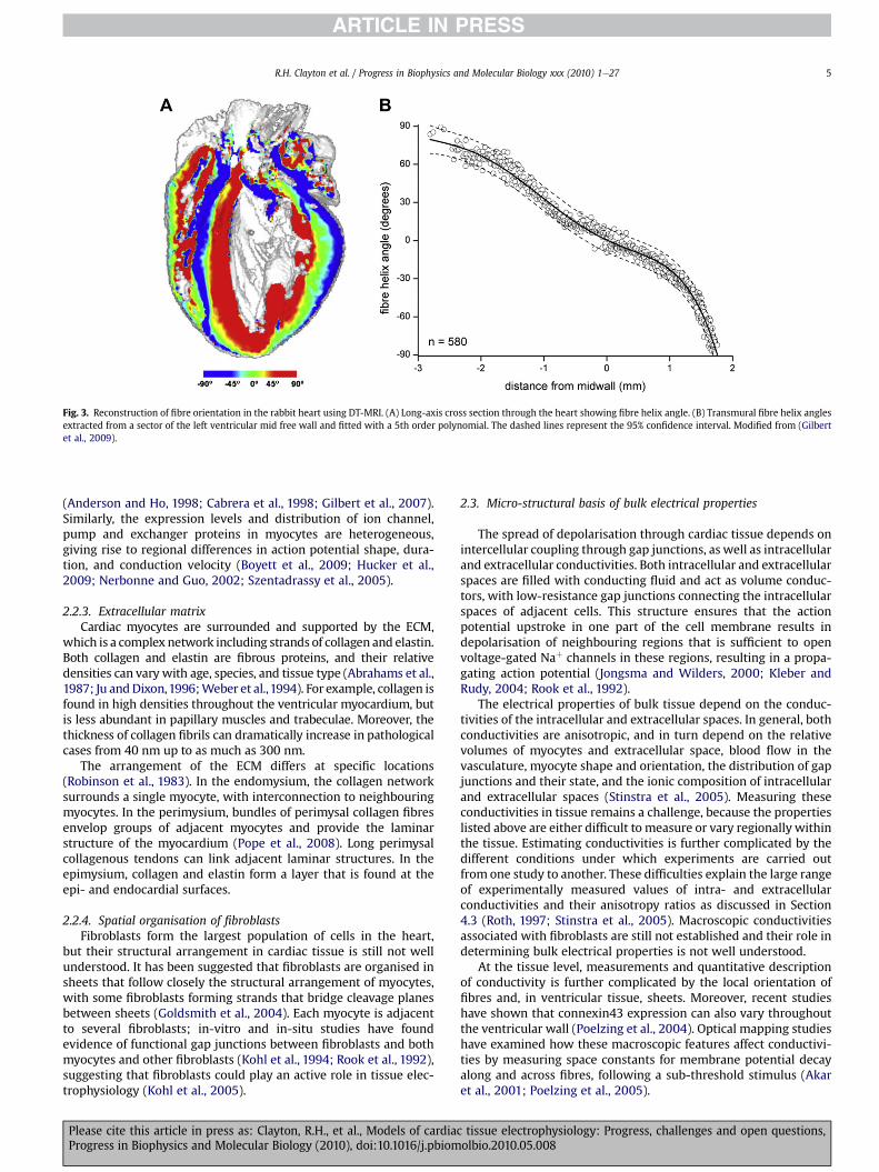
Fig. 3. Reconstruction of fibre orientation in the rabbit heart using DT-MRI. (A) Long-axis cross section through the heart showing fibre helix angle. (B) Transmural fibre helix anglesextracted from a sector of the left ventricular mid free wall and fitted with a 5th order polynomial. The dashed lines represent the 95% confidence interval. Modified from (Gilbertet al., 2009).
R.H. Clayton et al. / Progress in Biophysics and Molecular Biology xxx (2010) 1e27 5
(Anderson and Ho, 1998; Cabrera et al., 1998; Gilbert et al., 2007).Similarly, the expression levels and distribution of ion channel,pump and exchanger proteins in myocytes are heterogeneous,giving rise to regional differences in action potential shape, dura-tion, and conduction velocity (Boyett et al., 2009; Hucker et al.,2009; Nerbonne and Guo, 2002; Szentadrassy et al., 2005).
2.2.3. Extracellular matrixCardiac myocytes are surrounded and supported by the ECM,
which is a complex network including strands of collagen and elastin.Both collagen and elastin are fibrous proteins, and their relativedensities can vary with age, species, and tissue type (Abrahams et al.,1987; Ju andDixon,1996;Weber et al.,1994). For example, collagen isfound in high densities throughout the ventricular myocardium, butis less abundant in papillary muscles and trabeculae. Moreover, thethickness of collagen fibrils can dramatically increase in pathologicalcases from 40 nm up to as much as 300 nm.
The arrangement of the ECM differs at specific locations(Robinson et al., 1983). In the endomysium, the collagen networksurrounds a single myocyte, with interconnection to neighbouringmyocytes. In the perimysium, bundles of perimysal collagen fibresenvelop groups of adjacent myocytes and provide the laminarstructure of the myocardium (Pope et al., 2008). Long perimysalcollagenous tendons can link adjacent laminar structures. In theepimysium, collagen and elastin form a layer that is found at theepi- and endocardial surfaces.
2.2.4. Spatial organisation of fibroblastsFibroblasts form the largest population of cells in the heart,
but their structural arrangement in cardiac tissue is still not wellunderstood. It has been suggested that fibroblasts are organised insheets that follow closely the structural arrangement of myocytes,with some fibroblasts forming strands that bridge cleavage planesbetween sheets (Goldsmith et al., 2004). Each myocyte is adjacentto several fibroblasts; in-vitro and in-situ studies have foundevidence of functional gap junctions between fibroblasts and bothmyocytes and other fibroblasts (Kohl et al., 1994; Rook et al., 1992),suggesting that fibroblasts could play an active role in tissue elec-trophysiology (Kohl et al., 2005).
Please cite this article in press as: Clayton, R.H., et al., Models of cardiacProgress in Biophysics and Molecular Biology (2010), doi:10.1016/j.pbiom
2.3. Micro-structural basis of bulk electrical properties
The spread of depolarisation through cardiac tissue depends onintercellular coupling through gap junctions, as well as intracellularand extracellular conductivities. Both intracellular and extracellularspaces are filled with conducting fluid and act as volume conduc-tors, with low-resistance gap junctions connecting the intracellularspaces of adjacent cells. This structure ensures that the actionpotential upstroke in one part of the cell membrane results indepolarisation of neighbouring regions that is sufficient to openvoltage-gated Naþ channels in these regions, resulting in a propa-gating action potential (Jongsma and Wilders, 2000; Kleber andRudy, 2004; Rook et al., 1992).
The electrical properties of bulk tissue depend on the conduc-tivities of the intracellular and extracellular spaces. In general, bothconductivities are anisotropic, and in turn depend on the relativevolumes of myocytes and extracellular space, blood flow in thevasculature, myocyte shape and orientation, the distribution of gapjunctions and their state, and the ionic composition of intracellularand extracellular spaces (Stinstra et al., 2005). Measuring theseconductivities in tissue remains a challenge, because the propertieslisted above are either difficult to measure or vary regionally withinthe tissue. Estimating conductivities is further complicated by thedifferent conditions under which experiments are carried outfrom one study to another. These difficulties explain the large rangeof experimentally measured values of intra- and extracellularconductivities and their anisotropy ratios as discussed in Section4.3 (Roth, 1997; Stinstra et al., 2005). Macroscopic conductivitiesassociated with fibroblasts are still not established and their role indetermining bulk electrical properties is not well understood.
At the tissue level, measurements and quantitative descriptionof conductivity is further complicated by the local orientation offibres and, in ventricular tissue, sheets. Moreover, recent studieshave shown that connexin43 expression can also vary throughoutthe ventricular wall (Poelzing et al., 2004). Optical mapping studieshave examined how these macroscopic features affect conductivi-ties by measuring space constants for membrane potential decayalong and across fibres, following a sub-threshold stimulus (Akaret al., 2001; Poelzing et al., 2005).
tissue electrophysiology: Progress, challenges and open questions,olbio.2010.05.008

Fig. 4. Schematic representation of wave-front of different curvature and directions oflocal currents.
R.H. Clayton et al. / Progress in Biophysics and Molecular Biology xxx (2010) 1e276
3. Action potential propagation
Despite the complex structure described above, at the macro-scopic scale cardiac tissue behaves as a functional syncytium,supporting propagating waves of depolarisation and repolarisation.The biophysics of this process has been reviewed extensivelyelsewhere (Kleber and Rudy, 2004; Plonsey and Barr, 2000). In thissection we examine this continuum approximation and review theproperties of action potential propagation in 1-, 2-, and 3-D.
3.1. Continuum approximation
At the cellular scale, there is a delay between the depolarisation ofa myocyte and its neighbours that has been attributed to the effect ofgap junctions. This is evidence that at this scale action potentialpropagation is a discrete process (Kleber and Rudy, 2004). However, atlarger spatial scales depolarisation appears to propagate smoothly(Durrer et al.,1970). A common and important assumption underlyingcardiac tissue electrophysiology is that in some cases the discretenature of cardiac propagation may be neglected, and propagationcan be considered as continuous, and this leads to a simplifiedmathematical description of the tissue (see Section 4). Support for thisassumption comes from experiments in cultured neonatal ratventricular cells. In 1-D strands of these cells with a single-cell thick-ness, gap junction delaywas found to be around 118 ms, accounting forabout half of the total conduction time.However, in strands containingseveral cell layers this delay was smaller and amounted to only 22% ofthe total conduction time (Fast and Kleber, 1993).
3.2. 1-D propagation
In the continuous limit, 1-D propagation is characterised bya single parameter, the conduction velocity (CV). This parameter isdetermined by many factors, the most important of them beingmembrane excitability (mainly depending on the magnitude of thefast Naþ current, see Table 1) and the conductivities of cardiac tissue.Typical values of CV measured longitudinal to the cell axis arebetween 1.7 and 2.5 m s�1 in the conduction system, and between0.48 and 0.61 m s�1 in the ventricles (Kleber and Rudy, 2004). Bothlowering excitability and decreasing tissue conductivity result inreduction of CV and may therefore cause propagation block. CV alsodepends on the degree to which cardiac tissue has repolarised, anddecreases as pacing cycle length shortens, a feature known as CVrestitution. At cycle lengths of longer than about 1.5 times the actionpotential duration (APD), CV approaches a maximal saturated value.At shorter cycle lengths, CV usually decreases gradually because thedepolarisation wavefront encounters tissue that has not fully repo-larised. In some cases CV increases as cycle length is decreased.This phenomenon is called supernormality, and has been observedin Purkinje fibres, in some cardiac cell cultures, or under certainconditions (Chialvo et al., 1990; Endresen and Amlie,1989; Endresenet al., 1987). The slope of the CV restitution curve is an importantcharacteristic of myocardium, which is believed to influence theonset of dynamical heterogeneity in the heart (Mironov et al., 2008).
3.3. 2-D propagation
In 2-D, the 1-D features of cardiac action potential propagationare augmented by the effects of tissue anisotropy and wavefrontcurvature.
Anisotropic propagation results from the organisation of cardiacmyocytes into fibres as described in Section 2. Longitudinal prop-agation along the principal axis of a fibre is faster than transversepropagation, resulting in axially symmetric anisotropy (Girouardet al., 1996). Since most gap junctions are located at the ends of
Please cite this article in press as: Clayton, R.H., et al., Models of cardiaProgress in Biophysics and Molecular Biology (2010), doi:10.1016/j.pbiom
myocytes, gap junction coupling in the transverse direction islimited (Hoyt et al., 1989). Both these factors contribute to anisot-ropy; however, amodelling study by Spach et al. (Spach et al., 2000)showed that myocyte shape is as important (or more so) as changesin gap junction distribution. Anisotropic propagation is charac-terised by two principal values of CV: a longitudinal CV parallel tofibres and a transverse CV orthogonal to fibres.
The second important characteristic of 2-D propagation iscurvature. Fig. 4 displays three typical shapes of a wavefront: plane,convex and concave. The effects of curvature on wave propagationcan be explained by the following simple arguments. A planewavefront conserves its length locally and thus each depolarisedcell needs to depolarise only one cell in front of it. In contrast thelength of a convex wavefront steadily increases, and therefore thecurrent initiating depolarisation spreads to a larger area than thatfor a plane front. As a result, convex fronts propagate more slowlythan a plane front. Conversely, a concave front decreases its lengthduring propagation, resulting in faster propagation. The depen-dency of velocity on curvature is an important factor determiningnormal and abnormal (re-entrant) wave propagation in cardiactissue. Curvature effects are often also referred to as current-to-loador source-sink mismatch and may be important for propagationthrough Purkinje-muscle junctions.
Direct experimental measurement of the relationship betweenCV and curvature in real cardiac tissue remains difficult because ofregional differences in CV and restitution (Nash et al., 2006; Yueet al., 2005), and because both curvature and anisotropy affectthe velocity of wave propagation (Bernus et al., 2004). Fig. 5 showsan example of an indirect study of the curvature effects (Cabo et al.,1994), showing that the propagation velocity of a wave througha thin isthmus decreases as the isthmus width decreases. Numer-ical experiments performed by Cabo et al. confirmed that thedecrease in velocity could largely be ascribed to the effect ofwavefront curvature. They also showed that the radius of curvatureof the wavefront could be estimated as half of the isthmus width.Therefore, the plot of dependency of velocity on the inverseisthmuswidth shown in Fig. 5 represents the influence of curvatureon the velocity of wave propagation. Cabo et al. also founda particular size of the isthmus for which wave propagation wasblocked, varying from 0.5 mm to 2.7 mm depending on thestimulation frequency. Based on these data, the minimal radius ofa semicircular propagating wave is between 0.25 and 1.3 mm, andwaves with a greater curvature cannot propagate. These figures areconsistent with earlier estimates of 0.2 mm for critical radius, basedon stimulation of cardiac tissue by electrodes of different sizes(Lindemans and Zimmerman, 1979).
c tissue electrophysiology: Progress, challenges and open questions,olbio.2010.05.008

R.H. Clayton et al. / Progress in Biophysics and Molecular Biology xxx (2010) 1e27 7
3.4. 3-D propagation
Propagation in 3-D is influenced by tissue anisotropy andcurvature in a similar way to 2-D propagation. There is emergingevidence that 3-D propagation is modulated by the fibre-sheetstructure of cardiac tissue (see Section 2.2.1), which extends axiallysymmetric anisotropic propagation with two principal values of CVto orthotropic propagation with three principal values of CV in thefibre direction, normal to fibre direction and in the sheet plane, andnormal to the sheet plane. High-density intramural electricalmapping of active wave propagation in porcine ventricular tissue(Caldwell et al., 2009) has shown clear evidence of orthotropicpropagation, with local conduction velocities of 0.67, 0.3, and0.17 m s�1 in a ratio of 4.3:1.8:1.0 along the three main directionscoinciding with the local micro-structure directions.
4. Mathematical description of cardiac tissueelectrophysiology
Models of cardiac tissue electrophysiology encode informationabout excitability at the cell level and electrical conduction at thetissue level to enable quantitative description of action potentialpropagation. In discretemodels the granular nature of cardiac tissueis characterised by an explicit representation of individual cells,whereas in continuous models cardiac tissue is treated as a func-tional syncytium. Both approaches involve a choice of parameters aswell as a description of tissue geometry. In this section we reviewdifferent types of cardiac tissue model, and discuss the choice ofparameters and tissue geometry.
4.1. Models of discrete cardiac tissue
Mathematical descriptions of discrete cardiac tissue includesimple cellular automaton (CA) models, coupled map lattices (CML)(Holden and Zhang, 1993), and lattices of coupled ordinary differ-ential equations (or CODE lattice) (Winslow et al., 1993).
Cellular automata (CA) are simplified descriptions of cardiactissue, in which each cell has a finite number of states. At each time
Fig. 5. Relation between the isthmus width and the velocity of the wavefront. Experimentsof the isthmus (A) and for isthmus widths of 2.26 mm (B) and 0.88 mm (C). D conduction v
Please cite this article in press as: Clayton, R.H., et al., Models of cardiacProgress in Biophysics and Molecular Biology (2010), doi:10.1016/j.pbiom
step the state of each cell is updated to a new state that depends onits previous state and the state of its neighbours. This simplicityenabled CA models of cardiac action potential propagation to beformulated in mathematical terms (Wiener and Rosenblueth, 1946)and implemented on some of the earliest computers (Moe et al.,1964). CA updating rules have been developed to generatesmoothly curved wavefronts that can take into account the effectsof wavefront curvature described in Section 3.3 above (Bub et al.,2002; Gerhardt et al., 1990), and the effects of tissue anisotropy(Hall Barbosa, 2003). CA models are computationally cheap toimplement, and have been used to examine the properties of spiralwaves and in particular the effects of tissue heterogeneity (Bubet al., 2002; Greenberg and Hastings, 1978; Moe et al., 1964;Smith and Cohen, 1984). However, a major limitation of a CAapproach is the discrete states that each cell can occupy, whichmakes it difficult to implement rate-dependent effects.
Coupled map lattices (CML) are a development of a CA approachin which states are continuous, but are updated by interactionswithin a lattice (Waller and Kapral, 1984). Each interaction can beallocated a different coupling strength, enabling anisotropic prop-agation to be modelled (Holden and Zhang, 1993).
A further refinement of the CML approach is to couple the ordi-nary differential equations (ODEs) describing the kinetics of anindividual cell using resistors that represent gap junction connec-tions. This type ofmodel has been used to examine propagation in 1Dfibres (Rudy, 1995; Shaw and Rudy, 1995, 1997b; Viswanathan et al.,1999) and 2D sheets (Winslow et al., 1993). A variant of thisapproach is tomodel cardiac tissue as a network of connected cables,which can be used to represent 3D tissue anisotropy (Vigmond andLeon, 1999). A particular strength of these approaches is that it ispossible to construct detailedmodels of tissue architecture at the celllevel, so that the effect of discrete gap junction conductances andcellular anisotropy can be studied (Roberts et al., 2008; Spach et al.,1992, 2000; Stinstra et al., 2006). However, without careful optimi-sation these approaches can be computationally expensive (Vigmondand Leon, 1999) and they also require description of cell size andcapacitance, the composition of the extracellular space, as well asdata on the location and conductance of individual gap junctions.
in sheep myocardium. Fibres are directed horizontally. Isochronal maps before creationelocity vs inverse isthmus width. Reproduced from (Cabo et al., 1994) with permission.
tissue electrophysiology: Progress, challenges and open questions,olbio.2010.05.008

R.H. Clayton et al. / Progress in Biophysics and Molecular Biology xxx (2010) 1e278
4.2. Continuous approximation of cardiac tissue
At the tissue scale cardiac tissue behaves as a functionalsyncytium of electrically coupled cells (Section 3.1). A homogeni-sation of the discrete representation of cardiac tissue as a resistornetwork can be applied to derive a continuous description (Neu andKrassowska, 1993), and its idealised electrical behaviour may beconsidered as an excitablemedium in 1-, 2-, or 3-D, where excitablecells are coupled diffusively via transmembrane voltage Vm (Keenerand Sneyd, 1998)
4.2.1. Bidomain modelBidomain models represent cardiac tissue as a syncytium
composed of intracellular and extracellular domains. It is assumedthat both domains are overlapping and continuous, but separatedby the cell membrane. The bidomain model of cardiac tissue isbased on current flow, distribution of electrical potential and theconservation of charge and current (Henriquez, 1993).
The description of each domain is based on a generalisedversion of Ohm’s law defining the relationship between the electricfield E (in V m�1) derived from the potential fðVÞ, the currentdensity J (A m�2), and the conductivity tensor G (S m�1):
E ¼ �VfJ ¼ G E ¼ �G Vf
(1)
Considering the intracellular and extracellular spaces specifi-cally, we have:
Ji ¼ �GiVfiJe ¼ �GeVfe
(2)
Where Ji and Je are the intra- and extracellular current densities, Giand Ge are intra- and extracellular conductivity tensors respec-tively, and fi and fe are the electrical potential in the intracellularand extracellular spaces.
Considering the conservation of current and charge, andassuming only membrane related sources in the intra- and extra-cellular spaces we can write divergence equations:
V$Ji ¼ �Im; V$Je ¼ ImV$ðJi þ JeÞ ¼ 0; (3)
where Im (A m�3) is transmembrane current per unit volume,which is composed of a capacitive component with units Am�2 andan ionic component iion resulting from current flow through ionchannels, pumps and exchangers in the cell membrane, with unitsA m�2.
Im ¼ bm
�Cm
dVm
dtþ iion
�; (4)
Here bm (m�1) is the surface area-to-volume ratio of a cardiaccell, Cm (F m�2) the specific cell membrane capacitance, and Vm thetransmembrane voltage, which is given by:
Vm ¼ fi � fe (5)
Combining Equations (2)e(5), we obtain:
V$GiðVVm þ VfeÞ ¼ bm
�Cm
vVmvt þ iion
�V$ððGi þ GeÞVfeÞ ¼ �V$ðGiVVmÞ
(6)
Equation (6) represents the bidomain model of cardiac tissue.Assuming that the extracellular space is bounded and there is noelectric current flowing from the extracellular space to adjacentspaces homogeneous Neuman (no-flux) boundary conditions canbe implemented at the boundary G as:
Please cite this article in press as: Clayton, R.H., et al., Models of cardiaProgress in Biophysics and Molecular Biology (2010), doi:10.1016/j.pbiom
G : n$ðGiVfiÞ ¼ n$ðGiVðVm þ feÞÞ ¼ 0G : n$ðGeVfeÞ ¼ 0;
(7)
where n is the outward normal to the boundary G.The conductivity tensors in Equation (5) (Gi and Ge) are deter-
mined by the anisotropy of cardiac tissue, and their componentsdepend on the tissue conductivities as well as the local orientationof tissue within the coordinate system of the model.
4.2.2. Monodomain modelThe bidomain model of cardiac tissue (Equation (6)) can be
simplified by assuming that the anisotropy of the intracellularand extracellular spaces is the same, i.e. that the conductivity inthe extracellular space is proportional to the intracellularconductivity:
Ge ¼ lGi (8)
where l is a scalar, representing the ratio between the conductivityof the intra- and extracellular spaces.
Substituting equation (8) into equation (6), we have:
V$l
1þ lGiVVm ¼ bm
�Cm
vVm
vtþ iion
�(9)
If we introduce an effective conductivity G ¼ ðlÞ=ð1þ lÞGi, weobtain the monodomain model of cardiac tissue as:
V$G VVm ¼ bm
�Cm
vVm
vtþ iion
�; (10)
With the no-flux boundary condition:
G : n$ðG VVmÞ ¼ 0 (11)
The monodomain equation (10) is often written as.
vVm
vt¼ V$DVVm � iion
Cm(12)
where D (m2 s�1) is a diffusion tensor or scalar diffusion coefficient.In the case of axially symmetric anisotropy, where diffusion in alldirections orthogonal to the fibre direction is assumed to be thesame, propagation is described by two values of the diffusioncoefficient; a longitudinal coefficient D1 for propagation alongfibres, and a transverse coefficient D2 for propagation orthogonal tothe fibres. If the fibre direction is given by the vector f, the diffusiontensor can be written:
D ¼ D2Iþ ðD1 � D2ÞffT (13)
where I is the identity matrix, and fT is the transpose of f. Theelements ofD can bewritten as follows (Panfilov and Keener,1995):
dij ¼�D2 þ ðD1 � D2Þfifj ði ¼ jÞðD1 � D2Þfifj ðisjÞ (14)
For orthotropic anisotropy with principal directions longitudinal tofibres in the sheet plane, normal to fibres in the sheet plane, andnormal to the sheet plane, the diffusion tensor is given by (Colli-Franzone et al., 2005):
D ¼ D1ffT þ D2ss
T þ D3nnT (15)
where D1, D2 and D3 are diffusion coefficients longitudinal to fibres,normal to fibres in the sheet plane, and normal to both fibres andsheets, and f, s and n are unit vectors in the corresponding direc-tions. Since f, s and n are orthonormal
c tissue electrophysiology: Progress, challenges and open questions,olbio.2010.05.008

R.H. Clayton et al. / Progress in Biophysics and Molecular Biology xxx (2010) 1e27 9
ffT þ ssT þ nnT ¼ I; (16)
where I is the identitymatrix. Hence equation (15) can be rewritten as
D ¼ D2I þ ðD1 � D2ÞffT þ ðD3 � D2ÞnnT ; (17)
which reduces to equation (13) for axially symmetric anisotropywhere D3 ¼ D2.
4.2.3. Comparison between bidomain and monodomain modelsIf there is no injection of current into the extracellular space,
descriptions of action potential propagation provided by mono-domain and bidomain models are close to each other even under thecondition of unequal anisotropy ratio in the extracellular and intra-cellular spaces (Colli-Franzone et al., 2005). A recent study (Potse et al.,2006) compared patterns of action potential propagation simulatedusing monodomain and bidomain models. They found that in theabsence of external stimuli, the patterns obtained with the mono-domain model were almost identical to those obtained with a bido-main model. Similarly, Roth examined spiral wave tip trajectories inmonodomain and bidomain models and found that in most cases thetrajectories were similar in both cases (Roth, 2001). Themonodomainmodel is a single PDE, andnumerical solutions are easier to obtain (seeSection 6). However, the bidomain model provides a more detaileddescription of cardiac tissue, and the separation of intracellular andextracellular spaces is necessary to accommodate the injection ofcurrent into the extracellular space during external stimulation anddefibrillation (Trayanova, 2006). During defibrillation, the unequalanisotropy of the intracellular and extracellular spaces plays animportant role in generating virtual electrodes that are essential forsuccessful defibrillation (Wikswo et al., 1995). The bidomain modelcan be expanded to include further domains, and one example of thisapproach is a model that includes an additional domain representingfibroblasts (Sachse et al., 2009).
4.3. Parameters
The key parameters that determine the conduction properties ofa tissue model are elements of the effective diffusion tensor or thediffusion coefficient, which in turn depend on the tissue conduc-tivities, surface-to-volume ratio and specific capacitance (Winfree,1998).
As described in Section 2.3, the intracellular and extracellularconductivities are determined by the local tissue micro-structureand the composition of the intracellular and extracellular spaces,which vary within the tissue and are further modified by local bloodflow. Measurements of longitudinal conductivity span the rangeof 0.17e0.45 Sm�1 for intracellular space, and 0.12e0.62 Sm�1 forextracellular space; corresponding measurements for transverseconductivity range from 0.019 to 0.06 Sm�1 for intracellular spaceand 0.08e1.74 Sm�1 for extracellular space (Stinstra et al., 2005).Typically, the values for conductivities chosen for simulation studieslie within these ranges, and result in a plausible CV (Colli-Franzoneet al., 2005). However, detailed models of cardiac micro-structuredeveloped with the purpose of reconstructing the effect of cardiacmicro-structure on conductivity indicate that there may not bedefinitive values of conductivity, but rather a range of typical valueswith local variation (Stinstra et al., 2005).
Experimental measurements of surface-to-volume ratio rangefrom 2400 to 8900 cm�1 depending on species and developmentalstage (Bers, 2008). Typical values chosen for tissue models rangefrom 1000 to 5000 cm�1 (Colli-Franzone et al., 2005; Keener andBogar, 1998; TenTusscher et al., 2004; Xu and Guevara, 1998),which for an idealised cylinder correspond to a value of around 2divided by the cell radius (Winfree, 1998).
Please cite this article in press as: Clayton, R.H., et al., Models of cardiacProgress in Biophysics and Molecular Biology (2010), doi:10.1016/j.pbiom
Specific membrane capacitance is usually measured to be in therange 1e10 mF cm�2. This value combined with typical values ofconductivities and cellular dimensions gives CV within the physio-logical range. Themembrane capacitance of the squid giant axonwasestimated to be about 1 mF cm�2 based onmeasurements of the timecourse of membrane current following a sub-threshold voltage step(Curtis and Cole, 1938). However, similar studies in cardiac Purkinjefibres yielded a higher value of around 10 mF cm�2 (Fozzard, 1966;Schoenberg et al., 1975). This higher value can be attributed to twocomponents of the membrane capacitance, a surface componentassociated with the outer sarcolemma and a deep componentassociatedwith clefts in the cell membrane (Fozzard,1966). The cleftcapacitance charges only slowly, and so is thought to make onlya small contribution to actionpotential propagation. Since about 90%of the ventricular cell membrane is located within clefts and thet-tubule system, a specific membrane capacitance of 1 mF cm�2 isjustified (Noble, 1979) although some tissue models have useda higher value of 2 mF cm�2 (TenTusscher et al., 2004).
A common approach taken to determine suitable parametervalues for a computational model is to vary the conductivities (ordiffusion coefficients) to ensure that CVs within the observed rangeare achieved. However, because these parameters have an impor-tant effect not only on CV but also on features such as conductionblock, a more rigorous approach based on experimental work isneeded to determine the range of suitable values and how, forexample, these are modified by disease processes.
4.4. Tissue geometries and imaging data
Models of cardiac tissue can be implemented with either ide-alised geometries (1-D strand, 2-D sheet or 3-D box), or anatomi-cally detailed geometries based on reconstructions from dissectionor imaging data (2-D slice, 3-D slab or whole organ). A simplifiedgeometry enables propagation to be studied in the absence ofanatomical detail, whereas more detailed geometrical models withhigh spatial resolution enable the role of anatomical structures tobe evaluated.
Based on various experimental techniques that include histology,confocal microscopy, magnetic resonance imaging (MRI) and diffu-sion tensor MRI (DT-MRI), realistic anatomic structures of cardiactissue with high spatial resolutions have been reconstructed forsingle myocytes (Savio-Galimberti et al., 2008), the sino-atrial node(Dobrzynski et al., 2005), atrial tissue (Lasher et al., 2009), thewholeatrium (Seemann et al., 2006), and ventricle (Hsu et al.,1998; Nielsenet al., 1991). Furthermore anatomically detailed models of canine(Nielsen et al., 1991), rabbit (Vetter and McCulloch, 1998), pig(Stevens and Hunter, 2003) and mouse (Sampson and Henriquez,2005) ventricular anatomy defined on a finite-element mesh havebeen constructed, aswell as highly detailedmodels of small portionsof ventricular tissue imaged at spatial resolutions of around 1 mm(Pope et al., 2008; Sands et al., 2005). Whole ventricle anatomicalmodels combining histology data at resolutions of around 20 mmwithMRI data at a resolution of 100 mmare being developed (Gilbertet al., 2009; Plank et al., 2009), alongwith whole heart models usingMRI data at a resolution of 120 mm (Cherry and Fenton, 2008).
Diffusion tensor MRI (DT-MRI) provides a non-invasive tool toreconstruct the anatomical structures and fibre orientation ofcardiac tissue, especially in the ventricles. This technique measuresthe Brownian motion of protons, which reflects to some extentthe fibre structure of the tissue because the motion of protons isconstrained by the cell membrane. The use of this approach forventricular tissue has been validated by its correlation with histo-logical data (Holmes et al., 2000; Hsu et al., 1998; Scollan et al.,2000). DT-MRI provides a diffusion tensor with its primary eigen-vector being correlated to cardiac fibre orientation, whilst the
tissue electrophysiology: Progress, challenges and open questions,olbio.2010.05.008

Table 1Maximum conduction velocity (CVmax), maximum upstroke velocity (dv/dtmax), andmaximum conductance sodium current (gNa) in tissue for 14 different models, inorder of decreasing CVmax. Although overall CVmax and gNa decreases as dv/dtmax
decreases, neither value can be used alone to predict CVmax. The value of gNa is notavailable for the Nygren et al. model because it uses the Goldman-Hodgkin-Katzformulation for INa that uses membrane permeability rather than conductance. In allcases, the explicit Euler method is used with a spatial resolution of 0.01 cm, timestep of 0.01 ms, and diffusion coefficient of 0.001 cm2/ms. CV and dv/dtmax aremeasured at the center of a 1 cm-long cable paced at a cycle length of 1000 ms.
Model Reference CVmax
(cm/s)dv/dtmax
(V/s) intissue
gNa(nS/pF)
Luo-Rudy I (Luo and Rudy, 1991) 64.7 275 23Shannon et al. (Shannon et al., 2004) 61.8 263 16Iyer et al. (Iyer et al., 2004) 60.1 266 56.32Faber-Rudy (Faber and Rudy, 2000) 59.4 246 16ten Tusscher et al. (TenTusscher
et al., 2004)59.4 227 14.838
Priebe-Beuckelmann (Priebe andBeuckelmann,1998)
58.7 247 16
Mahajan et al. (Mahajan et al., 2008) 52.2 172 12Fox et al. (Fox et al., 2002) 51.3 196 12.8Courtemanche et al. (Courtemanche
et al., 1998)50.4 127 7.8
Beeler-Reuter (Beeler andReuter, 1977)
47.6 110 4
Hund-Rudy (Hund and Rudy, 2004) 47.3 133 8.25Pandit et al. (Pandit et al., 2001) 45.1 103 8Nygren et al. (Nygren et al., 1998) 39.5 83 n/aBondarenko et al. (Bondarenko et al., 2004) 39.1 94 13
R.H. Clayton et al. / Progress in Biophysics and Molecular Biology xxx (2010) 1e2710
secondary and tertiary eigenvectors being have been proposed torelate to the orientation of cardiac sheets (Helm et al., 2005).However, the typical spatial resolution available with DT-MRI maynot be sufficient to correctly identify the sheet structure in ventric-ular tissue because there may be more than one sheet orientationwithin the volume of tissue that produces the DT-MRI signal (Gilbertet al., 2007).
At the cell scale there are often discontinuities in tissue structure,especially at the junctions between two distinctive tissue regions,such as at the junction of the crista terminalis and the pectinatemuscle in the right atrium, the junction of the right ventricular freewall and ventricular septum, and Purkinje-ventricular junctions.At these locations fibre orientation changes abruptly, and thesechanges not only complicate the process of obtaining a mesh fromDT-MRI data, but may also require special numerical treatment.
5. Integration of cell and tissue models of cardiacelectrophysiology
As described above, models of tissue electrophysiology integrateindividual cells together into a given structure, where each cell iselectrically connected tomultiple neighbours. The coupling betweencells is generally represented as a diffusive process, which gives riseto an electrotonic (diffusive) current between neighbouring cells.This electrotonic current can modify cellular electrophysiology bothquantitatively andqualitatively in both real tissue andmodels. In thissection, we present some of the issues associated with simulatingcardiac tissue electrophysiology and discuss their implications.
5.1. Emergent properties in tissue
When individual cells are coupled together to form tissue,new properties that have no single-cell equivalents emerge. In thissection we describe some of these fundamental properties anddiscuss their importance to tissue behaviour.
5.1.1. Liminal lengthIn a single cell, an action potential can be elicited from a stim-
ulus provided the stimulus current has sufficient magnitude andduration to raise the membrane potential above its threshold ofexcitability. In tissue, neighbouring cells that are initially polarisedact as current sinks and so counteract a stimulus current. Thus,enough current must be injected to raise the membrane potentialof neighbouring cells above threshold, while also ensuring that themembrane potential at the stimulus site is not decreased belowthreshold by electrotonic current flow to polarised neighbours.This requirement introduces a new spatial scale to the stimulationprocess; a large enough region of tissue must be stimulated directlyfor a wave to develop and propagate. Although the length (in 1-D)necessary to initiate propagation depends on the magnitude andduration of the injected current, there is a minimum length belowwhich no combination of stimulus strength and duration canproduce a propagating wave. This minimum length is called theliminal length, which in cardiac tissue is typically of the order of1 mm (Fozzard and Schoenberg, 1972; Noble, 1972; Rushton, 1937).The corresponding concepts of liminal area and liminal volumeapply in 2- and 3-D, respectively. Stimulating a region of size belowthe liminal length (or area or volume) ensures that the stimulus willdissipate without producing a propagating wave, even if the samemagnitude and duration of the stimulus current successfully elicitsan action potential in a single cell with the same electrophysio-logical characteristics. This concept is related to the safety factor forpropagation (Kleber and Rudy, 2004; Shaw and Rudy, 1997a).Because different models may have different thresholds of excit-ability, liminal length is a model-dependent property.
Please cite this article in press as: Clayton, R.H., et al., Models of cardiaProgress in Biophysics and Molecular Biology (2010), doi:10.1016/j.pbiom
5.1.2. Minimum cycle length for propagationAnother important property associated with excitable tissue is
the appearance of a minimum cycle length that can be used toachieve successful wave propagation. This is also known as theeffective refractory period (ERP). In a single cell, any stimulus currentcauses a change of themembrane voltage, evenwithin the refractoryperiod. However, in tissue a response may occur at the site ofstimulation, but neighbouring tissue within the refractory periodwill not be able to sustain awave, and propagationwill fail. As tissueis paced faster, there is less recovery time before the next beat, sothat there is a minimum cycle length at which the tissue can bepaced and below which propagation failure occurs. As an example,Fig. 6 shows the minimum cycle length for propagation in tissuecompared to the minimum cycle length achievable in a single cell(using a stimulus currentmagnitude of twice diastolic threshold) forthe Nygren et al. model for human atrial cells (Nygren et al., 1998)and the Fox et al. model for canine ventricular cells (Fox et al., 2002).It is possible to pace the Nygren et al. model at extremely short cyclelengths in a single cell, whereas when embedded in a tissue modelpropagation fails for cycle lengths below 320 ms. Similarly, the Foxet al. model can be paced at a minimum cycle length of 90 ms fora single cell, but propagation fails for cycle lengths below 190 ms ina tissue model. Because the diastolic interval characterises therecovery time following an action potential, the minimum cyclelength for propagation is associated with the minimum diastolicinterval for propagation. This important property of real tissue maynot be captured by simplified or generic models (see Section 6).
5.1.3. Conduction velocityWith the introduction of propagation in tissue, the property of
CV emerges. CV is related to the strength of cell-to-cell coupling,and scales as the square root of the diffusion coefficient D(see equation (12)). CV is also determined by the characteristics ofthe action potential upstroke. Although the maximum upstrokevelocity (maximum dVm/dt) is loosely correlated with maximumCV, it is not especially useful as a velocity predictor, as can be seen
c tissue electrophysiology: Progress, challenges and open questions,olbio.2010.05.008

Fig. 6. Rate adaptation plots show the minimum cycle length for propagation in tissuecompared to the minimum cycle length in a single cell. For a stimulus amplitude twicediastolic threshold, much smaller cycle lengths can be achieved in single cells (grey,solid) than in tissue (black, dashed) because action potentials do not always propagatein tissue. Dynamical properties therefore can be significantly different in tissue, as inthe Fox et al. model, where the range of cycle lengths exhibiting alternans is reduced intissue and no return to the 1:1 response at short cycle lengths is observed.
Fig. 7. Effects of cell coupling on action potential amplitude. In single cells (grey, solid),depolarizing current is applied directly to the cell. In tissue away from the stimulus site(black, dashed), the stimulating current is mediated through the electrotonic currentcoupling neighbouring cells. Because the electrotonic current includes contributionsfrom neighbours not yet depolarised, action potential amplitudes in tissue aregenerally smaller than those in single cells. Action potentials are shown for a cyclelength of 500 ms for the Pandit et al. and Shannon et al. models and for a cycle lengthof 1000 ms for the Luo-Rudy I, Priebe-Beuckelmann, and Courtemanche et al. models.
R.H. Clayton et al. / Progress in Biophysics and Molecular Biology xxx (2010) 1e27 11
in Table 1. For example, the CV obtained for the Fox et al. model forcanine ventricular cells (Fox et al., 2002) is only 8 percent largerthan that obtained with the Hund-Rudy model for canine ventric-ular cells (Hund and Rudy, 2004); however, maximum dVm/dt forthe Fox et al. model is 47 percent larger than that of the Hund-Rudymodel. Similarly, the CV obtained using the ten Tusscher et al.model for human ventricular cells (TenTusscher et al., 2004) is thesame as the CV obtained with the Faber-Rudy model of guinea-pigventricular cells (Faber and Rudy, 2000), with a dVm/dtmax that is 8percent smaller; and the CVs of the Nygren et al. (Nygren et al.,1998) and Bondarenko et al. (Bondarenko et al., 2004) modelsdiffer by only 1 percent, although dVm/dtmax is 13 percent larger forthe Bondarenko et al. model. In addition, the CV of the Courte-manche et al. model for human atrial cells (Courtemanche et al.,1998) is 7 percent larger than that of the Hund-Rudy model,whereas maximum dVm/dt is actually 5 percent smaller for theCourtemanche et al. model. The value of maximumdVm/dt has beenshown to depend not only on membrane kinetics but also onelectrotonic currents from neighbouring cells (Spach et al., 1992).CV also is correlated loosely with the maximum Naþ conductancegNa, with exceptions such as the relatively high conductance valuesfor the Iyer et al. (Iyer et al., 2004) and Bondarenko et al.(Bondarenko et al., 2004) models and the relatively low value forthe Beeler-Reuter model (Beeler and Reuter, 1977).
5.2. Electrotonic current-mediated differences in dynamics
The flow of current within the tissue resulting from regionaldifferences in potential can have an important influence on localdynamics and excitation.
5.2.1. Decreased action potential amplitude and shapeWhen an action potential propagates in tissue, cells outside the
stimulus region are brought above the excitability threshold purelythrough electrotonic currents from neighbouring depolarised cells.In addition, neighbouring cells that have not yet been depolarisedremove current from cells that are beginning to depolarise. Asa result, the action potential amplitude in tissue is generally reducedcompared to the amplitude in single cells (or in tissue regions wherea stimulating current is injected directly). Fig. 7 shows decreasedaction potential amplitude in tissue compared to single cells for five
Please cite this article in press as: Clayton, R.H., et al., Models of cardiacProgress in Biophysics and Molecular Biology (2010), doi:10.1016/j.pbiom
different models (Courtemanche et al., 1998; Luo and Rudy, 1991;Pandit et al., 2001; Priebe and Beuckelmann, 1998; Shannon et al.,2004). Amplitude reductions of up to 20% have been observed inmodels (Bueno-Orovio et al., 2008).
The effects of reduced action potential amplitude can havesignificant consequences. Fig. 8 shows how reduced action poten-tial amplitudes in tissue can affect transmembrane currents and theoverall action potential morphology and duration. In this case, thereduction in amplitude leaves the maximum voltage attainedoutside the stimulus region below the threshold for activation ofthe L-type Ca2þ current. As a result, the action potential plateaudevelops later and in a different manner, resulting in actionpotential prolongation away from the stimulus site.
Experimental and modelling studies have shown that electro-tonic current can affect the shape of the action potential. One study(Conrath et al., 2004) showed that in some models of cardiac tissuethe duration of propagating action potential is up to 80 ms shorterthan action potential duration in uncoupled cells. Moreover, earlierexperimental studies on dog hearts showed that the repolarisation
tissue electrophysiology: Progress, challenges and open questions,olbio.2010.05.008

Fig. 8. Effect of the decrease in action potential amplitude in tissue on action potentialmorphology. Within the region directly stimulated (red traces from red locationsindicated at top), the action potential retains a large amplitude, but the amplitude isdecreased by more than 35 mV outside the stimulated region (black traces from blacklocations indicated at top). As a result, the L-type calcium current (iCa,L) is not activatednormally outside the stimulus region because the voltage Vm is insufficient to open thed-gate. Hund-Rudy model (Hund and Rudy, 2004) in a 1.8-cm-long cable with thestimulus applied within the first 0.3 cm.
Fig. 9. Differences in alternans magnitude between single cells (grey, solid) and tissue(black, dashed). In the Fox et al. model, the alternans increases in tissue, while in theMajahan et al. model, the alternans magnitude decreases. Cycle lengths shown are190 ms for the Fox et al. model and 150 ms for the Mahajan et al. model.
R.H. Clayton et al. / Progress in Biophysics and Molecular Biology xxx (2010) 1e2712
phase of the action potential can be modulated by the activationsequence or distance from pacing site (Abildskov, 1976; Osaka et al.,1987). The APD was found to progressively decrease as the wavemoved away from the stimulation site, and this effect was morepronounced in directions transverse to the local fibre orientation(Osaka et al., 1987). Evidence of this negative linear correlationbetween APD and activation time has been found in several animalspecies including humans (Hanson et al., 2009). However, theamount by which APD decreases as a function of activation timemay be species-dependent due to different expressions of ioniccurrents underlying the repolarisation phase, as recently suggestedin a computational study by Sampson and Henriquez (Sampson andHenriquez, 2005).
5.2.2. Changes in restitution, alternans, and memoryRestitution is the rate adaptation of cardiac cells and tissue,
alternans is beat to beat alternation in action potential shape andduration, and memory is the extent to which a particular actionpotential depends on the sequence of preceding beats. In tissue,a number of important dynamical properties associated with resti-tution, alternans, and memory may be altered by the presence ofelectrotonic currents. Fig. 9 shows an example of how alternansproperties can change in tissue. For the Fox et al. model (Fox et al.,2002), the magnitude of alternans (difference between long andshort APDs for one cycle length) is increased in tissue, but 2:1 block
Please cite this article in press as: Clayton, R.H., et al., Models of cardiaProgress in Biophysics and Molecular Biology (2010), doi:10.1016/j.pbiom
occurs at a relatively long cycle length, thereby reducing the range ofcycle lengths over which alternans is experienced. For the Mahajanet al. model (Mahajan et al., 2008), the alternans magnitude isdecreased in tissue over range of cycle lengths that experiencealternans. A number of differences in other properties, includingrestitution curve shape and slope, alternans onset cycle length andmagnitude, and memory amplitude, have been described in detail(Bueno-Orovio et al., 2008; Cherry and Evans, 2008; Cherry andFenton, 2007; Cherry et al., 2008; Nygren et al., 1998; Ten Tusscheret al., 2006).
Electrotonic effects can influence not only the presence andcharacteristics of alternans but also on the stability of re-entrantwaves. As an example, Fig. 10 shows the Fox et al. model in twocases: the original parameter set and with the conductance gKrdoubled. Doubling gKr eliminates alternans in a single cell; however,electrotonic effects cause the alternans to reappear in tissue, similarto the case with original parameter values. In 2-D, the originalparameters give rise to a stable spiral wave, whereas doubling gKrleads to sustained spiral wave breakup from the same initialconditions. The output of models that are intended to be predictiveshould therefore be interpreted carefully. Interventions that appearpromising in single cells may not produce the same results intissue, and indeed may lead to less desirable electrophysiologicalbehaviour.
6. Numerical implementation of cardiac tissue models
This section addresses numerical issues related to modelling ofcardiac tissue electrophysiology; in particular we discuss discretespace and discrete time representations of the continuous
c tissue electrophysiology: Progress, challenges and open questions,olbio.2010.05.008

Fig. 10. Differences in dynamics between the Fox et al. model with original parameters and a modification where the maximal conductance gKr is doubled. With the modification,alternans is eliminated in a single cell (0-D) but reappears in tissue (1-D), and a spiral wave initiated in two-dimensional tissue experiences sustained breakup. 2-D domain is30 cm � 30 cm; both spiral wave simulations are shown after 6.3 s of simulation time.
R.H. Clayton et al. / Progress in Biophysics and Molecular Biology xxx (2010) 1e27 13
bidomain and monodomain equations and the types of mesh usedto represent tissue geometry. Furthermore, we provide an overviewof the techniques used for numerical simulations of tissueelectrophysiology.
6.1. Modelling approaches
As described earlier, models of cardiac tissue electrophysiologyare based on reactionediffusion-systemswhere the reaction processis attributed to the cellular actionpotential, and the diffusionprocessrepresents current flow between cells. Most modelling approachesincluding mono-, bi-, or multidomain models assume that cardiactissue behaves as a functional syncytium (Henriquez and Papazogou,1996; Sachse et al., 2009), and the numerical implementationsdiscussed in this section are related to these approaches.
6.2. Discrete representations of tissue geometry
Discrete models of tissue geometry can be constructed in variousways. Standard spatial models include 2- and 3-D tissue segments,ventricles andwhole heart geometries. Thesemodels are commonlycomposed of discrete volume elements, for example tetrahedra(Bourgault et al., 2003; Vigmond et al., 2002) and hexahedra (ColliFranzone and Pavarino, 2004; Seemann et al., in press). Eachelement type has advantages and disadvantages. For instance,a mesh assembled from uniform cubic voxels can be derived easilyfrom imaging data, but does not reconstruct curved surfaces such asthe epicardium effectively. In contrast, irregular tetrahedral meshescan improve the representation of surfaces, butmesh generation canbe more difficult and the numerical methods associated withirregular meshes can result in higher computational costs.
Common approaches are to construct a regular mesh withuniform elements, or an irregular mesh with elements of uniformtype, but different shape and size. Hybrid meshes, with uniform and
Please cite this article in press as: Clayton, R.H., et al., Models of cardiacProgress in Biophysics and Molecular Biology (2010), doi:10.1016/j.pbiom
non-uniform elements used to represent less important regions, andgrid refinement at critical points, border zones or at areas oflarge gradients in physical fields have been considered. Meshes thataccurately represent complicated tissue anatomy while meeting therequirements of computational methods are difficult to construct,and this area remains an important challenge. Various meshgeneration tools have been developed in other engineering fields,and some of these tools may be useful for generating models oftissue geometry. Recent work aims at providing meshes with highspatial accuracy that are appropriate for the computationalmethodsused in simulation of tissue electrophysiology (Prassl et al., 2009).Adaptive mesh generation during the simulation has also beensuggested to decrease calculation time (Deuflhard et al., 2009) (seeSection 7.4).
6.3. Numerical methods for discrete space
The bidomain (equation (6)) and monodomain (equations (10)and (12)) models are based on Ohm’s law and Poisson’s equationfor stationary electrical currents. In this section, we will focus onnumerical approaches for solving Poisson’s equation, which allowfor a sound description of the anisotropic electrical properties ofcardiac tissue. Mathematically, Poisson’s equation is a set of partialdifferential equations (PDEs), and analytical solutions exist only forspecial cases. In general, numerical techniques implemented oncomputers are used for solving these PDEs. To approximate the PDEs,they are transformed into linear systems of equations (LSE) of theform Axþ b ¼ 0 using discretisation techniques. In cardiac elec-trophysiology, the Finite Difference Method (FDM), the FiniteVolumeMethod (FVM) and the Finite ElementMethod (FEM) are themost commonly applied techniques to transform Poisson’s equationinto LSEs. Less often, the Boundary Element Method, mesh freemethods, and spectralmethods have been used (Bueno-Orovio et al.,2006; Chinchapatnam et al., 2009; Fischer et al., 2000).
tissue electrophysiology: Progress, challenges and open questions,olbio.2010.05.008

R.H. Clayton et al. / Progress in Biophysics and Molecular Biology xxx (2010) 1e2714
The FDM is based on approximating the partial derivatives ofPDEs by difference quotients. The underlying spatial mesh isusually structured, so for example in 3-D the mesh consists ofuniform cubic voxels. The FDM has been applied by many groupsover many years for studies of tissue electrophysiology, for exam-ples see (Potse et al., 2006; Pullan et al., 2005; Seemann et al., inpress). This approach can be generalised for grids with irregularspacing (Trew et al., 2005b). The advantage of the FDM is thestraightforward and easy implementation of the method. Thedisadvantage is that it is difficult to describe smooth surfaces suchas the surface of the heart without steps, so that it becomes difficultto implement boundary conditions.
The FEM provides an alternative way of representing discretespace. Potentially complex cardiac tissue geometry is divided intosmall sub-domains, called “finite elements”, which can be describedand handled in a simplified manner. These finite elements can havenon-uniform size and shape. Inside each element, the solution isapproximated by an interpolation function. The FEM is widely usedbecause of its well-developed theory allowing for variable meshrepresentations and interpolation of material properties. A partic-ular advantage is that complex, curved geometries can be modelledaccurately, see for example (Vigmond et al., 2002). The FEMmethodhas been used by many groups in this research field, especially forsimulations with anatomically detailed geometry. The disadvantageof the FEM is the higher effort compared to the FDM both fordevelopment and running the software.
For the FVM, volume integrals in a PDE involving a divergencecomponent are converted to surface integrals applying the diver-gence theorem. This method is conservative because the fluxentering a volume is identical to the one leaving the neighbouringvolume. The FVM can be applied easily to unstructured meshes. Italso has been used in bidomain modelling (Courdiere and Pierre,2006; Harrild and Henriquez, 1997; Jacquemet and Henriquez,2005; Trew et al., 2005a). Since the method is conservative,a coupling between excitation propagation in the heart and theelectrical field in the body can be easily achieved (Courdiere andPierre, 2006).
The FDM, FVM and FEM applied to Poisson’s equations lead tolarge sparse, symmetric matrices. Methods for solving these aredescribed in Section 6.4 onwards.
Simulations of action potential propagation require boundaryconditions, including permanent conditions at tissue boundaries asdescribed in equations (7) and (11) (usually Neumann or no-fluxboundary conditions) and possibly time dependent boundaryconditions, which can be current or voltage stimuli in the intra- andextracellular space at given points in time. Boundary conditions(Dirichlet boundary conditions) associated with setting potentialscan be applied to the LSE after its generation by changing therespective entries or line and column in the matrix and therespective entry in the right hand side vector. With the bidomainmodel, a permanent zero potential is frequently set in the extra-cellular space at the boundary. For complex tissue geometriesno-flux boundary conditions can be implemented using a phase-field approach (Fenton et al., 2005).
6.4. Implicit, explicit and semi-implicit solutionschemes for discrete time
Explicit, implicit, and semi-implicit (also called IMEX) methodscan be used to solve the equations describing the time dependenceof action potential propagation. The choice of numerical methodinfluences the stability, computational cost and the accuracy of theimplemented model.
Explicit methods have been used extensively (Barr and Plonsey,1984; Henriquez and Plonsey, 1990; Penland et al., 2002; Pollard
Please cite this article in press as: Clayton, R.H., et al., Models of cardiaProgress in Biophysics and Molecular Biology (2010), doi:10.1016/j.pbiom
and Barr, 1991; Roth, 2001; Vigmond et al., 2002), because they areeasy to implement. However, even though the computational cost foreach time step is low in an explicitmethod, the time stepmayneed tobe small to guarantee stability for the diffusion operator (Press et al.,1992); specifically, the condition Dt � Dx2/(2dD) must be satisfied,where d is the system dimension (1, 2, or 3), D is the diffusion coef-ficient, Dt is the time step, and Dx the space step. Implicit schemescan be stable with longer time steps (Bourgault et al., 2003; Hookeet al., 1994; Munteanu et al., 2009; Murillo and Cai, 2004), butrequire solution of a non-linear systemof equations at each time step,and so are more computationally expensive. A good compromisebetween these two methods is a semi-implicit IMEX scheme, inwhich some terms of the equations (e.g., the linear terms) are solvedimplicitly and the remaining terms (e.g., the non-linear terms) aresolved explicitly. The stability constraints of IMEX methods are lessdemanding than fully explicit methods but more than fully implicitmethods. Since the linear terms are usually solved implicitly in anIMEX method, a solution of a linear system is required at each timestep. The IMEXmethods can be classified on the basis of the order ofapproximation they can achieve. First-order methods are the mostcommon (Colli Franzone and Pavarino, 2004; Keener and Bogar,1998), although higher order IMEX methods have been proposed.An extensive stability analysis of IMEX methods for solving thebidomain equations coupled with the FitzHugh-Nagumo kineticmodel can be found in (Ethier and Bourgault, 2008), together witha comparison of computational costs.
6.5. Linear system solvers and preconditioners
Most numerical approaches to solve tissue electrophysiologymodels result in a linear system of equations. Linear solvers can beclassified as direct solvers and iterative solvers (Demmel, 1997).The first group is composed ofmethods that compute the solution ofthe linear system by direct manipulation of the original matrix, likecomputing a factorisation or a Gaussian elimination. In theory, thesemethods can be efficient and produce exact solutions. However,rounding errors can become significant, and exact solutions are notobtained. In addition, these methods require large amounts ofmemory and CPU time for the initial decomposition, although thefactorisation of the matrix needs to be computed only once if thematrix does not change during the time evolution. LU factorisation isone of the most common direct solvers, together with the Choleskyfactorisation for symmetricmatrices. Since one drawback of this kindof approach is that the pattern of the original sparse matrix can befilled during the process, a renumbering strategy can be employed.
The most widely used approach is iterative solving. Approxi-mate solutions of the linear system are computed until a stoppingcriterion (usually an error tolerance) is reached. Most publishedapproaches for discretisation of monodomain and bidomainmodels leads to symmetric matrices. In this case a common optionis the Conjugate Gradient method (CG) or its variations like theBiConjugate Gradient Stabilized (BICGSTAB) method. However insome cases, due to the formulation chosen and numerical schemeimplemented, non-symmetric matrices may arise (Gerardo-Giordaet al., 2009; Pennacchio and Simoncini, 2009) requiring an alter-native method to solve the system. In these cases Krylov subspacesolvers are extensively used, in particular the Generalised MinimalResidual Method (GMRES). We refer to (Saad, 2003) for detailsabout the algorithms mentioned.
To speedup the convergence of iterative solvers, several types ofpreconditioners can be used to transform the initial linear system intoan equivalent system whose associated matrix has better spectralproperties such as a lower condition number or improved clusteringof the Eigenvalues. One common option is to compute an incompleteLU (or an incomplete Cholesky) factorisation, preserving a pattern
c tissue electrophysiology: Progress, challenges and open questions,olbio.2010.05.008

R.H. Clayton et al. / Progress in Biophysics and Molecular Biology xxx (2010) 1e27 15
similar to that of the original matrix, and to use this new matrix asa preconditioner for the system. This approach has been shown tobe effective on serial architectures (Ethier and Bourgault, 2008).A comparison using a parallel architecture has shown that a CG solverpreconditioned by a domain decomposition parallelisation of anincomplete LU factorisation to be twice as fast as the same solverpreconditioned with a Jacobi preconditioner (Potse et al., 2006). Inanother study (Colli Franzone et al., 1998) a CG solver preconditionedby Symmetric Successive Over-Relaxation preconditioner was usedfor solving the eikonal model. In (Gerardo-Giorda et al., 2009) theauthors propose a specific preconditioner for the bidomain problemconsisting of amodel-based block triangular preconditioner obtainedafter a proper reformulation of the monodomain model. Thisapproach reduced the CPU time required by the incomplete LU pre-conditioned CG solver on serial architectures by 50%.
Multigrid methods have been employed for the solution ofmono- and bidomain models. Multigrid methods are based on thecombination of an iterative solver running on fine computationalgrids of the domain and a direct solver running on a coarse grid ofthe same domain (Hackbusch, 1985). The rationale behind thesemethods is that, while the high frequency component of the errorin the solution can be reduced quickly using an iterative solver ona fine grid, the low frequency (smooth) component of the error canbe reduced efficiently by using a direct solver on a coarse grid.. Byrepeating these steps both the high and the low frequencies of theresidual of the linear system can be reduced efficiently withina specified tolerance. Multigrid methods have been employed assolvers in sequential implementation (Keener and Bogar, 1998).They have been also used as preconditioners for other solvers (suchas CG) both in sequential (Sundnes et al., 2002) and in parallelimplementations (Weber dos Santos et al., 2004a). Multigridmethods can also be adapted for local changes in conductivity formodelling discontinuous tissue (Austin et al., 2006). Studies thatcompared multigrid preconditioners with standard incomplete LUpreconditioned CG in sequential and parallel implementations,demonstrated higher efficiency of multigrid preconditioners (Planket al., 2007; Weber dos Santos et al., 2004b). In parallel imple-mentations the optimal number of levels of the multigrid approachdepends on the number of processors and on the available memory(Vigmond et al., 2008).
In (Pavarino and Scacchi, 2008) the authors propose and analyzea Multilevel Additive Schwarz preconditioner for the bidomainsystem. This approach overcomes the multiplicative nature ofmultigrid methods, which limit the extent to which they will scalein parallel implementations. This type of preconditioner hasbeen shown to be both scalable and to offer a potentially optimalapproach on structured meshes (Munteanu et al., 2009).
6.6. Temporal and spatial resolution
Choosing an appropriate spatial and temporal resolution fora tissue model is a complex decision that depends on thenumerical method, cell electrophysiology model, diffusion coef-ficients and their anisotropy ratio, and geometrical properties ofthe tissue anatomy. It is important not only to guaranteenumerical stability, but also to ensure that the solution fulfilsrequirements regarding accuracy. The underlying motivation is toensure that rapid processes, such as the action potentialupstroke, are sufficiently resolved in time and, similarly, thatsharp spatial gradients, such as a propagating wavefront, aresufficiently resolved in space. The optimal way to consider theserequirements is to choose the resolution based on the features ofthe reactionediffusion properties. The upstroke velocity of theaction potential determines the upper limit of the time step, andthe size of the wavefront the space step. However, there is little
Please cite this article in press as: Clayton, R.H., et al., Models of cardiacProgress in Biophysics and Molecular Biology (2010), doi:10.1016/j.pbiom
consensus about what these values ought to be. Simulations oflarger tissue volumes typically employ a spatial resolution ofbetween 0.1 and 0.2 mm, with a time step of between 0.01 and0.02 ms for the diffusion component of the model, with a smallertime step for the cell membrane kinetics. A pragmatic approachis to compare the conduction velocity found using given spatialand temporal resolutions with a solution obtained using smalleror larger resolutions and to consider the resolution adequatewhen these solutions converge. However, this approach may notalways be practical, especially for 3-D simulations, where usinga spatial resolution twice as fine increases the computational costby a factor of eight. In addition, altering the spatial resolutionrequires remeshing, which can be a complicated and time-consuming procedure when non-uniform grids are used. Somemesh generation software may be unable to produce finermeshes because of restrictions on the number of elements.
In practice, a common procedure for ensuring converged resultsis to measure CV for different resolutions and to consider theresolutions used adequate when finer resolutions give close results(often, within five or ten percent). CV is quite sensitive to spatialresolution, much more so than other quantities like APD. Fig. 11shows examples of CV and APD as a function of spatial resolutionfor two models solved using an explicit Euler method with theFDM. In this case APD was insensitive to resolution for the valuesused. In contrast, CV was sensitive. The temporal resolution is alsoimportant; however, for the range of spatial resolutions consideredhere the time step was constrained by the stability criterion for theexplicit Euler method, and hence little error was incurred by thetime step selection. When using implicit or semi-implicit methods,larger time steps can be used without incurring instability in thenumerical method, but a time step that is too large, like a space stepthat is too large, can yield unacceptable errors in CV, as has beenshown previously (Cherry et al., 2003; Courtemanche, 1996).
In two or more dimensions, spatial resolution can play a signif-icant role in determining the system dynamics. With insufficientresolution, curved wave-fronts can develop corners; with standardFDMs on uniform Cartesian grids, these corners tend to develop inthe fronts at 45-degree angles from the coordinate axes. Fig. 12shows rotating spiral waves for the Nygren et al. model (Nygrenet al., 1998) obtained at different spatial resolutions with thesame temporal resolution. The spirals obtained using coarserresolutions show clear signs of under-resolution. The resolutionrequired to avoid these distortions varies frommodel to model anddepends largely on the steepness of the upstroke. For example,0.25 mm is an adequate resolution for the Nygren et al. model, asshown in Fig. 15, but for the Fox et al. model, which has a fasterupstroke, a finer resolution of 0.15 mm is required.
In addition to overt signs like wave-front distortions, spatialresolution also can affect other properties of spiral waves. Fig. 12also shows the spiral wave tip trajectories obtained for differentspatial resolutions during the last 5 s of a 60 s simulation. Whencoarser resolutions are used, the trajectory obtained increases insize; thewavelength decreases as well. In addition, although the tiptrajectory differences are relatively small, the spiral period dependsmore strongly on resolution. Here spiral periods of 352, 352, 358,and 391 ms are obtained for resolutions of 0.125, 0.25, 0.5, and1 mm, respectively. The coarsest resolution, then, gives a spiralperiod 11% longer than the finest resolution. This type of change inperiod can produce significant differences in dynamics for morecomplex spiral wave trajectories and consequently result in erro-neous stability or instability.
When anisotropy is introduced, additional resolutionconstraints apply. The safest choice is to use a resolution sufficientfor the smallest diffusion coefficient (or conductivity) of the system.It may be possible to use a resolution betweenwhat is sufficient for
tissue electrophysiology: Progress, challenges and open questions,olbio.2010.05.008
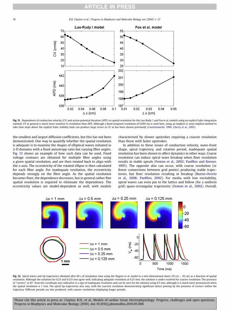
Fig. 11. Dependence of conduction velocity (CV) and action potential duration (APD) on spatial resolution for the Luo-Rudy 1 and Fox et al. models using an explicit Euler integrationmethod. CV in general is much more sensitive to resolution than APD. Although a fixed temporal resolution of 0.005 ms is used here, using an implicit or semi-implicit method totake time steps above the explicit Euler stability limit can produce large errors in CV as has been shown previously (Courtemanche, 1996; Cherry et al., 2003).
R.H. Clayton et al. / Progress in Biophysics and Molecular Biology xxx (2010) 1e2716
the smallest and largest diffusion coefficients, but this has not beendemonstrated. One way to quantify whether the spatial resolutionis adequate is to examine the shapes of elliptical waves initiated in2-D domains with a fixed anisotropy ratio but varying fibre angles.Fig. 13 shows an example of how such data can be used. Fixedvoltage contours are obtained for multiple fibre angles usinga given spatial resolution, and are then rotated back to align withthe x-axis. The eccentricity of the rotated ellipse is then calculatedfor each fibre angle. For inadequate resolution, the eccentricitydepends strongly on the fibre angle. As the spatial resolutionbecomes finer, the dependence decreases, but in general rather finespatial resolution is required to eliminate the dependence. Theeccentricity values are model-dependent as well, with models
Fig. 12. Spiral waves and tip trajectories obtained after 60 s of simulation time using the Nresolution. Although the solutions for 0.25 and 0.125 mm agree well, indicating adequate resof “corners” at 45� from the coordinate axes indicative is a sign of inadequate resolution andthe spatial resolution is 1 mm. The spiral tip trajectories also vary, with the coarsest resoltrajectory. Different periods are also produced, with coarser resolutions displaying longer p
Please cite this article in press as: Clayton, R.H., et al., Models of cardiaProgress in Biophysics and Molecular Biology (2010), doi:10.1016/j.pbiom
characterised by slower upstrokes requiring a coarser resolutionthan those with faster upstrokes.
In addition to these issues of conduction velocity, wave-frontshape, spiral trajectory, and rotation period, inadequate spatialresolution has been shown to affect dynamics in other ways. Coarseresolution can induce spiral wave breakup when finer resolutionresults in stable spirals (Fenton et al., 2002; Panfilov and Keener,1995). The opposite also can occur, with coarse resolution (orfewer connections between grid points) producing stable trajec-tories, but finer resolution resulting in breakup (Bueno-Orovioet al., 2008; Panfilov, 2002). For media with low excitability,spiral waves can even pin to the lattice and follow (for a uniformgrid) quasi-rectangular trajectories (Fenton et al., 2002). Overall,
ygren et al. model in a two-dimensional sheet (10 cm � 10 cm) as a function of spatialolution at 0.25 mm, the solution is under-resolved for coarser resolution. The presencecan be seen for the solution using 0.5 mm, although it is much more pronounced whenution demonstrating significant lattice pinning by the presence of corners within theeriods.
c tissue electrophysiology: Progress, challenges and open questions,olbio.2010.05.008

Fig. 13. Eccentricity of ellipsoids formed from a centrally located point stimulus as a function of uniform spatial resolution and constant fibre angle for the Fox et al. model in a two-dimensional sheet (3 cm � 3 cm). Anisotropy ratio is 5:1 (corresponding to diffusion coefficients of 0.001 and 0.0002 cm2 ms�1 along and across fibers, respectively). Top: contoursat �30 mV after 20 ms of propagation for a spatial resolution of 0.075 mm. Rotated contours are obtained by rotating the ellipses by the fibre angle to align with the x-axis. Bottom:Eccentricity is calculated for the rotated contours by taking the ratio of the width to the height. For the range of spatial resolutions used, the shape of the ellipsoids depends stronglyon the fibre angle, with a maximum eccentricity for fibers aligned to the coordinate axes. The dependence decreases as spatial resolution increases.
R.H. Clayton et al. / Progress in Biophysics and Molecular Biology xxx (2010) 1e27 17
although it is known that variations in spatial resolution can resultnot only in quantitative but also in qualitative dynamical differ-ences, there is at present no method to guarantee adequate reso-lution in the absence of such a comparison beyond reporting thata given solution agrees well with a solution obtained using finerspatial and temporal resolution.
6.7. Parallel implementation
Various parallel implementations of solvers have been developedand applied for simulations of tissue electrophysiology. For example,some simulators (Potse et al., 2006) are based on on OpenMP (OpenMulti-Processing, http://openmp.org/), an implementation of mul-tithreading that supports shared memory architectures. The mainadvantage of OpenMP is that implementations can be easily adaptedfor parallel execution, since the serial implementation needs only tobe slightly modified or can use OpenMP indirectly by interfacingwith specific linear algebra packages able to deal with OpenMP.An alternative approach is based onMPI (Message Passing Interface,http://www.mpi-forum.org/), a message-passing applicationprogramming interface that allows codes running on manycomputers to communicate with one another. In this case, specificcode has to be developed to dealwithMPI, but high scalability can beachieved. MPI based code can run on both distributed and sharedmemory architectures. The principal advantage of MPI is that itallows usage of systems with distributed memory architectures,which have only a fraction of the cost of systems with sharedmemory. Examples of this programming strategy applied to tissueelectrophysiology simulations can be found in (Colli Franzone andPavarino, 2004; Pavarino and Scacchi, 2008). An intermediateapproach involves developing hybrid code that can take advantageof computer architectures, which are a hybrid combination of sharedand distributed memory (Bordas et al., 2009).
Please cite this article in press as: Clayton, R.H., et al., Models of cardiacProgress in Biophysics and Molecular Biology (2010), doi:10.1016/j.pbiom
7. Strategies for reducing calculation time
A number of pragmatic approaches apart from standardnumerical methods can be used to reduce the time required to runsimulations of cardiac tissue electrophysiology. In the followingsectionwe describe some of the most effective and well establishedtechniques, as well as some more novel approaches.
7.1. Lookup tables
Many models of cellular electrophysiology require computa-tionally expensive functions, such as exponentials, logarithms, trig-onometric functions, and exponentiation to be evaluated. Thesefunctions often depend purely on Vm, and significant computationalspeedup can be obtained by pre-computing these values and storingthem in lookup tables that are used for subsequent evaluation(Victorri et al., 1985). The tables can be arranged as 1-D arrayscorresponding to a physiological range of voltages with a fineenough resolution to capture differences (generally on the order of0.01e0.1 mV) (Cherry et al., 2003). Often, combinations of theseexpensive functions can be evaluated and stored together so as tominimize their calculation within the time-stepping process.Although choosing thenearest voltage value represented in the tableis one possibility, linear interpolation can be used to interpolatebetween the two nearest voltage values represented in the table forincreased precision. Speedup achieved depends on the number andtype of functions replaced by lookup tables: the more computa-tionally expensive the function, the greater the resulting speedup.Exponentiation to non-integer powers are often themost expensive,followed by exponentials and trigonometric functions, althoughsystem architecture, compiler choice, and function argument rangesaffect the relative cost of evaluating such functions. Nevertheless, itis possible to achieve a speedup factor of up to 5 (Cherry et al., 2003).
tissue electrophysiology: Progress, challenges and open questions,olbio.2010.05.008

R.H. Clayton et al. / Progress in Biophysics and Molecular Biology xxx (2010) 1e2718
Lookup tables can also be used for computationally expensivefunctions that are dependent on other variables, such as intracel-lular calcium concentrations and even some transmembranecurrents. It is also possible to construct a 2D table for computa-tionally expensive functions that depend on two state variables.An example can be found in the Hund-Rudy model (Hund andRudy, 2004), where the d-gate is raised to a variable power.Because exponentiation is usually such a costly function to eval-uate, and because the two quantities involved have limited ranges(the d-gate varies strictly between 0 and 1, while the variable powertakes values between 1 and 9), use of a 2D lookup table can savecomputational time with little effort.
It is important to note that lookup tables may not be suitable forrepresenting certain quantities that exhibit large changes for smallchanges of function variables. If the table size needed to representsuch quantities is extremely large pre-calculation may be time-consuming, and memory requirements or memory access timesmay become prohibitive. An alternative is to use lookup tablevalues within certain values and to calculate the quantity directlyotherwise, which can be accomplished with a simple if- statementin the code. Careful coding of the model equations can further addto the benefits of lookup tables. This can be done using automatedtools, and can result in substantial speedup (Cooper et al., 2006). Inall cases, the application of lookup tables for a given model withina particular code should be evaluated to ensure acceptable accuracyis maintained.
7.2. Exponential solutions for gating variables
For explicit time-stepping schemes, speedups can be obtained inmany cases by integrating the gating variables for the cell modelODEs using exponential solutions (Maclachlan et al., 2007; Planket al., 2008; Rush and Larsen, 1978). This method applies to anindividual gating variable y represented by the ordinary differentialequation.
dydt
¼ ðyN � yÞ=sy_ (18)
With a steady-state value yN and the time constant sy. If voltage isconsidered to be non-varying over a single time step, this can besolved analytically over that time step as
ynþ1 ¼ ðyN � ynÞ exp��Dt=sy
�; (19)
where yN and sy are solved using Vn, the value of the membranepotential at the old time step, and Dt is the time step. Similarbenefits can also be achieved by integrating using a semi-implicitintegration scheme under the same assumption that yN and sy areconstant over the time step (Cherry et al., 2003; Whiteley, 2006).
The primary advantage of this method is to allow a larger timestep than a purely explicit integration scheme while retainingaccuracy. The value of the exponential can be pre-computed andstored in a lookup table to avoid computationally expensive func-tion evaluations. However, the technique solves the set of ODEs ina decoupled form, which makes the solution first-order-accurate,although it is possible to extend the approach to achieve second-order accuracy (Sundnes et al., 2009). In addition, calculation ofother variables, such as ionic concentrations, cannot benefit fromthis approach.
7.3. Operator splitting
The global system of PDEs and ODEs can be decoupled in severalways by operator splitting, with the aim of improving the compu-tational efficiency and reduce complex dependencies between the
Please cite this article in press as: Clayton, R.H., et al., Models of cardiaProgress in Biophysics and Molecular Biology (2010), doi:10.1016/j.pbiom
variables of the problem. In principle, these methods introducea splitting error that is independent of the numerical methods usedto solve the decoupled components. This additional error should bequantified, unless the split steps are re-iterated until a suitableconvergence condition is satisfied. In general, splitting techniquesthat alternate between two decoupled operators are first-order-accurate. However, second-order accuracy can be achieved byperforming two half-steps with one operator before and after a fullstep of the second operator (Strang, 1968).
In (Roth, 1991; Roth and Wikswo, 1994) an operator splittingtechnique on a finite-difference formulation is used to solve thebidomainproblemcoupledwith a PDE representing the surroundingbath. In (Qu and Garfinkel, 1999) a second-order-accurate splittingtechnique has been proposed to separate the solution of the mon-odomain PDE describing propagation in tissue from the Luo-Rudyphase I ODEs describing local membrane kinetics. However, the ODEsystemwas solved using the Rush-Larsen method (Rush and Larsen,1978), while the time advancing scheme for the PDE was an explicitforward Euler method, which resulted in the algorithm being onlyfirst-order-accurate overall. An operator splitting technique has alsobeen devised for solving the bidomain equations coupled witha description of volume conduction in the torso (Sundnes et al.,2005). In this study the authors proposed a general formulation ofoperator splitting, based on a q-rule, that can result in a second-orderscheme for a proper choice of the parameter. The ODE system wassolved using a third order Runge-Kutta scheme. The ODE systemitself can be decomposed using operator splittingmethods. Operatorsplitting techniques applied to the bidomain equations are reviewedin (Vigmond et al., 2008), where a three-step operator splitting fordecoupling the bidomain system that has been proven to beunconditionally stable is described.
7.4. Efficiency of adaptivity in space and time
A major challenge for increasing numerical efficiency is the useof adaptivity in space and time. Although there is a rich numericalliterature on adaptivity for bothfinite-difference and finite-elementmethods, dynamic and simultaneous adaptivity in space and time inwhich both the spatial and temporal resolution are varied locallyover the course of the simulation has only rarely been applied tocardiac electrophysiology studies (Cherry et al., 2000, 2003; ColliFranzone et al., 2006; Deuflhard et al., 2009; Trangenstein andKim, 2004). In contrast, dynamic temporal adaptivity has beenused more commonly (Allexandre and Otani, 2002; Qu andGarfinkel, 1999; Quan et al., 1998; Vigmond and Leon, 1999) anddynamic spatial adaptivity also has been used (Belhamadia et al.,2009; Pennacchio, 2004). The general idea behind dynamic space-time adaptivity is that the algorithm can increase or decrease spatialand temporal resolution locally during times when such adjust-ments are beneficial (e.g., higher resolution for action potentialupstrokes and associated sharp spatial gradients and lower reso-lution during quiescence). Developing dynamically adaptivemethods is a complex endeavour. Also, characterising accuracy andefficiency of such techniques is not straightforward. One significantchallenge in measuring the speedup provided by adaptive methodsis that their efficiency generally depends on the dynamical statesimulated, including thewavelength,wave-frontwidth, and densityof waves in the domain. For simple plane wave propagation withlong periods during which no wave is present in most of thedomain, adaptive methods can provide a speedup of more than anorder of magnitude. The efficiency gains in the presence of morecomplex spatiotemporal dynamics are generally more modest butstill can be significant. A spaceetimeeadaptive finite-differenceapproach was found to give a speedup of a factor of 20 for a planewave in a 2-D domain (Cherry et al., 2000) and 50 for a plane wave
c tissue electrophysiology: Progress, challenges and open questions,olbio.2010.05.008

R.H. Clayton et al. / Progress in Biophysics and Molecular Biology xxx (2010) 1e27 19
in a 3-D slabwith rotational anisotropy (Cherry et al., 2003), but stillmanaged a factor offive for a 2-D domainwith sustained spiralwavebreakup (Cherry et al., 2000), in all cases compared to a uniformmesh using the finest spatial and temporal resolutions available tothe adaptive algorithm.
A different spaceetimeeadaptive method using finite elementsgave a speedup factor of less than a of two in a two-dimensionaldomain during initiation and about one rotation of a spiral wave(Trangenstein and Kim, 2004). Another approach using an adaptivemultilevel FEM with a linearly implicit Rosenbrock discretisationwith stepsize control in time achieved a reduction of two orders ofmagnitude for both the number of computational nodes and thenumber of time steps taken for a single propagating plane wave ina 3-D domain using the bidomain equations (nomeasure of speedupwas given) (Colli Franzone et al., 2006). This approach recently wasextended to realistic cardiac geometries (Deuflhard et al., 2009).Similar work is underway to extend thework of Cherry et al. (Cherryet al., 2003) to realistic anatomies by combining it with the phase-field approach (Fenton et al., 2005), as shown in Fig.14. In addition toa dependence on the dynamical state, the speedup achieved alsodepends on the electrophysiology model: because the computa-tional costs associatedwith regridding andother numerical tasks arefixed, computationally more expensive models can benefit morefrom adaptivity than simpler models.
A related approach is to use increased spatial resolution only forthe variables associated with the rapidly varying INa. This techniquehas been shown to increase computational efficiency for planewavesimulations using the bidomain model with the Noble et al. guinea-pig ventricularmodel (Noble et al.,1998) by two orders ofmagnitude(Whiteley, 2008); however, it is unclear whether similar perfor-mance gains would be achieved for larger domains with morecomplex dynamical states containing multiple interacting waves.
7.5. Using graphics processing units for computation
The increased computational power and memory of graphicsprocessing units (GPUs), combined with decreasing costs, hasgenerated significant interest in utilising graphics hardware for otherapplications. GPUs have been optimised for traditional computergraphics, which is focused on highly data-parallel operations onfloating-point numbers, and provide less of an advantage for activi-ties outside this range, such as intensive memory communicationand integer and double precision calculations (Owens et al., 2007).Nevertheless, many computationally demanding calculations maybenefit from GPUs, which have already been applied to simulationsoutside graphics and visualisation applications, including cellularautomata, dendritic growth,fluid and gas dynamics, signal and imageprocessing, geometric computing, and reactionediffusion equations
Fig. 14. Adaptive mesh refinement in a 2-D slice of canine ventricles from (Nielsen et al., 19Rudy phase I model (Luo and Rudy, 1991). Right: Three-level grid structure corresponding togreen, and intermediate (0.25 mm) in yellow.
Please cite this article in press as: Clayton, R.H., et al., Models of cardiacProgress in Biophysics and Molecular Biology (2010), doi:10.1016/j.pbiom
(Owens et al., 2007). Recent work demonstrated significant speedupof simulations of cardiac electrophysiology equations in tissue (Satoet al., 2009; Vigmond et al., 2009). However, it remains to be seenhow effectively GPUs can be integrated into large-scale cardiacsimulations.
7.6. Use of simplified cell models for representing tissue
Large-scale tissue simulations may involve millions of gridpoints. To improve computational tractability in these cases,simplified cell models represent an alternative to more biophysi-cally detailed models. Whereas detailed cell models may include30e100 variables and tens to hundreds of equations, simplifiedmodels generally include between two and ten variables anda correspondingly reduced number of equations. Such a reductionin the computational workload can translate into a speedup of oneor more orders of magnitude, although this depends on how thediffusion calculation is implemented (see Section 6). In some cases,reduced models also have less restrictive spatial and temporalresolution requirements and can achieve further computationalsavings by using coarser resolutions.
Simplified models used for cardiac electrophysiology tissuesimulations generally fall into three categories: direct reductions ofmore detailed models, generic models, and phenomenologicalmodels. Below we discuss each of these types of simplifications,including the advantages and limitations of each approach.
7.6.1. Reductions of detailed modelsReduced models are developed by reducing the complexity of
a detailed model through any of several means. Rapidly varyingvariables, such as the m-gate governing activation of INa, may beeliminated adiabatically by using the steady-state value, therebyassuming that the gate instantaneously achieves the steady-statevalue associated with a given voltage. This approach not onlyeliminates a variable but also eliminates a significant source ofstiffness in the differential equations, so that larger time steps canbe used in the numerical scheme. However, unacceptably largeerrors can result. For example, eliminating them-gate adiabaticallyin a reduction of the Priebe-Beuckelmann model results in signif-icantly decreased conduction velocity (Bernus et al., 2002). Addi-tional gating variables also can be eliminated either by removingslowly varying gates or by combining variables, such as the twovoltage-dependent inactivation gates used inmany representationsof INa (Bernus et al., 2002). Another common approach to reducethe number of variables is to treat many or all intracellular ionconcentrations as constant. Although this can produce computa-tionally desirable benefits, such as decreased accommodation timeafter a change in the pacing cycle length, treating concentrations as
91). Left: Voltage plot 160 ms after a stimulus applied in the lower left using the Luo-the voltage plot. Coarse mesh regions (0.5 mm) are shown in white, fine (0.125 mm) in
tissue electrophysiology: Progress, challenges and open questions,olbio.2010.05.008

Fig. 15. Original TNNP and the 4-variable model fit to it (see text for details). A. Resti-tution curves for the TNNP model (grey) and the 4-variable fit. B. Spiral wave at threetimes during a single rotation for the TNNP model (left) and the 4-variable fit (right).
R.H. Clayton et al. / Progress in Biophysics and Molecular Biology xxx (2010) 1e2720
constants can remove other important properties, such as memory.Finally, currents with small values, such as background currents orthe sarcolemmal Ca2þ pump, in some cases may be eliminated(Mahajan et al., 2008). For models involving detailed Markovrepresentations of currents, reductions can be accomplished bysubstitution with less complex Hodgkin-Huxley-style descriptions,by using rapid equilibrium assumptions, or by replacing ODEs withalgebraic equations when transients are fast enough to be consid-ered instantaneous (Henry and Rappel, 2004; Hinch, 2004; Planket al., 2008).
The main advantage of reduced models is that they improvecomputational tractability while retaining a similar structure asthe more detailed model. In this way, it is easier to incorporatebiophysical variations, such as spatial gradients in channelexpression, the effects of pharmaceutical agents, and electrophys-iological remodeling effects. However, the reduction may reduce oreliminate important properties of real cells and tissue such asmemory. Furthermore, the reduction process must be undertakenseparately for each detailed model to ensure that the desiredproperties are preserved with each variable eliminated.
7.6.2. Generic modelsGeneric excitable media models, such as the FitzHugh-Nagumo
(FHN) model (Fitzhugh, 1961), have been used to illustrate prop-erties of wave propagation for many years. The FHN model itself isa 2-variable reduction of the Hodgkin-Huxley model (Hodgkin andHuxley, 1952) and in its original form produces action potentialsmore like neural action potentials. The FHN model can be modifiedto produce longer action potentials and to eliminate hyper-polarisation that occurs during the depolarisation phase. However,the FHN model and others like it generally lack other importantproperties, including realistic APD restitution, memory, and a non-zero minimum cycle length (and corresponding APD) below whichblock occurs. Without these more realistic properties, genericmodels often are of limited use and are generally restricted toshowing the types of qualitative behaviour that may occur withmore detailed models. As computational resources have increased,generic models have been used less often. Nevertheless, one of themain advantages of generic models remains, which is the relativeease with which analytical results can be obtained.
7.6.3. Phenomenological modelsA third approach to reducing computational complexity is to
develop phenomenological models that aim to reproduce keydynamical properties of cells and tissue without including as muchbiophysical detail. For example, rather than including the descrip-tions of ten or more ion channels, pumps, and exchangers,a phenomenological model may focus on “summary” inward andoutward currents that rely on fewer variables and equations. TheFenton-Karma model (Fenton and Karma, 1998) and its subsequentextensions (Bueno-Orovio et al., 2008; Cherry and Fenton, 2004)are examples of phenomenological models. Originally, it included 3variables and 12 parameters together with three currents, repre-senting the fast inward, slow inward, and slow outward currents.Extended versions of the model have increased the number ofvariables to 4 and the number of parameters to 28 (Bueno-Orovioet al., 2008); these additions allow spike-and-dome action poten-tial shapes to be reproduced. Among the dynamical properties thatare incorporated are the threshold of excitation, maximumupstroke velocity, action potential shape, APD, APD and CV resti-tution curves, and the minimum cycle length (greater than zero)below which propagation fails.
Because thesemodels focus on reproducingdynamical propertiesrather than detailed ion currents, they can be fit directly from tissue-level experimental data that measure these quantities, rather than
Please cite this article in press as: Clayton, R.H., et al., Models of cardiaProgress in Biophysics and Molecular Biology (2010), doi:10.1016/j.pbiom
from patch-clamp data obtained from isolated cells. The smallnumber of variables and parameters makes parameter fitting muchmore straightforward, as many parameters govern or contribute toonly a single property. In addition to fitting experimental data, themodel can be used to fit data from more biophysically detailedmodels (Bueno-Orovio et al., 2008). For example, as shown in Fig.15,
c tissue electrophysiology: Progress, challenges and open questions,olbio.2010.05.008

R.H. Clayton et al. / Progress in Biophysics and Molecular Biology xxx (2010) 1e27 21
themodel can successfully reproduce the restitution curve and spiralwave dynamics of the ten Tusscher et al. model for human ventric-ular cells (TenTusscher et al., 2004). The Priebe-Beuckelmannmodel(Priebe and Beuckelmann, 1998) also has been fit (Bueno-Orovioet al., 2008).
In addition to improving computational tractability, one of theadvantages of phenomenological models is the relative ease withwhich specific dynamical properties can be implemented. As anexample, this model has been used to examine the effects ofdifferent action potential shapes on tissue dynamics (Cherry andFenton, 2004). In this application, two different parameter setswere developed to produce action potentials with different shapesbut with identical restitution and bifurcation properties in isolatedcells for APDmeasured to 80 percent (ormore) of repolarisation (seeFig. 16A and Fig. 16B). In tissue, bothmodels also produce alternans,but CV restitution curves with more gradual slopes over a widerrange of cycle lengths are able to reduce or even eliminate alternansfor the model that repolarises more quickly. This can prevent spiralwave breakup that occurs for the other model, as shown in Fig. 16C.
A disadvantage of this approach is that reproducing the effects ofion channel blockers and other modifications of specific electro-physiological changes is not as straightforward. However, it is possibleto perform a newparameterfitting to data obtained in the presence ofthe channel blocker, an approach that may be useful for novel phar-maceutical agents whose effects are incompletely characterised.
7.7. Choosing an appropriate cellular electrophysiology model
Most recentmodels of cardiac cellular electrophysiology have beendesigned to represent cells from specific regions (e.g., atria, ventricles,Purkinje) and in some cases sub-regions (e.g., ventricular epicardial,endocardial, midmyocardial) of the heart, as well as particular animalspecies (e.g., mouse, rat, guinea pig, rabbit, canine, and human). Thenumber of models described in the literature has increased signifi-cantly in the recent past. However, a model for a specific combinationof species and region may still not be available. Also, more than one
Fig. 16. Electrotonic effects on alternans and spiral wave stability. A. Bifurcation diagram shoand Fenton, 2004). APD values below 80 percent of repolarisation match almost exactly, as500 ms. B. Matching restitution functions for the same two parameter sets in isolated celventricular anatomy (Vetter and McCulloch, 1998). Although the APD restitution curves foreffects of the steep APD restitution curve slope in tissue for Model 2 and produces a stable
Please cite this article in press as: Clayton, R.H., et al., Models of cardiacProgress in Biophysics and Molecular Biology (2010), doi:10.1016/j.pbiom
model representing the particular combination of species and heartregion of interest may be available, and in some cases only a smallsubset ofmodels reproduce the properties of interest for a given study.For instance, alternans at fast pacing rates is a common phenomenonacross a number of species, but not all models of such species showalternans. The Fox et al. canine ventricularmodel (Fox et al., 2002) hasalternans, but the earlier Winslow et al. canine ventricular model(Winslow et al., 1999) on which it is based does not. Among rabbitventricular models, the Mahajan et al. model (Mahajan et al., 2008)has alternans, but the earlier Shannon et al. model (Shannon et al.,2004), from which it derives most of its current formulations, doesnot. Thus, not all models are appropriate for investigating specificphenomena, and in some cases there may be no model that displaysbehaviours observed experimentally.
Furthermore, most cellular level models are validated forspecific conditions, including temperature, pacing rate, ionicconcentrations, sex, and age. The model may not be applicableoutside these conditions. Most models are constructed using iso-lated cell data only and so may not be able to reconstruct theproperties and behaviour of cells in tissue. It is also important tonote that modifying a model by changing parameter values orequations may compromise the original validation.
Simplified models can be appropriate in many cases, especiallyfor tissue studies. In many cases tissue phenomena such as spiralwave breakup are model-independent and can be obtained usingany model with similar characteristics. In addition, simplifiedmodels avoid problems associated with large numbers of param-eters whose values may not be known with great precision andwith large number of variables whose individual effects cannoteasily be discerned. Ideally, models should be as simple as neededto explain the phenomena of interestdbut no simpler.
8. Discussion
Integrated models of cardiac tissue electrophysiology are beingused in basic science investigations including studies of defibrillation
wing APD as a function of cycle length in an isolated cell for two parameter sets (Cherryshown for the inset action potentials at cycle lengths of 290 ms (during alternans) andls. C. Two views of spiral wave dynamics for the two parameter sets within a rabbitthe two models are identical in isolated cells, CV restitution in tissue modulates thespiral wave.
tissue electrophysiology: Progress, challenges and open questions,olbio.2010.05.008

R.H. Clayton et al. / Progress in Biophysics and Molecular Biology xxx (2010) 1e2722
(Rodriguez et al., 2005) and specific arrhythmias such as ventricularfibrillation (Ten Tusscher et al., 2009). Recently, highly detailedanatomical (Li et al., 2008; Plank et al., 2009) and patient specificmodels (Pop et al., 2009) have been introduced. Despite this prog-ress, and increasing acceptance ofmodels as research tools, there arefew examples where models of cardiac electrophysiology have beenuseful in clinical applications. The reality falls short of the vision ofthe Cardiac Physiome Project, and there are some important issuesthat are not fully resolved;we have highlighted some of these in thisreview, andwe continue this discussion below. From a philosophicalpoint of view it is important to recognise that the purpose ofmodelling cardiac tissue electrophysiology is not only to reconstructobserved phenomena at ever greater levels of detail, but also toexplain themechanisms that underlie them and hence to predict theeffects of changing parameters and other components of the model.All models involve assumptions and simplifications, and the tissuemodels reviewed here are simplifications of real cardiac tissue, itsmicro-structure and electrophysiological properties. For a particularmodel to be useful, itmust capture enough detail at a particular levelof complexity to offer a mechanistic insight that complementsexperimental work.
The heart is a complex multi-scale and multiphysics system,where cellular electrophysiology is coupled to metabolism andtension development, as well as to tissue mechanics and fluiddynamics. Fully coupled and integrative models of the heart are partof the vision for the cardiac Physiome, and although progress in thisarea is being made (Jie et al., 2010; Nickerson et al., 2006; Niedererand Smith, 2007; Panfilov et al., 2007), these large-scale modelsremain a significant future challenge. Within the narrower scope oftissue electrophysiological models, there are a number of challengesand open questions; we summarise and discuss some of these below.
8.1. Challenges
The increasing availability of high performance computingresources has a huge impact on computational research, in partic-ular enabling simulations with more biophysical detail and higherthroughput. These technological advances are driving the efforts incardiac computational research towards more detailed and inte-gratedmodels, with the goal of patient specific electrophysiologicalsimulations that can be used clinically (Neal and Kerckhoffs, 2010;Plank et al., 2009; Pop et al., 2009). Balancing the amount ofanatomical and biophysical detail against the time and datacapacity required to perform simulations is an important issue.Underlying these considerations is the validity of the assumptionsthat are embedded in tissue electrophysiology models.
Understanding the functional relationship between the discretestructure and continuum behaviour of cardiac tissue at microscopicand macroscopic levels is a significant challenge. As describedearlier in Section 6, most models of cardiac tissue approximate thespatial domain as a continuum even though observation of cardiactissue shows clearly that it is fundamentally discrete, composed ofcells of different sizes and shapes that couple together. Althoughseveral approaches for representing cells discretely have beendeveloped (Roberts et al., 2008; Spach et al., 2004; Stinstra et al.,2006), it is not yet known when such treatment is necessary. Itwill be important in the future to identify at what spatial resolutionand under what conditions the continuum approximation is nolonger valid for both monodomain and bidomain models, in tissueswith anisotropy and, in 3-D, orthotropic fibre-sheet structure. Afurther consideration is the diverse cellular composition of cardiactissue. Most current models of tissue electrophysiology representonly myocytes and neglect other cell types such as fibroblasts.
A challenge common to modelling communities is the level ofmethodological detail required for a scientific publication that
Please cite this article in press as: Clayton, R.H., et al., Models of cardiaProgress in Biophysics and Molecular Biology (2010), doi:10.1016/j.pbiom
describes a model of cardiac tissue electrophysiology. How muchdetail is necessary to describe a model of cardiac tissue electro-physiology, so that the results cannot only be conceived by a reader,but also reproduced for validation? Recently the MIRIAM (Le Novereet al., 2005) and MIASE (http://www.ebi.ac.uk/compneur-srv/miase/index.html) standards have been proposed, which specify the type ofinformation that should be includedwhen describing computationalexperiments at the cellular and sub-cellular level. Specific guidelinesfor models at the cardiac cell level have been proposed, whichemphasise careful documentation of the underlying biophysics,relation of the model parameters to experimental data, numericalmethods, and consistency of units in the model (Smith et al., 2007).
Although many of the simulation studies cited in this reviewreport details and provenance of models and parameters, thisattention to detail is not always the case. With rapid increases inavailable computing power and increasingly detailed simulationcodes, it is possible to overlook the assumptions and parametervalues that underlie a particular tissue scale model. On the basis ofthe issues raised in this review, we would propose that thefollowing tentative list of should be included in descriptions oftissue electrophysiology models:
� Description of the model for cardiac cellular electrophysiology,referenced to the original cell model publication or modelimplementation such as the CellML model version curated athttp://www.cellml.org, and with any changes to parametersjustified and highlighted.
� Description of the tissue electrophysiology model, referencedto the original model publication and with a list of parametersthat includes conductivities (or diffusion coefficients) andcorresponding CV justified and linked to experimentalmeasurements where possible.
� Numerical methods used to obtain solutions to the equations forcell and tissue electrophysiology, with evidence of numericalstabilitysuchasconsistentCVwithchanges in timeandspacestep.
� Links to source code, libraries, and other resources used inimplementation of the model.
� Details of tissue geometry, including the source of anatomicallydetailed models, details of the meshing procedure and anyimage processing operations carried out on raw imaging data,information on spatial resolution, and, for FEM approaches,element size and shape.
� Evidence that tissue dynamics have been validated againsteither experimental data or comparable modelling data. Forinstance APD and CV restitution curves can be compared withother published data to demonstrate convergence.
This list is not intended to be proscriptive; rather our aim is topropose criteria to ensure that models of cardiac tissue electro-physiology are described in a consistent way:
8.2. Open questions
Different models of tissue electrophysiology involve differentassumptions and simplifications, yet there is no generally acceptedframework for choosing an appropriate combination of cellularelectrophysiology model, tissue model, geometrical model, andnumerical method. As discussed in Section 7.7, there is evidencethat the choice of cellular electrophysiology model can influencetissue-level behaviours such as the stability of re-entry, howevereach of the areas listed below remain as open questions.
� Choice of tissue model. As discussed above (Section 4.2.3)both monodomain and bidomain tissue models can producealmost identical propagation sequences providing that no
c tissue electrophysiology: Progress, challenges and open questions,olbio.2010.05.008

R.H. Clayton et al. / Progress in Biophysics and Molecular Biology xxx (2010) 1e27 23
current is injected into the extracellular space (Potse et al.,2006). However, there remain unresolved issues such as therole that boundary conditions play in the behaviour of re-entrant waves (Sambelashvilli and Efimov, 2004).
� Numerical approach. We have discussed the differentnumerical approaches that have been used to solve the equa-tions of tissue electrophysiology, yet there are few studieswhere the influence of the numerical approach has beenstudied in a systematic way. A particular issue here is thepossible influence of irregular and hybrid meshes on complexemergent behaviours such as re-entrant waves.
� Benchmarks for numerical methods and implementations.Related to the two questions described above is the lack ofsuitable benchmarks by which different tissue models andnumerical approaches could be compared. A set of exampleproblems, with accepted solutions, would be one solution.
� Parameters and parameter sensitivity. There are not wellaccepted values for parameters, such as the tissue conductivi-ties, which are important in determining the behaviour ofa propagating action potential. Morework is needed not only toestablish the biophysical basis for these parameters, but also tounderstand how sensitive the emergent phenomena discussedin section 5 are to variability in these parameters.
� Methods for representing detailed and heterogeneous tissuemicro-structure. Cardiac tissue is a complex composite tissue,that is full of heterogeneity, yet it appears to behave as a func-tional syncytium. The roles played by fibroblasts, voids, and thefibre-sheet architecture of cardiac tissue inpropagation of normaland abnormal activation remain unexplored in detail. Further-more, appropriate parameters and boundary conditions for rep-resenting this complex structure are not well accepted, and it islikely that new numerical methods will need to be developed tohandle abrupt changes in the orientation of fibres and sheets.
� Representation of pathological structure and function. Thisreview has concentrated on models of normal tissue, althoughmodels of cardiac tissue electrophysiology have been widelyused to study arrhythmias, which are usually associated withpathology. Robust methods and parameters to representconditions including ischaemia, infarction, and heart failure arean important area that is not yet fully developed.
� Validation against experimental data. While it can be possibleto validate simulated conduction velocity, activation sequenceand restitution against published experimental data, many ofthe phenomena investigated using tissue models, such as thecriteria for stable or unstable re-entry, are difficult to verifyexperimentally. As we have suggested above, a useful startingpoint for this process is to carefully compare model behaviourto experimental datawhere theyexist, and to include this link inpublicationswheremodels of tissue electrophysiology are used.
8.3. Conclusion
Models of cardiac tissue electrophysiology have played animportant role in advancing our understanding of action potentialpropagation in the heart. Despite significant progress in the eval-uation of modelling approaches, the efficient numerical treatmentof models, and development of novel approaches, there areimportant challenges to be overcome for application and validationof these approaches in fields outside of their established domain.
Acknowledgements
This paper was conceived and drafted during a workshop on theCardiac Physiome Project held at the Newton Institute for
Please cite this article in press as: Clayton, R.H., et al., Models of cardiacProgress in Biophysics and Molecular Biology (2010), doi:10.1016/j.pbiom
Mathematical Sciences at the University of Cambridge in July 2009and funded by the Newton Institute, the UK Biotechnology andBiological Sciences Research Council, and the Wellcome Trust. FBSacknowledges support from the Nora Eccles Treadwell Foundation.AVP acknowledges support from the IHES and the James SimonsFoundation. We thank Daniel Olmos Liceaga from the Departa-mento de Matemáticas, Universidad de Sonora, México for helpfulcomments on the manuscript. EMC and FHF acknowledge supportfrom the National Science Foundation through grants no. 0824399(EMC) and 0800793 (FHF and EMC) and through TeraGrid resourcesprovided by the Pittsburgh Supercomputing Center.
References
Abildskov, J.A., 1976. Effects of activation sequence on the local recovery ofventricular excitability in the dog. Circulation Research 38, 240e243.
Abrahams, C., Janicki, J.S., Weber, K.T., 1987. Myocardial hypertrophy in Macacafascicularis. Structural remodeling of the collagen matrix. Laboratory Investi-gation 56, 676e683.
Adler, C.P., Ringlage, W.P., Bohm, N., 1981. [DNA content and cell number in heartand liver of children. Comparable biochemical, cytophotometric and histolog-ical investigations (author’s transl)]. Pathology, Research and Practice 172,25e41.
Akar, F.G., Roth, B.J., Rosenbaum, D.S., 2001. Optical measurement of cell-to-cellcoupling in intact heart using subthreshold electrical stimulation. AmericanJournal of Physiology, Heart Circulatory Physiology 281, H533eH542.
Allexandre,D., Otani,N.F., 2002. Accuracyand speedconsiderations of a newnumericalmethod for modeling cardiac excitation. Computers in Cardiology 29, 9e12.
Anderson, R.H., Ho, S.Y., 1998. The architecture of the sinus node, the atrioven-tricular conduction axis, and the internodal atrial myocardium. Journal ofCardiovascular Electrophysiology 9, 1233e1248.
Austin, T.M., Trew, M.L., Pullan, A.J., 2006. Solving the cardiac bidomain equationsfor discontinuous conductivities. IEEE Transactions on Biomedical Engineering53, 1265e1272.
Banerjee, I., Yekkala, K., Borg, T.K., Baudino, T.A., 2006. Dynamic interactionsbetween myocytes, fibroblasts, and extracellular matrix. Annals of New YorkAcademy Sciences 1080, 76e84.
Banerjee, I., Fuseler, J.W., Price, R.L., Borg, T.K., Baudino, T.A., 2007. Determination ofcell types and numbers during cardiac development in the neonatal and adultrat and mouse. American Journal of Physiology, Heart Circulatory Physiology293, H1883eH1891.
Barr, R.C., Plonsey, R., 1984. Propagation of excitation in an idealized anisotropictwo-dimensional tissue. Biophysical Journal 45, 1191e1202.
Beeler, G.W., Reuter, H., 1977. Reconstruction of the action potential of ventricularmyocardial fibres. Journal of Physiology 268, 177e210.
Belhamadia, Y., Fortin, A., Bourgault, Y., 2009. Towards accurate numerical methodfor monodomain models using a realistic heart geometry. MathematicalBiosciences 220, 89e101.
Bernus, O., Wilders, R., Zemlin, C.W., Verschelde, H., Panfilov, A.V., 2002. Acomputationally efficient electrophysiological model of human ventricular cells.Americal Journal of Physiology (Heart and Circulatory Physiology) 282,H2296eH2308.
Bernus, O., Wellner, M., Pertsov, A.M., 2004. Intramural wave propagation in cardiactissue: asymptotic solutions and cusp waves. Physical Review E 70, 061913.
Bers, D.M., 2008. Excitation-Contraction Coupling and Cardiac Contractile Force.Springer, Dordrecht.
Bondarenko, V.E., Szigeti, G.P., Bett, G.C.L., Kim, S., Rasmusson, R.L., 2004. Computermodel of action potential of mouse ventricular myocytes. American Journal ofPhysiology (Heart and Circulatory Physiology) 287, H1378eH1403.
Bordas, R., Carpenteri, B., Fotia, G.,Maggio, F., Nobes, R., Pitt-Francis, J., Southern, J., 2009.Simulation of cardiac electrophysiology on next-generation high-performancecomputers. Philosophical Transactions of the Royal Society A 367, 1951e1969.
Bourgault, Y., Ethier, M., LeBlanc, V.G., 2003. Simulation of electrophysiologicalwaves with an unstructured finite element method. M2AN 37, 649e661.
Boyett, M.R., Tellez, J.O., Dobrzynski, H., 2009. The sinoatrial node:Its complex structureand unique ion channel gene program. In: Zipes, D.P., Jalife, J. (Eds.), Cardiac Elec-trophysiology: From Cell to Bedside, fifth ed. Saunders, Philadelphia, pp. 127e138.
Brette, F., Orchard, C., 2003. T-tubule function in mammalian cardiac myocytes.Circualtion Research 92, 1182e1192.
Bub, G., Shrier, A., Glass, L., 2002. Spiral wave generation in heterogenous excitablemedia. Physical Review Letters 88, 058101-1e058101-4.
Bueno-Orovio, A., Perez-Garcia, V.M., Fenton, F.H., 2006. Spectral methods forpartial differential equations in irregular domains: the spectral smoothedboundary method. SIAM Journal on Scientific Computing 28, 886e900.
Bueno-Orovio, A., Cherry, E.M., Fenton, F.H., 2008. Minimal model for human ventric-ular action potentials in tissue. Journal of Theoretical Biology 253, 544e560.
Burton, R.A.B., Plank, G., Schneider, J.E., Grau, V., Ahammer, H., Keeling, S.J., Lee, J.,Smith, N.P., Gavaghan, D., Trayanova, N., Kohl, P., 2006. 3-Dimensional models ofindividual cardiac histo-anatomy: tools and challenges. Annals of the New YorkAcademy of Sciences 1380, 301e319.
tissue electrophysiology: Progress, challenges and open questions,olbio.2010.05.008

R.H. Clayton et al. / Progress in Biophysics and Molecular Biology xxx (2010) 1e2724
Cabo, C., Pertsov, A.M., Baxter, W.T., Davidenko, J.M., Gray, R.A., Jalife, J., 1994.Wavefront curvature as a cause of slow conduction and block in isolated cardiacmuscle. Circulation Research 75, 1014e1028.
Cabrera, J.A., Sanchez-Quintana, D., Ho, S.Y., Medina, A., Anderson, R.H., 1998. Thearchitecture of the atrial musculature between the orifice of the inferior cavalvein and the tricuspid valve: the anatomy of the isthmus. Jouranl of Cardio-vascular Electrophysiology 9, 1186e1195.
Caldwell, B.J., Trew, M.L., Sands, G.B., Hooks, D.A., LeGrice, I.J., Smaill, B.H., 2009.Three distinct directions of intramural activation reveal nonuniform side-to-side electrical coupling of ventricular myocytes. Circulation Arrhythmia andElectrophysiology 2, 433e440.
Camelliti, P., Borg, T.K., Kohl, P., 2005. Structural and functional characterisation ofcardiac fibroblasts. Cardiovascuar Research 65, 40e51.
Campbell, S.E., Gerdes, A.M., Smith, T.D., 1987. Comparison of regional differences incardiac myocyte dimensions in rats, hamsters, and guinea pigs. AnatomicalRecord 219, 53e59.
Chen, L.S., Chen, P.-S., 2009. Nerve sprouting and cardiac arrhythmias. In: Zipes, D.P.,Jalife, J. (Eds.), Cardiac Electrophysiology: From Cell to Bedside, fifth ed. Saun-ders, Philadelphia, pp. 381e390.
Cherry, E.M., Evans, S.J., 2008. Properties of two human atrial cell models in tissue:restitution, memory, propagation, and reentry. Journal of Theoretical Biology254, 674e690.
Cherry, E., Fenton, F., 2004. Suppression of alternans and conduction blocks despitesteep APD restitution: electrotonic, memory, and conduction velocity restitu-tion effects. American Journal of Physiology (Heart and Circulatory Physiology)286, 2332e2341.
Cherry, E.M., Fenton, F.H., 2007. A tale of two dogs: analyzing two models of canineventricular electrophysiology. American Journal of Physiology (Heart andCirculatory Physiology) 292, H43eH55.
Cherry, E.M., Fenton, F.H., 2008. Visualization of spiral and scroll waves in simulatedand experimental cardiac tissue. New Journal of Physics 10, 125016.
Cherry, E.M., Greenside, H.S., Henriquez, C.S., 2000. A space-time adaptive methodfor simulating complex cardiac dynamics. Physical Review Letters 84,1343e1346.
Cherry, E.M., Greenside, H.S., Henriquez, C.S., 2003. Efficient simulation of three-dimensional anisotropic cardiac tissue using an adaptive mesh refinementmethod. Chaos 13, 853e865.
Cherry, E.M., Hastings, H.M., Evans, S.J., 2008. Dynamics of human atrial cellmodels: restitution, memory, and intracellular calcium dynamics in single cells.Progress in Biophysics & Molecular Biology 98, 24e37.
Chialvo, D.R., Michaels, D.C., Jalife, J., 1990. Supernormal excitability as a mechanismof chaotic dynamics of activation in cardiac Purkinje fibers. CardiovascularResearch 66, 525e545.
Chinchapatnam, P., Rhode, K., Ginks, M., Nair, P., Razavi, R., Arridge, S.,Sermesant, M., 2009. Voxel based adaptive meshless method for cardiac ele-trophysiology simulation. LNCS Functional Imaging and Modeling of the Heart5528.
Clayton, R.H., Panfilov, A.V., 2008. A guide to modelling cardiac electrical activity inanatomically detailed ventricles. Progress in Biophysics & Molecular Biology 96,19e43.
Colli Franzone, P., Pavarino, L., 2004. A parallel solver for reaction-diffusion systemsin computational electrocardiology. Mathematical Models and Methods inApplied Sciences 14, 883e911.
Colli Franzone, P., Guerri, L., Pennacchio, M., Taccardi, B., 1998. Spread of excitationin 3-D models of the anisotropic cardiac tissue. II. Effects of fiber architectureand ventricular geometry. Mathematical Biosciences 147, 131e171.
Colli Franzone, P., Deuflhard, P., Erdmann, B., Lang, J., Pavarino, L.F., 2006. Adaptivityin space and time for reaction-diffusion systems in electrocardiology. SIAMJournal on Scientific Computing 28, 942e962.
Colli Franzone, P., Pavarino, L., Taccardi, B., 2005. Simulating patterns of excitation,repolarization and action potential duration with cardiac Bidomain and Mon-odomain models. Mathematical Biosciences 197, 35e66.
Conrath, C.E., Wilders, R., Coronel, R., De Bakker, J.M.T., Taggart, P., De Groot, J.R.,Opthof, T., 2004. Intercellular coupling through gap junctions masks M cells inthe human heart. Cardiovascular Research 62, 407e414.
Cooper, P.J., McKeever, S., Garny, A., 2006. On the application of partial evaluation tothe optimisation of cardiac eletrophysiological simulations. In: In ACMSymposium on Partial Evaluation and Semantics Based Program Manipulation.ACM, Charleston South Carolina, pp. 12e20.
Courdiere, Y., Pierre, C., 2006. Stability and convergence of a finite volume methodfor two systems of reaction-diffusion equations in electro-cardiology. NonlinearAnalysis: Real World Applications 7, 916e935.
Courtemanche, M., Ramirez, R.J., Nattel, S., 1998. Ionic mechanisms underlyinghuman atrial action potential properties: insights from a mathematical model.American Journal of Physiology (Heart and Circulatory Physiology) 275,H301eH321.
Courtemanche, M., 1996. Complex spiral wave dynamics in a spatially distributedionic model of cardiac electrical activity. Chaos 6, 579e600.
Curtis, H.J., Cole, K.S., 1938. Transverse electrical impedance of the squid giant axon.Journal of General Physiology 21, 757e765.
Davies, P.F., 2009. Hemodynamic shear stress and the endothelium in cardiovascularpathophysiology. Nature Clinical Practice Cardiovascular Medicine 6, 16e26.
Delmar, M., Sorgen, P., 2009. Molecular organization and regulation of the cardiacgap junction channel connexin 43. In: Zipes, D.P., Jalife, J. (Eds.), Cardiac Elec-trophysiology: From Cell to Bedside, fifth ed. Saunders, Philadelphia, pp. 85e92.
Please cite this article in press as: Clayton, R.H., et al., Models of cardiaProgress in Biophysics and Molecular Biology (2010), doi:10.1016/j.pbiom
Demmel, J., 1997. Applied Numerical Algebra. SIAM, Philadelphia.Deuflhard, P., Erdmann, B., Roitzsch, R., Lines, G.T., 2009. Adaptive finite element
simulation of ventricular fibrillation dynamics. Computing and Visualization inScience 12, 201e205.
Dierckx, H., Benson, A.P., Gilbert, S.H., Ries, M.E., Holden, A.V., Verschelde, H.,Bernus, O., 2009. Intravoxel fibre structure of the left ventricular free wall andposterior left-right ventricular insertion site in canine myocardium using Q-ballimaging. Lecture Notes in Computer Science 5528, 495e504.
Dobrzynski, H., Li, J., Tellez, J., Greener, I.D., Nikolski, V.P., Wright, S.E., Parson, S.H.,Jones, S.A., Lancaster, M.K., Yamamoto, M., Honjo, H., Takagishi, Y., Kodama, I.,Efimov, I.R., Billeter, R., Boyett, M.R., 2005. Computer three-dimensionalreconstruction of the sinoatrial node. Circulation 111, 846e854.
Durrer, D., van Dam, R.T., Freud, G.E., Janse, M.J., Meijler, F.L., Arzbaecher, R.C., 1970.Total excitation of the isolated human heart. Circulation 41, 899e912.
Efimov, I.R., Nikolski, V.P., Salama, G., 2004. Optical imaging of the heart. CirculationResearch 286, H2183eH2194.
Endresen, K., Amlie, J.P., 1989. Electrical restitution and conduction intervals ofventricular premature beats in man: influence of heart rate. PACE 12,1347e1354.
Endresen, K., Amlie, J.P., Forfang, K., Simonsen, S., Jensen, O., 1987. Monophasicaction potentials in patients with coronary artery disease: reproducibility andelectrical restitution and conduction at different stimulation rates. Cardiovas-cular Research 21, 696e702.
Ethier, M., Bourgault, Y., 2008. Semi-implicit time-discretization schemes for thebidomain model. SIAM Journal on Numerical Analysis 46, 2443e2468.
Faber, G.M., Rudy, Y., 2000. Action potential and contractility changes in Naþ (i)overloaded cardiac myocytes: a simulation study. Biophysical Journal 78,2392e2404.
Fast, V.G., Kleber, A.G., 1993. Macroscopic conduction in cultured strands ofneonatal rat heart cells measured with voltage-sensitive dyes. CirculationResearch 73, 914e925.
Fawcett, D.W., McNutt, N.S., 1969. The ultrastructure of the cat myocardium. I.Ventricular papillary muscle. Journal of Cell Biology 42, 1e45.
Fenton, F., Karma, A., 1998. Vortex dynamics in three-dimensional continuousmyocardium with fibre rotation: filament instability and fibrillation. Chaos 8,20e47.
Fenton, F.H., Cherry, E.M., Hastings, H.M., Evans, S.J., 2002. Multiple mechanisms ofspiral wave breakup in a model of cardiac electrical activity. Chaos 12, 852e892.
Fenton, F., Cherry, E., Karma, A., Rappel, W.-J., 2005. Modeling wave propagation inrealistic heart geometries using the phase-field method. Chaos 15, 013502.
Fischer, G., Tilg, B., Modre, R., Huiskamp, G.J., Fetzer, J., Rucker, W., Wach, P., 2000. Abidomain model based BEM-FEM coupling formulation for anisotropic cardiactissue. Annals of Biomedical Engineering 28, 1229e1243.
Fitzhugh, R., 1961. Impulses and physiological states in theoretical models of nervemembrane. Biophysical Journal 1, 445e466.
Fox, J.J., McHarg, J.L., Gilmour, R.F., 2002. Ionic mechanism of electrical alternans.American Journal of Physiology (Heart and Circulatory Physiology) 282,H516eH530.
Fozzard, H.A., Schoenberg, M., 1972. Strengtheduration curves in cardiac Purkinjefibres: effects of liminal length and charge distribution. Journal of Physiology226, 593e618.
Fozzard, H.A., 1966. Membrane capacity of the cardiac Purkinje fibre. Journal ofPhysiology 182, 255e267.
Gerardo-Giorda, L., Mirabella, L., Nobile, F., Perego, M., Veneziani, A., 2009. A model-based block-triangular preconditioner for the bidomain system in electro-cardiology. Journal of Computational Physics 228, 3625e3639.
Gerdes, A.M., Moore, J.A., Hines, J.M., Kirkland, P.A., Bishop, S.P., 1986. Regionaldifferences in myocyte size in normal rat heart. Anatomical Record 215,420e426.
Gerhardt, M., Schuster, H., Tyson, J.J., 1990. A cellular automaton model of excitablemedia including curvature and dispersion. Science 247, 1563e1566.
Gerneke, D.A., Sands, G.B., Ganesalingam, R., Joshi, P., Caldwell, B.J., Smaill, B.H.,Legrice, I.J., 2007. Surface imaging microscopy using an ultramiller for largevolume 3D reconstruction of wax- and resin-embedded tissues. MicroscopyResearch and Technique 70, 886e894.
Gilbert, S.H., Benson, A.P., Li, P., Holden, A.V., 2007. Regional localisation of leftventricular sheet structure: integration with current models of cardiac fibre,sheet and band structure. European Journal of Cardiothoracic Surgery 32,231e249.
Gilbert, S.H., Bernus, O., Holden, A.V., Benson, A.P., 2009. A quantitative comparisonof the myocardial fibre orientation in the rabbit as determined by histology andby diffusion Tensor-MRI. Lecture Notes in Computer Science 5528, 49e57.
Girouard, S.D., Pastore, J.M., Laurita, K.R., Gregory, K.W., Rosenbaum, D.S., 1996.Optical mapping in a new guinea pig model of ventricular tachycardia revealsmechanisms for multiple wavelengths in a single reentrant circuit. Circulation93, 603e613.
Goldsmith, E.C., Hoffman, A., Morales, M.O., Potts, J.D., Price, R.L., McFadden, A.,Rice, M., Borg, T.K., 2004. Organization of fibroblasts in the heart. Develop-mental Dynamics 230, 787e794.
Gray, R.A., Pertsov, A.M., Jalife, J., 1998. Spatial and temporal organization duringcardiac fibrillation. Nature 392, 75e78.
Greenberg, J.M., Hastings, S.P., 1978. Spatial patterns for discrete models of diffusionin excitablemedia. SIAM Journal on Applied Mathematics 34, 515e523.
Gumbiner, B.M., 1996. Cell adhesion: the molecular basis of tissue architecture andmorphogenesis. Cell 84, 345e357.
c tissue electrophysiology: Progress, challenges and open questions,olbio.2010.05.008

R.H. Clayton et al. / Progress in Biophysics and Molecular Biology xxx (2010) 1e27 25
Hackbusch, W., 1985. Multi-grid Methods and Applications. Springer, New York.Hall Barbosa, C.R., 2003. Simulation of a plane wavefront propagating in cardiac
tissue using a cellular automata model. Physics in Medicine and Biology 48,4151e4164.
Hanson, B., Sutton, P., Elameri, N., Gray, M., Critchley, H., Gill, J.S., Taggart, P., 2009.Interaction of activation-repolarization coupling and restitution properties inhumans. Circulation Arrhythmia and Electrophysiology 2, 162e170.
Harrild, D.M., Henriquez, C.S., 1997. A finite volume model of cardiac propagation.Annals of Biomedical Engineering 28, 315e334.
Helm, P., Faisal, M., Miller, M.I., Winslow, R.L., 2005. Measuring and mapping cardiacfiber and laminar architecture using diffusion tensor imaging. Annals of theNew York Academy of Sciences 1047, 296.
Henriquez, C.S., Papazogou, A.A., 1996. Using computer models to understand theroles of tissue structure and membrane dynamics in arrhythmogenesis.Proceedings of the IEEE 84, 334e354.
Henriquez, C.S., Plonsey, R., 1990. Simulation of propagation along a cylindricalbundle of cardiac tissue. II. Results of simulation. IEEE Transactions onBiomedical Engineering 37, 861e875.
Henriquez, C.S., 1993. Simulating the electrical behavior of cardiac tissue using thebidomain model. Critical Reviews in Biomedical Engineering 21, 1e77.
Henry, H., Rappel, W.-J., 2004. The role of M cells and the long QT syndrome incardiac arrhythmias: simulation studies of reentrant excitations usinga detailed electrophysiological model. Chaos 14, 172e182.
Hinch, R., 2004. A mathematical analysis of the generation and termination ofcalcium sparks. Biophysical Journal 86, 1293e1307.
Hodgkin, A.L., Huxley, A.F., 1952. A quantitative description of membrane currentand its application to conduction and excitation in nerve. Journal of Physiology(London) 117, 500e544.
Holden, A.V., Zhang, H., 1993. Modelling propagation and re-entry in anisotropicand smooth heterogeneous cardiac tissue. Journal of the Chemical SocietyFaraday Transactions 89, 2833e2837.
Holmes, A.A., Scollan, D.F., Winslow, R.L., 2000. Direct histological validation ofdiffusion tensor MRI in formaldehyde-fixed myocardium. Magnetic Resonancein Medicine 44, 157e161.
Hooke, N., Henriquez, C.S., Lanzkron, P., Rose, D., 1994. Linear algebraic trans-formations of the bidomain equations: implications for numerical methods.Mathematical Biosciences 120, 127e145.
Hort, W., 1957a. [Micrometric examination of guinea pig hearts of different width.].Verh Dtsch Ges Kreislaufforsch 23, 343e346.
Hort, W., 1957b. [Research on muscle fiber dilation and the structure of the rightheart chamber wall in a guinea pig.]. Virchows Archiv 329, 694e731.
Hort, W., 1960. [Macroscopic and micrometric research on the myocardium of theleft ventricle filled to varying degrees.]. Virchows Archives of PathologyAnatomy and Physiology and Clinical Medicine 333, 523e564.
Hoyt, R.H., Cohen, M.L., Saffitz, J.E., 1989. Distribution and three-dimensionalstructure of intercellular junctions in canine myocardium. Circulation Research64, 563e574.
Hsu, E.W., Muzikant, A.L., Matulevicius, S.A., Penland, R.C., Henriquez, C.S., 1998.Magnetic resonance myocardial fiber-orientation mapping with direct histo-logical correlation. American Journal of Physiology (Heart and CirculatoryPhysiology) 274, 1627e1634.
Hucker, W.J., Ripplinger, C.M., Fleming, C.P., Fedorov, V.V., Rollins, A.M., Efimov, I.R.,2008. Bimodal biophotonic imaging of the structure-function relationship incardiac tissue. Journal of Biomedical Optics 13, 054012.
Hucker, W.J., Dobrzynski, H., Efimov, I.R., 2009. Mechanisms of atrioventricular Nodalexcitability and propagation. In: Zipes, D.P., Jalife, J. (Eds.), Cardiac Electrophysi-ology: From Cell to Bedside, fifth ed. Saunders, Philadelphia, pp. 249e258.
Hund, T.J., Rudy, Y., 2004. Rate dependence and regulationof actionpotential and calciumtransient in a canine cardiac ventricular cell model. Circulation 110, 4008e4074.
Iyer, V., Mazhari, R., Winslow, R.L., 2004. A computational model of the human left-ventricular epicardial myocyte. Biophysical Journal 87, 1507e1525.
Jacquemet, V., Henriquez, C.S., 2005. Finite volume stiffness matrix for solvinganisotropic cardiac propagation in 2-D and 3-D unstructured meshes. IEEETransactions on Biomedical Engineering 52, 1490e1492.
Jie, X., Gurev, V., Trayanova, N., 2010. Mechanisms of mechanically induced spon-taneous arrhythmias in acute regional ischaemia. Circulation Research 106,185e192.
Jongsma, H.J., Wilders, R., 2000. Gap junctions in cardiovascular disease. CirculationResearch 86, 1193e1197.
Ju, H., Dixon, I.M., 1996. Extracellular matrix and cardiovascular diseases. CanadianJournal of Cardiology 12, 1259e1267.
Keener, J.P., Bogar, K., 1998. A numerical method for the solution of the bidomainequations in cardiac tissue. Chaos 8, 234e241.
Keener, J.P., Sneyd, J., 1998. Mathematical Physiology. Springer-Verlag, New York.Kleber, A.G., Rudy, Y., 2004. Basic mechanisms of cardiac impulse propagation and
associated arrhythmias. Physiological Reviews 84, 431e488.Kohl, P., Kamkin, A.G., Kiseleva, I.S., Noble, D., 1994. Mechanosensitive fibroblasts in
the sino-atrial node region of rat heart: interaction with cardiomyocytes andpossible role. Experimental Physiology 79, 943e956.
Kohl, P., Camelliti, P., Burton, F.L., Smith, G.L., 2005. Electrical coupling of fibroblastsand myocytes: relevance for cardiac propagation. Journal of Electrocardiology38, 45e50.
Lampe, P.D., Fishman, G.I., 2009. Gap junction distribution and regulation in theheart. In: Zipes, D.P., Jalife, J. (Eds.), Cardiac Electrophysiology: From Cell toBedside, fifth ed. Saunders, Philadelphia, pp. 307e316.
Please cite this article in press as: Clayton, R.H., et al., Models of cardiacProgress in Biophysics and Molecular Biology (2010), doi:10.1016/j.pbiom
Lasher, R.A., Hitchcock, R.W., Sachse, F.B., 2009. Towards modeling of cardiac micro-structure with catheter-based confocal microscopy: a novel approach for dyedelivery and tissue characterization. IEEE Transactions on Medical Imaging 28,1156e1164.
Le Novere, N., Finney, A., Hucka, M., Bhalla, U.S., Campagne, F., Collado-Vides, J.,Crampin, E.J., Halstead, M., Klipp, E., Mendes, P., Nielsen, P.M., Sauro, H.,Shapiro, B., Snoep, J.L., Spence, H.D., Wanner, B.L., 2005. Minimum informationrequested in the annotation of biochemical models (MIRIAM). NatureBiotechnology 23, 1509e1515.
LeGrice, I.J., Smaill, B.H., Chai, L.Z., Edgar, S.G., Gavin, J.B., Hunter, P.J., 1995. Laminarstructure of the heart: ventricular myocyte arrangement and connective tissuearchitecture in the dog. American Journal of Physiology (Heart and CirculatoryPhysiology) 269, 571e582.
Li, J., Greener, I.D., Inada, S., Nikolski, V.P., Yamamoto, M., Hancox, J.C., Zhang, H.,Billeter, R., Efimov, I.R., Dobrzynski, H., Boyett, M.R., 2008. Computer three-dimensional reconstruction of the atrioventricular node. Circulation Research102, 975e985.
Lindemans, F.W., Zimmerman, A.E., 1979. Acute voltage, charge, and energythresholds as functions of electrode size for electrical stimulation of the canineheart. Cardiovascular Research 13, 383e391.
Lodish, H., Berk, A., Matsudaira, P., Kaiser, C.A., Krieger, M., Scott, M.P., Zipursky, S.L., Darnell, J., 2003. Molecular Cell Biology. W.H. Freeman and Company,New York.
Lunkenheimer, P.P., Redmann, K., Kling, N., Jiang, X., Rothaus, K., Cryer, C.W.,Wubbeling, F., Niederer, P., Heitz, P.U., Ho, S.Y., Anderson, R.H., 2006. Three-dimensional architecture of the left ventricular myocardium. The AnatomicalRecord. Part A, Discoveries in Molecular, Cellular, and Evolutionary Biology 288,565e578.
Luo, C.-H., Rudy, Y., 1991. A model of the ventricular cardiac action potential.Depolarization, repolarization and their interaction. Circulation 68,1501e1526.
Maclachlan, M.C., Sundnes, J., Spiteri, R.J., 2007. A comparison of non-standardsolvers for ODEs describing cellular reactions in the heart. Computer Methodsin Biomechanics and Biomedical Engineering 10, 317e326.
Mahajan, A., Shiferaw, Y., Sato, D., Baher, A., Olcese, R., Xie, L., Yang, M., Chen, P.,Restrepo, J.G., Karma, A., Garfinkel, A., Qu, Z., Weiss, J.N., 2008. A rabbitventricular action potential model replicating cardiac dynamics at rapid heartrates. Biophysical Journal 94, 392e410.
Mironov, S., Jalife, J., Tolkacheva, E.G., 2008. Role of conduction velocity restitutionand short-term memory in the development of action potential durationalternans in isolated rabbit hearts. Circulation 118, 17e25.
Moe, G.K., Rheinboldt, W.C., Abildskov, J.A., 1964. A computer model of atrialfibrillation. American Heart Journal 67, 200e220.
Munteanu, M., Pavarino, L.F., Scacchi, S., 2009. A scalable Newton-Krylov-Schwarzmethod for the bidomain reactionediffusion system. SIAM Journal on ScientificComputing 31, 3861e3883.
Murillo, M., Cai, X.-C., 2004. A fully implicit parallel algorithm for simulating thenon-linear electrical activity of the heart. Numerical Linear Algebra withApplications 11, 261e277.
Nash, M.P., Bradley, C.P., Sutton, P.M., Clayton, R.H., Kallis, P., Hayward, M.P.,Paterson, D.J., Taggart, P., 2006. Whole heart action potential duration restitu-tion properties in cardiac patients: a combined clinical and modelling study.Experimental Physiology 91, 339e354.
Neal, M.L., Kerckhoffs, R., 2010. Current progress in patient-specific modeling.Breifings in Bioinformatics 11, 111e126.
Nerbonne, J.M., Guo, W., 2002. Heterogeneous expression of voltage-gated potas-sium channels in the heart: roles in normal excitation and arrhythmias. Journalof Cardiovascular Electrophysiology 13, 406e409.
Neu, J.C., Krassowska, W., 1993. Homogenization of syncytial tissues. CriticalReviews in Biomedical Engineering 21, 137e199.
Nickerson, D.P., Nash, M.P., Nielsen, P.M., Smith, N.P., Hunter, P.J., 2006. Computa-tional multiscale modeling in the physiome project: modeling cardiac elec-tromechanics. IBM Journal of Research and Development 50, 617e630.
Niederer, S.A., Smith, N.P., 2007. An improved numerical method for strong couplingof excitation and contraction models in the heart. Progress in Biophysics &Molecular Biology 96, 90e111.
Nielsen, P.M.F., LeGrice, I.J.E., Smaill, B.H., Hunter, P.J., 1991. Mathematical model ofgeometry and fibrous structure of the heart. American Journal of Physiology(Heart and Circulatory Physiology) 260, H1365eH1378.
Noble, D., Varghese, A., Kohl, P., Noble, P., 1998. Improved guinea pig ventricular cellmodel incorporating a dyadic space, IKr and IKs, and length and tensiondependent processes. Canadian Journal of Cardiology 14, 123e134.
Noble, D., 1972. The relation of Rushton’s ‘liminal length’ for excitation to the restingand active conductances of excitable cells. Journal of Physiology 226, 573e591.
Noble, D., 1979. The Initiation of the Heartbeat. Oxford University Press, Oxford.Nygren, A., Fiset, C., Firek, L., Clark, J.W., Lindblad, D.S., Clark, R.B., Giles, W.R., 1998.
Mathematical model of an adult human atrial cell: the role of Kþ currents inrepolarization. Circulation Research 82, 63e81.
Olivetti, G., Melissari, M., Capasso, J.M., Anversa, P., 1991. Cardiomyopathy of theaging human heart: myocyte loss and reactive cellular hypertrophy. CirculationResearch 68, 1560e1568.
Olivetti, G., Cigola, E., Maestri, R., Corradi, D., Lagrasta, C., Gambert, S.R., Anversa, P.,1996. Aging, cardiac hypertrophy and ischemic cardiomyopathy do not affectthe proportion of mononucleated and multinucleated myocytes in the humanheart. Journal of Molecular and Cellular Cardiology 28, 1463e1477.
tissue electrophysiology: Progress, challenges and open questions,olbio.2010.05.008

R.H. Clayton et al. / Progress in Biophysics and Molecular Biology xxx (2010) 1e2726
Osaka, T., Kodama, I., Tsuboi, N., Toyama, J., Yamada, K., 1987. Effects of activationsequence and anisotropic cellular geometry on the repolarization phase ofaction potential in the dog ventricles. Circulation 76, 226e236.
Owens, J.D., Luebke, D., Govindaraju, N., Harris, M., Kruger, J., Lefohn, A.E., Purcell, T.J., 2007. A survey of general-purpose computation on graphics hardware.Computer Graphics Forum 26, 80e113.
Pandit, S.V., Clark, R.B., Giles, W.R., Demir, S.S., 2001. A mathematical model ofaction potential heterogeneity in adult rat left ventricular myocytes. BiophysicalJournal 81, 3029e3051.
Panfilov, A.V., Keener, J.P., 1995. Re-entry in three-dimensional Fitzhugh-Nagumomedium with rotational anisotropy. Physica D 84, 545e552.
Panfilov, A.V., Keldermann, R.H., Nash, M.P., 2007. Drift and breakup of spiral wavesin reaction-diffusion-mechanics systems. Proceedings of the National Academyof Sciences of the United States of America 104, 7922e7926.
Panfilov, A.V., 2002. Sprial breakup in an array of coupled cells: the role of theintercellular conductance. Physical Review Letters 88, 118101.
Pavarino, L.F., Scacchi, S., 2008. Multilevel additive Schwarz preconditioners for thebidomain reactionediffusion system. SIAM Journal on Scientific Computing 31,420e443.
Penland, R.C., Harrild, D.M., Henriquez, C.S., 2002. Modeling impulse propagationand extracellular potential distributions in anisotropic cardiac tissue usinga finite volume element discretization. Computing and Visualization in Science4, 215e226.
Pennacchio, M., Simoncini, V., 2009. Algebraic multigrid preconditioners for thebidomain reaction-diffusion system. Applied Numerical Mathematics 59,3033e3050.
Pennacchio, M., 2004. The mortar finite element method for the cardiac “bidomain”model of extracellular potential. Journal of Scientific Computing 20, 191e210.
Peskin, C.S., 1989. Fiber architecture of the left ventricular wall: an asymptoticanalysis. Communications in Pure and Applied Mathematics 42, 79e113.
Plank, G., Liebmann, M., Weber dos Santos, R., Vigmond, E., Haase, G., 2007. Alge-braic multigrid preconditioner for the cardiac bidomain model. IEEE Trans-actions on Biomedical Engineering 54, 585e596.
Plank, G., Zhou, L., Greenstein, J.L., Cortassa, S., Winslow, R.L., O’Rourke, B.,Trayanova, N., 2008. From mitochondrial ion channels to arrhythmias in theheart: computational techniques to bridge the spatio-temporal scales. Philo-sophical Transactions of the Royal Society A 366, 3381e3409.
Plank, G., Burton, R.A.B., Hales, P., Bishop, M.J., Mansoori, T., Bernabeu, M.O.,Garny, A., Prassl, A.J., Bollensdorff, C., Mason, F., Mahmood, F., Rodriguez, B.,Grau, V., Schneider, J.E., Gavaghan, D., Kohl, P., 2009. Generation of histo-anatomically representative models of the individual heart: tools and applica-tion. Philosophical Transactions of the Royal Society A 367, 2257e2292.
Plonsey, R., Barr, R.C., 2000. Bioelectricity. A Quantitative Approach. KluwerAcademic, New York.
Poelzing, S., Akar, F.G., Baron, E., Rosenbaum, D.S., 2004. Heterogeneous connexin43expression produces electrophysiological heteroegeneities across the ventric-ular wall. American Journal of Physiology (Heart and Circulatory Physiology)296, H2001eH2009.
Poelzing, S., Roth, B.J., Rosenbaum, D.S., 2005. Optical measurements reveal natureof intercellular coupling across ventricular wall. American Journal of Physiology(Heart and Circulatory Physiology) 289, H1428eH1435.
Pollard, A.E., Barr, R.C., 1991. Computer simulations of activation in an anatomicallybased model of the human ventricular conduction system. IEEE Transactions onBiomedical Engineering 38, 982e996.
Pop, M., Sermesant, M., Lepiller, D., Truong, M.V., McVeigh, E.R., Crystal, E., Dick, A.,Delingette, H., Ayache, N., Wright, G.A., 2009. Fusion of optical imaging and MRIfor the evaluation and adjustment of macroscopic models of cardiac electro-physiology: a feasibility study. Medical Image Analysis 13, 370e380.
Pope, A.J., Sands, G.B., Smaill, B.H., LeGrice, I.J., 2008. Three-dimensional transmuralorganization of perimysial collagen in the heart. American Journal of Physiology(Heart and Circulatory Physiology) 295, H1243eH1252.
Potse, M., Dube, B., Richer, J., Vinet, A., Gulrajani, R.M., 2006. A comparison ofmonodomain and bidomain reaction-diffusion models for action potentialpropagation in the human heart. IEEE Transactions on Biomedical Engineering53, 2425e2435.
Prassl, A.J., Kickinger, F., Ahammer, H., Grau, V., Schneider, J.E., Hofer, E., Vigmond, E.,Trayanova, N.A., 2009. Automatically generated, anatomically accurate meshesfor cardiac electrophysiology problems. IEEE Transactions on BiomedicalEngineering 53, 2425e2435.
Press, W.H., Flannery, B.P., Teukolsky, S.A., Vetterling, W.T., 1992. Numerical Recipesin C. The Art of Scientific Computing. Cambridge University Press, Cambridge.
Priebe, L., Beuckelmann, D.J., 1998. Simulation study of cellular electrical propertiesin heart failure. Circulation Research 82, 1206e1223.
Pullan, A., Cheng, L.K., Buist, M.L., 2005. Mathematically Modelling the EletricalActivity of the Heart: From Cell to Body Surface and Back Again. World Scien-tific, New Jersey.
Qu, Z.L., Garfinkel, A., 1999. An advanced algorithm for solving partial differentialequation in cardiac conduction. IEEE Transactions on Biomedical Engineering46, 1166e1168.
Quan, W., Evans, S.J., Hastings, H.M., 1998. Efficient integration of a realistic two-dimensional cardiac tissue model by domain decomposition. IEEE Transactionson Biomedical Engineering 45, 372e385.
Roberts, S.F., Stinstra, J.G., Henriquez, C.S., 2008. Effect of nonuniform interstitialspace properties on impulse propagation: a discrete multidomain model.Biophysical Journal 95, 3724e3737.
Please cite this article in press as: Clayton, R.H., et al., Models of cardiaProgress in Biophysics and Molecular Biology (2010), doi:10.1016/j.pbiom
Robinson, T.F., Cohen-Gould, L., Factor, S.M., 1983. Skeletal framework of mamma-lian heart muscle. Arrangement of inter- and pericellular connective tissuestructures. Laboratory Investigation 49, 482e498.
Rodriguez, B., Li, L., Eason, J.C., Efimov, I.R., Trayanova, N., 2005. Differences betweenleft and right ventricular chamber geometry affect cardiac vulnerability toelectric shocks. Circulation Research 97, 168e175.
Rook, M.B., van Ginneken, A.C., de Jonge, B., el Aoumari, A., Gros, D., Jongsma, H.J.,1992. Differences in gap junction channels between cardiac myocytes, fibro-blasts, and heterologous pairs. American Journal Physiology 263, C959eC977.
Roth, B.J., Wikswo, J.P., 1994. Electrical stimulation of cardiac tissue: a bidomainmodel with active membrane properties. IEEE Transactions on BiomedicalEngineering 41, 232e240.
Roth, B.J., 1991. Action potential propagation in a thick strand of cardiac muscle.Circulation Research 68, 162e173.
Roth, B.J., 1997. Electrical conductivity values used with the bidomain model ofcardiac tissue. IEEE Transactions on Biomedical Engineering 44, 326e328.
Roth, B.J., 2001. Meandering of spiral waves in anisotropic cardiac tissue. Physica D150, 127e136.
Rudy, Y., 1995. Reentry e insights from theoretical simulations in a fixed Pathway.Journal of Cardiovascular Electrophysiology 6, 294e312.
Rush, S., Larsen, H., 1978. A practical algorithm for solving dynamic membraneequations. IEEE Transactions on Biomedical Engineering 25, 389e392.
Rushton, W.A.H., 1937. Initiation of the propagated disturbance. Proceedings of theRoyal Society of London B 124, 210e243.
Saad, Y., 2003. Iterative Methods for Sparse Linear Systems, second ed. Society forIndustrial and Applied Mathematics, Philadelphia.
Sachse, F.B., Moreno, A.P., Seemann, G., Abildskov, J.A., 2009. A model of electricalconduction in cardiac tissue including fibroblasts. Annals of Biomedical Engi-neering 37, 874e889.
Sambelashvilli, A., Efimov, I.R., 2004. Dynamics of virtual electrode-induced scroll-wave reentry in a 3D bidomain model. American Journal of Physiology (Heartand Circulatory Physiology) 287, H1570eH1581.
Sampson, K.J., Henriquez, C.S., 2005. Electrotonic influences on action potentialduration dispersion in small hearts: a simulation study. American Journal ofPhysiology (Heart and Circulatory Physiology) 289, H350eH360.
Sands, G.B., Gerneke, D.A., Hooks, D.A., Green, C.R., Smaill, B.H., Legrice, I.J., 2005.Automated imaging of extended tissue volumes using confocal microscopy.Microscopy Research and Technique 67, 227e239.
Sato, D., Xie, Y., Weiss, J.N., Qu, Z., Garfinkel, A., Sanderson, A.R., 2009. Accelerationof cardiac tissue simulations with graphic processing units. Medical and Bio-logical Engineering and Computing 47, 1011e1015.
Satoh, H., Delbridge, L.M., Blatter, L.A., Bers, D.M., 1996. Surface:volume relationshipin cardiac myocytes studied with confocal microscopy and membrane capaci-tance measurements: species-dependence and developmental effects.Biophysical Journal 70, 1494e1504.
Savio-Galimberti, E., Frank, J., Inoue, M., Goldhaber, J.I., Cannell, M.B., Bridge, J.H.,Sachse, F.B., 2008. Novel features of the rabbit transverse tubular systemrevealed by quantitative analysis of three-dimensional reconstructions fromconfocal images. Biophysical Journal 95, 2053e2062.
Schoenberg, M., Dominguez, G., Fozzard, H.A., 1975. Effect of diameter onmembrane capacity and conductance of sheep cardiac Purkinje fibres. Journal ofGeneral Physiology 65, 441e458.
Scollan, D.F., Holmes, A., Zhang, J., Winslow, R.L., 2000. Reconstruction of cardiacventricular geometry and fibre orientation using magnetic resonance imaging.Annals of Biomedical Engineering 28, 934e944.
Seemann, G., Hoper, C., Sachse, F.B., Dössel, O., Holden, A.V., Zhang, H., 2006.Heterogeneous three-dimensional anatomical and electrophysiological model ofhuman atria. Philosophical Transactions of the Royal Society A 364, 1465e1481.
Seemann, G., Sachse, F.B., Karl, K., Weiss, D.L., Heuveline, V. and Dössel, O. Frame-work for modular, flexible and efficient solving the cardiac bidomain equationusing PETSc. Progress in Industrial Mathematics at ECMI 2008, in press.
Shannon, T.R., Wang, F., Puglisi, J.L., Weber, C., Bers, D.M., 2004. A mathematicaltreatment of integrated Ca dynamics within the ventricular myocyte.Biophysical Journal 87, 3351e3371.
Shaw, R.M., Rudy, Y., 1995. The vulnerable window for unidirectional block incardiac tissue: characterisation and dependence on membrane excitability andintercellular coupling. Journal of Cardiovascular Electrophysiology 6, 115e131.
Shaw, R.M., Rudy, Y., 1997a. Electrophysiologic effects of acute myocardial ischemiae a mechanistic investigation of action potential conduction and conductionfailure. Circulation Research 80, 124e138.
Shaw, R.M., Rudy, Y., 1997b. Ionic mechanisms of propagation in cardiac tissue e
roles of the sodium and L-type calcium currents during reduced excitability anddecreased gap junction coupling. Circulation Research 81, 727e741.
Smith, J.M., Cohen, R.J., 1984. Simple finite-element model accounts for a wide range ofcardiacdysrhythmias. Proceedings of theNationalAcadamyof Sciences 81, 233e237.
Smith, N.P., Crampin, E.J., Niederer, S.A., Bassingthwaighte, J.B., Beard, D.A., 2007.Computational biology of cardiac myocytes: proposed standards for the phys-iome. Journal of Experimental Biology 210, 1576.
Smith, R.M., Matiukas, A., Zemlin, C.W., Pertsov, A.M., 2008. Nondestructive opticaldetermination of fiber organization in intact myocardial wall. Microsc Res Tech71, 510e516.
Spach, M.S., Heidlage, J.F., Darken, E.R., Hofer, E., Raines, K.H., Starmer, C.F., 1992.Cellular Vmax reflects both membrane properties and the load presented byadjoining cells. American Journal of Physiology (Heart and Circulatory Physi-ology) 263, H1855eH1863.
c tissue electrophysiology: Progress, challenges and open questions,olbio.2010.05.008
R.H. Clayton et al. / Progress in Biophysics and Molecular Biology xxx (2010) 1e27 27
Spach, M.S., Heidlage, J.F., Dolber, P.C., Barr, R.C., 2000. Electrophysiological effects ofremodeling cardiac gap junctions and cell size: experimental and model studiesof normal cardiac growth. Circulation Research 86, 302e311.
Spach, M.S., Heidlage, J.F., Barr, R.C., Dolber, P.C., 2004. Cell size and communication:role in structural and electrical development and remodeling of the heart. HeartRhythm 1, 500e515.
Stevens, C., Hunter, P., 2003. Sarcomere length changes in a 3D mathematicalmodel of the pig ventricles. Progress in Biophysics and Molecular Biology 82,229e241.
Stinstra, J.G., Hopenfeld, B., MacLeod, R.S., 2005. On the passive cardiac conductivity.Annals of Biomedical Engineering V33, 1743e1751.
Stinstra, J.G., Roberts, S.F., Pormann, J.B., Macleod, R.S., Henriquez, C.S., 2006. Amodel of 3D propagation in discrete cardiac tissue. Computers in Cardiology 33,41e44.
Strang, G., 1968. On the construction and comparison of difference schemes. SIAMJournal on Numerical Analysis 5, 506e517.
Streeter, D.D.J., Spotnitz, H.M., Patel, D.P., Ross, J.J., Sonnenblick, E.H., 1969. Fiberorientation in the canine left ventricle during diastole and systole. CirculationResearch 24, 339e347.
Sundnes, J., Lines, G.T., Mardal, K.A., Tveito, A., 2002. Multigrid block pre-conditioning for a coupled system of partial differential equations modeling theelectrical activity in the heart. Computer Methods in Biomechanics andBiomedical Engineering 5, 397e409.
Sundnes, J., Lines, G.T., Tveito, A., 2005. An operator splitting method for solving thebidomain equations coupled to a volume conductor model for the torso.Mathematical Biosciences 194, 233e248.
Sundnes, J., Artebrant, R., Skavhaug, O., Tveito, A., 2009. A second-order algortihmfor solving dynamic cell membrane equations. IEEE Transactions on BiomedicalEngineering 56.
Szentadrassy, N., Banyasz, T., Biro, T., Szabo, G., Toth, B.I., Magyar, J., Lazar, J.,Varro, A., Kovacs, L., Nanasi, P.P., 2005. Apicoebasal inhomogeneity in distri-bution of ion channels in canine and human ventricular myocardium. Cardio-vascular Research 65, 851e860.
Ten Tusscher, K.H.W.J., Bernus, O., Hren, R., Panfilov, A.V., 2006. Comparison ofelectrophysiological models for human ventricular cells and tissues. Progress inBiophysics & Molecular Biology 90, 326e345.
Ten Tusscher, K.H.W.J., Mourad, A., Nash, M.P., Clayton, R.H., Bradley, C.P.,Paterson, D.J., Hayward, M.P., Panfilov, A.V., Taggart, P., 2009. Organization ofventricular fibrillation in the human heart: experiments and models. Experi-mental Physiology 94, 553e562.
TenTusscher, K.H.W.J., Noble, D., Noble, P.J., Panfilov, A.V., 2004. A model for humanventricular tissue. American Journal of Physiology (Heart and CirculatoryPhysiology) 286, H1573eH1589.
Trangenstein, J.A., Kim, C., 2004. Operator splitting and adaptive mesh refinementfor the Luo-Rudy I model. Journal of Computational Physics 196, 645e679.
Trayanova, N., 2006. Defibrillation of the heart: insights into mechanisms frommodelling studies. Experimental Physiology 91, 323e337.
Trew, M., Grice, I.L., Smaill, B., Pullan, A., 2005a. A finite volume method formodeling discontinuous electrical activation in cardiac tissue. Annals ofBiomedical Engineering V33, 590e602.
Trew, M.L., Smaill, B.H., Bullivant, D.P., Hunter, P.J., Pullan, A.J., 2005b. A generalizedfinite difference method for modeling cardiac electrical activation on arbitrary,irregular computational meshes. Mathematical Biosciences 198, 169e189.
van Kempen, M.J., Fromaget, C., Gros, D., Moorman, A.F., Lamers, W.H., 1991. Spatialdistribution of connexin43, the major cardiac gap junction protein, in thedeveloping and adult rat heart. Circulation Research 68, 1638e1651.
Please cite this article in press as: Clayton, R.H., et al., Models of cardiacProgress in Biophysics and Molecular Biology (2010), doi:10.1016/j.pbiom
Vetter, F.J., McCulloch, A.D., 1998. Three dimensional analysis of regional cardiacfunction: a model of rabbit ventricular anatomy. Progress in Biophysics &Molecular Biology 69, 157e183.
Victorri, B., Vinet, A., Roberge, F.A., Drouhard, J.P., 1985. Numerical integration in thereconstruction of cardiac action potentials using Hodgkin-Huxley-type models.Computers and Biomedical Research 18, 10e23.
Vigmond, E.J., Leon, L.J., 1999. Computationally efficient model for simulatingelectrical activity in cardiac tissue with fiber rotation. Annals of BiomedicalEngineering 27, 160e170.
Vigmond, E., Aguel, F., Trayanova, N., 2002. Computational techniques for solvingthe bidomain equations in three dimensions. IEEE Transactions on BiomedicalEngineering 49, 1260e1269.
Vigmond, E., Weber dos Santos, R., Prassl, A.J., Deo, M., Plank, G., 2008. Solvers for thecardiac bidomain equations. Progress in Biophysics &Molecular Biology 96, 3e18.
Vigmond, E.J., Boyle, P.M., Leon, L., Plank, G., 2009. Near-real-time simulations ofbioelectric activity in small mammalian hearts using graphical processing units.Conference Proceedings: IEEE Engineering in Medicine and Biology Society 1,3290e3293.
Viswanathan, P.C., Shaw, R.M., Rudy, Y., 1999. Effects of I-Kr and I-Ks heterogeneityon action potential duration and its rate dependence e a simulation study.Circulation 99, 2466e2474.
Waller, I., Kapral, R., 1984. Spatial and temporal structure in systems of couplenonlinear oscillators. Physical Review A 30, 2047e2055.
Weber dos Santos, R., Plank, G., Bauer, S., Vigmond, E., 2004a. Parallel multigridpreconditioner for the cardiac bidomain model. IEEE Transactions on Biomed-ical Engineering 51, 1960e1968.
Weber dos Santos, R., Plank, G., Bauer, S., Vigmond, E., 2004b. Preconditioningtechniques for the bidomain equations. Lecture Notes in Computational Scienceand Engineering 40, 571e580.
Weber, K.T., Sun, Y., Tyagi, S.C., Cleutjens, J.P., 1994. Collagen network of themyocardium: function, structural remodeling and regulatory mechanisms.Journal of Molecular and Cellular Cardiology 26, 279e292.
Whiteley, J.P., 2006. An efficient numerical technique for the solution of the mon-odomain and bidomain equations. IEEE Transactions on Biomedical Engineering53, 2139e2147.
Whiteley, J.P., 2008. An efficient technique for the numerical solution of the bido-main equations. Annals of Biomedical Engineering 36, 1398e1408.
Wiener, N., Rosenblueth, A., 1946. The mathematical formations of the problem ofconduction of impulse in a network of connected elements, specifically incardiac muscle. Archivos del Instituto de Cardiologa de Mexico 16, 205e265.
Wikswo, J.P., Lin, S.F., Abbas, R.A., 1995. Virtual electrodes in cardiac tissue:a common mechanism for anodal and cathodal stimulation. Biophysical Journal73, 2195e2210.
Winfree, A.T., 1998. A spatial scale factor for electrophysiological models ofmyocardium. Progress in Biophysics & Molecular Biology 69, 185e203.
Winslow, R.L., Varghese, A., Noble, D., Adlakha, C., Hoythya, A., 1993. Generation andpropagation of ectopic beats induced by spatially localized NaeK pump inhibitionin atrial networkmodels. Proceedings of the Royal Society of London B 254, 55e61.
Winslow, R.L., Rice, J., Jafri, M.S., Marban, E., O’Rourke, B., 1999. Mechanisms ofaltered excitation-contraction coupling in canine tachycardia-induced heartfailure, II: model studies. Circulation Research 84, 571e586.
Xu, A., Guevara, M.R., 1998. Two forms of spiral-wave re-entry in an ionic model ofischaemic myocardium. Chaos 8, 157e174.
Yue, A.M., Franz, M.R., Roberts, P.R., Morgan, J.M., 2005. Global endocardial electricalrestitution in human right and left ventricles determined by noncontactmapping. Journal of the American College of Cardiology 46, 1067e1075.
tissue electrophysiology: Progress, challenges and open questions,olbio.2010.05.008