1 introduction to biophysics of receptors biophysics of hearing and vestibular sense lectures on...
TRANSCRIPT
1
Introduction to biophysics of
receptors
Biophysics of hearing and
vestibular sense
Lectures on Medical Biophysics
Department of Biophysics, Medical Faculty, Masaryk University in Brno
2
Lecture outline
• General features of sensory perception
• Perception of sound– Properties of sound– Biophysical function of the ear
• Biophysical function of the vestibular system
3
Biophysics of sensory perception
Sensory perception – reception and perception of information from outer and inner medium.
From outer medium: Vision, hearing, smell, taste and sense of touch
From inner medium: information on position, active and passive movement (vestibular organ, nerve-endings in the musculoskeletal system ). Also: changes in composition of inner medium and pain.
Complex feelings: hunger, thirst, fatigue etc.
4
Categorising receptorsa) According to the acting energy:mechanoceptorsthermoceptorschemoceptorsphotoceptors- adequate and inadequate stimuli
b) According to the complexity:free nerve-endings (pain)sensory bodies (sensitive nerve fibre + fibrous envelope - cutaneous sensation)sensory cells (parts of sensory organs) - specificitynon-specific: receptors of pain - react on various stimuli.
c) According to the place of origin and way of their reception:- teleceptors (vision, hearing, smell),- exteroceptors (from the body surface - cutaneous sensation, taste),- proprioceptors, in muscles, tendons, joints - they inform about body position and
movement,- interoceptors - in inner organs
In biophysics, the receptors are energy transducers above all.
5
Conversion function of receptors
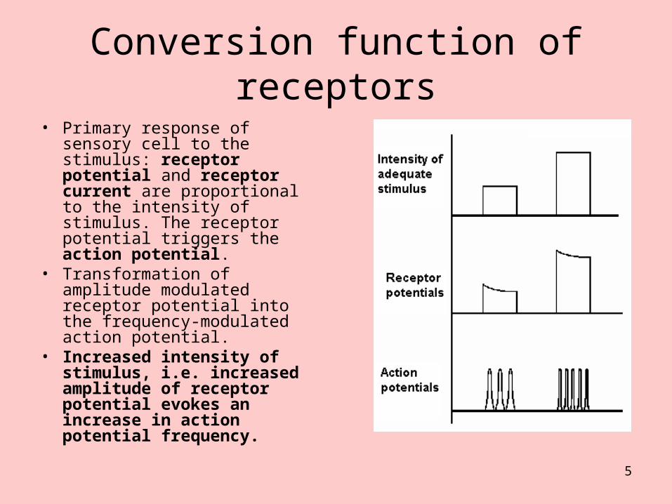
• Primary response of sensory cell to the stimulus: receptor potential and receptor current are proportional to the intensity of stimulus. The receptor potential triggers the action potential.
• Transformation of amplitude modulated receptor potential into the frequency-modulated action potential.
• Increased intensity of stimulus, i.e. increased amplitude of receptor potential evokes an increase in action potential frequency.
6
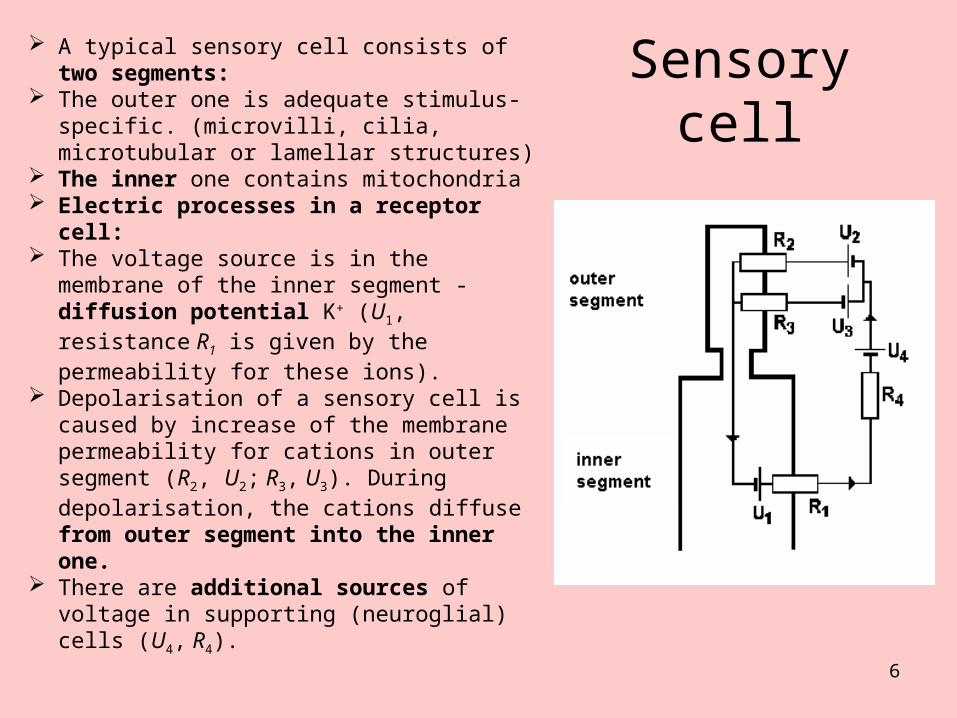
Sensory cell A typical sensory cell consists of two
segments: The outer one is adequate stimulus-
specific. (microvilli, cilia, microtubular or lamellar structures)
The inner one contains mitochondria Electric processes in a receptor cell: The voltage source is in the membrane
of the inner segment - diffusion potential K+ (U1, resistance R1 is given by the permeability for these ions).
Depolarisation of a sensory cell is caused by increase of the membrane permeability for cations in outer segment (R2, U2; R3, U3). During depolarisation, the cations diffuse from outer segment into the inner one.
There are additional sources of voltage in supporting (neuroglial) cells (U4, R4).
7
Biophysical relation between the stimulus and sensation
• The intensity of sensation increases with stimulus intensity non-linearly. It was presumed earlier the sensation intensity is proportional to the logarithm of stimulus intensity (Weber-Fechner law). Intensity of sensation is IR, intensity of stimulus is IS, then:
IR = k1 . log(IS).• Today is the relation expressed exponentially (so-called
Stevens law): IR = k2 . IS
a,• k1, k2 are the proportionality constants, a is an exponent specific
for a sense modality. The Stevens law expresses better the relation between the stimulus and sensation at very low or high stimulus intensities.
8
Adaptation
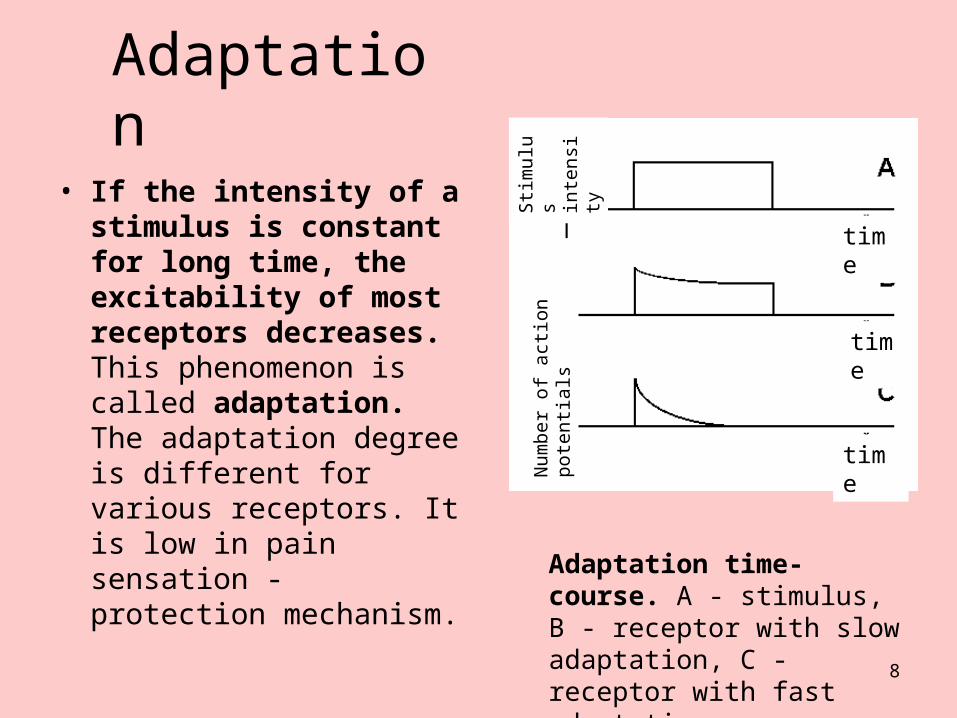
• If the intensity of a stimulus is constant for long time, the excitability of most receptors decreases. This phenomenon is called adaptation. The adaptation degree is different for various receptors. It is low in pain sensation - protection mechanism.
Adaptation time-course. A - stimulus, B - receptor with slow adaptation, C - receptor with fast adaptation
time
time
time
Num
ber
of
actio
n po
tent
ials
Stim
ulus
inte
nsity
9
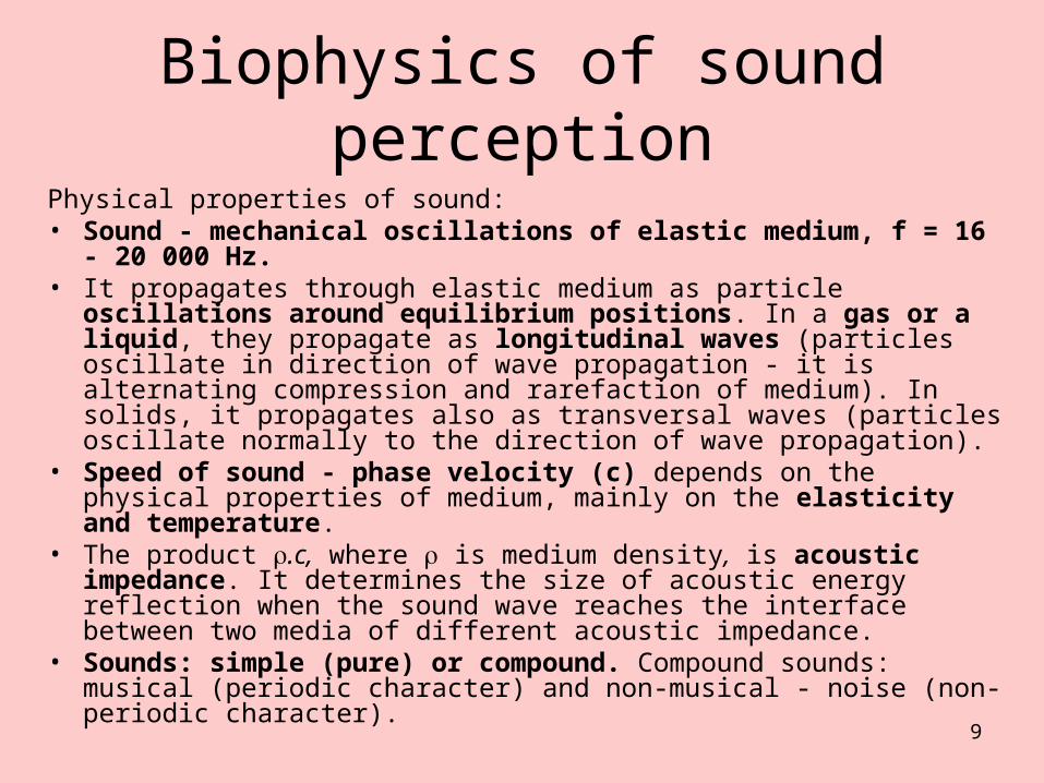
Biophysics of sound perceptionPhysical properties of sound:• Sound - mechanical oscillations of elastic medium, f = 16 - 20 000
Hz.• It propagates through elastic medium as particle oscillations around
equilibrium positions. In a gas or a liquid, they propagate as longitudinal waves (particles oscillate in direction of wave propagation - it is alternating compression and rarefaction of medium). In solids, it propagates also as transversal waves (particles oscillate normally to the direction of wave propagation).
• Speed of sound - phase velocity (c) depends on the physical properties of medium, mainly on the elasticity and temperature.
• The product .c, where is medium density, is acoustic impedance. It determines the size of acoustic energy reflection when the sound wave reaches the interface between two media of different acoustic impedance.
• Sounds: simple (pure) or compound. Compound sounds: musical (periodic character) and non-musical - noise (non-periodic character).
10
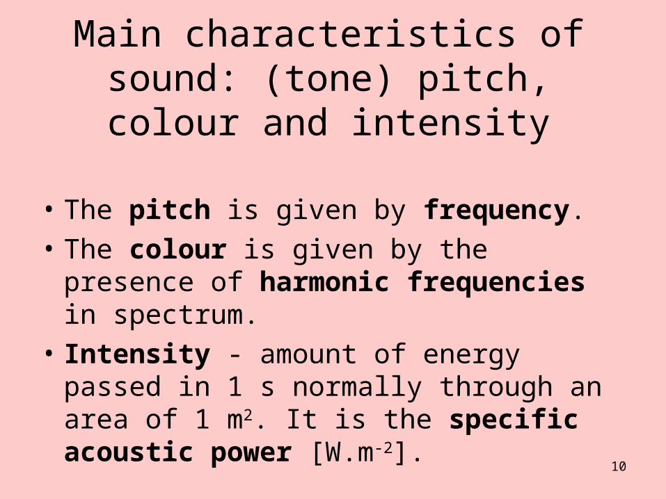
Main characteristics of sound: (tone) pitch, colour and intensity
• The pitch is given by frequency.
• The colour is given by the presence of harmonic frequencies in spectrum.
• Intensity - amount of energy passed in 1 s normally through an area of 1 m2. It is the specific acoustic power [W.m-2].
11
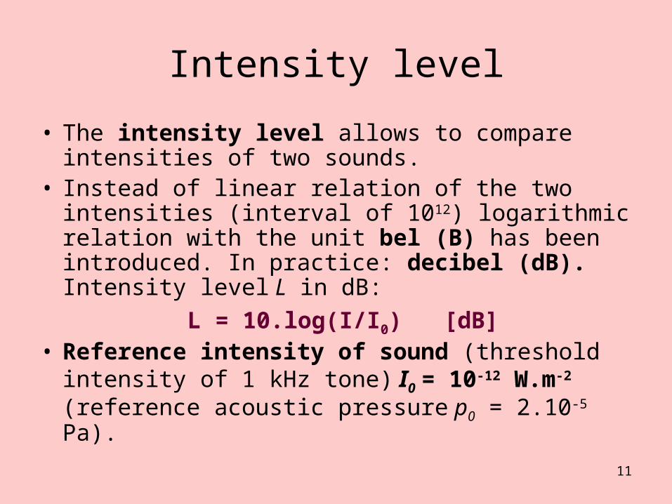
Intensity level
• The intensity level allows to compare intensities of two sounds.
• Instead of linear relation of the two intensities (interval of 1012) logarithmic relation with the unit bel (B) has been introduced. In practice: decibel (dB). Intensity level L in dB:
L = 10.log(I/I0) [dB]• Reference intensity of sound (threshold
intensity of 1 kHz tone) I0 = 10-12 W.m-2 (reference acoustic pressure p0 = 2.10-5 Pa).

12
Loudness, hearing field
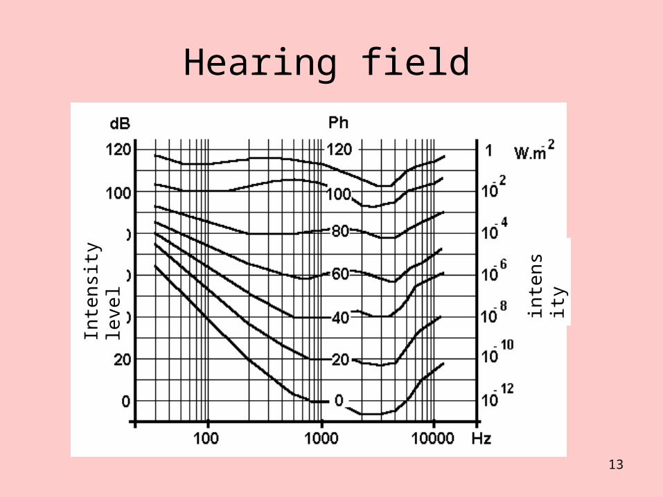
• Loudness is subjectively felt intensity approx. proportional to the logarithm of the physical intensity change of sound stimulus. The ear is most sensitive for frequencies of 1-5 kHz. The loudness level is expressed in phones (Ph). 1 phone corresponds with intensity level of 1 dB for the reference tone (1 kHz). For the other tones, the loudness level differs from the intensity level. 1 Ph is the smallest difference in loudness, which can be resolved by ear. For 1 kHz tone, an increase of loudness by 1 Ph needs an increase of physical intensity by 26%.
• The unit of loudness is son. 1 son corresponds (when hearing by both ears) with the hearing sensation evoked by reference tone of 40 dB.
• Loudness is a threshold quantity.• When connecting in a graph the threshold intensities of audible
frequencies, we obtain the zero loudness line (zero isophone). For any frequency, it is possible to find an intensity at which the hearing sensation changes in pain - pain threshold line in a graph. The field of intensity levels between hearing threshold and pain threshold in frequency range of 16 - 20 000 Hz is the hearing field.
13
Hearing fieldIn
ten
sity
leve
l
inte
nsity
14
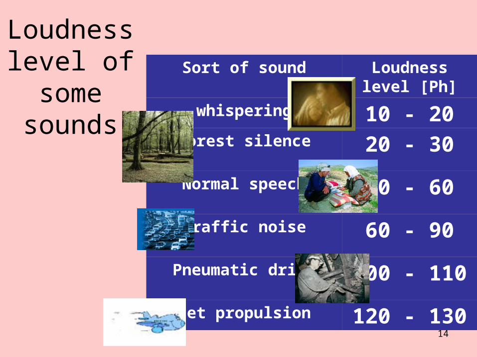
Loudness level of some
sounds
Sort of sound Loudness level [Ph]
whispering 10 - 20Forest silence 20 - 30
Normal speech 40 - 60
Traffic noise 60 - 90
Pneumatic drill 100 - 110
Jet propulsion 120 - 130
15
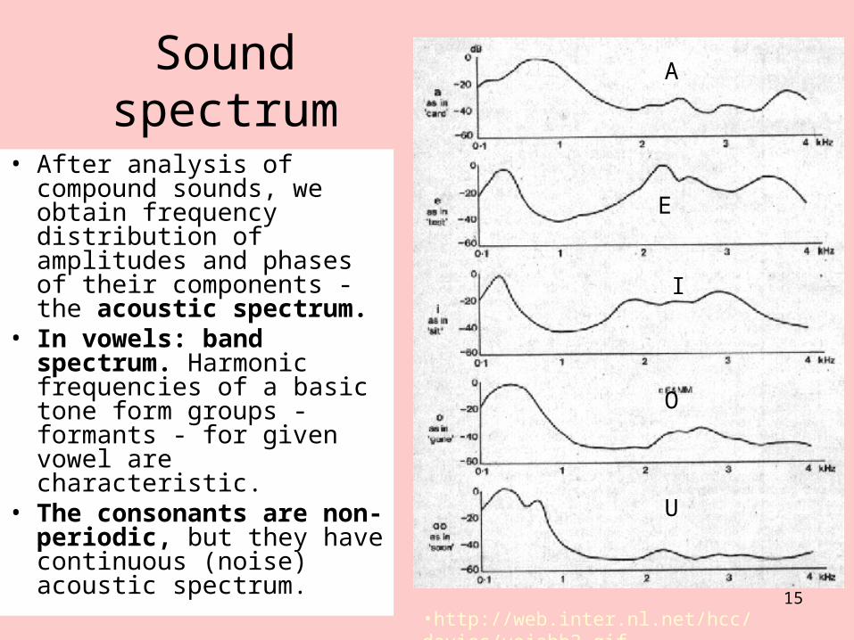
Sound spectrum
• After analysis of compound sounds, we obtain frequency distribution of amplitudes and phases of their components - the acoustic spectrum.
• In vowels: band spectrum. Harmonic frequencies of a basic tone form groups - formants - for given vowel are characteristic.
• The consonants are non-periodic, but they have continuous (noise) acoustic spectrum.
A
E
I
O
U
•http://web.inter.nl.net/hcc/davies/vojabb2.gif

16
Biophysical function of the earThe ear consists of outer, middle and inner ear.
• Transmission of sounds into inner ear is done by outer and middle ear.• Outer ear: auricle (ear pinna) and external auditory canal. Optimally
audible sounds come frontally under the angle of about 15 measured away the ear axis.
• Auditory canal is a resonator. It amplifies the frequencies 2-6 kHz with maximum in range of 3-4 kHz, (+12 dB). The canal closure impairs the hearing by 40 - 60 dB.
• Middle ear consists of the ear-drum (~ 60 mm2) and the ossicles – maleus (hammer), incus (anvil) and stapes (stirrup). Manubrium malei is connected with drum, stapes with foramen ovale (3 mm2). Eustachian tube equalises the pressures on both sides of the drum.
• A large difference of acoustic impedance of the air (3.9 kPa.s.m-1) and the liquid in inner ear (15 700 kPa.s.m-1) would lead to large intensity loss (about 30 dB). It is compensated by the ratio of mentioned areas and by the change of amplitude and pressure of acoustic waves (sound waves of the same intensity have large amplitudes and low pressure in the air, small amplitudes and high pressure in a liquid). Transmission of acoustic oscillations from the drum to the smaller area of oval foramen increases pressure 20x.
17
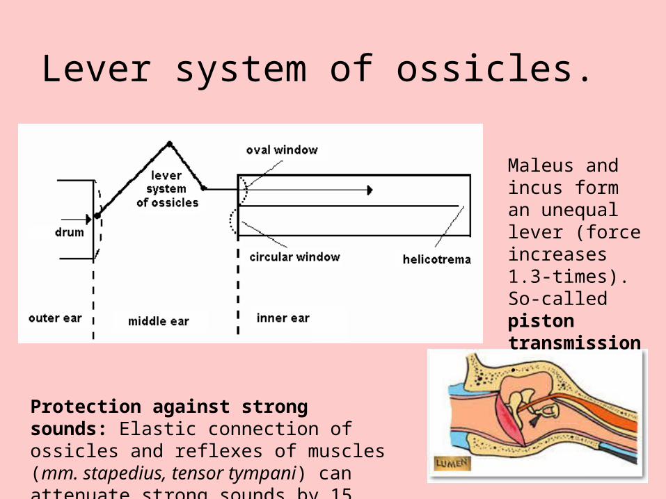
Lever system of ossicles.
Protection against strong sounds: Elastic connection of ossicles and reflexes of muscles (mm. stapedius, tensor tympani) can attenuate strong sounds by 15 dB.
Maleus and incus form an unequal lever (force increases 1.3-times). So-called piston transmission.
18
Mechanism of reception of acoustic signals
• The inner ear is inside the petrous bone and contains the receptors of auditory and vestibular analyser.
• The auditory part is formed by a spiral, 35 mm long bone canal - the cochlea. The basis of cochlea is separated from the middle ear cavity by a septum with two foramina.
• The oval foramen is connected with stapes, the circular one is free.
• Cochlea is divided into two parts by longitudinal osseous lamina spiralis and elastic membrana basilaris. Lamina spiralis is broadest at the basis of cochlea, where the basilar membrane is narrowest, about 0.04 mm (0.5 mm at the top of cochlea).
• The helicotrema connects the space above (scala vestibuli) and below the basilar membrane (scala tympani).
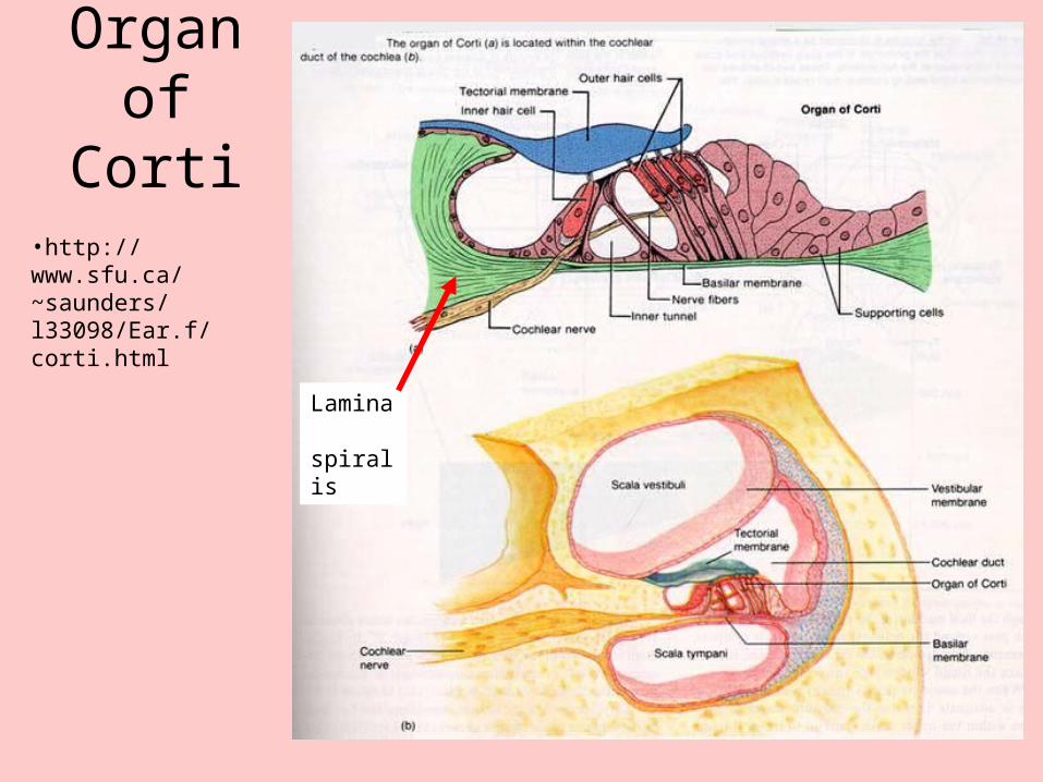
19
Organ of Corti
•http://www.sfu.ca/~saunders/l33098/Ear.f/corti.html
Lamina spiralis
20
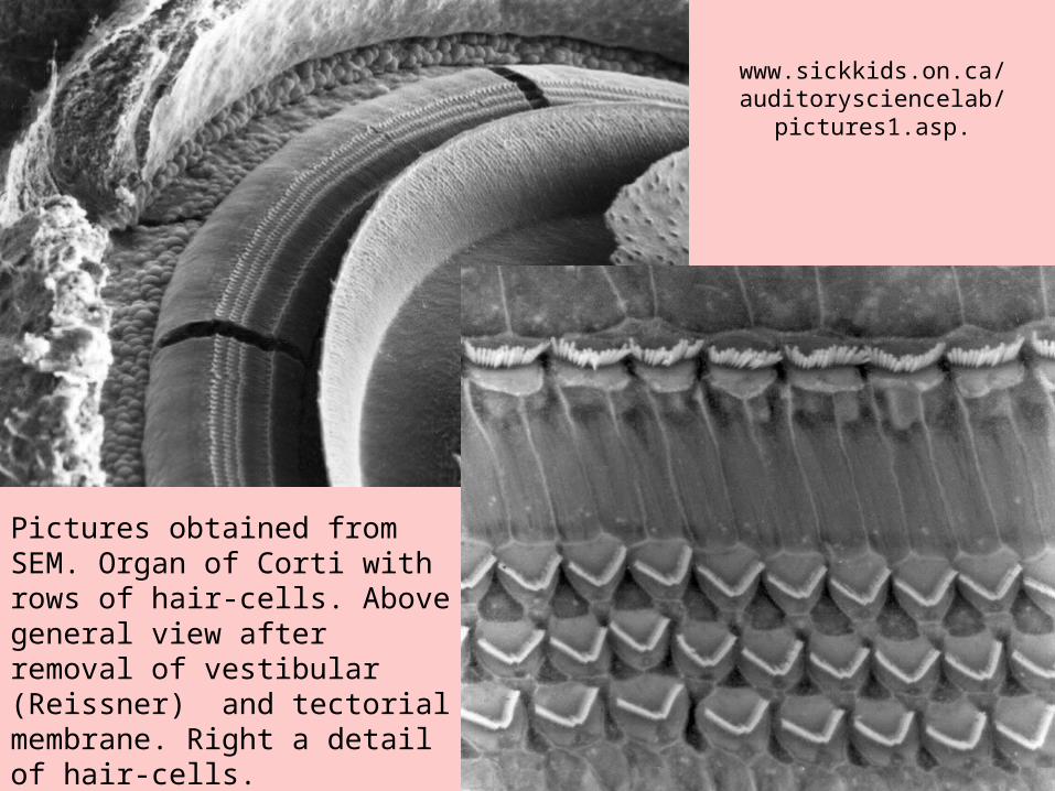
www.sickkids.on.ca/auditorysciencelab/
pictures1.asp.
Pictures obtained from SEM. Organ of Corti with rows of hair-cells. Above general view after removal of vestibular (Reissner) and tectorial membrane. Right a detail of hair-cells.
21
Organ of Corti• Perilymph - ionic composition like liquor, but it has 2x more
proteins. Endolyph - protein content like liquor, but only 1/10 of Na+ ions and 30x more K+ ions - like intracellular liquid.
• Sensory cells of Corti's organ: hair-cells (inner and outer). In cochlea there are about 4000 inner and about 20000 outer hair-cells.
• sensory hairs (cilia) - stereocilia, deformed by tectorial membrane. Bending of hairs towards lamina spiralis leads to depolarisation, bending away lamina spiralis causes hyperpolarisation.
• About 95% neurons begin on inner cells (20 axons on one inner cell), about 5% neurons begin on outer cells - nerve-endings of 10 outer cells are connected in 1 axon. There are about 25 - 30 000 axons in auditory nerve.
22
Mechanism of sound perception: Békésy theory of travelling wave.
• Békésy theory of travelling wave: Sound brings the basilar membrane into oscillations, and the region of maximum oscillation shifts with increasing frequency from the top to the basis of cochlea.
• The receptor system in cochlea performs probably a preliminary frequency analysis. The further processing is done in cerebral auditory centres.
• Sound comes to the receptors in three ways: air (main), bone (the hearing threshold is by about 40 dB higher) and through circular foramen – small importance.
23
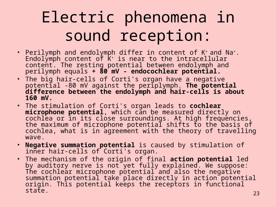
Electric phenomena in sound reception:
• Perilymph and endolymph differ in content of K+ and Na+. Endolymph content of K+ is near to the intracellular content. The resting potential between endolymph and perilymph equals + 80 mV - endocochlear potential.
• The big hair-cells of Corti's organ have a negative potential -80 mV against the periplymph. The potential difference between the endolymph and hair-cells is about 160 mV.
• The stimulation of Corti's organ leads to cochlear microphone potential, which can be measured directly on cochlea or in its close surroundings. At high frequencies, the maximum of microphone potential shifts to the basis of cochlea, what is in agreement with the theory of travelling wave.
• Negative summation potential is caused by stimulation of inner hair-cells of Corti's organ.
• The mechanism of the origin of final action potential led by auditory nerve is not yet fully explained. We suppose: The cochlear microphone potential and also the negative summation potential take place directly in action potential origin. This potential keeps the receptors in functional state.
24
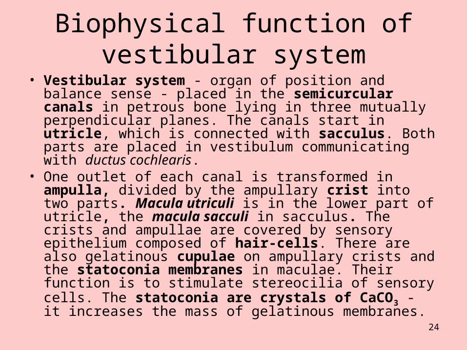
Biophysical function of vestibular system
• Vestibular system - organ of position and balance sense - placed in the semicurcular canals in petrous bone lying in three mutually perpendicular planes. The canals start in utricle, which is connected with sacculus. Both parts are placed in vestibulum communicating with ductus cochlearis.
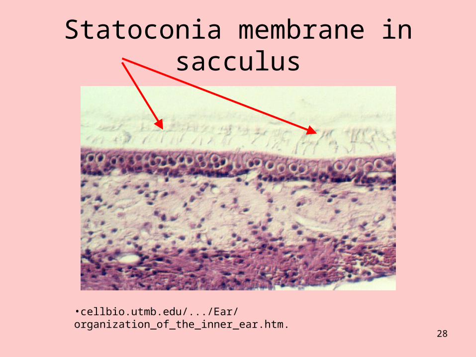
• One outlet of each canal is transformed in ampulla, divided by the ampullary crist into two parts. Macula utriculi is in the lower part of utricle, the macula sacculi in sacculus. The crists and ampullae are covered by sensory epithelium composed of hair-cells. There are also gelatinous cupulae on ampullary crists and the statoconia membranes in maculae. Their function is to stimulate stereocilia of sensory cells. The statoconia are crystals of CaCO3 - it increases the mass of gelatinous membranes.
25
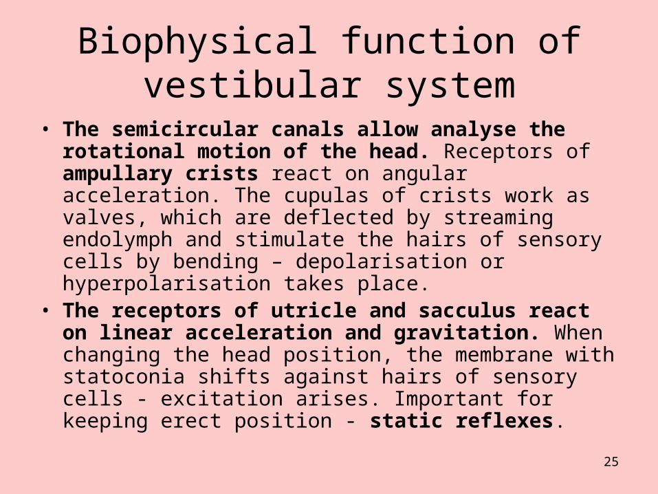
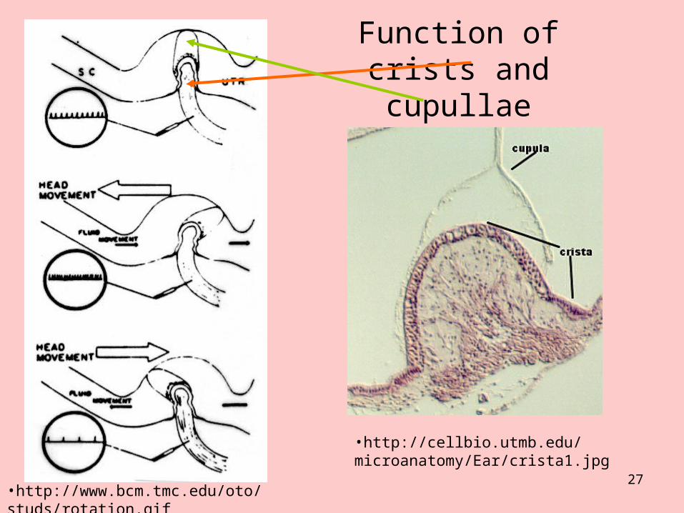
• The semicircular canals allow analyse the rotational motion of the head. Receptors of ampullary crists react on angular acceleration. The cupulas of crists work as valves, which are deflected by streaming endolymph and stimulate the hairs of sensory cells by bending – depolarisation or hyperpolarisation takes place.
• The receptors of utricle and sacculus react on linear acceleration and gravitation. When changing the head position, the membrane with statoconia shifts against hairs of sensory cells - excitation arises. Important for keeping erect position - static reflexes.
Biophysical function of vestibular system
26
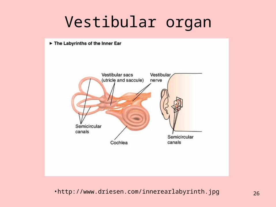
Vestibular organ
•http://www.driesen.com/innerearlabyrinth.jpg
27
Function of crists and cupullae
•http://www.bcm.tmc.edu/oto/studs/rotation.gif
•http://cellbio.utmb.edu/microanatomy/Ear/crista1.jpg
28
Statoconia membrane in sacculus
•cellbio.utmb.edu/.../Ear/ organization_of_the_inner_ear.htm.
29
Author: Vojtěch Mornstein
Content collaboration and language revision: Ivo Hrazdira, Carmel J. Caruana
Presentation design: - - -
Last revision: May 2012