professional skills review gastrointestinal...
TRANSCRIPT

Kingdom of BahrainArabian Gulf University
College of Medicine and Medical Sciences
Prepared by: Ali Jassim Alhashli
Based on: Macleod’s Clinical Examination; 13th Ed
Professional Skills ReviewGastrointestinal System

Surface Anatomy of The Abdomen
• Surface anatomy of liver and spleen:
– Liver:
• Upper border: right 5th
intercostal space at midclavicularline on full expiration.
• Lower border: right costal marginon full inspiration.
– Spleen: left 9th-11th ribs atmidaxillary line.
• The abdomen is divided to 9 regions asshown in the image.

• Anorexia and weight loss:
– Anorexia: loss of appetite.
– Weight loss occurs due to reduced energy intake NOT increased energyexpenditure. Net calorie deficit of 1000 kcal/day leads to weight loss of nearly1 kg/week.
– Energy requirement:
• Males: 2500 kcal/day.
• Females: 2000 kcal/day.
• Painful mouth:
– Etiology:
• Vitamin deficiencies: iron, folate, vitamin B12 and vitamin C.
• Crohn’s disease.
• Dermatological disorders.
• Heartburn and reflux: retrosternal discomfort radiating upwards(Gastroesophageal Reflux Disease GERD).
History

• Dyspepsia: dyspepsia which worsens with an empty stomach and is relieved by food → classical symptomsof peptic ulcer disease (duodenal ulcer).
• Odynophagia: pain with swallowing which occurs with:
– Esophageal ulcer.
– Esophageal candidiasis.
• Abdominal pain:
– Fever, nausea/vomiting and colicky pain in right iliac fossa → appendicitis.
– Pain in left iliac fossa → diverticulitis.
– Severe pain in right hypochondrium → cholecystitis.
– Periumbilical pain radiating to the back → pancreatitis.
– Colicky flank pain (which can move from loin to groin) → renal stone.
– Males with abdominal pain may have → testicular torsion.
– Epigastric pain triggered by stress, spicy/fatty food or NSAIDs → Peptic Ulcer Disease (PUD).
• Dysphagia:
– Difficulty in swallowing.
– Etiology:
• Tonsillitis, pharyngitis and mouth ulcers.
• Bulbar or pseudobulbar palsy.
• Achalasia or pharyngeal pouch. Achalasia is failure of Lower Esophageal Sphincter (LES) to relaxresulting in dilation of esophagus above the sphincter with regurgitation of food.
• Esophageal cancer.
History
• Nausea and vomiting:
– Non-bile stained vomit: gastric outlet obstruction (projectile vomiting).
– Bile-stained vomit: obstruction distal to pylorus.
• Wind and flatulance:
– Flatus: mixture of swallowed air and colonic bacterial fermintation of poorly absorbedcarbohydrates.
• Abdominal distention:
– Remember the 5 F’s: Fat, Flatus, Fluid, Feces and Fetus.
– Acites:
• Definition: it is accumulation of fluid in peritoneal cavity.
• Etiology:
– Transudative: cirrhosis and portal hypertension.
– Exudative: intra-abdominal malignancy.
• Diarrhea:
– Frequent passage of loose stools.
– Normal bowel habit: 3 times/day – once/3days.
– Steatorrhea (fat in stool): due to fat malabsoprtion; stool is greasy/bulky and difficult to flush away.
– Bloody diarrhea:
• Infective gastroenteritis.
• Inflammatory Bowel Disease (IBD).
• Colonic ischemia.
History

• Hematemesis: vomiting blood (fresh red or coffee ground when blood is coming from thestomach).
• Melena:
– Passage of tarry black stool due to upper GI bleeding (melena occurs when bleeding exceeds≥ 100 ml).
– Etiology:
• Mallory-Weiss tear.
• Esophageal varices.
• Esophagitis and esophageal cancer.
• Gastritis.
• Gastric cancer.
• PUD.
• Jaundice:
– Yellowish discoloration of skin, sclera and mucous membranes due to hyperbilirubinemia.
– Etiology:
• Hemolysis.
• Viral or autoimmune hepatitis.
• Cirrhosis.
• Congenital.
• Primary Biliary Cirrhosis (PBC).
History

Physical Examination of Gastrointestinal System
• What are the first things to make sure of before starting yourexamination?
– Introduce yourself to the patient.
– Explain for him what you are going to do.
– Take his permission.
– Make sure of privacy.
• What is your next step?
– Vital signs:
• Blood pressure (ideal: 120/80; hypotension ≤ 90/60; hypertension≥ 140/90).
• Temperature (36.5 – 37.5 C).
• Respiratory rate (12-16 breaths/ minute).
• Pulse: rate, rhythm, volume and character (notice that condition ofvessel wall is of no clinical significance).
Physical Examination of Gastrointestinal System
• Make sure of patient’s position:
– Patient is lying supine on bed with a small pillow placed under his head to relax muscles of anteriorabdominal wall.
– Patient should empty his urinary bladder bedore physical examination.
– Expose the abdomen from xyphisternum to pubic symphysis.
• Comment on the general appearance of the patient:
– Conscious, alert?
– Pain or respiratory distress?
– Cachectic or obese?
– Connected to any devices?
• Then, start by your general inspection:
– Hands:
• Clubbing of fingers? → this occurs with liver cirrhosis, Inflammatory Bowel Disease (IBD) andceliac disease (don’t forget to memorize grades of clubbing).
• Koilonychia (spoon-shaped nails) which occurs with iron-deficiency anemia.
• Leukonychia (white nails) → this occurs due to hypoalbuminemia, celiac disease or nephroticsyndrome.
• Palmar erythema or spider navi → occurring due to increased estrogen levels when patient hasliver failure (normally breakdown of sex steroids occurs in the liver).
• Peripheral cyanosis and tobacco staining.
• Fine tremor or flapping tremor (which might occur with hepatic encephalopathy).
• Dupuytren’s contracture (indicating alcohol liver disease).
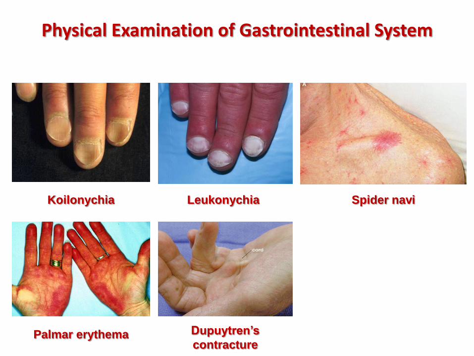
Physical Examination of Gastrointestinal System
Koilonychia Leukonychia Spider navi
Palmar erythema Dupuytren’s
contracture

• General inspection (continued):
– Face and neck:
• Pallor of conjunctiva → anemia.
• Jaundiced sclera → hyperbilirubinemia.
• Central cyanosis (bluish discoloration of themouth).
• Tongue:
– Pale and smooth: iron-deficiency anemia.
– Red beefy tongue: vitamin B12 or folicacid deficiency.
• Angular stomatitis (inflammation of one orboth corners of the mouth due to riboflavindeficiency).
• Remember to check cervical lymph nodes:gastric and pancreatic cancers causeenlargement of the left supraclavicular lymphnode (this is known as troisier’s sign =Virchow’s node).
Physical Examination of Gastrointestinal System

• Inspection of the abdomen:
• The abdomen is not distended (normally it is flat or slightly scaphoid). If the abdomen is distended, how would you differentiate if this is due to obesity, ascites or a localized mass?
– Obesity: abdomen looks rounded and the umbilicus is usually sunken.
– Ascites: umbilicus is flat or everted.
– Localized mass: there is asymmetrical enlargement of the abdomen.
• It is symmetrical and moving with respiration. What is the normal breathing pattern in males and females?
– Males: abdomino-thoracic.
– Females: Thoracoabdominal.
• Umbilicus is centrally located (normally it is everted).
• There is no rash, no discoloration, normal hair distribution.
• There are no scars. How would you know if the patient had a previous laparoscopy?
– There would be a small infraumbilical incision.
• No dilated veins can be seen. When do you see dilated veins?
– Portal hypertension (producing what is known as “caput medusae”).
– Inferior vena cava obstruction.
Physical Examination of Gastrointestinal System

Obesity Ascites Localized mass
Male/female hair
distribution
Laparoscopy scars Dilated abdominal
veins

• Inspection of the abdomen (continued):
• There are no obvious masses and no visible peristalsis. Mention twoconditions in which a patient has visible peristalsis.
– Intestinal obstruction (large or small bowel obstruction).
– Pyloric obstruction.
• How would you confirm the presence of a hernia?
– Ask the patient to cough → a bulge will appear in the abdomendue to the defect in abdominal wall.
• There is no striae. How would you differentiate between a normaland abnormal striae.
– Normal: silver-white striae (stretch marks).
– Abnormal: purple striae (can be due to Cushing’s syndrome) ornormal conditions such as obesity (new stretch marks) andpregnancy.
Physical Examination of Gastrointestinal System


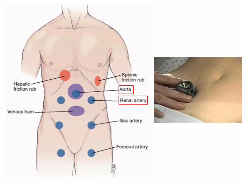
• Auscultation of the abdomen:
• Use the diaphragm of your stethoscope.
• What are going to auscultate for?
– Bowel sounds:
• Divide the abdomen into 4 quadrants and start by listening over the rightlower quadrant.
• What would be the normal finding?
– Gurgling noises (due to normal peristaltic activity of the gut). Normalrange: 6-12/ minute.
• When would you find increased bowel sounds?
– When there is intestinal obstruction.
• When would you find absent bowel sounds?
– Paralytic ileus or peritonitis.
– Listen 1 cm above the umbilicus for abdominal aorta bruits (due to: atheromatousaorta, aneurysmal aorta or superior mesenteric artery stenosis).
– Listen 2 cm above and lateral to the umbilicus (on both sides) for renal artery bruits(due to stenosis).
– Also listen over femoral artery (on both sides) and the liver.
Physical Examination of Gastrointestinal System

• Palpation of the abdomen:
• Palpation of the abdomen is divided to:
– Superficial palpation.
– Deep palpation.
• Superficial palpation:
– Before palpating patient’s abdomen, ask if he has any pain (area of tenderness) and keep it as the last area to bepalpated.
– Don’t forget to maintain eye contact with your patient and watch for any facial expressions.
– Use your right hand, keep it warm and start your (LIGHT) palpation from the right iliac fossa in a systemic pattern (INA CIRCLE).
– What are you looking for:
• It there rigidity or guarding?What is the difference between them?
– Rigidity: abdominal muscles are tight or hard as a board.
– Guarding: Involuntary protection of the area which is being examined.
• Is there any tenderness (site of tenderness and differentials?)
• Temperature.
• Are there any palpable masses? If yes, how would you describe it?
– Location.
– Size.
– Mobility.
– Shape.
– Consistency.
– Superficial or deep to muscles? How to differentiate?
Physical Examination of Gastrointestinal System

• Palpation of the abdomen (continued):
• Deep palpation (liver, spleen and kidneys):
– Palpation of the liver:
• Place your right hand flat on the skin over the right iliac fossa.
• Ask the patient to breath deeply through the mouth.
• Press on the abdomen with each inspiration taken by the patient.
• Move your hand progressively up the abdomen (1-2 cm) between each breath thepatient takes.
• What would you expect?
– Normal: the liver is impalpable (but in children you might feel it).
– Abnormal: if you feel the edge of the liver you have to describe the following
» Size (how many cm below the costal margin).
» Surface: is it smooth or irregular (same for the edge).
» Consistency: is it soft or hard?
» Tenderness.
– What are you differentials for hepatomegaly?
» Alcoholic liver disease.
» Hepatitis.
» Hepatocellular carcinoma.
» Right heart failure.
» Leukemia and lymphoma.
Physical Examination of Gastrointestinal System
• Palpation of the abdomen (continued):
• Deep palpation (liver, spleen and kidneys)… continued:
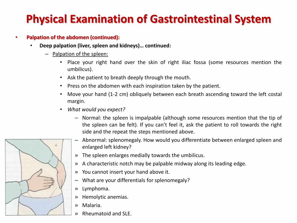
– Palpation of the spleen:
• Place your right hand over the skin of right iliac fossa (some resources mention theumbilicus).
• Ask the patient to breath deeply through the mouth.
• Press on the abdomen with each inspiration taken by the patient.
• Move your hand (1-2 cm) obliquely between each breath ascending toward the left costalmargin.
• What would you expect?
– Normal: the spleen is impalpable (although some resources mention that the tip ofthe spleen can be felt). If you can’t feel it, ask the patient to roll towards the rightside and the repeat the steps mentioned above.
– Abnormal: splenomegaly. How would you differentiate between enlarged spleen andenlarged left kidney?
» The spleen enlarges medially towards the umbilicus.
» A characteristic notch may be palpable midway along its leading edge.
» You cannot insert your hand above it.
– What are your differentials for splenomegaly?
» Lymphoma.
» Hemolytic anemias.
» Malaria.
» Rheumatoid and SLE.
Physical Examination of Gastrointestinal System
• Palpation of the abdomen (continued):
• Deep palpation (liver, spleen and kidneys)…continued:
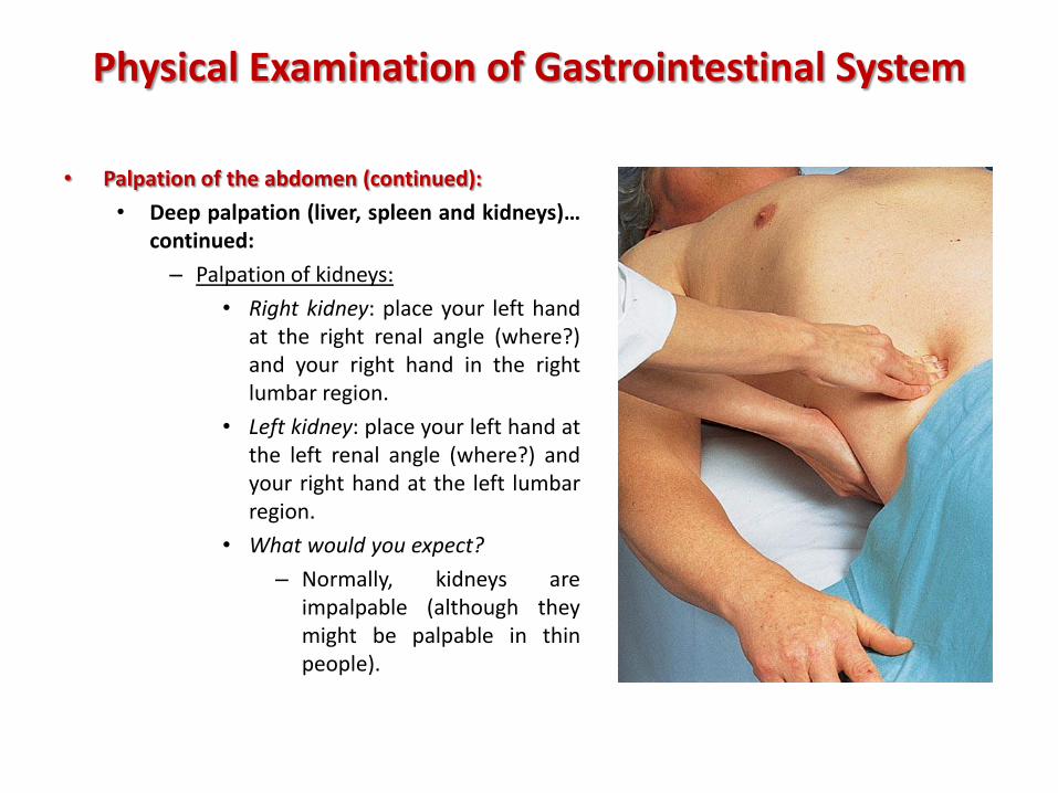
– Palpation of kidneys:
• Right kidney: place your left handat the right renal angle (where?)and your right hand in the rightlumbar region.
• Left kidney: place your left hand atthe left renal angle (where?) andyour right hand at the left lumbarregion.
• What would you expect?
– Normally, kidneys areimpalpable (although theymight be palpable in thinpeople).
Physical Examination of Gastrointestinal System

• Percussion of the abdomen:
• Percussion for what?
– Liver.
– Spleen.
– Urinary bladder.
– Ascites.
• Percussion of the liver:
– Ask the patient to hold his breath in full expiration (why?)
– Start percussing downward from the 2nd intercostal space in mid-clavicular line until hearing dullnesswhich indicates for the upper border of the liver (usually in the 5th intercostal space).
– Percuss/palpate from right iliac fossa upward to reach the lower edge of the liver.
– Measure liver span with a meter (normal: 10 cm ± 2).
• Percussion of the spleen:
– Start percussing at the left mid-axillary line descending down to the costal margin.
– Normally, you will hear dullness between 9-11th ribs.
• Percussion of urinary bladder:
– Percuss in the midline starting below the umbilicus and descending until you reach the pubicsymphysis.
– What would you expect?
• Normal: resonance (because we asked the patient to empty his bladder before examination).
• Abnormal: dullness. How would you differentiate if it is tumor from the bladder or abdominaltumor?
– All dullness: tumor from bladder extending up to the abdomen.
– Dullness in the upper part but tympanic near pubic symphysis: tumor from abdomen.
Physical Examination of Gastrointestinal System


• Percussion of the abdomen (continued):
• Ascites:
– Mild ascites (shifting dullness):
• Start percussing in the midline from epigastric area and descending downuntil reaching the hypogastric region.
• Then percuss right and left flanks. If there is ascites, fluid will accumulate inflanks due to effect of gravity and you will hear dullness.
• Suppose you heard dullness in the right flank. To confirm the presence offluid, ask the patient to turn to the left side. Wait for 10 seconds and thenpercuss the right flank again and the sound will be converted into tympanic.
– Gross ascites (fluid thrill):
• Ask an assistant (nurse or patient himself) to place the ulnar border of hishand on the patient’s abdomen in the midline.
• Place the palm of one hand on the patient’s right flank and tap with otherhand fingers on patient’s left flank.
• Notice that only fluids in peritoneal cavity elicit such a thrill.
• When ending your examination, don’t forget to……….
– Say: “I will end my examination with PR exam”.
– Thank the patient and cover him.
Physical Examination of Gastrointestinal System
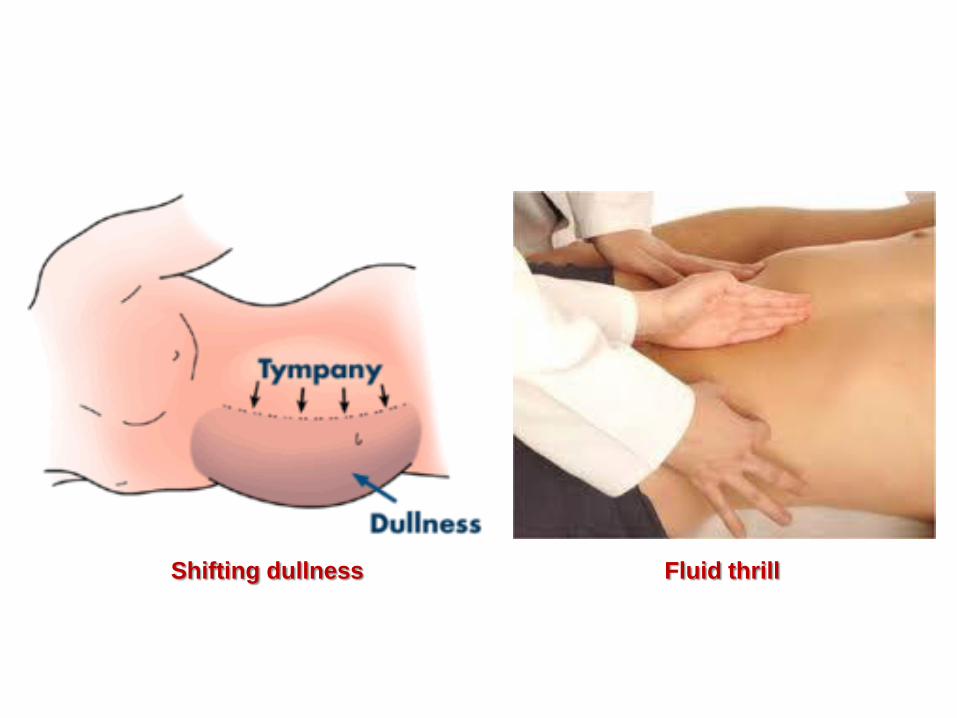
Shifting dullness Fluid thrill