prof. jan bakker md, phd chair dept intensive care adults ... · optimization and lactate...
TRANSCRIPT

EGDTlactate
Prof. Jan Bakker MD, PhD
Chair dept Intensive Care Adults
www.intensivecare.me
Tuesday, March 20, 12

20
30
40
50
60
70
80
90
100
>3 >4 >5 >6 >7 >8 >9 >10
Hospital mortality (%)
Lactate mmol/L
n=156
2006-2008r=0.99
Tuesday, March 20, 12

Intensive Care Med (2007) 33:1892–1899DOI 10.1007/s00134-007-0680-5 O R I G I N A L
Michael D. HowellMichael DonninoPeter ClardyDaniel TalmorNathan I. Shapiro
Occult hypoperfusion and mortality in patientswith suspected infection
Received: 29 June 2006Accepted: 30 March 2007Published online: 6 July 2007© Springer-Verlag 2007
Electronic supplementary materialThe online version of this article(doi:10.1007/s00134-007-0680-5) containssupplementary material, which is availableto authorized users.
This article is discussed in the editorialavailable at: http://dx.doi.org/10.1007/s00134-007-0679-y
M. D. Howell (!) · P. ClardyBeth Israel Deaconess Medical Center andHarvard Medical School, KB-23, Divisionof Pulmonary and Critical Care Medicine,Department of Medicine,330 Brookline Avenue, Boston 02215, MA,USAe-mail: [email protected].: +1-617-3596331Fax: +1-617-7542350
M. Donnino · N. I. ShapiroBeth Israel Deaconess Medical Center andHarvard Medical School, Department ofEmergency Medicine,Boston MA, USA
D. TalmorBeth Israel Deaconess Medical Center andHarvard Medical School, Department ofAnesthesia, Critical Care and PainMedicine,Boston MA, USA
Abstract Objective: To determine,in the early stages of suspectedclinically significant infection, theindependent relationship of thepresenting venous lactate level to28-day in-hospital mortality. Design:Prospective, observational cohortstudy. Setting: Urban, universitytertiary-care hospital. Patients: Onethousand two hundred and eightyseven adults admitted through theemergency department who had clin-ically suspected infection and a lac-tate measurement. Measurementsand results: Seventy-three [5.7%(95% CI 4.4–6.9%)] patients died inthe hospital within 28 days. Lactatelevel was strongly associated with28-day in-hospital mortality in uni-variate analysis (p < 0.0001). Whenstratified by blood pressure, lactateremained associated with mortality(p < 0.0001). Normotensive patients
with a lactate level ! 4.0 mmol/l hada mortality rate of 15.0% (6.0–24%).Patients with either septic shockor lactate ! 4.0 mmol/l had a mor-tality rate of 28.3% (21.3–35.3%),which was significantly higher thanthose who had neither [mortality of2.5% (1.6–3.4%), p < 0.0001]. Ina model controlling for age, bloodpressure, malignancy, platelet count,and blood urea nitrogen level, lactateremained strongly associated withmortality. Patients with a lactatelevel of 2.5–4.0 mmol/l had ad-justed odds of death of 2.2 (1.1–4.2);those with lactate ! 4.0 mmol/l had7.1 (3.6–13.9) times the odds ofdeath. The model had good discrim-ination (AUC = 0.87) and was wellcalibrated. Conclusions: In patientsadmitted with clinically suspectedinfection, the venous lactate levelpredicts 28-day in-hospital mortalityindependent of blood pressure andadds significant prognostic informa-tion to that provided by other clinicalpredictors.
Keywords Sepsis · Infection · Lacticacid/lactate · Triage · Prognosis ·Risk assessment
Introduction
Severe sepsis and septic shock afflict about three-quartersof a million patients in the USA annually, resultingin approximately 215,000 deaths [1]. After decades of
negative sepsis trials, Rivers et al. published a randomized,controlled trial of an early goal-directed hemodynamicmanagement strategy for emergency department patientswith sepsis which resulted in a decrease in absolute mor-tality from 46% to 31% [2]. Since approximately one-third
Emergency Room
Tuesday, March 20, 12

J Crit Care 2011;26:435
Persistent sepsis-induced hypotension without hyperlactatemia may not constitute a real septic shock. Our results support the need to review the current definition of septic shock. Hyperlactatemia could represent an objective parameter worth to be explored as a potential diagnostic criterion for septic shock
Tuesday, March 20, 12

Copyright @ 2009 by the Shock Society. Unauthorized reproduction of this article is prohibited.
MULTICENTER STUDY OF EARLY LACTATE CLEARANCE AS ADETERMINANT OF SURVIVAL IN PATIENTS WITH PRESUMED SEPSIS
Ryan C. Arnold,* Nathan I. Shapiro,† Alan E. Jones,‡ Christa Schorr,§Jennifer Pope,† Elisabeth Casner,‡ Joseph E. Parrillo,§ R. Phillip Dellinger,§
Stephen Trzeciak,* and on behalf of the Emergency Medicine Shock ResearchNetwork (EMShockNet) Investigators
*Department of Emergency Medicine, UMDNJ-Robert Wood Johnson Medical School at Camden, CooperUniversity Hospital, Camden, New Jersey; †Department of Emergency Medicine, Beth Israel DeaconessMedical Center Boston, Massachusetts; ‡Department of Emergency Medicine, Carolinas Medical Center,Charlotte, North Carolina; and §Department of Medicine, Divisions of Cardiovascular Disease and CriticalCare Medicine, UMDNJ-Robert Wood Johnson Medical School at Camden, Cooper University Hospital,
Camden, New Jersey
Received 31 Oct 2008; first review completed 05 Nov 2008; accepted in final form 19 Nov 2008
ABSTRACT—We sought to determine (a) if early lactate clearance is associated with improved survival in emergencydepartment patients with severe sepsis and (b) the concordance between central venous oxygen saturation (ScvO2)optimization and lactate clearance during early sepsis resuscitation. Within a multicenter shock research network that usesquantitative resuscitation for severe sepsis, we analyzed prospectively collected registries of consecutive emergencydepartment patients diagnosed with severe sepsis at three urban hospitals. Inclusion criteria are as follows: (a) age olderthan 17 years, (b) two or more systemic inflammation criteria, (c) systolic blood pressure 90 mmHg or less after fluidchallenge or initial lactate of 4mmol/L or greater, and (d ) initial and repeat lactate measurement within 6 h of resuscitationinitiation. We stratified patients into two groups defined a priori based on previously published data: (a) lactateclearanceVrepeat lactate decrease by 10% or greater from initial (or both initial and repeat levels e2.0 mmol/L), and (b)lactate non-clearanceVrepeat lactate decrease by less than 10% from initial. The primary outcome was in-hospitalmortality. Among 166 patients, lactate non-clearance occurred in 15 (9%) of 166. Mortality was 60% for lactate non-clearance versus 19% for lactate clearance, P G 0.001. On multivariate analysis, lactate non-clearance was anindependent predictor of death (odds ratio, 4.9 [confidence interval, 1.5Y15.9]). We found discordance between ScvO2optimization and lactate clearance; 79% of lactate non-clearance had concomitant ScvO2 of 70% or greater. In thismulticenter cohort of sepsis patients, failing to clear lactate during resuscitation carried a high risk of death, and ScvO2optimization did not reliably exclude lactate non-clearance. These data provide rationale for a clinical trial of lactateclearance as a distinct end point of early sepsis resuscitation.
KEYWORDS—Shock, severe sepsis, resuscitation, lactic acid, emergency medicine
INTRODUCTION
Severe sepsis is the most common cause of death incritically ill patients (1). The effectiveness of early resuscita-tion is an important determinant of sepsis survival (2).Specifically, early quantitative resuscitation, the use of astructured set of cardiovascular interventions targeting pre-defined hemodynamic end points, can have a profound effecton hospital mortality (3). Currently, international consensustreatment guidelines recommend a quantitative resuscitationstrategy that includes targeting central venous oxygen satu-ration (ScvO2) of 70% or greater in the first 6 h of severesepsis therapy (4). However, the optimal end points of sepsisresuscitation remain controversial (5, 6).Serum lactate elevation is an important marker of impaired
tissue perfusion in patients with sepsis and is often elevated even
in the absence of arterial hypotension (7). Numerous studiesshow that a single early lactate measurement has importantprognostic significance and predicts mortality in populations ofpatients with infection (8, 9). A small number of studies havepreviously reported that serial measurements of lactate havepotential prognostic value during conventional sepsis manage-ment (10Y12); however, it is not known if lactate clearance isassociated with survival in the context of medical centers thathave adopted and routinely perform aggressive quantitativeresuscitation for severe sepsis. In addition, it is not known iflactate clearance is important independent of other recommen-ded quantitative resuscitation end points, specifically ScvO2.
The objectives of this study were to determine (a) if earlylactate clearance is associated with improved survival inemergency department (ED) patients with severe sepsis and(b) the concordance between ScvO2 optimization and lactateclearance during early sepsis resuscitation.
METHODS
Setting and study designWe analyzed prospectively collected registries of consecutive ED patients
diagnosed with severe sepsis at three urban hospitals. The three hospitals arepart of a multicenter research collaborative (Emergency Medicine ShockResearch Network) (13), where each institution uses ED-based protocolYdirected quantitative resuscitation for patients with severe sepsis (14Y16).The registries were compiled between 2004 and 2007 and were approved
35
SHOCK, Vol. 32, No. 1, pp. 35Y39, 2009
Address reprint requests to Ryan C. Arnold, MD, Department of EmergencyMedicine UMDNJ-Robert Wood Johnson Medical School at Camden CooperUniversity Hospital, One Cooper Plaza, Camden, NJ 08103. Email: [email protected].
Funding: Dr Trzeciak’s effort to this project was supported by a grant from theNational Institutes of Health/National Institutes of General Medical Sciences(K23GM83211). Dr Jones’ effort to this project was supported by a grant from theNational Institutes of Health/National Institutes of General Medical Sciences(K23GM76652).DOI: 10.1097/SHK.0b013e3181971d47Copyright ! 2009 by the Shock Society
Copyright @ 2009 by the Shock Society. Unauthorized reproduction of this article is prohibited.
10:1 necessary for multivariate modeling (21, 22); and (c) the multivariatemodel would need to, at a minimum, include the following covariates thathave previously been shown to differentiate sepsis survivors from non-survivors: arterial hypotension (23), failure to achieve ScvO2 70% or greater(2), and lactate non-clearance (12). To accrue the necessary 30 cases ofmortality to test these three covariates in a multivariate model, we estimatedthat a minimum of 120 total cases would be necessary.
RESULTS
Characteristics of study cohort
There were 166 subjects who met inclusion criteria. Theoverall mortality rate was 23% (38/166). Table 1 showscharacteristics of the entire cohort. Of the 166 subjects, 110came from center 1, 22 came from center 2, and 34 came fromcenter 3. Using multivariate analysis, there was no apparentcenter effect on in-hospital mortality.
The quantitative resuscitation algorithm targeting ScvO270% or greater was successfully initiated in the ED in 148(89%) of 166 study subjects. Study patients received anaverage of 4.4 L (95% CI, 4.1Y4.7 L) of i.v. crystalloid fluidduring their ED resuscitation. Lactate non-clearance occurredin 15 (9%) of 166, and the remainder (151/166, 91%) clearedlactate.
Lactate clearance versus lactate non-clearance
Table 2 displays data for comparison between lactateclearance and lactate non-clearance groups. Mortality was60% in the lactate non-clearance group versus 19% in thelactate clearance group (proportion difference, 41% [95% CI,19%Y63%; P G 0.001]). Vasopressor use was not significantlydissimilar between the groups (lactate non-clearance: 11/15 or73%, and lactate clearance: 87/151 or 58%; P = 0.39). Figure 2displays the Kaplan-Meier curves for survival fractions overtime. The curves diverge significantly by log-rank test (P =0.003). There was no significant difference in achievement of
TABLE 1. Characteristics of the study cohort (n = 166)
Age, mean (SD), y 66 (15)
Sex, % female, n (%) 83 (50)
Suspected source of infection in ED, n (%)Urinary tract 46 (28)
Pneumonia 22 (13)
Skin/soft tissue 23 (14)
Intra-abdominal 14 (8)
Line/device 10 (6)
Other 7 (4)Unknown primary source 44 (27)
Signs of systemic inflammation, n (%)
Body temperature 938-C or G36-C 131 (79)
Heart rate 990 beats/min 140 (84)
Respiratory rate 920 breaths/min 137 (83)
White blood cell count, 912,000 or G4,000/2L 121 (73)
SBP G90 mmHg despite i.v. fluids, n (%) 91 (55)
Initial serum lactate 94 mmol/L, n (%) 90 (54)
Vasopressor use in ED, n (%) 39 (23)
Individual organ failures, n (%)
Cardiovascular 63 (44)
Pulmonary 28 (17)
Renal 54 (33)
Hepatic 14 (8)
Coagulopathy 23 (14)
Acidosis 140 (84)Total SOFA score, mean (SD) 3.6 (2.6)
Mortality, n (%) 38 (23)
SOFA, Sequential Organ Failure Assessment; ED, EmergencyDepartment.
TABLE 2. Lactate clearance versus lactate non-clearance
Lactateclearance(n = 151)
Lactatenon-clearance
(n = 15) P
Age, mean (SD), y 67 (15) 62 (16) 0.22
SBP G90 mmHg despite i.v.fluids, n (%)
50 (33) 13 (87) 0.02
Initial serum lactate, mean (SD) 4.5 (2.7) 3.9 (1.7) 0.08
Serial serum lactate, mean (SD) 2.3 (1.8) 5.1 (2.9) G0.001
Vasopressor usage, n (%) 87 (58) 11 (73) 0.39
Individual organ failure, n (%)
Cardiovascular 50 (33) 13 (87) G0.001
Pulmonary 25 (17) 3 (20) 0.94
Renal 48 (32) 6 (40) 0.73
Hepatic 12 (8) 2 (13) 0.86
Coagulopathy 18 (12) 5 (33) 0.06
Total SOFA score, mean (SD) 3.6 (2.6) 4.1 (2.3) 0.47
Continuous ScvO2 monitoring,n (%)
134 (81) 14 (93) 0.42
ScvO2 Q70% achieved 114 (85) 11 (79) 0.84
Mortality, n (%) 29 (19) 9 (60) G0.001
Characteristics of study subjects with lactate clearance (910%) and non-clearance (G10%) over the first 6 h (n = 166).SOFA, Sequential Organ Failure Assessment; ED, EmergencyDepartment.
FIG. 2. Kaplan-Meier survival curves. This graph depicts survival curvesover time for lactate clearance (lactate decrease by Q10%) and lactate non-clearance (lactate decrease G10%) groups. The curves diverge significantlyby log-rank test (P = 0.003).
TABLE 3. Lactate clearance and ScvO2 goals
Lactate non-clearance Lactate clearance Total
ScvO2 G70% 3 20 23
ScvO2 Q70% 11 114 125
14 134 148
Among 148 subjects in whom ScvO2 was targeted as an end point ofresuscitation, this table depicts the lack of concordance between lactateclearance and ScvO2. There was no evidence of a relationship betweenlactate clearance and ScvO2 (Fisher exact test, P = 0.457).
SHOCK JULY 2009 EARLY LACTATE CLEARANCE IN SEPSIS RESUSCITATION 37ORdeath: 4.9 (1.5-15.9)
Copyright @ 2009 by the Shock Society. Unauthorized reproduction of this article is prohibited.
the ScvO2 goal between the two groups (85% lactate clearancevs. 79% lactate non-clearance; proportional difference, 6%[95% CI, j14% to 26%]; P = 0.84). Table 3 is a 2 ! 2 tablecomparing the concordance of ScvO2 measurement withlactate clearance. There was no evidence of a relationshipbetween lactate clearance and ScvO2 (Fisher exact test, P =0.457). We found discordance between the two measure-ments. Specifically, 79% of lactate non-clearance had con-comitant ScvO2 70% or greater.
Survivors versus nonsurvivors
Table 4 compares survivors with nonsurvivors. On bivariateanalysis, there were four factors that were significantlydifferent (P G 0.05) between survivors and nonsurvivors: (a)initial cardiovascular organ failure (initial SBP G90 mmHg),(b) persistent hypotension (SBP G90 mmHg) despite intra-venous fluids, (c) maximum ScvO2 less than 70%, and (d )lactate non-clearance. Table 5 shows the results for the multi-variate logistic regression model. Because ScvO2 had a signif-icant association with mortality in the bivariate analysis, welimited the regression model to include only subjects in whomScvO2 was measured continuously (n = 148). We omitted one ofthe two hypotension-related covariates (initial SBP G90 mmHg)from inclusion in the model on the grounds that it could becollinear with the persistent hypotension covariate. Lactate non-clearance was found to be a strong independent predictor of in-hospital mortality (odds ratio, 4.9; 95% CI, 1.5Y15.9).
DISCUSSION
In this multicenter sample of ED patients with severesepsis, we found that early lactate clearance was a strongindependent predictor of in-hospital death. In addition,optimization of ScvO2 during resuscitation was not sufficientto exclude lactate non-clearance. This is the first study toevaluate the impact of lactate clearance on survival in thepresence of a protocol-directed quantitative resuscitation
algorithm for the treatment of patients with severe sepsis,and to date, this is the largest study evaluating lactateclearance in patients with sepsis (10Y12).
The cause of an elevated serum lactate in patients withsepsis can be multifactorial. Although lactate elevation mayresult from acute tissue hypoperfusion and anaerobic metab-olism (24), other possible causes may include (a) sepsis-induced impairment of pyruvate-dehydrogenase enzymeactivity (25), (b) increased lactate production via catechol-amine-driven pathways (26Y28), and (c) decreased lactateclearance due to hepatic dysfunction (29, 30). However,regardless of etiology of an elevated serum lactate, lactateelevation in sepsis has been consistently linked to increasedmortality (7Y12). Although initial reports of lactate clearancein sepsis were published more than 15 years ago (10),evaluating lactate trends during resuscitation is not yet partof the current consensus recommendations for sepsis manage-ment (4) and is not routinely performed in practice. Whileoptimizing ScvO2 could in theory be a single comprehensivemonitor of the systemic balance between global oxygendelivery and consumption in sepsis patients with circulatoryshock (2, 4), our data show that assessment of lactateclearance is important as a predictor of mortality independentof achievement of ScvO2 goals and that tracking ScvO2 doesnot reliably reflect the effectiveness of lactate clearanceduring resuscitation. Therefore, these data suggest that seriallactate measurement may provide unique and importantinformation on resuscitation effectiveness.
We acknowledge important limitations in interpreting theseresults. First, this is a nonexperimental observational studyand as such can detect only the association between lactateclearance and mortality but cannot establish cause and effect.Second, because performing serial lactate measurement inpatients with sepsis is not a mandatory practice in our centersand measurements are performed at the discretion of theclinician, this could potentially represent a source of selectionbias. Third, there was a relatively low incidence (9%) oflactate non-clearance in this cohort. This may be attributableto highly aggressive resuscitation practices in the EDs of theparticipating centers (14Y16). In the only other ED-basedstudy of lactate clearance in severe sepsis, Nguyen et al. (12)reported a lactate non-clearance rate of 29% using the samecutoffs for clearance and non-clearance used in this study. Onepotential explanation for this disparity is the aggressiveness ofcrystalloid resuscitation administered in the first 6 h, 4.4 L
TABLE 4. Survivors versus nonsurvivors (n = 166)
Survivors(n = 128)
Nonsurvivors(n = 38) P
Age, mean (SD), y 66 (15) 66 (16) 1.00
SBP G90 mmHg despite i.v.fluids, n (%)
42 (33) 21 (55) 0.02
Initial serum lactate, mean (SD) 4.3 (2.6) 4.7 (2.8) 0.41
Serial serum lactate, mean (SD) 2.2 (1.6) 3.6 (2.8) G0.001
Individual organ failure, n (%)
Cardiovascular 42 (33) 21 (55) 0.02
Pulmonary 20 (16) 8 (21) 0.64
Renal 43 (34) 11 (29) 0.70
Hepatic 9 (7) 5 (13) 0.40
Coagulopathy 15 (12) 8 (21) 0.26
Total SOFA score, mean (SD) 3.6 (2.6) 3.7 (2.7) 0.84
Continuous ScvO2 monitoring,n (%)
112 (87) 36 (95) 0.28
ScvO2 Q70% achieved, n (%) 99 (88) 26 (72) 0.03
Lactate clearance Q10%, n (%) 122 (95) 29 (76) 0.001
TABLE 5. Multivariate logistic regression analysis
Variable CoefficientOddsratio
95% CIs for oddsratio
Lactate non-clearance 1.59 4.9 1.5Y15.9
Maximum ScvO2 G70% 1.05 2.7 1.1Y7.6
Hypotension despite fluidchallenge
0.10 1.1 0.5Y2.5
Factors associated with increased mortality on bivariate analysis wereevaluated with in-hospital mortality as the dependent variable (n = 148)Logistic regression model was limited to subjects for which ScvO2 wasmeasured continuously (148/166).lactate non-clearance = less than 10% decrease in repeat lactate value;hypotension = SBP G90 mmHg after a 20-mL/kg i.v. fluid bolus.
38 SHOCK VOL. 32, NO. 1 ARNOLD ET AL.
clearance >10%
clearance <10%
Tuesday, March 20, 12

Prognostic Value of Blood Lactate Levels: Does the ClinicalDiagnosis at Admission Matter?Tim C. Jansen, MD, Jasper van Bommel, MD, PhD, Paul G. Mulder, PhD, Alexandre P. Lima, MD,Ben van der Hoven, MD, Johannes H. Rommes, MD, PhD, Ferdinand T. F. Snellen, MD,and Jan Bakker, MD, PhD
Background: Hyperlactatemia andits reduction after admission in the inten-sive care unit (ICU) have been related tosurvival. Because it is unknown whetherthis equally applies to different groups ofcritically ill patients, we compared theprognostic value of repeated lactate levels(a) in septic patients versus patients withhemorrhage or other conditions generallyassociated with low-oxygen transport (LT)(b) in hemodynamically stable versus un-stable patients.
Methods: In this prospective obser-vational two-center study (n ! 394 pa-tients), blood lactate levels at admission to
the ICU (LacT0) and the reduction of lac-tate levels from T ! 0 to T ! 12 hours("LacT0–12) and from T ! 12 to T ! 24hours ("LacT12–24), were related to in-hospital mortality.
Results: Reduction of lactate was as-sociated with a lower mortality only in thesepsis group ("LacT0–12: hazard ratio[HR] 0.34, p ! 0.004 and "LacT12–24: HR0.24, p ! 0.003), but not in the LT group("LacT0–12; HR 0.78, p ! 0.52 and"LacT12–24; HR 1.30, p ! 0.61). The prog-nostic values of LacT0, "LacT0–12, and"LacT12–24 were similar in hemodynami-cally stable and unstable patients (p ! 0.43).
Conclusions: Regardless of the hemo-dynamic status, lactate reduction duringthe first 24 hours of ICU stay is associatedwith improved outcome only in septicpatients, but not in patients with hemor-rhage or other conditions generally asso-ciated with LT. We hypothesize that inthis particular group a reduction in lac-tate is not associated with improved out-come due to irreversible damage at ICUadmission.
Key Words: Lactate, Hyperlactatemia,Hemorrhage, Sepsis, Prognosis, Intensivecare.
J Trauma. 2009;66:377–385.
Blood lactate levels are often determined in critically illpatients, because repeated measurements identify thosepatients who are at risk for multiple organ failure and
death.1–3 In these patients, hyperlactatemia is thought to bepredominantly caused by impaired organ perfusion. Althoughthis condition is often associated with hemodynamic instabil-ity, hyperlactatemia can also occur during stable hemody-namic conditions, in which case it is considered to be due tooccult hypoperfusion.4–6 Treatment aims at correction oftissue perfusion, resulting in decreased lactate levels andimproved patient outcome.7
With this purpose, monitoring of lactate levels is used ina broad range of patients, including trauma as well as sepsispatients.8,9 The generalized use of lactate in critically illpatients has given rise to a universal concept that a highlactate level is bad and that a decrease in lactate levels is
good, despite considerable differences in underlying disease(e.g., hemorrhage vs. sepsis) and hemodynamic status withinthe patient population. In fact, it is unknown whether thesefactors, which can be clinically evaluated at admission, in-fluence the risk of death associated with initial lactate levelsand their course during therapy. Therefore, it was our objec-tive to compare the prognostic value of repeated lactate levelsafter intensive care unit (ICU) admission in different catego-ries of patients: (a) in septic patients versus patients withhemorrhage and other conditions generally associated withlow-oxygen transport (LT) and (b) in hemodynamically sta-ble versus unstable patients.
PATIENTS AND METHODSStudy Design and Patients
In this prospective observational two-center study, weenrolled all consecutive unscheduled patients admitted to thegeneral ICU of two Dutch university-affiliated hospitals fromMay 2000 to April 2002. In patients, who were readmittedduring the study period only data of the first admission wereused. This study was approved by the Medical Ethical Com-mittees of both hospitals and informed consent was waived.
Patient ClassificationTo categorize patients according to their disease at ad-
mission to the ICU, we used prespecified admission diag-noses derived from the Acute Physiology and Chronic HealthEvaluation (APACHE) III scoring system.10 In this way,patients were classified as SEPSIS (all conditions associated
Submitted for publication July 23, 2007.Accepted for publication December 7, 2007.Copyright © 2009 by Lippincott Williams & WilkinsFrom the Departments of Intensive Care (T.C.J., J.v.B., A.P.L.,
B.v.d.H., J.B.) and Epidemiology and Biostatistics (P.G.M.), Erasmus MCUniversity Medical Center, Rotterdam, The Netherlands; Department ofIntensive Care (J.H.R.), Gelre Hospitals, Lukas site, Apeldoorn, The Neth-erlands; and Department of Cardiothoracic Anesthesiology and IntensiveCare (F.T.F.S.), Isala Clinics, Zwolle, The Netherlands.
Address for reprints: Jan Bakker, MD, PhD, Department of Intensive Care,Erasmus MC University Medical Center, (room Hs - 3.20), P.O. Box 2040, 3000CA, Rotterdam, The Netherlands; email: [email protected].
DOI: 10.1097/TA.0b013e3181648e2f
The Journal of TRAUMA! Injury, Infection, and Critical Care
Volume 66 • Number 2 377
hampered oxygen utilization at the tissue level. On the basisof our results, we can only conclude that if these processescan somehow be resolved within the first 24 hours of ICUadmission, thereby leading to decreased lactate levels, patientsurvival in this group is improved.
The different time course of lactate levels in the SEPSISand LT nonsurvivors could possibly be explained by differ-ences in the pathogenesis of hyperlactatemia, although mech-anisms such as catecholamine-stimulated Na-K-ATP-aseactivation have been described in hemorrhage as well.27–29
Another hypothesis could be that the SEPSIS patients suf-fered a less severe and more continuous insult from theirdisease compared with the LT patients. As a result organdamage and dysfunction might be more reversible and lessrelated to patient survival. This is supported by our observa-tion that the extent of organ dysfunction in SEPSIS survivorsand nonsurvivors became similar after 24 hours (i.e., survi-vors had similar organ dysfunction but somehow this wasreversible), whereas the exact opposite happened in LT pa-tients. Reversal of organ dysfunction in septic patients hasbeen suggested to be part of a protective regulatory process,which induces a temporary hypometabolic state resemblinghibernation that may protect the cells from dying and allowthe possibility of functional recovery.30
With regard to the patients’ hemodynamic status, wefound that the prognostic value of hyperlactatemia was sim-ilar in hemodynamically stable and instable patients, alsowithin the SEPSIS and LT groups. This means that, regard-less of hemodynamics, reduction or normalization of lactatewas not associated with improved outcome in LT patients.This finding might be in contradiction with previous studiesin trauma patients with occult hypoperfusion,5,7 in which along time to normalization was associated with a highermortality. Besides potential variation in time before ICUadmission, this discrepancy could perhaps be explained bythe higher mortality in our LT patients (29% vs. 3–11%5,7),possibly corresponding with more irreversible organ damageat admission.
Our study has several limitations. First, the groupclassification might have changed during the 24 hours. Forinstance, a trauma patient with a hemorrhagic shock at ad-mission could have developed ischemia-reperfusion damageor sepsis within 24 hours. However, for clinical relevance wewere interested in the course of lactate concentration in dif-ferent categories of patients who can be easily identifiedimmediately at admission. Although this classification might
Fig. 4. Receiver operating characteristic (ROC) curves of lactatelevels at the different time points to predict in-hospital death. Thearrows depict the progression of the prognostic value of lactateduring the first 24 hours of ICU admission. The area under the ROCvalue increased from T ! 0 to T ! 24 in the SEPSIS group (T ! 0,0.52, p ! 0.73; T ! 12, 0.62, p ! 0.049; T ! 24, 0.68, p ! 0.004)whereas it decreased in the LT group (T ! 0, 0.71, p ! 0.002; T !12, 0.69, p ! 0.004; T ! 24, 0.59, p ! 0.15).
Table 3 Cox Proportional Hazard Model: HazardRatios (95% CI) for In-Hospital Death for the LactateLevel at ICU Admission (LacT0) and the Reduction From0 to 12 h (!LacT0–12) and 12 to 24 h (!LacT12–24)
Sepsis Low TO2
LacT0 2.72 (1.42–5.20), p ! 0.003 2.43 (1.14–5.16), p ! 0.021!LacT0–12 0.34 (0.16–0.71), p ! 0.004 0.78 (0.36–1.67), p ! 0.52!LacT12–24 0.24 (0.10–0.61), p ! 0.003 1.30 (0.48–3.49), p ! 0.61
Because of logarithmic transformation, interpretation of the haz-ard ratios is as follows: in the SEPSIS group, each decrement spec-ified as a 10% increase of the 0 to 12 h lactate ratio (!LacT0–12)causes a "10% ("16 to "3%) change of the mortality hazard. Each10% increase of !LacT12"24 causes a "13% ("20 to "5%) change.In the LT group, the corresponding (nonsignificant) changes in mor-tality are "2% ("9 to #5%) for !LacT0–12 and #3% ("7 to #13%) for!LacT12–24. Each 10% reduction in LacT0 causes a "10% ("16 to"4%) change of the mortality hazard in SEPSIS and "9% ("16 to"1%) in LT.
Hyperlactatemia and Clinical Diagnosis
Volume 66 • Number 2 383
Tuesday, March 20, 12

Review Article
Blood lactate monitoring in critically ill patients: A systematichealth technology assessment*
Tim C. Jansen, MD; Jasper van Bommel, MD, PhD; Jan Bakker, MD, PhD
Measurement of lactate inhuman blood was first de-scribed by Scherer in 1843when he described a lethal
case of fulminant septic shock due topuerperal fever in a young woman (1).Blood lactate monitoring is performedfrequently in critically ill patients, usu-ally aiming to detect tissue hypoxia (2).However, other processes not related totissue hypoxia and subsequent anaerobicmetabolism can also result in increasedblood lactate levels (3), complicatingclinical interpretation and therapy incases of raised lactate levels. The use ofblood lactate monitoring remains contro-versial, which is reflected by its variableclinical use in different hospitals world-wide: Some hospitals routinely measure
it whereas others hardly do so. Becausethe clinical benefit of blood lactate mon-itoring in critically ill patients has neverbeen subjected to rigorous clinical eval-uation, the question remains: Should weroutinely monitor lactate in the criticallyill and if so, when should we measureit? What would be the therapeutic con-sequences? and Would this improve pa-tient outcome? To address these con-troversies, we performed a systematichealth technology assessment (HTA)(4 – 6), which includes eight key ques-tions (6) (Table 1).
METHODS
Data Sources
PubMed and other databases of Englishand non-English language literature (up toApril 2008) were used: the Cochrane CEN-TRAL Register of Controlled Trials, Co-chrane Database of Systematic Reviews, Da-tabase of Abstracts of Reviews of Effects, theHTA Database, and NHS Economic Evalua-tion Database. Information on ongoing clin-ical trials was derived from the U.S. NationalInstitutes of Health Web site (http://www.clinicaltrials.gov).
Study Selection
We performed a systematic search for lac-tate (Medical Subject Heading !MeSH" terms“lactic acid” or “lactic acidosis”), in combina-tion with critically ill patients as the targetpatient population (MeSH terms “intensivecare units,” “critical care,” “critical illness,”“hospital emergency service,” “emergencymedicine,” or “postoperative care”). Refer-ences of retrieved literature were reviewedmanually for additional relevant material.
Out of the retrieved information, two re-viewers (T.C.J. and J.vB.) independently se-lected studies to be included in this HTA onthe basis of relevance for answering the eightkey questions. Disagreements were resolvedby consensus. General exclusion criteria were:no original research, case reports, and articlesdescribing D-lactate or lactate concentrationin other fluids than whole blood or plasma.
For each key question (Table 1), separateinclusion criteria were defined.
Question I
To evaluate how accurate lactate measure-ment is in ideal controlled conditions, we firstincluded studies that evaluated the accuracy ofthe measurement itself by comparison with agold standard (arterial blood as reference site
*See also p. 2858.From the Department of Intensive Care, Erasmus
MC University Medical Center, Rotterdam, Netherlands.The authors have not disclosed any potential con-
flicts of interest.For information regarding this article, E-mail:
[email protected] © 2009 by the Society of Critical Care
Medicine and Lippincott Williams & Wilkins
DOI: 10.1097/CCM.0b013e3181a98899
Objective: To decide whether the use of blood lactate moni-toring in critical care practice is appropriate. We performed asystematic health technology assessment as blood lactate mon-itoring has been implemented widely but its clinical value incritically ill patients has never been evaluated properly.
Data Source: PubMed, other databases, and citation review.Study Selection: We searched for lactate combined with crit-
ically ill patients as the target patient population. Two reviewersindependently selected studies based on relevance for the follow-ing questions: Does lactate measurement: 1) perform well in alaboratory setting? 2) provide information in a number of clinicalsituations? 3) relate to metabolic acidosis? 4) increase workers’confidence? 5) alter therapeutic decisions? 6) result in benefit topatients? 7) result in similar benefits in your own setting? 8)result in benefits which are worth the extra costs?
Data Extraction and Synthesis: We concluded that blood lac-tate measurement in critically ill patients: 1) is accurate in termsof measurement technique but adequate understanding of the
(an)aerobic etiology is required for its correct interpretation; 2)provides not only diagnostic but also important prognostic infor-mation; 3) should be measured directly instead of estimated fromother acid-base variables; 4) has an unknown effect on healthcareworkers’ confidence; 5) can alter therapeutic decisions; 6) couldpotentially improve patient outcome when combined with a treat-ment algorithm to optimize oxygen delivery, but this has onlybeen shown indirectly; 7) is likely to have similar benefits incritical care settings worldwide; and 8) has an unknown cost-effectiveness.
Conclusions: The use of blood lactate monitoring has a placein risk-stratification in critically ill patients, but it is unknownwhether the routine use of lactate as a resuscitation end pointimproves outcome. This warrants randomized controlled studieson the efficacy of lactate-directed therapy. (Crit Care Med 2009;37:2827–2839)
KEY WORDS: health technology assessment; lactate; hyperlac-tatemia; ICU; cost-effectiveness; efficacy; systematic review
2827Crit Care Med 2009 Vol. 37, No. 10
Conclusions: The use of blood lactate monitoring has a place in risk-stratification in critically ill patients, but it is unknown whether the routine use of lactate as a resuscitation endpoint improves outcome. This warrants randomized controlled studies on the efficacy of lactate-directed therapy.
Tuesday, March 20, 12

CARING FOR THECRITICALLY ILL PATIENT
Lactate Clearance vs Central VenousOxygen Saturation as Goalsof Early Sepsis TherapyA Randomized Clinical TrialAlan E. Jones, MDNathan I. Shapiro, MD, MPHStephen Trzeciak, MD, MPHRyan C. Arnold, MDHeather A. Claremont, BFAJeffrey A. Kline, MDfor the Emergency Medicine ShockResearch Network (EMShockNet)Investigators
THE RATE OF SEVERE SEPSIS HOS-pitalizations has doubled dur-ing the last decade with esti-mates indicating that at least
750 000 persons are affected annuallyin the United States.1-3 Approxi-mately, 500 000 patients with severesepsis in the United States annually areinitially treated in emergency depart-ments.4 The Surviving Sepsis Cam-paign international consensus guide-lines recommend protocol-driventreatment that uses quantitative resus-citation for emergency department pa-tients with severe sepsis and septicshock.5
Quantitative resuscitation refers tothe use of an explicit protocol that tar-gets predefined physiological or labo-ratory goals to be achieved within thefirst several hours. This concept waspioneered by Shoemaker et al6 to treathigh-risk surgical patients. Results ofa recent meta-analysis indicated a sur-vival benefit associated with the use of
See also p 777 and Patient Page.
AuthorAffiliations:DepartmentofEmergencyMedicine,Carolinas Medical Center, Charlotte, North Carolina(Drs JonesandKlineandMsClaremont);DepartmentofEmergencyMedicineandCenter forVascularBiologyRe-search, Beth Israel Deaconess Medical Center, Boston,Massachusetts (Dr Shapiro); and Departments of Medi-cine,DivisionofCriticalCareMedicine (DrTrzeciak), andEmergencyMedicine (DrsTrzeciakandArnold),Cooper
University Hospital, Camden, New Jersey.Corresponding Author: Jeffrey A. Kline, MD, Depart-ment of Emergency Medicine, 1000 Blythe Blvd, MEB304, Carolinas Medical Center, Charlotte, NC 28203([email protected]).Caring for the Critically Ill Patient Section Editor: DerekC. Angus, MD, MPH, Contributing Editor, JAMA([email protected]).
Context Goal-directed resuscitation for severe sepsis and septic shock has been re-ported to reduce mortality when applied in the emergency department.
Objective To test the hypothesis of noninferiority between lactate clearance and cen-tral venous oxygen saturation (ScvO2) as goals of early sepsis resuscitation.
Design, Setting, and Patients Multicenter randomized, noninferiority trial involv-ing patients with severe sepsis and evidence of hypoperfusion or septic shock whowere admitted to the emergency department from January 2007 to January 2009 at1 of 3 participating US urban hospitals.
Interventions We randomly assigned patients to 1 of 2 resuscitation protocols. TheScvO2 group was resuscitated to normalize central venous pressure, mean arterial pres-sure, and ScvO2 of at least 70%; and the lactate clearance group was resuscitated tonormalize central venous pressure, mean arterial pressure, and lactate clearance of atleast 10%. The study protocol was continued until all goals were achieved or for up to6 hours. Clinicians who subsequently assumed the care of the patients were blindedto the treatment assignment.
Main Outcome Measure The primary outcome was absolute in-hospital mortal-ity rate; the noninferiority threshold was set at ! equal to !10%.
Results Of the 300 patients enrolled, 150 were assigned to each group andpatients were well matched by demographic, comorbidities, and physiological fea-tures. There were no differences in treatments administered during the initial 72hours of hospitalization. Thirty-four patients (23%) in the ScvO2 group died while inthe hospital (95% confidence interval [CI], 17%-30%) compared with 25 (17%;95% CI, 11%-24%) in the lactate clearance group. This observed difference betweenmortality rates did not reach the predefined !10% threshold (intent-to-treat analysis:95% CI for the 6% difference, !3% to 15%). There were no differences intreatment-related adverse events between the groups.
Conclusion Among patients with septic shock who were treated to normalize cen-tral venous and mean arterial pressure, additional management to normalize lactateclearance compared with management to normalize ScvO2 did not result in signifi-cantly different in-hospital mortality.
Trial Registration clinicaltrials.gov Identifier: NCT00372502JAMA. 2010;303(8):739-746 www.jama.com
©2010 American Medical Association. All rights reserved. (Reprinted) JAMA, February 24, 2010—Vol 303, No. 8 739
at Erasmus MC - Univ of Rotterdam on February 25, 2010 www.jama.comDownloaded from
current as of February 25, 2010. Online article and related content
http://jama.ama-assn.org/cgi/content/full/303/8/739
. 2010;303(8):739-746 (doi:10.1001/jama.2010.158) JAMA
Alan E. Jones; Nathan I. Shapiro; Stephen Trzeciak; et al.
Clinical Trialas Goals of Early Sepsis Therapy: A Randomized Lactate Clearance vs Central Venous Oxygen Saturation
Supplementary material http://jama.ama-assn.org/cgi/content/full/303/8/739/DC1
eFigure
Correction Contact me if this article is corrected.
Citations Contact me when this article is cited.
This article has been cited 1 time.
Topic collections
Contact me when new articles are published in these topic areas.Hematology/ Hematologic Malignancies; Hematology, Other; Infectious Diseases Randomized Controlled Trial; Prognosis/ Outcomes; Emergency Medicine; Bacterial Infections; Critical Care/ Intensive Care Medicine; Adult Critical Care;
the same issueRelated Articles published in
. 2010;303(8):804.JAMAHuan J. Chang et al. Sepsis
. 2010;303(8):777.JAMARoger J. Lewis. Disassembling Goal-Directed Therapy for Sepsis: A First Step
http://pubs.ama-assn.org/misc/[email protected]
http://jama.com/subscribeSubscribe
[email protected]/E-prints
http://jamaarchives.com/alertsEmail Alerts
at Erasmus MC - Univ of Rotterdam on February 25, 2010 www.jama.comDownloaded from
CARING FOR THECRITICALLY ILL PATIENT
Lactate Clearance vs Central VenousOxygen Saturation as Goalsof Early Sepsis TherapyA Randomized Clinical TrialAlan E. Jones, MDNathan I. Shapiro, MD, MPHStephen Trzeciak, MD, MPHRyan C. Arnold, MDHeather A. Claremont, BFAJeffrey A. Kline, MDfor the Emergency Medicine ShockResearch Network (EMShockNet)Investigators
THE RATE OF SEVERE SEPSIS HOS-pitalizations has doubled dur-ing the last decade with esti-mates indicating that at least
750 000 persons are affected annuallyin the United States.1-3 Approxi-mately, 500 000 patients with severesepsis in the United States annually areinitially treated in emergency depart-ments.4 The Surviving Sepsis Cam-paign international consensus guide-lines recommend protocol-driventreatment that uses quantitative resus-citation for emergency department pa-tients with severe sepsis and septicshock.5
Quantitative resuscitation refers tothe use of an explicit protocol that tar-gets predefined physiological or labo-ratory goals to be achieved within thefirst several hours. This concept waspioneered by Shoemaker et al6 to treathigh-risk surgical patients. Results ofa recent meta-analysis indicated a sur-vival benefit associated with the use of
See also p 777 and Patient Page.
AuthorAffiliations:DepartmentofEmergencyMedicine,Carolinas Medical Center, Charlotte, North Carolina(Drs JonesandKlineandMsClaremont);DepartmentofEmergencyMedicineandCenter forVascularBiologyRe-search, Beth Israel Deaconess Medical Center, Boston,Massachusetts (Dr Shapiro); and Departments of Medi-cine,DivisionofCriticalCareMedicine (DrTrzeciak), andEmergencyMedicine (DrsTrzeciakandArnold),Cooper
University Hospital, Camden, New Jersey.Corresponding Author: Jeffrey A. Kline, MD, Depart-ment of Emergency Medicine, 1000 Blythe Blvd, MEB304, Carolinas Medical Center, Charlotte, NC 28203([email protected]).Caring for the Critically Ill Patient Section Editor: DerekC. Angus, MD, MPH, Contributing Editor, JAMA([email protected]).
Context Goal-directed resuscitation for severe sepsis and septic shock has been re-ported to reduce mortality when applied in the emergency department.
Objective To test the hypothesis of noninferiority between lactate clearance and cen-tral venous oxygen saturation (ScvO2) as goals of early sepsis resuscitation.
Design, Setting, and Patients Multicenter randomized, noninferiority trial involv-ing patients with severe sepsis and evidence of hypoperfusion or septic shock whowere admitted to the emergency department from January 2007 to January 2009 at1 of 3 participating US urban hospitals.
Interventions We randomly assigned patients to 1 of 2 resuscitation protocols. TheScvO2 group was resuscitated to normalize central venous pressure, mean arterial pres-sure, and ScvO2 of at least 70%; and the lactate clearance group was resuscitated tonormalize central venous pressure, mean arterial pressure, and lactate clearance of atleast 10%. The study protocol was continued until all goals were achieved or for up to6 hours. Clinicians who subsequently assumed the care of the patients were blindedto the treatment assignment.
Main Outcome Measure The primary outcome was absolute in-hospital mortal-ity rate; the noninferiority threshold was set at ! equal to !10%.
Results Of the 300 patients enrolled, 150 were assigned to each group andpatients were well matched by demographic, comorbidities, and physiological fea-tures. There were no differences in treatments administered during the initial 72hours of hospitalization. Thirty-four patients (23%) in the ScvO2 group died while inthe hospital (95% confidence interval [CI], 17%-30%) compared with 25 (17%;95% CI, 11%-24%) in the lactate clearance group. This observed difference betweenmortality rates did not reach the predefined !10% threshold (intent-to-treat analysis:95% CI for the 6% difference, !3% to 15%). There were no differences intreatment-related adverse events between the groups.
Conclusion Among patients with septic shock who were treated to normalize cen-tral venous and mean arterial pressure, additional management to normalize lactateclearance compared with management to normalize ScvO2 did not result in signifi-cantly different in-hospital mortality.
Trial Registration clinicaltrials.gov Identifier: NCT00372502JAMA. 2010;303(8):739-746 www.jama.com
©2010 American Medical Association. All rights reserved. (Reprinted) JAMA, February 24, 2010—Vol 303, No. 8 739
at Erasmus MC - Univ of Rotterdam on February 25, 2010 www.jama.comDownloaded from
ing and real-time calibration andtroubleshooting that can divert atten-tion from the patient.28 We thus sub-mit that the need exists for a simplerand more generalizable method tomonitor the adequacy of tissue oxy-gen delivery as a research imperative inthe treatment of patients with severe in-fection. Our results address this un-met need by providing data that jus-tify the use of lactate clearance insteadof continuous ScvO2 monitoring.
The sample size assumed a mortalityrate of 25% in the active comparator(ScvO2) group. The observed mortalitywas 23% in the ScvO2 group, indicatingthat we maintained approximately 80%power to detect a true difference. Al-though our observed overall mortalityrate (20%) is lower than the 37% over-all rate reported in the sentinel emer-gency department–based resuscitationstudy,9 it is nearly identical to mortalityrates that both individual investigatorsfrom our group22-24 and others29 have pre-viously reported in effectiveness stud-ies conducted in more heterogeneouspopulations. We believe that that themortality rate represents a contempora-neous and accurate estimate of the truemortality rate for patients with severesepsis and septic shock treated with anearly quantitative resuscitation proto-col in the emergency department.
Several limitations of our study war-rant discussion. By its design, thegroups could not be blinded, allowingfor possible treatment bias. Our proto-col was designed with safeguards tominimize this potential effect. For ex-ample, every participant received iden-tical central venous catheters so thatgroup assignment would not be easilyidentifiable. Also, investigators in-volved with the study were not al-lowed to provide care for the partici-pants or influence their care in the ICU.Second, we did not have a method toassess whether an indicated therapeu-tic action was performed in response toa parameter below the intended goal(eg, if central venous pressure was 4mm Hg, we did not record whether afluid bolus was given). Rather, we onlyassessed for compliance with indi-
vidual treatment goals during the studytreatment period. Third, this study wasconducted at 3 institutions that had es-tablished emergency department–based quantitative resuscitation pro-grams for sepsis prior to initiation of thestudy. Therefore, our results may notbe generalizable to centers that do notroutinely perform early quantitative re-suscitation. Fourth, if other influ-ences on care were ignored, it could be
suggested that the potential differencein protocol actions directly attribut-able to using lactate clearance vs ScvO2
was small, because only 10% of pa-tients went on to receive dobutamineor packed red blood cell transfusion.Fifth, we did not have a mechanism toquery ICU admission for potentiallymissed cases; thus, we may have missedpatients who met criteria because aclinical alert was not activated. Finally,
Table 4. Administered Treatments and Resuscitation Goals
Intervention, h
No. (%) of Patients
PValuea
Lactate Clearance Group(n = 150)
ScvO2 Group(n = 150)
Crystalloid volume, mean (SD), L0-!6 4.5 (2.36) 4.3 (2.21) .556-72 12.4 (6.15) 11.8 (6.41) .44
Vasopressor administration0-!6 108 (72) 113 (75) .606-72 100 (67) 108 (72) .45
Dobutamine administration0-!6 5 (3) 8 (5) .576-72 10 (7) 13 (9) .66
PRBC transfusion0-!6 11 (7) 5 (3) .206-72 35 (23) 31 (21) .78
Mechanical ventilation0-!6 40 (27) 39 (26) .996-72 69 (46) 75 (50) .56
Activated protein C0-!6 0 06-72 3 (2) 2 (1) .68
Parenteral corticosteroids0-!6 18 (12) 26 (17) .256-72 59 (39) 51 (34) .40
Abbreviations: PRBC, packed red blood cell; ScvO2, central venous oxygen saturation.aContinuous variables are compared using unpaired t test; categorical variables, using "2 test except activated protein C
which was analyzed using Fisher exact test.
Table 5. Hospital Mortality and Length of Stay
Variable
Lactate ClearanceGroup
(n = 150)ScvO2 Group
(n = 150)
ProportionDifference (95%
ConfidenceInterval)
PValueb
In-hospital mortality, No. (%)aIntent to treat 25 (17) 34 (23) 6 (!3 to 15)Per protocol 25 (17) 33 (22) 5 (!3 to 14)
Length of stay, mean (SD), dICU 5.9 (8.46) 5.6 (7.39) .75Hospital 11.4 (10.89) 12.1 (11.68) .60
Hospital complicationsVentilator-free days, mean (SD) 9.3 (10.31) 9.9 (11.09) .67Multiple organ failure, No. (%) 37 (25) 33 (22) .68Care withdrawn, No. (%) 14 (9) 23 (15) .15
Abbreviations: ICU, intensive care unit; ScvO2, central venous oxygen saturation.aPrimary study end point.bContinuous data are compared using an unpaired t test; categorical variables, using the "2 test.
LACTATE CLEARANCE IN SEPSIS RESUSCITATION
©2010 American Medical Association. All rights reserved. (Reprinted) JAMA, February 24, 2010—Vol 303, No. 8 745
at Erasmus MC - Univ of Rotterdam on February 25, 2010 www.jama.comDownloaded from
Tuesday, March 20, 12

ScvO2 and LactateScvO2≦60% § n=15
3!
3.5!
4!
4.5!
5!
5.5!
6!
6.5!
7!
45! 50! 55! 60! 65! 70!
Lact
ate
mm
ol/L!
ScvO2 %!
p<0.01
T0
T2
Tuesday, March 20, 12

Early Lactate-Guided Therapy in Intensive CareUnit PatientsA Multicenter, Open-Label, Randomized Controlled Trial
Tim C. Jansen1, Jasper van Bommel1, F. Jeanette Schoonderbeek3, Steven J. Sleeswijk Visser4,Johan M. van der Klooster5, Alex P. Lima1, Sten P. Willemsen2, and Jan Bakker1, for the LACTATE study group*
1Department of Intensive Care, Erasmus MC University Medical Centre, Rotterdam, The Netherlands; 2Department of Biostatistics, UniversityMedical Centre Rotterdam, Rotterdam, The Netherlands; 3Department of Intensive Care, Ikazia Hospital, Rotterdam, The Netherlands; 4Departmentof Intensive Care, Reinier de Graaf Hospital, Delft, The Netherlands; and 5Department of Intensive Care, St. Franciscus Gasthuis, Rotterdam,The Netherlands
Rationale: It is unknown whether lactate monitoring aimed to de-crease levels during initial treatment in critically ill patients improvesoutcome.Objectives: To assess the effect of lactate monitoring and resuscita-tion directed at decreasing lactate levels in intensive care unit (ICU)patients admittedwith a lactate level of greater than or equal to 3.0mEq/L.Methods: Patients were randomly allocated to two groups. In thelactate group, treatment was guided by lactate levels with theobjective to decrease lactate by 20% or more per 2 hours forthe initial 8 hours of ICU stay. In the control group, the treatmentteam had no knowledge of lactate levels (except for the admissionvalue) during this period. The primary outcome measure washospital mortality.Measurements and Main Results: The lactate group received morefluids and vasodilators. However, there were no significant differ-ences in lactate levels between the groups. In the intention-to-treatpopulation (348 patients), hospital mortality in the control groupwas 43.5% (77/177) compared with 33.9% (58/171) in the lactategroup (P 5 0.067). When adjusted for predefined risk factors,hospital mortality was lower in the lactate group (hazard ratio,0.61; 95% confidence interval, 0.43–0.87; P 5 0.006). In the lactategroup, Sequential Organ Failure Assessment scores were lowerbetween 9 and 72 hours, inotropes could be stopped earlier, andpatients could be weaned from mechanical ventilation and dis-charged from the ICU earlier.Conclusions: In patients with hyperlactatemia on ICU admission,lactate-guided therapy significantly reduced hospital mortalitywhen adjusting for predefined risk factors. As this was consistentwith important secondary endpoints, this study suggests that initiallactate monitoring has clinical benefit.Clinical trial registered with www.clinicaltrials.gov (NCT00270673).
Keywords: lactate; shock; central venous oxygenation; early goaldirected therapy; oxygen delivery
Increased blood lactate levels have been associated withsignificant morbidity and mortality ever since their first de-scription in 1843 by Scherer (1). Many studies have emphasizedthe prognostic importance of either a single lactate level (2) orlimited lactate reduction during treatment (3–5). Interestingly,the prognostic value of lactate levels seems to be independentfrom the underlying critical illness (6) or the presence of shockor organ failure (7).
Despite this strong and already long-lasting predictive powerof lactate levels, little evidence exists on what interventionswould benefit patients with increased lactate levels or a failureto reduce lactate (8). Earlier studies have shown that improvinglactate metabolism by the administration of dichloroacetatedecreases lactate levels but does not result in improved outcomein critically ill patients (9, 10). This could indicate that thedetrimental outcome associated with increased lactate levels ordelayed reduction is more likely related to the underlying causethan to the hyperlactatemia itself.
Both experimental (11) and clinical studies (12, 13) haveemphasized tissue hypoxia, characterized by supply-dependentoxygen consumption, as a cause of increased lactate levels.These findings would support therapy aimed at improving thebalance between the demand for oxygen by the tissues and thedelivery of oxygen to the tissues, by increasing oxygen deliveryand/or decreasing oxygen demand, in patients with increasedlactate levels or a failure to reduce lactate. However, as otherprocesses, not related to anaerobic metabolism, can also resultin increased blood lactate levels (14, 15), the efficacy of thelatter approach could be limited. In the literature, the efficacy of
AT A GLANCE COMMENTARY
Scientific Knowledge on the Subject
Increased blood lactate levels have been associated withsignificant morbidity and mortality. Nevertheless, it isunknown whether monitoring of lactate aimed to decreaselevels during initial treatment in critically ill patientsimproves outcome.
What This Study Adds to the Field
In patients with hyperlactatemia on ICU admission, lactatemonitoring followed by targeted treatment significantlyreduced ICU length of stay. In addition, ICU and hospitalmortality were reduced when adjusting for predefined riskfactors. This study suggests that initial treatment aimed atreducing lactate levels has clinical benefit.
(Received in original form December 23, 2010; accepted in final form May 11, 2010)
Supported by Pulsion Medical Systems AG (Munich, Germany), which providedthe CeVOX central venous oximetry monitors and catheters, and by RocheDiagnostics (Mannheim, Germany), which provided Accutrend hand-held lactateanalyzers. Sponsors have not been involved in the design of the study and havehad no access to the data, nor were they involved in the analysis of the data orcontent of this article.
* A complete list of members may be found at the end of the article.
Correspondence and requests for reprints should be addressed to Jan BakkerM.D., Ph.D., Department of Intensive Care, Erasmus MC University MedicalCentre, PO Box 2040 – Room Hs 324, 3000 CA Rotterdam, The Netherlands.E-mail: [email protected]
This article has an online supplement, which is accessible from this issue’s table ofcontents at www.atsjournals.org
Am J Respir Crit Care Med Vol 182. pp 752–761, 2010Originally Published in Press as DOI: 10.1164/rccm.200912-1918OC on May 12, 2010Internet address: www.atsjournals.org
LACTATE study group
D Gommers1, B v.d Hoven1, W Thijsse1, C Groeninx Van Zoelen1, J Weigel1, P Gerritsen1, B v.d Berg1, J Lenoble1, D Reis Miranda1, J Rischen1, B. Dellen1, M Zijnen1, C Ince1, E Kompanje1, C Birsak1, H de Geus1, J Epker1, M Muller1, W Mol1, W in t Veld1, C. Bruning1, E forman1, E Klijn1, P Mulder2, M Middelkoop3, J Zandee3, Wilma Smit3, G Burggraaff3, I Meynaar4, L Dawson4, M v Spreuwel4, P Tangkau4, E Salm✝4 5, M. Ruijters4, N Verburg4, R. Kleijn4, A Rietveld5, P de Feiter5 and A Brouwers5
Ikazia hospital RotterdamSt. Fransiscus Gasthuis hospital RotterdamReinier de Graaf hospital DelftErasmus MC Rotterdam
ClinicalTrial.gov number NCT00270673
Early Lactate-Guided Therapy in Intensive CareUnit PatientsA Multicenter, Open-Label, Randomized Controlled Trial
Tim C. Jansen1, Jasper van Bommel1, F. Jeanette Schoonderbeek3, Steven J. Sleeswijk Visser4,Johan M. van der Klooster5, Alex P. Lima1, Sten P. Willemsen2, and Jan Bakker1, for the LACTATE study group*
1Department of Intensive Care, Erasmus MC University Medical Centre, Rotterdam, The Netherlands; 2Department of Biostatistics, UniversityMedical Centre Rotterdam, Rotterdam, The Netherlands; 3Department of Intensive Care, Ikazia Hospital, Rotterdam, The Netherlands; 4Departmentof Intensive Care, Reinier de Graaf Hospital, Delft, The Netherlands; and 5Department of Intensive Care, St. Franciscus Gasthuis, Rotterdam,The Netherlands
Rationale: It is unknown whether lactate monitoring aimed to de-crease levels during initial treatment in critically ill patients improvesoutcome.Objectives: To assess the effect of lactate monitoring and resuscita-tion directed at decreasing lactate levels in intensive care unit (ICU)patients admittedwith a lactate level of greater than or equal to 3.0mEq/L.Methods: Patients were randomly allocated to two groups. In thelactate group, treatment was guided by lactate levels with theobjective to decrease lactate by 20% or more per 2 hours forthe initial 8 hours of ICU stay. In the control group, the treatmentteam had no knowledge of lactate levels (except for the admissionvalue) during this period. The primary outcome measure washospital mortality.Measurements and Main Results: The lactate group received morefluids and vasodilators. However, there were no significant differ-ences in lactate levels between the groups. In the intention-to-treatpopulation (348 patients), hospital mortality in the control groupwas 43.5% (77/177) compared with 33.9% (58/171) in the lactategroup (P 5 0.067). When adjusted for predefined risk factors,hospital mortality was lower in the lactate group (hazard ratio,0.61; 95% confidence interval, 0.43–0.87; P 5 0.006). In the lactategroup, Sequential Organ Failure Assessment scores were lowerbetween 9 and 72 hours, inotropes could be stopped earlier, andpatients could be weaned from mechanical ventilation and dis-charged from the ICU earlier.Conclusions: In patients with hyperlactatemia on ICU admission,lactate-guided therapy significantly reduced hospital mortalitywhen adjusting for predefined risk factors. As this was consistentwith important secondary endpoints, this study suggests that initiallactate monitoring has clinical benefit.Clinical trial registered with www.clinicaltrials.gov (NCT00270673).
Keywords: lactate; shock; central venous oxygenation; early goaldirected therapy; oxygen delivery
Increased blood lactate levels have been associated withsignificant morbidity and mortality ever since their first de-scription in 1843 by Scherer (1). Many studies have emphasizedthe prognostic importance of either a single lactate level (2) orlimited lactate reduction during treatment (3–5). Interestingly,the prognostic value of lactate levels seems to be independentfrom the underlying critical illness (6) or the presence of shockor organ failure (7).
Despite this strong and already long-lasting predictive powerof lactate levels, little evidence exists on what interventionswould benefit patients with increased lactate levels or a failureto reduce lactate (8). Earlier studies have shown that improvinglactate metabolism by the administration of dichloroacetatedecreases lactate levels but does not result in improved outcomein critically ill patients (9, 10). This could indicate that thedetrimental outcome associated with increased lactate levels ordelayed reduction is more likely related to the underlying causethan to the hyperlactatemia itself.
Both experimental (11) and clinical studies (12, 13) haveemphasized tissue hypoxia, characterized by supply-dependentoxygen consumption, as a cause of increased lactate levels.These findings would support therapy aimed at improving thebalance between the demand for oxygen by the tissues and thedelivery of oxygen to the tissues, by increasing oxygen deliveryand/or decreasing oxygen demand, in patients with increasedlactate levels or a failure to reduce lactate. However, as otherprocesses, not related to anaerobic metabolism, can also resultin increased blood lactate levels (14, 15), the efficacy of thelatter approach could be limited. In the literature, the efficacy of
AT A GLANCE COMMENTARY
Scientific Knowledge on the Subject
Increased blood lactate levels have been associated withsignificant morbidity and mortality. Nevertheless, it isunknown whether monitoring of lactate aimed to decreaselevels during initial treatment in critically ill patientsimproves outcome.
What This Study Adds to the Field
In patients with hyperlactatemia on ICU admission, lactatemonitoring followed by targeted treatment significantlyreduced ICU length of stay. In addition, ICU and hospitalmortality were reduced when adjusting for predefined riskfactors. This study suggests that initial treatment aimed atreducing lactate levels has clinical benefit.
(Received in original form December 23, 2010; accepted in final form May 11, 2010)
Supported by Pulsion Medical Systems AG (Munich, Germany), which providedthe CeVOX central venous oximetry monitors and catheters, and by RocheDiagnostics (Mannheim, Germany), which provided Accutrend hand-held lactateanalyzers. Sponsors have not been involved in the design of the study and havehad no access to the data, nor were they involved in the analysis of the data orcontent of this article.
* A complete list of members may be found at the end of the article.
Correspondence and requests for reprints should be addressed to Jan BakkerM.D., Ph.D., Department of Intensive Care, Erasmus MC University MedicalCentre, PO Box 2040 – Room Hs 324, 3000 CA Rotterdam, The Netherlands.E-mail: [email protected]
This article has an online supplement, which is accessible from this issue’s table ofcontents at www.atsjournals.org
Am J Respir Crit Care Med Vol 182. pp 752–761, 2010Originally Published in Press as DOI: 10.1164/rccm.200912-1918OC on May 12, 2010Internet address: www.atsjournals.orgTuesday, March 20, 12
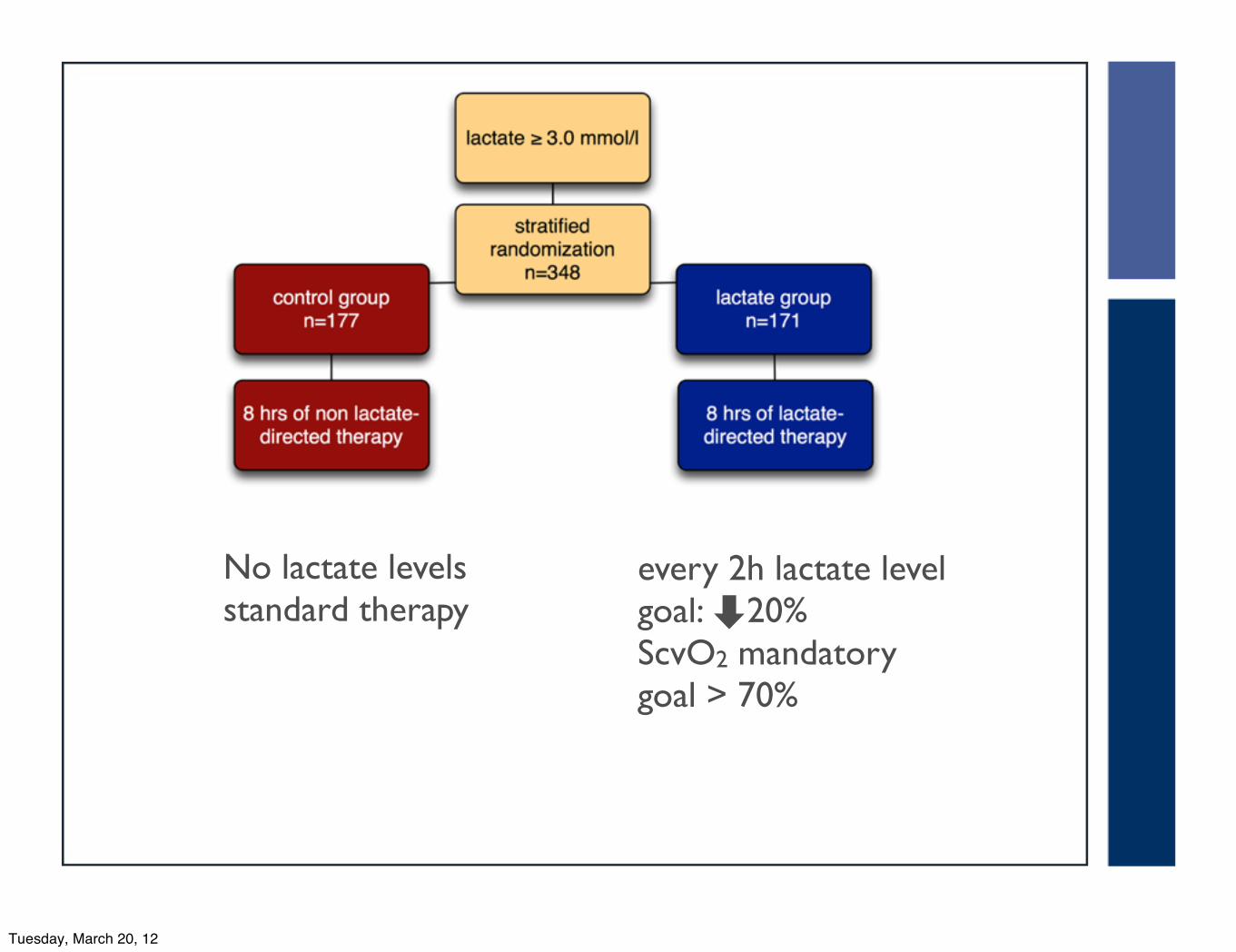
No lactate levelsstandard therapy
every 2h lactate levelgoal: ⬇20%ScvO2 mandatorygoal > 70%
Tuesday, March 20, 12

MA
P>
60
mm
Hg
Tuesday, March 20, 12
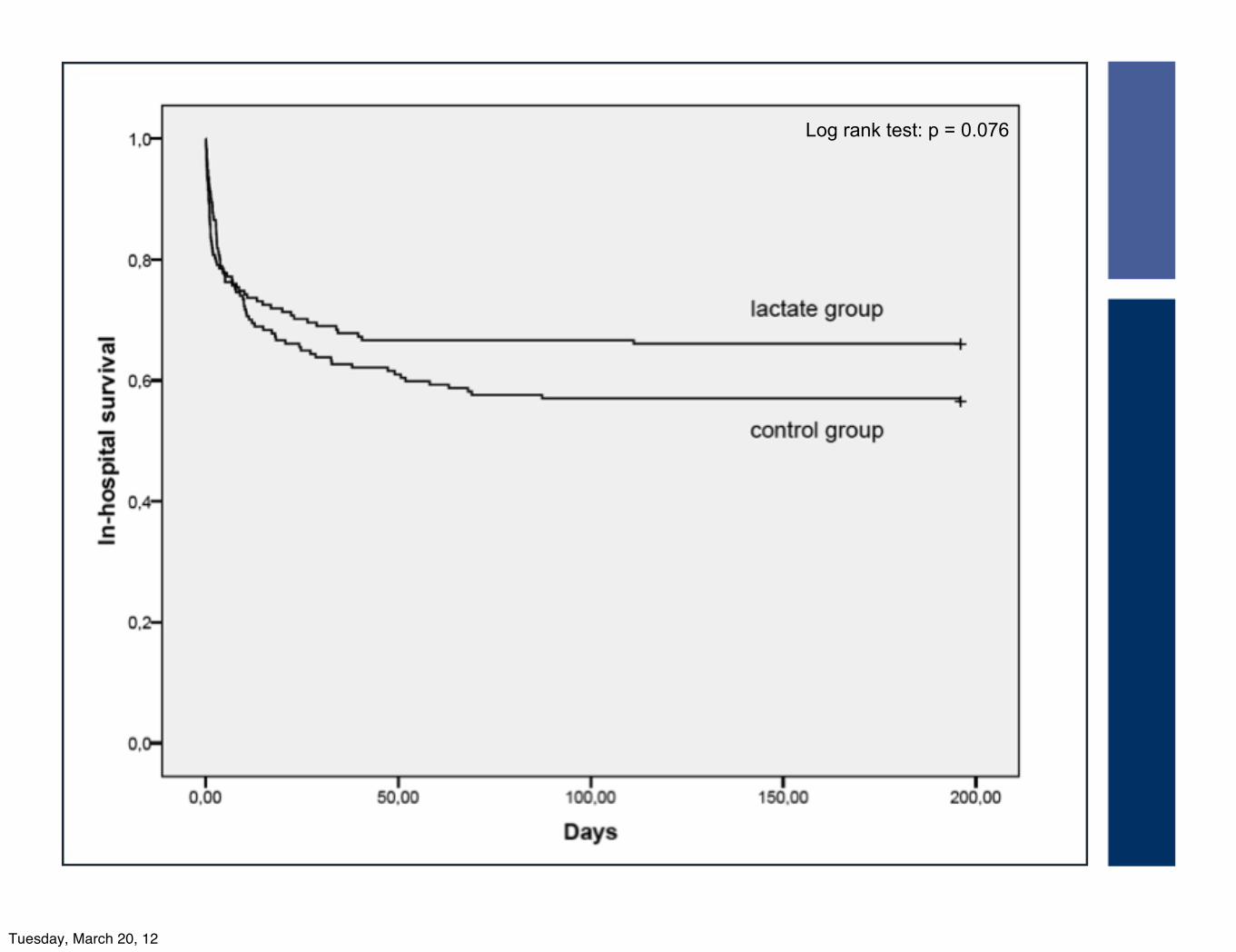
Log rank test: p = 0.076
Tuesday, March 20, 12

Subgroups
opnameduur_ZH120,00100,0080,0060,0040,0020,000,00
Cum
Sur
viva
l
1 ,0
0,8
0,6
0,4
0,2
0,0
controle groep therapie-censored
early lactate- directed therapy-censored
controle groep therapieearly lactate- directed therapy
randomisatie_therapie
Survival Functions
Page 1
opnameduur_ZH120,00100,0080,0060,0040,0020,000,00
Cum
Sur
viva
l
1 ,0
0,8
0,6
0,4
0,2
0,0
controle groep therapie-censored
early lactate- directed therapy-censored
controle groep therapieearly lactate- directed therapy
randomisatie_therapie
Survival Functions
Page 1
non sepsis sepsis
Tuesday, March 20, 12

Cox’s proportional hazards analysis
‣ICU Mortality
•HR 0.66 (0.45-0.98) p=0.037
‣Hospital mortality
•HR 0.61 (0.43-0.87) p=0.006
Tuesday, March 20, 12

Organ failure
**
SOFA score Control (n=177) Protocol (n=171)
Baseline
End of protocol(T=8h)
Observation time(T=9-72h)
6.3 (5.6-7.1) 6.4 (5.6-7.1)
7.2 (6.5-7.9) 6.9 (6.2-7.6)
7.0 (6.3-7.7) 6.4 (5.7-7.2)**
Tuesday, March 20, 12

Resource useWeaning and ICUstay
Tuesday, March 20, 12
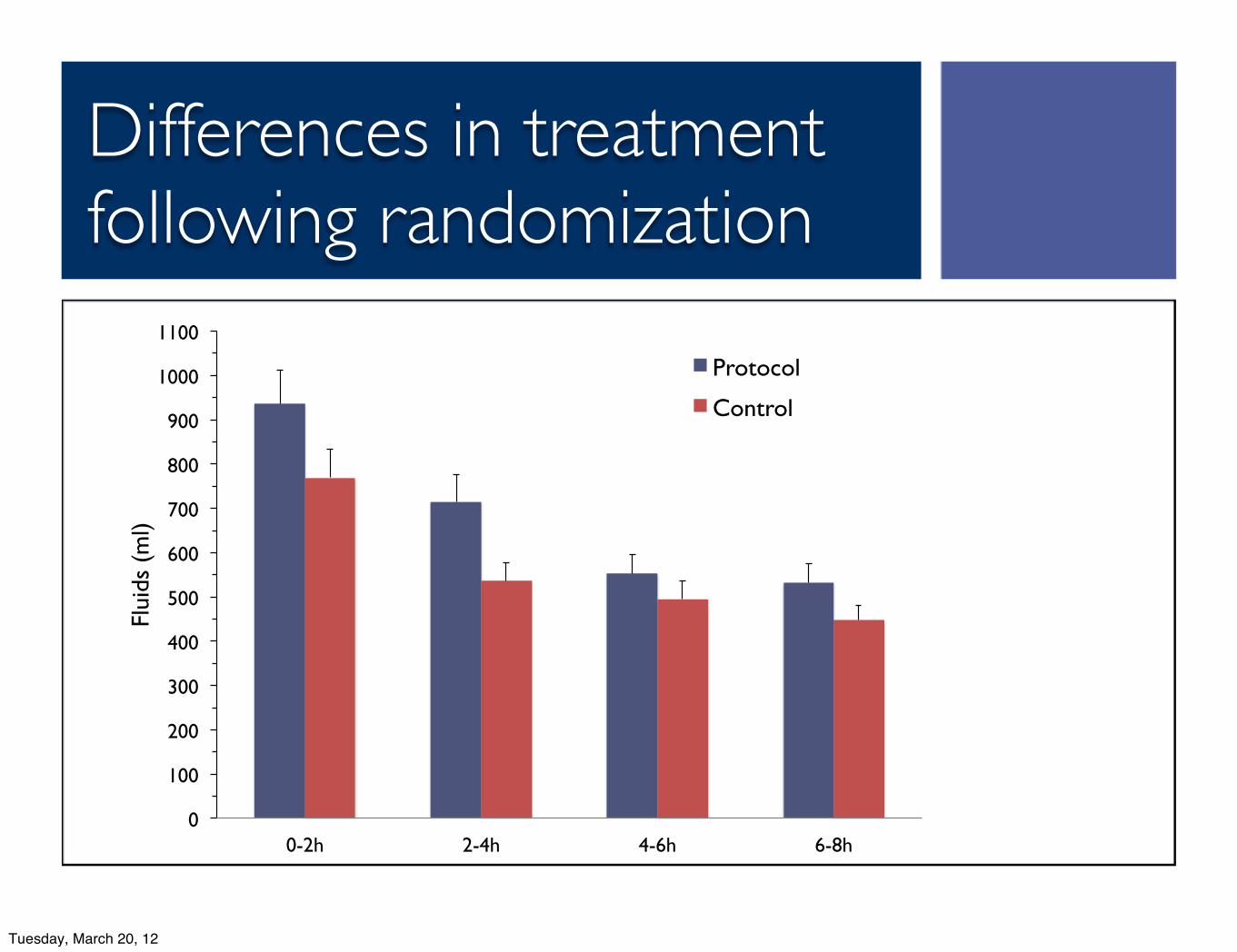
Differences in treatment following randomization
0!
100!
200!
300!
400!
500!
600!
700!
800!
900!
1000!
1100!
0-2h! 2-4h! 4-6h! 6-8h!
Flui
ds (
ml)!
Protocol!
Control!
Tuesday, March 20, 12

0
10
20
30
40
50
60
70
80
>3 >4 >5 >6 >7 >8 >9 >10 >11 >12 >13
ProtocolControl
Lactate mmol/L
R2=0.83R2=0.97
Survival in relation to baseline lactate level
%
Tuesday, March 20, 12
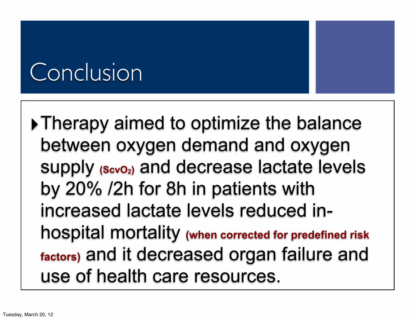
Conclusion
‣Therapy aimed to optimize the balance between oxygen demand and oxygen supply (ScvO2) and decrease lactate levels by 20% /2h for 8h in patients with increased lactate levels reduced in-hospital mortality (when corrected for predefined risk
factors) and it decreased organ failure and use of health care resources.
Tuesday, March 20, 12