product information and manual -...
TRANSCRIPT

Instructions for Use
Interleukin-33 ELISA
Enzyme immunoassay for the quantitative determination of human Interleukin-33 (IL-33) in human serum, plasma
and cell culture supernatants.
BE53331
12x8
2-8°C
I B L I N T E R N A T I O N A L G M B H Flughafenstrasse 52a Phone: +49 (0)40-53 28 91-0 [email protected] D-22335 Hamburg, Germany Fax: +49 (0)40-53 28 91-11 www.IBL-International.com
05.06.14 (23)
TABLE OF CONTENTS
1 Intended Use 32 Summary 33 Principles of the Test 54 Reagents Provided 75 Storage Instructions – ELISA Kit 86 Specimen Collection and Storage Instructions 87 Materials Required But Not Provided 98 Precautions for Use 109 Preparation of Reagents 1210 Test Protocol 1611 Calculation of Results 2012 Limitations 2313 Performance Characteristics 2414 Ordering Information 2815 Reagent Preparation Summary 2916 Test Protocol Summary 30

3
1 Intended Use
The human IL-33 ELISA is an enzyme-linked immunosorbent assay for the quantitative detection of human IL-33. The human IL-33 ELISA is for research use only. Not for diagnostic or therapeutic procedures.
2 Summary
IL-33 is the 11th and most recently discovered member of the IL-1 family of cytokines, which furthermore includes IL-1 alpha, IL-1 beta, IL-18 and IL-1Ra. As with IL-1 beta and IL-18, the synthesized 30 kDa propeptide IL-33 lacks a clear signal peptide for direct processing via the endoplasmic reticulum and Golgi apparatus. In vivo, caspase-1 cleaves the pro-IL-1 beta and pro-IL-18 bioactive forms, an essential step for their subsequent secretion. The mechanism for IL-33 might be similar, but the process has not been fully elucidated yet. In vitro, caspase-1 can cleave human IL-33, which results in a 20-22 kDa mature form. In humans, IL-33 mRNA is predominantly found in dermal fibroblasts, bronchial and small airway epithelial cells and smooth muscle cells of skin and lung tissues. Later, cellular mRNA expression has been observed in adipocytes, synovial fibroblasts, high endothelial venules, and endothelial cells. Furthermore, IL-33 is expressed in fibroblastic reticular cells of lymphoid tissues, skin keratinocytes, epithelial cells of stomach, tonsillar crypts and salivary glands, cardiac fibroblasts and cardiomyocytes. In most cells the predominant localization of IL-33 is nuclear rather than cytoplasmic. IL-33 specifically binds to IL1RL-1, which is known also as ST2, and is part of the IL-1R family. IL-33 promotes the release of Th2-associated cytokines from in vitro polarized human and murine Th2 cells, and also acts as a Th2 chemotactic factor. IL-33-promoting cytokine production activates human basophils and probably also regulates their migration. IL-33 influence on activation, degranulation, enhanced adhesion and survival of eosinophils has been shown. Mast cells are well-studied responders to IL-33. The cytokine increases synthesis of IL-6, IL-13, IL-1 beta, TNF alpha, prostaglandin D2 and MCP-1 by primary bone marrow-derived mast cells. Other cells activated by IL-33 are cardiomyocytes, glial cells and CD34+ cells.

4
Studies have shown a role of ST2/IL-33 in asthma. Serum levels of soluble ST2 are increased in patients with acute exacerbation of asthma. IL-33 has been shown to exacerbate allergic airway inflammation by promoting IL-5+ T cells. Increased serum levels were also detected in patients with rhinitis and atopic patients with anaphylaxis. IL-33 gene polymorphism appears to be associated with allergic rhinitis and Alzheimer’s disease. IL-33 protein is expressed in normal and diseased synovium, but diseased tissue displays higher levels of expression. It has been demonstrated that soluble ST2 plays a role in inflammatory responses in arthritis. Increased levels can be found in synovial fluid in RA patients compared to osteoarthritis controls. Furthermore, patients suffering from other rheumatic diseases such as systemic lupus erythematosus, progressive systemic sclerosis, Wegener’s granulomatosis and Behcet’s disease show increased serum IL-33 concentrations.

5
3 Principles of the Test
An anti-human IL-33 coating antibody is adsorbed onto microwells.
Figure 1
Human IL-33 present in the sample or standard binds to antibodies adsorbed to the microwells.
Figure 2
First Incubation
Following incubation unbound biological components are removed during a wash step. A biotin-conjugated anti-human IL-33 antibody is added and binds to human IL-33 captured by the first antibody.
Figure 3
Following incubation unbound biotin-conjugated anti-human IL-33 antibody is removed during a wash step. Streptavidin-HRP is added and binds to the biotin-conjugated anti-human IL-33 antibody.
Figure 4
Streptavidin-HRP -
Standard or Sample
Biotin-Conjugate
Standard or Sample
Coating Antibody
Second Incubation
Coated Microwell
Third Incubation
6
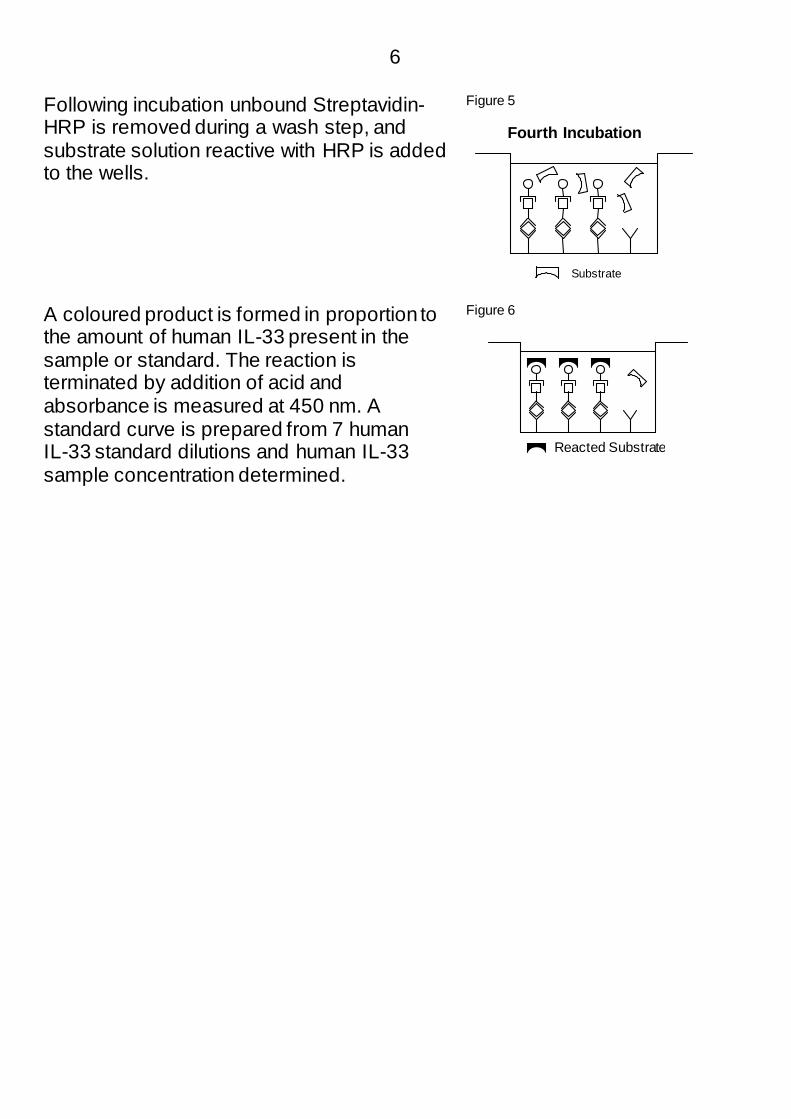
Following incubation unbound Streptavidin-HRP is removed during a wash step, and substrate solution reactive with HRP is added to the wells.
Figure 5
A coloured product is formed in proportion to the amount of human IL-33 present in the sample or standard. The reaction is terminated by addition of acid and absorbance is measured at 450 nm. A standard curve is prepared from 7 human IL-33 standard dilutions and human IL-33 sample concentration determined.
Figure 6
Reacted Substrate
Substrate
Fourth Incubation
7
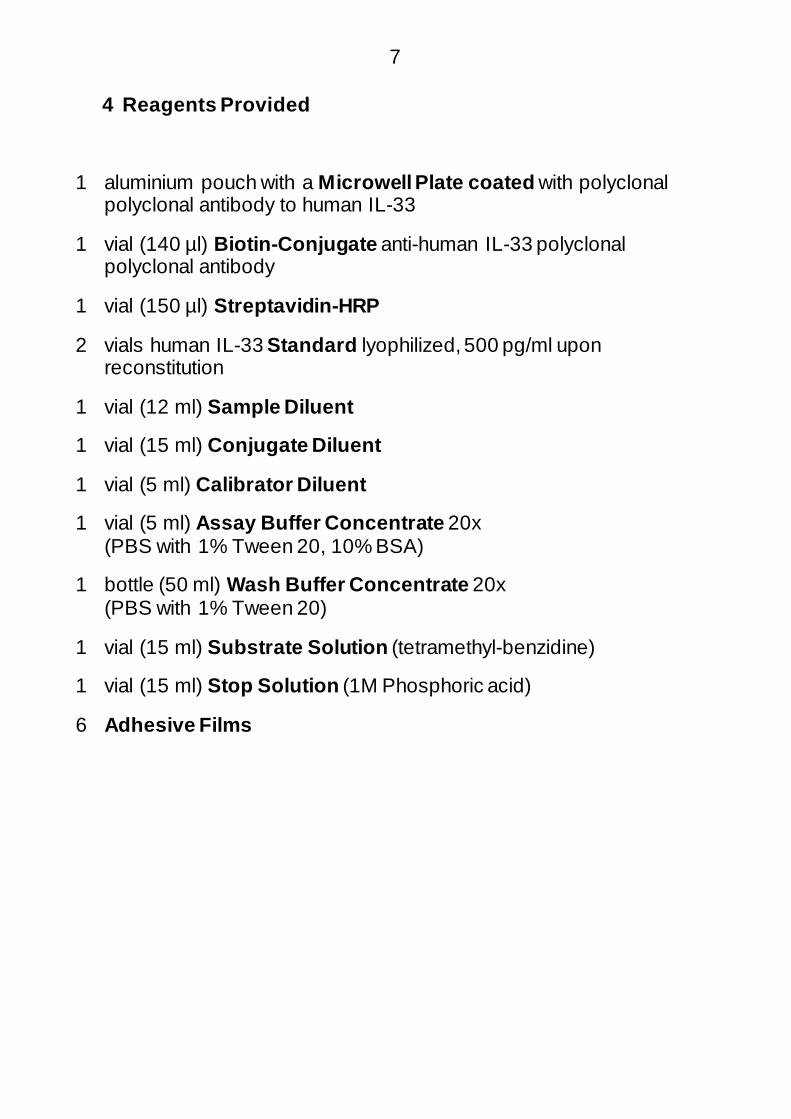
4 Reagents Provided
1 aluminium pouch with a Microwell Plate coated with polyclonal polyclonal antibody to human IL-33
1 vial (140 µl) Biotin-Conjugate anti-human IL-33 polyclonal polyclonal antibody
1 vial (150 µl) Streptavidin-HRP
2 vials human IL-33 Standard lyophilized, 500 pg/ml upon reconstitution
1 vial (12 ml) Sample Diluent
1 vial (15 ml) Conjugate Diluent
1 vial (5 ml) Calibrator Diluent
1 vial (5 ml) Assay Buffer Concentrate 20x (PBS with 1% Tween 20, 10% BSA)
1 bottle (50 ml) Wash Buffer Concentrate 20x (PBS with 1% Tween 20)
1 vial (15 ml) Substrate Solution (tetramethyl-benzidine)
1 vial (15 ml) Stop Solution (1M Phosphoric acid)
6 Adhesive Films
8
5 Storage Instructions – ELISA Kit
Store kit reagents between 2° and 8°C. Immediately after use remaining reagents should be returned to cold storage (2° to 8°C). Expiry of the kit and reagents is stated on labels.
Expiry of the kit components can only be guaranteed if the components are stored properly, and if, in case of repeated use of one component, this reagent is not contaminated by the first handling.
6 Specimen Collection and Storage Instructions
Cell culture supernatant, serum and plasma (citrate, heparin) were tested with this assay. Other biological samples might be suitable for use in the assay. Remove serum or plasma from the clot or cells as soon as possible after clotting and separation.
Samples containing a visible precipitate must be clarified prior to use in the assay. Do not use grossly hemolyzed or lipemic specimens.
Samples should be aliquoted and must be stored frozen at -20°C to avoid loss of bioactive human IL-33. If samples are to be run within 24 hours, they may be stored at 2° to 8°C (for sample stability refer to 13.5). Avoid repeated freeze-thaw cycles. Prior to assay, the frozen sample should be brought to room temperature slowly and mixed gently.

9
7 Materials Required But Not Provided
− 5 ml and 10 ml graduated pipettes
− 5 µl to 1000 µl adjustable single channel micropipettes with disposable tips
− 50 µl to 300 µl adjustable multichannel micropipette with disposable tips
− Multichannel micropipette reservoir
− Beakers, flasks, cylinders necessary for preparation of reagents
− Device for delivery of wash solution (multichannel wash bottle or automatic wash system)
− Microplate shaker
− Microwell strip reader capable of reading at 450 nm (620 nm as optional reference wave length)
− Glass-distilled or deionized water
− Statistical calculator with program to perform regression analysis
10
8 Precautions for Use
− All reagents should be considered as potentially hazardous. We therefore recommend that this product is handled only by those persons who have been trained in laboratory techniques and that it is used in accordance with the principles of good laboratory practice. Wear suitable protective clothing such as laboratory overalls, safety glasses and gloves. Care should be taken to avoid contact with skin or eyes. In the case of contact with skin or eyes wash immediately with water. See material safety data sheet(s) and/or safety statement(s) for specific advice.
− Reagents are intended for research use only and are not for use in diagnostic or therapeutic procedures.
− Do not mix or substitute reagents with those from other lots or other sources.
− Do not use kit reagents beyond expiration date on label.
− Do not expose kit reagents to strong light during storage or incubation.
− Do not pipette by mouth.
− Do not eat or smoke in areas where kit reagents or samples are handled.
− Avoid contact of skin or mucous membranes with kit reagents or specimens.
− Rubber or disposable latex gloves should be worn while handling kit reagents or specimens.
− Avoid contact of substrate solution with oxidizing agents and metal.
− Avoid splashing or generation of aerosols.
− In order to avoid microbial contamination or cross-contamination of reagents or specimens which may invalidate the test use disposable pipette tips and/or pipettes.
− Use clean, dedicated reagent trays for dispensing the conjugate and substrate reagent.

11
− Exposure to acid inactivates the conjugate.
− Glass-distilled water or deionized water must be used for reagent preparation.
− Substrate solution must be at room temperature prior to use.
− Decontaminate and dispose specimens and all potentially contaminated materials as they could contain infectious agents. The preferred method of decontamination is autoclaving for a minimum of 1 hour at 121.5°C.
− Liquid wastes not containing acid and neutralized waste may be mixed with sodium hypochlorite in volumes such that the final mixture contains 1.0% sodium hypochlorite. Allow 30 minutes for effective decontamination. Liquid waste containing acid must be neutralized prior to the addition of sodium hypochlorite.
12
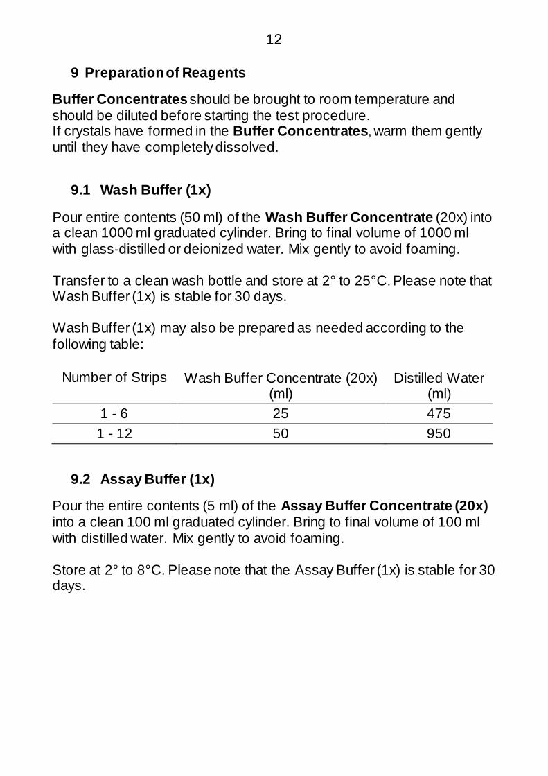
9 Preparation of Reagents
Buffer Concentrates should be brought to room temperature and should be diluted before starting the test procedure. If crystals have formed in the Buffer Concentrates, warm them gently until they have completely dissolved.
9.1 Wash Buffer (1x)
Pour entire contents (50 ml) of the Wash Buffer Concentrate (20x) into a clean 1000 ml graduated cylinder. Bring to final volume of 1000 ml with glass-distilled or deionized water. Mix gently to avoid foaming.
Transfer to a clean wash bottle and store at 2° to 25°C. Please note that Wash Buffer (1x) is stable for 30 days.
Wash Buffer (1x) may also be prepared as needed according to the following table:
Number of Strips Wash Buffer Concentrate (20x) (ml)
Distilled Water (ml)
1 - 6 25 475 1 - 12 50 950
9.2 Assay Buffer (1x)
Pour the entire contents (5 ml) of the Assay Buffer Concentrate (20x) into a clean 100 ml graduated cylinder. Bring to final volume of 100 ml with distilled water. Mix gently to avoid foaming.
Store at 2° to 8°C. Please note that the Assay Buffer (1x) is stable for 30 days.
13
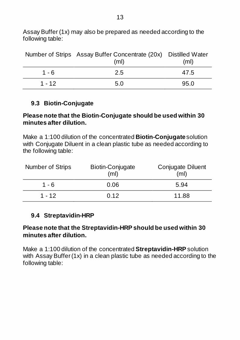
Assay Buffer (1x) may also be prepared as needed according to the following table:
Number of Strips Assay Buffer Concentrate (20x) (ml)
Distilled Water (ml)
1 - 6 2.5 47.5
1 - 12 5.0 95.0
9.3 Biotin-Conjugate
Please note that the Biotin-Conjugate should be used within 30 minutes after dilution.
Make a 1:100 dilution of the concentrated Biotin-Conjugate solution with Conjugate Diluent in a clean plastic tube as needed according to the following table:
Number of Strips Biotin-Conjugate (ml)
Conjugate Diluent (ml)
1 - 6 0.06 5.94
1 - 12 0.12 11.88
9.4 Streptavidin-HRP
Please note that the Streptavidin-HRP should be used within 30 minutes after dilution.
Make a 1:100 dilution of the concentrated Streptavidin-HRP solution with Assay Buffer (1x) in a clean plastic tube as needed according to the following table:

14
Number of Strips Streptavidin-HRP (ml)
Assay Buffer (1x) (ml)
1 - 6 0.06 5.94
1 - 12 0.12 11.88
9.5 Human IL-33 Standard
Reconstitute human IL-33 standard by addition of Calibrator Diluent Reconstitution volume is stated on the label of the standard vial. Swirl or mix gently to insure complete and homogeneous solubilization (concentration of reconstituted standard = 500 pg/ml). Allow the standard to reconstitute for 10-30 minutes. Mix well prior to making dilutions.
After usage remaining standard cannot be stored and has to be discarded.
9.5.1 Standard Dilution
Label 6 tubes, one for each standard point. S2, S3, S4, S5, S6, S7 Then prepare 1:2 serial dilutions for the standard curve as follows: Pipette 150 µl of Calibrator Diluent into each tube. Pipette 150 µl of reconstituted standard (serves as highest standard S1 = 500 pg/ml) into the first tube, labelled S2, and mix (concentration of standard 2 = 250 pg/ml). Pipette 150 µl of this dilution into the second tube, labelled S3, and mix thoroughly before the next transfer. Repeat serial dilutions 4 more times thus creating the points of the standard curve (see Figure 7).
Calibrator Diluent serves as blank.
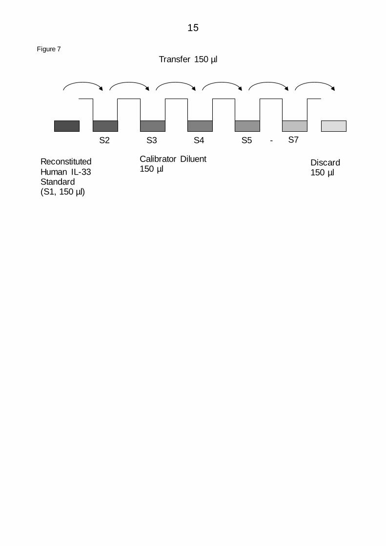
15
Figure 7 Transfer 150 µl
Reconstituted Human IL-33 Standard (S1, 150 µl)
S2 S3 S4 S5 - S7
Calibrator Diluent 150 µl
Discard 150 µl

16
10 Test Protocol
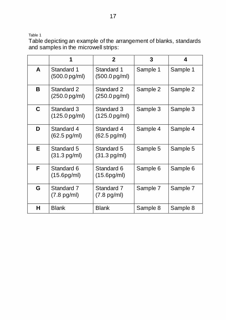
a. Determine the number of microwell strips required to test the desirednumber of samples plus appropriate number of wells needed forrunning blanks and standards. Each sample, standard, blank andoptional control sample should be assayed in duplicate. Removeextra microwell strips from holder and store in foil bag with thedesiccant provided at 2°-8°C sealed tightly.
b. Wash the microwell strips twice with approximately 400 µl WashBuffer per well with thorough aspiration of microwell contentsbetween washes. Allow the Wash Buffer to sit in the wells for about10 – 15 seconds before aspiration. Take care not to scratch thesurface of the microwells.After the last wash step, empty wells and tap microwell strips onabsorbent pad or paper towel to remove excess Wash Buffer. Usethe microwell strips immediately after washing. Alternativelymicrowell strips can be placed upside down on a wet absorbentpaper for not longer than 15 minutes. Do not allow wells to dry.
c. Add 50 µl of Sample Diluent to all wells.
d. Add 50 µl of each prepared Standard dilution in duplicate to thecorresponding standard well (see Table 1).
e. Add 50 µl Calibrator Diluent to the blank wells.
f. Add 50 µl µl of each sample in duplicate to the sample wells (seeTable 1).
17
Table 1 Table depicting an example of the arrangement of blanks, standards and samples in the microwell strips:
1 2 3 4 A Standard 1
(500.0 pg/ml) Standard 1 (500.0 pg/ml)
Sample 1 Sample 1
B Standard 2 (250.0 pg/ml)
Standard 2 (250.0 pg/ml)
Sample 2 Sample 2
C Standard 3 (125.0 pg/ml)
Standard 3 (125.0 pg/ml)
Sample 3 Sample 3
D Standard 4 (62.5 pg/ml)
Standard 4 (62.5 pg/ml)
Sample 4 Sample 4
E Standard 5 (31.3 pg/ml)
Standard 5 (31.3 pg/ml)
Sample 5 Sample 5
F Standard 6 (15.6pg/ml)
Standard 6 (15.6pg/ml)
Sample 6 Sample 6
G Standard 7 (7.8 pg/ml)
Standard 7 (7.8 pg/ml)
Sample 7 Sample 7
H Blank Blank Sample 8 Sample 8

18
g. Cover with an adhesive film and incubate at room temperature(18° to 25°C) for 2 hours, on a microplate shaker set at 400 rpm.(Shaking is absolutely necessary for an optimal testperformance.)
h. Prepare Biotin-Conjugate (see Preparation of Biotin-Conjugate 9.3).
i. Remove adhesive film and empty wells. Wash microwell strips 6times according to point b. of the test protocol. Proceed immediatelyto the next step.
j. Add 100 µl of Biotin-Conjugate to all wells.
k. Cover with an adhesive film and incubate at room temperature(18° to 25°C) for 1 hour, on a microplate shaker set at 400 rpm.(Shaking is absolutely necessary for an optimal testperformance.)
l. Prepare Streptavidin-HRP (refer to Preparation of Streptavidin-HRP9.4).
m. Remove adhesive film and empty wells. Wash microwell strips 6times according to point b. of the test protocol. Proceed immediatelyto the next step.
n. Add 100 µl of diluted Streptavidin-HRP to all wells, including theblank wells.
o. Cover with an adhesive film and incubate at room temperature(18° to 25°C) for 1 hour, on a microplate shaker set at 400 rpm.(Shaking is absolutely necessary for an optimal testperformance.)
p. Remove adhesive film and empty wells. Wash microwell strips 6times according to point b. of the test protocol. Proceed immediatelyto the next step.
q. Pipette 100 µl of TMB Substrate Solution to all wells.
r. Incubate the microwell strips at room temperature (18° to 25°C) forabout 30 min. Avoid direct exposure to intense light.

19
The colour development on the plate should be monitored and the substrate reaction stopped (see next point of this protocol) before positive wells are no longer properly recordable. Determination of the ideal time period for colour development has to be done individually for each assay.
It is recommended to add the stop solution when the highest standard has developed a dark blue colour. Alternatively the colour development can be monitored by the ELISA reader at 620 nm. The substrate reaction should be stopped as soon as Standard 1 has reached an OD of 0.9 – 0.95.
s. Stop the enzyme reaction by quickly pipetting 100 µl of StopSolution into each well. It is important that the Stop Solution isspread quickly and uniformly throughout the microwells to completelyinactivate the enzyme. Results must be read immediately after theStop Solution is added or within one hour if the microwell strips arestored at 2 - 8°C in the dark.
t. Read absorbance of each microwell on a spectro-photometer using450 nm as the primary wave length (optionally 620 nm as thereference wave length; 610 nm to 650 nm is acceptable). Blank theplate reader according to the manufacturer's instructions by using theblank wells. Determine the absorbance of both the samples and thestandards.

20
11 Calculation of Results
− Calculate the average absorbance values for each set of duplicate standards and samples. Duplicates should be within 20 per cent of the mean value.
− Create a standard curve by plotting the mean absorbance for each standard concentration on the ordinate against the human IL-33 concentration on the abscissa. Draw a best fit curve through the points of the graph (a 5-parameter curve fit is recommended).
− To determine the concentration of circulating human IL-33 for each sample, first find the mean absorbance value on the ordinate and extend a horizontal line to the standard curve. At the point of intersection, extend a vertical line to the abscissa and read the corresponding human IL-33 concentration.
− If instructions in this protocol have been followed, samples have not been diluted and the concentration read from the standard curve must not be multiplied by a dilution factor.
− Calculation of samples with a concentration exceeding standard may result in incorrect, low human IL-33 levels. Such samples require further external predilution according to expected human IL-33 values with Sample Diluent in order to precisely quantitate the actual human IL-33 level.
− It is suggested that each testing facility establishes a control sample of known human IL-33 concentration and runs this additional control with each assay. If the values obtained are not within the expected range of the control, the assay results may be invalid.
− A representative standard curve is shown in Figure 8. This curve cannot be used to derive test results. Each laboratory must prepare a standard curve for each group of microwell strips assayed.

21
Figure 8 Representative standard curve for human IL-33 ELISA. Human IL-33 was diluted in serial 2-fold steps in Calibrator Diluent. Do not use this standard curve to derive test results. A standard curve must be run for each group of microwell strips assayed.

22
Table 2 Typical data using the human IL-33 ELISA Measuring wavelength: 450 nm Reference wavelength: 620 nm
Standard
Human IL-33 Concentration
(pg/ml) O.D. at 450 nm
Mean O.D. at 450 nm
C.V. (%)
1 500.0 2.489 2.536 2.6 2.583
2 250.0 1.257 1.366 11.3 1.475
3 125.0 0.801 0.806 0.9 0.811
4 62.5 0.449 0.446 1.0 0.442
5 31.3 0.259 0.262 1.7 0.265
6 15.6 0.183 0.184 1.0 0.185
7 7.8 0.123 0.132 9.6 0.141
Blank 0 0.074 0.078 7.2 0.082
The OD values of the standard curve may vary according to the conditions of assay performance (e.g. operator, pipetting technique, washing technique or temperature effects). Furthermore shelf life of the kit may affect enzymatic activity and thus colour intensity. Values measured are still valid.
23
12 Limitations
− Since exact conditions may vary from assay to assay, a standard curve must be established for every run.
− Bacterial or fungal contamination of either screen samples or reagents or cross-contamination between reagents may cause erroneous results.
− Disposable pipette tips, flasks or glassware are preferred, reusable glassware must be washed and thoroughly rinsed of all detergents before use.
− Improper or insufficient washing at any stage of the procedure will result in either false positive or false negative results. Empty wells completely before dispensing fresh wash solution, fill with Wash Buffer as indicated for each wash cycle and do not allow wells to sit uncovered or dry for extended periods.

24
13 Performance Characteristics
13.1 Sensitivity
The limit of detection of human IL-33 defined as the analyte concentration resulting in an absorbance significantly higher than that of the dilution medium (mean plus 2 standard deviations) was determined to be 0.2 pg/ml (mean of 6 independent assays).
13.2 Reproducibility
13.2.1 Intra-assay
Reproducibility within the assay was evaluated in 3 independent experiments. Each assay was carried out with 6 replicates of 8 serum samples containing different concentrations of human IL-33. 2 standard curves were run on each plate. Data below show the mean human IL-33 concentration and the coefficient of variation for each sample (see Table 3). The calculated overall intra-assay coefficient of variation was 4.7%.

25
Table 3 The mean human IL-33 concentration and the coefficient of variation for each sample
Sample Experiment
Mean Human IL-33 Concentration
(pg/ml)
Coefficient of Variation
(%) 1 1 131.89 3.4
2 142.36 1.2 3 141.34 2.5
2 1 64.37 4.0 2 64.28 3.1 3 71.24 3.5
3 1 29.71 1.3 2 30.30 4.9 3 32.68 6.1
4 1 13.37 9.5 2 12.40 6.4 3 13.21 6.5
5 1 123.94 5.0 2 137.08 3.2 3 144.29 5.6
6 1 65.01 6.5 2 60.92 5.5 3 73.34 5.0
7 1 28.04 1.6 2 25.09 5.3 3 30.19 4.2
8 1 11.18 7.5 2 9.37 8.0 3 11.00 3.1

26
13.2.2 Inter-assay
Assay to assay reproducibility within one laboratory was evaluated in 3 independent experiments. Each assay was carried out with 6 replicates of 8 serum samples containing different concentrations of human IL-33. 2 standard curves were run on each plate. Data below show the mean human IL-33 concentration and the coefficient of variation calculated on 18 determinations of each sample (see Table 4). The calculated overall inter-assay coefficient of variation was 6.9%.
Table 4 The mean human IL-33 concentration and the coefficient of variation of each sample
Sample
Mean Human IL-33 Concentration
(pg/ml)
Coefficient of Variation
(%) 1 138.53 4.2 2 66.63 6.0 3 30.89 5.1 4 13.00 4.0 5 135.11 7.6 6 66.42 9.5 7 27.77 9.2 8 10.52 9.5
13.3 Spike Recovery
The spike recovery was evaluated by spiking 3 levels of human IL-33 into serum, plasma and cell culture supernatant. Recoveries were determined with 4 replicates each. For recovery results see Table 5.

27
Table 5
Sample matrix
Spike high Spike medium Spike low
Range (%)
Mean (%)
Range (%)
Mean (%)
Range (%)
Range (%)
Serum 74 – 88 79 64 - 79 69 66 – 77 67 Plasma (citrate)
59 – 73 68 46 - 66 57 29 – 46 38
Plasma (heparin)
79 – 85 82 78 – 87 82 69 – 87 76
Cell culture supernatant
92 90 73
13.4 Dilution Parallelism
Serum, plasma and cell culture supernatant samples with different levels of human IL-33 were analysed at serial 2 fold dilutions with 4 replicates each. The recovery ranged from 78% to 118% with an overall recovery of 99% (see Table 6). For recovery data see Table 6.
Table 6
Sample matrix Recovery of Exp. Val. Range (%) Mean (%)
Serum 92 - 118 101 Plasma (citrate) 91 - 111 99 Plasma (heparin) 78 - 99 89 Cell culture supernatant
106 - 107 107
28
13.5 Sample Stability
13.5.1 Freeze-Thaw Stability
Aliquots of serum samples were stored at -20°C and thawed 5 times, and the human IL-33 levels determined. There was no significant loss of human IL-33 immunoreactivity detected by freezing and thawing.
13.5.2 Storage Stability
Aliquots of serum samples were stored at -20°C, 2-8°C, room temperature (RT) and at 37°C, and the human IL-33 level determined after 24 h. There was no significant loss of human IL-33 immunoreactivity detected during storage at -20, 2-8°C and RT.
13.6 Specificity
The assay detects both natural and recombinant human IL-33. The cross reactivity and interference of circulating factors of the immune system was evaluated by spiking these proteins at physiologically relevant concentrations into a human IL-33 positive sample. There was no cross reactivity or interference detected, notably not with IL-1 alpha, IL-1 beta and IL-18.
13.7 Expected Values
There were no detectable human IL-33 levels found. Elevated human IL-33 levels depend on the type of immunological disorder.
14 Ordering Information
See last page.

29
15 Reagent Preparation Summary
15.1 Wash Buffer (1x) Add Wash Buffer Concentrate 20x (50 ml) to 950 ml distilled water.
Number of Strips Wash Buffer Concentrate (ml) Distilled Water (ml) 1 - 6 25 475
1 - 12 50 950
15.2 Assay Buffer (1x) Add Assay Buffer Concentrate 20x (5 ml) to 95 ml distilled water.
Number of Strips Assay Buffer Concentrate (ml) Distilled Water (ml) 1 - 6 2.5 47.5
1 - 12 5.0 95.0
15.3 Biotin-Conjugate Make a 1:100 dilution of Biotin-Conjugate in Conjugate Diluent:
Number of Strips Biotin-Conjugate (ml) Conjugate Diluent (ml) 1 - 6 0.06 5.94
1 - 12 0.12 11.88
15.4 Streptavidin-HRP Make a 1:100 dilution of Streptavidin-HRP in Assay Buffer (1x):
Number of Strips Streptavidin-HRP (ml) Assay Buffer (1x) (ml) 1 - 6 0.06 5.94
1 - 12 0.12 11.88
15.5 Human IL-33 Standard Reconstitute lyophilized human IL-33 standard with Calibrator Diluent. (Reconstitution volume is stated on the label of the standard vial.)

30
16 Test Protocol Summary
1. Determine the number of microwell strips required.2. Wash microwell strips twice with Wash Buffer.3. Add 50µl Sample Diluent to all wells.4. Add 50 µl of prepared Standard dilutions in duplicate to standard
wells.5. Add 50 µl Calibrator Diluent to blank wells.6. Add 50 µl sample in duplicate to sample wells.7. Cover microwell strips and incubate 2 hours at room temperature
(18°to 25°C). (Shaking is absolutely necessary for an optimal testperformance.)
8. Prepare Biotin-Conjugate.9. Empty and wash microwell strips 6 times with Wash Buffer.10. Add 100 µl Biotin-Conjugate to all wells.11. Cover microwell strips and incubate 1 hour at room temperature
(18°to 25°C). (Shaking is absolutely necessary for an optimaltest performance.)
12. Prepare Streptavidin-HRP.13. Empty and wash microwell strips 6 times with Wash Buffer.14. Add 100 µl diluted Streptavidin-HRP to all wells.15. Cover microwell strips and incubate 1 hour at room temperature
(18°to 25°C). (Shaking is absolutely necessary for an optimaltest performance.)
16. Empty and wash microwell strips 6 times with Wash Buffer.17. Add 100 µl of TMB Substrate Solution to all wells.18. Incubate the microwell strips for about 30 minutes at room
temperature (18°to 25°C).19. Add 100 µl Stop Solution to all wells.20. Blank microwell reader and measure colour intensity at 450 nm.
Note: If instructions in this protocol have been followed, samples have not been diluted and the concentration read from the standard curve must not be multiplied by a dilution factor.


Symbols / Symbole / Symbôles / Símbolos / Símbolos / Σύμβολα
Symbols Version 4.5 / 2015-12-07
REF Cat.-No.: / Kat.-Nr.: / No.- Cat.: / Cat.-No.: / N.º Cat.: / N.–Cat.: / Αριθμός-Κατ.:
LOT Lot-No.: / Chargen-Bez.: / No. Lot: / Lot-No.: / Lote N.º: / Lotto n.: / Αριθμός -Παραγωγή:
Use by: / Verwendbar bis: / Utiliser à: / Usado por: / Usar até: / Da utilizzare entro: / Χρησιμοποιείται από: No. of Tests: / Kitgröße: / Nb. de Tests: / No. de Determ.: / N.º de Testes: / Quantità dei tests: / Αριθμός εξετάσεων:
CONC Concentrate / Konzentrat / Concentré / Concentrar / Concentrado / Concentrato / Συμπύκνωμα
LYO Lyophilized / Lyophilisat / Lyophilisé / Liofilizado / Liofilizado / Liofilizzato / Λυοφιλιασμένο
IVD In Vitro Diagnostic Medical Device. / In-vitro-Diagnostikum. / Appareil Médical pour Diagnostics In Vitro. / Dispositivo Médico para Diagnóstico In Vitro. / Equipamento Médico de Diagnóstico In Vitro. / Dispositivo Medico Diagnostico In vitro. / Ιατρική συσκευή για In-Vitro ∆ιάγνωση. Evaluation kit. / Nur für Leistungsbewertungszwecke. / Kit pour évaluation. / Juego de Reactivos para Evaluació. / Kit de avaliação. / Kit di evaluazione. / Κιτ Αξιολόγησης. Read instructions before use. / Arbeitsanleitung lesen. / Lire la fiche technique avant emploi. / Lea las instrucciones antes de usar. / Ler as instruções antes de usar. / Leggere le istruzioni prima dell’uso. / ∆ιαβάστε τις οδηγίες πριν την χρήση. Keep away from heat or direct sun light. / Vor Hitze und direkter Sonneneinstrahlung schützen. / Garder à l’abri de la chaleur et de toute exposition lumineuse. / Manténgase alejado del calor o la luz solar directa. / Manter longe do calor ou luz solar directa. / Non esporre ai raggi solari. / Να φυλάσσεται μακριά από θερμότητα και άμεση επαφή με το φως του ηλίου. Store at: / Lagern bei: / Stocker à: / Almacene a: / Armazenar a: / Conservare a: / Αποθήκευση στους:
Manufacturer: / Hersteller: / Fabricant: / Productor: / Fabricante: / Fabbricante: / Παραγωγός:
Caution! / Vorsicht! / Attention! / ¡Precaución! / Cuidado! / Attenzione! / Προσοχή!
Symbols of the kit components see MATERIALS SUPPLIED. Die Symbole der Komponenten sind im Kapitel KOMPONENTEN DES KITS beschrieben.
Voir MATERIEL FOURNI pour les symbôles des composants du kit. Símbolos de los componentes del juego de reactivos, vea MATERIALES SUMINISTRADOS.
Para símbolos dos componentes do kit ver MATERIAIS FORNECIDOS. Per i simboli dei componenti del kit si veda COMPONENTI DEL KIT.
Για τα σύμβολα των συστατικών του κιτ συμβουλευτείτε το ΠΑΡΕΧΟΜΕΝΑ ΥΛΙΚΑ.
COMPLAINTS: Complaints may be submitted initially written or vocal. Subsequently they need to be filed including the test performance and results in writing in case of analytical reasons.
WARRANTY: The product is warranted to be free from material defects within the specific shelf life and to comply with product specifications delivered with the product. The product must be used according to the Intended use, all instructions given in the instructions for use and within the product specific shelf life. Any modification of the test procedure or exchange or mixing of components of different lots could negatively affect the results. These cases invalidate any claim for replacement.
LIMITATION OF LIABILITY: IN ALL CIRCUMSTANCES THE EXTENT OF MANUFACTURER’S LIABILITY IS LIMITED TO THE PURCHASE PRICE OF THE KIT(S) IN QUESTION. IN NO EVENT SHALL MANUFACTURER BE LIABLE FOR ANY INCIDENTAL OR CONSEQUENTIAL DAMAGES, INCLUDING DAMAGES FOR LOST PROFITS, LOST SALES, INJURY TO PERSON OR PROPERTY OR ANY OTHER INCIDENTAL OR CONSEQUENTIAL LOSS.
IBL International GmbH Flughafenstr. 52A, 22335 Hamburg, Germany
Tel.: + 49 (0) 40 532891 -0 Fax: -11 E-MAIL: [email protected] WEB: http://www.IBL-International.com