printed britain proceedings anatomical society great ... · a concomitant increase in numbers of...
TRANSCRIPT
J. Anat. (1982), 134, 3, pp. 595-631 595Printed in Great Britain
Proceedings of the Anatomical Society ofGreat Britain and Ireland
DECEMBER 1981
The Annual General Meeting of the Society for the Session 1981/2 was held on 10 and 11December 1981 at Charing Cross Hospital Medical School, London. The Meeting included aSymposium on 'Modern Aspects of Medical Imaging'. The following are authors' abstractsof communications and demonstrations.
1. Experimental model for studying degeneration in unmyelinated fibres. By C. L. CRAWFORD andP. M. D. HARDWICKE. Departments of Anatomy, Charing Cross Hospital Medical School,London, and Biology, Brandeis University, Massachusetts, U.S.A.
There are several available animal models for studying Wallerian degeneration and segmentaldemyelination in peripheral nerves: Agents such as 6-hydroxydopamine cause degeneration inautonomic unmyelinated fibres. However, there are few experimental models for the study ofdegeneration in somatic unmyelinated fibres.We injected Dutch Bantam rabbits in the footpad with a suspension of human sural nerve or
dorsal roots plus Freund's complete adjuvant. Six months to 4 years later the rabbits were skin-tested in the back with 0-2 ml 1-10 % dilution of suspension of sural nerve or dorsal roots insaline. Several rabbits responded by developing a papule at 5 weeks. Histologically cells werefound resembling the epithelioid cells occurring in granulomatous diseases such as leprosy,tuberculosis or sarcoidosis. Electron microscopic studies showed that some of these cells hadinfiltrated the cutaneous nerves and there was degeneration of unmyelinated fibres. Axons ofmyelinated fibres remained normal.The antigen producing this nerve damage does not appear to be a component of myelin. Very
high doses of myelin used in skin tests do not produce epithelioid cells. Instead the antigenappears to reside in the low speed 'nuclear' fraction. Membranes may be obtained from thenuclear fraction of dorsal roots by means of a discontinuous sucrose gradient. When skin-testedthese produce damage to unmyelinated fibres. Also deoxycholate-extracted material from thenuclear pellet in doses of 1*5 ,ug produces severe unmyelinated fibre damage on skin testing.The microsomal and mitochondrial subfractions are negative. The antigen may possibly be aprotein or glycoprotein of the Schwann cell plasma membrane.
2. Demyelination: a morphological and electrophysiological correlation. By M. N. GHABRIEL*,G. ALLT, L. SEDAL, M. J. G. HARRISON and JENNIFER R. SMALL. Departments of Anatomyand Reta Lila Weston Institute of Neurological Studies, The Middlesex Hospital MedicalSchool, London and *Department of Anatomy, Charing Cross Hospital Medical School,LondonIn clinical neurology the comparison of the amplitude of muscle action potential induced by
nerve stimulation above, and below, localised nerve lesions is widely used as a measure of theseverity of local demyelination (Kraft, Practical Electromyography, ed. Johnson; Williams &Wilkins; Baltimore, 1980). The validity of this commonly utilised assumption has been testedin an experimental model.
Demyelination was induced by a local injection of lysophosphatidyl choline (LPC, 4-5 ,tt1, 1 %)into the rat tibial nerve at the level of the popliteal fossa in 42 rats. In each animal the contra-lateral nerve served as a control and in 9 animals the tibial nerve was injected with normal salineinstead of LPC (Ghabriel & Allt, Acta neuropathol. 52, 1980). After perfusion fixation, thenerves were examined at the site of injection at 6 days by light and electron microscopy. LPC-injected nerves showed variable degrees of demyelination, the extent of which was estimated ona four-point scale by two independent observers using teased nerve fibre preparations andtransverse nerve sections. Control nerves, whether from the contralateral limb, or after salineinjection, revealed no demyelination.
Electrophysiologically the amplitude of responses was recorded from the small foot musclesduring stimulation of the tibial nerve at two sites separated by the lesion: proximally at the

Proceedings of the Anatomical Society ofsciatic notch and distally at the ankle, and the two measurements were expressed as a ratio.Recordings were made both before, and up to, 6 days after LPC and saline injections', and alsofrom contralateral nerves. The normal ratio of amplitude (0-81±0-08) did not change signifi-cantly after saline injection or in the contralateral nerves. After LPC injection, due to a fall inthe size of the response to proximal stimulation, a mean decrease of 0-27 ± 0-21 occurred in theamplitude ratio. Comparison of the physiological and histological results indicates that whileclinical recordings usually provide a reliable indication of the presence of demyelination they donot accurately reflect the severity of demyelination.
3. Autonomic ganglia in the wall of the adult human urinary bladder. By C. J. GILPIN, J. S. DIXON,S. A. THOMPSON and J. A. GOSLING. Department of Anatomy, University of ManchesterWhilst it is generally agreed that autonomic ganglia occur around the bladder base, and in the
vicinity of the ureterovesical junctions, there is considerable doubt concerning their distributionthroughout the remainder of the adult bladder wall. The present study has employed histologicaland neurohistochemical techniques to determine the arrangement, distribution and morpho-logical characteristics of autonomic ganglia in the dome and lateral walls of the human urinarybladder. Additional data-have been provided by electron microscopy. Biopsy specimens ofnormalhuman urinary bladders were obtained either trans-urethrally or during open surgical procedureson the bladder wall.
Ganglia containing 1-20 neurons were consistently observed in the adventitia, within themuscularis and in submucosal locations in specimens obtained from both the dome and lateralwalls. Histochemical evaluation of these ganglia revealed intense acetylcholinesterase activity inall nerve cell bodies and their associated pericellular networks of nerve fibres. Neurons andnerve fibres containing catecholamines were never observed in relation to these intramuralganglia. Using the electron microscope numerous presumptive axosomatic synapses were en-countered in which the axon terminals contained small (50 nm diameter) agranular and large(100 nm diameter) granulated vesicles.The morphological findings were discussed in relation to various functional aspects of the
human urinary bladder. In addition the clinical relevance of the present findings was consideredwith particular reference to surgical transection and denervation of the urinary bladder.
4. Baroreceptors in the pulmonary trunk of the domestic fowl. By A. A. M. TAHA (introduced byA. S. KING). Department of Veterinary Anatomy, University ofLiverpool (Fig. 1)
Baroreceptor activity has been recorded from the pulmonary arteries of mammals (Coleridgeand Kidd, J. Physiol. 150, 1960), and mechanoreceptor activity has been recorded from thepulmonary artery of birds (Jones, Comp. Biochem. Physiol. 28, 1969). Nerve fibres have beendemonstrated by silver techniques in the media and adventitia of the pulmonary arteries in bothbirds and mammals, but the presence of baroreceptors has not been established electron micro-scopically.The pulmonary trunks from 13 normal domestic fowls were fixed by perfusion and immersion
in Karnovsky's solution, and processed for electron microscopy. Toluidine blue thick sectionsrevealed faintly stained modified regions (Fig. 1, M) in the media and adventitia. Electron micro-scopic examination of these regions disclosed various types of axonal endings. The majoritywere enlarged, irregular in shape, and characterised by many mitochondria, some of which wereabnormal (Fig. 1, arrows). The endings were covered partly by Schwann cells and partly by anextensive basal lamina to which fine collagen fibres were attached. A few of the endings weretotally bare except for a basal lamina. Since these endings closely resemble ultrastructurally thebaroreceptors of the mammalian carotid sinus and aortic arch, they are interpreted as pulmonarybaroreceptors.Other axonal endings in the modified regions contained small (30-50 nm) or large (60-120 nm)
dense-cored granular vesicles, and were considered to be aminergic or peptidergic efferentendings. Yet others contained many clear vesicles (30-50 nm), and were interpreted as cholinergicefferent endings. Sometimes the endings with large dense-cored granular vesicles were separatedfrom the baroreceptors by no more than a wide basal lamina, thus suggesting the possibilityof modulation of the stimulus threshold of the baroreceptors.
596

Great Britain and Ireland
Fig. 1
5. Gliogenesis in the fetal rabbit spinal cord. By R. R. STURROCK. Department of Anatomy,University of Dundee, Scotland
Gliogenesis was studied quantitatively in grey matter, and presumptive white matter, in thefetal rabbit spinal cord from E12 to E30. Glia could be identified in the ventral grey matter atE12 and in the ventral and lateral white matter at E14. A rapid increase in the number of glialcells occurred between E20 and E30, especially in white matter.Counts of mitotic cells indicated that the rapid increase in glial number in white matter was
mainly due to division of cells within the white matter, although there was probably also somemigration of cells from grey to white matter. Ependymal mitosis contributed little, if anything,to glial production between E20 and E30.
All types of glial cells (oligodendrocytes, astrocytes, microglia and glioblasts) appeared todivide during the rapid phase of gliogenesis. Pyknotic cells were few in number except in theventral grey matter at E16, when most of the pyknotic cells appeared to be neurons.There was no evidence at any stage of development of a germinal layer in the spinal cord,
similar to the subventricular or subependymal layer which is present in the developing forebrain.
6. Selective neuronal survival in long-term cell cultures of chick embryo spinal cord. By P. L.DEBBAGE. Department of Anatomy and Embryology, and Centre for Neuroscience, UniversityCollege London (Fig. 2)
In cultures of mechanically dissociated central nervous tissue, numbers of neurons survive.They extend processes, are known to form synaptic contacts, and synthesise neurotransmitters.But their survival, in contrast to that reported for explant cultures of similar origin, is typicallylimited to a few weeks. In cell cultures of chick embryo spinal cord, the majority of well ramifiedneurons disappear from the cultures during the third and fourth weeks in vitro, and there occurs
597

598 Proceedings of the Anatomical Society of
Cell body
q
1
.
0 25 mm
Fig. 2
a concomitant increase in numbers of microglia, which are active phagocytes. Thus oldercultures appear to consist of extensive multilayered mats of non-neuronal cells, with a fewmicroglia, and in this condition they can be maintained for many further weeks.
In these older cultures there remains, however, a small number of surviving neurons, repre-senting at maximum no more than 1 % of the neurons originally established in the cultures, andmaintaining extensively ramified neurites (Fig. 2) for as long as 16 weeks. They have been seenroutinely in many batches of cultures after silver staining and by immunohistochemical demon-stration of markers including neurofilament protein (Fig. 2) and synaptosomal membrane com-ponents. That these neurons form a single population is suggested by their distinctive form,a major feature of which is that each cell extends at least one fine neurite of great length.Studies in progress aim to demonstrate that these cells share a common origin in the embryonicspinal cord. Their possible identity is considered.
7. Cartographic display of cell migrations into cortical areas of the developing mouse telencephalon.By I. H. M. SMART. Department of Anatomy, Medical Sciences Institute, The University,Dundee
Changes in the depth and nuclear crowding pattern of the intermediate layer were recorded onmaps of the developing mouse telencephalon made on each day of prenatal life between ElO andE16. Production of neurons for future cortical areas commenced about E12 in two quite separatelocations: one on the medial wall associated with the hippocampus, and one on the lateral wallrostrally producing isocortical neurons. By E14 neuron release had spread from the two originalsites over the entire surface of the ventricular layer to form a continuous intermediate layer.During E14 a plate of crowded immature cells (the result of increased neuron release from theperiventricular germinal layer) appeared in the outer intermediate layer of the isocortical areaand spread over the lateral telencephalic wall passing at E15 over the telencephalic roof intothe medial wall. By E16 this type of neuron release had extended as far down the medial wall asthe anterior hippocampus, induseum griseum and subiculum which together formed a rostro-caudal band of tissue uninfiltrated by cortical plate. The significance of the cortical platemigration in terms of cell production mechanisms was discussed.
I
1
I1
I
9,
r

Great Britain and Ireland
8. A Golgi analysis of the hyperstriatum ventrale in the chick. By P. BRADLEY and G. HORN.*Department of Anatomy, University of Newcastle upon Tyne and, *Department of Zoology,University of Cambridge (Fig. 3)The intermediate and medial parts of the hyperstriatum ventrale (IMHV) of the domestic
chick have been implicated in the learning process of imprinting (Horn et al. Brain Res. 168,1979). The length of the synaptic apposition zones in this region was increased in chicks whichhad been imprinted (Bradley et al. Exp. Brain Res. 41, 1981). The afferent and efferent con-
Fig. 3
599

Proceedings of the Anatomical Society ofnections of IHMV have been described (Bradley & Horn, J. Anat. 128, 1979; Bradley & Horn,J. Physiol. 278, 1978; Davies & Bradley, J. Anat. 133, 1981) but the cellular organisation of thisregion has not previously been studied. We report here the results of a Golgi analysis of theneuronal components of IMHV.Camera lucida drawings of a total of 140 Golgi-impregnated neurons from the IMHV of ten
light-reared Ross 1 chicks 1 day old were analysed. Cells were classified according to size,shape of soma and number of primary dendrites arising from the soma. Fifty six (40 %) of thecells were large multipolar neurons (Fig. 3 A). The dendrites of these cells displayed no specificorientation although the dendritic tree was least extensive posteroventrally. The origin andcourse of the axons of these cells were highly variable. Twenty seven (20 %) of the cells were largepyramid shaped neurons (Fig. 3 C) which showed a definite dorsomedial orientation of theapical dendrite. The axons of these cells ran in a direction opposite to that of the apical dendriteand commonly bifurcated in a 'pitchfork' fashion prior to leaving IMHV. Twenty one (15 %) ofthe cells had large ovoid cell bodies with a maximum of four primary dendrites (Fig. 3 B). Theaxons of these cells divided into a number of branches soon after leaving the cell body. The longaxes of these cells tended to be orientated dorsoventrally. The remainder of the cells wereclassified as small pyramidal or stellate neurons. No specific lamination of the cells was observed.
4
9. The distribution of acetylcholinesterase in the chick telencephalon. By B. J. MCCABE, G. HORNand G. MCGRATH.* Department of Zoology, University of Cambridge, and *Department ofAnatomy, University of Bristol (Fig. 4)
Acetylcholinesterase activity in the forebrain hemispheres of the chick changes markedlyduring development (Marchand et al. FEBSLett. 78, 1977) and is influenced by visual experience(Haywood et al. Brain Res. 92, 1975). The anatomical distribution of enzyme activity is notknown and was investigated in the present study.
Fourteen Ross 1 chicks were anaesthetised 1-2 days post-hatching with sodium thiopentone.Eight birds were anaesthetised 6 weeks after hatching. Brains were fixed by intracardiac per-fusion with 20 % formalin in 154 mM-Na2SO4 followed by immersion in 10 % neutral formalin
Fig. 4. HA, hyperstriatum accessorium; HD,hyperstriatumdorsale; HV,hyperstriatumventrale;N, neostriatum; E, ectostriatum; PA, paleostriatum augmentatum; LPO, lobus parolfactorius.Scale x 4.
600
4

Great Britain and Irelandat 4 °C for 18 h. Frozen sections 60 or 80,um thick were stained for acetylcholinesterase (Lewis,Bibliotheca anat. 2, 1961). Incubation time was 3-5 h at room temperature (pH 5 6). Sectionsincubated at the same time with butyrylthiocholine iodide were unstained. Results were con-firmed on fresh frozen sections cut from two additional brains from 1 day old chicks.
Stained cell body profiles were observed in hyperstriatum accessorium, hyperstriatum intra-calatus/dorsale, neostriatum and the olfactory bulb (particularly the mitral cell layer). Inaddition to stained cell body profiles, diffuse staining (presumably of neuropil) occurred inhyperstriatum ventrale (in which dorsal and ventral laminae could be distinguished), paleo-striatum augmentatum, paleostriatum primitivum, lobus parolfactorius, dorsal archistriatum,septal nuclei, caudal neostriatum and ectostriatum (Fig. 4). Nucleus basalis contained densediffuse staining. Intensely stained cell bodies and fibres were observed in the nucleus intra-peduncularis.
In chickens aged 6 weeks the overall pattern of staining was similar to that in the 1-2 day oldchick.
10. The projection of the somatosensory cortex on the superior colliculus of the rat. By H. N.RAMADAN and K. E. WEBSTER. Department of Anatomy, King's College London
The ascending somatosensory input to the intermediate and deep layers of the rat superiorcolliculus is organised to produce a body map congruent with the visual field (Webster &Ramadan, J. Anat. 133, 1981). A second source of somatic information to the colliculus is thesomatosensory cortex. We have examined the organisation of this system by using the Fink andHeimer technique after making small cortical lesions; and by exploiting the uptake and transportof [3H]amino acids or horseradish peroxidase (HRP) injected into cortex or colliculus (Ramadan& Webster, J. Anat. 133, 1981).The Fink and Heimer and autoradiographic niaterial shows the corticotectal axons to arise
from perikarya deep to layer IV of the ipsilateral somatosensory cortex, and to end in the inter-mediate and deep layers of the colliculus, which the axons enter from its rostral and lateralborders. Like those from the spinotectal tract (Antonetty & Webster, J. comp. Neurol. 163,1975), the terminals aggregate into clusters separated by relatively empty areas. It is thereforepossible that the two inputs interdigitate rather than converge. Axons from medial cortex (tailand hindlimb - Welker, J. comp. Neurol. 166 1976) end in the caudal and medial colliculus;those from face areas distribute laterally and rostrally; those from intermediate cortex endbetween those two zones.The HRP studies confirm that the descending projection arises from pyramidal cells in layer V
of the ipsilateral primary and secondary somatosensory cortices. Although its significance isdismissed by Wise & Jones (Brain Res. 133, 1977), the mode of entry to the colliculus of cortico-tectal axons makes inevitable the uptake of HRP by axons of passage in all but extremely caudalor medial injections. Only when due allowance is made for this is it possible to confirm themapping described above. The 'descending' somatic map occupies the same collicular layer as,and is congruent with, the map derived from ascending projections.
11. The combined effects of nutrition and environmental diversity on body and brain growth in rats.By P. G. BHIDE and K. S. BEDI. Department of Anatomy, University of Aberdeen
Both undernutrition and environmental enrichment during early life are known to causealterations in various brain characteristics. However, there are few reports on the interactioneffects of nutrition and environment on brain structure. This report is based on such an in-vestigation.
Rats were undernourished from the 16th day of gestation to 25 postnatal days of age (byrestriction of their mothers' diet to about 50 % of that eaten by well-fed controls) and thenweaned on to an ad libitum diet. Around 35 days of age, 12 previously undernourished (PU) malepups were assigned to an 'enriched environmental condition' (EC) and 12 to an 'isolatedenvironmental condition' (IC). Well-fed controls were similarly assigned. The EC consisted of12 rats living together in a large (48 x 36 x 62cm3) metal cage containing 'toys' which werechanged daily. The IC animals were singly housed in small (21 x 18 x 33 cm3) opaque plasticcages without 'toys'. After 30 days in these conditions all rats were killed by perfusion with10 % formol saline.
601

Proceedings of the Anatomical Society ofIn both the well-fed and previously undernourished groups, the EC rats were significantly
lighter (P < 0-01) than the IC rats. The 'forebrains' of the EC rats were about 4 % heavier(P < 0 03) than those of the IC. The thicknesses of the occipital cortex, measured on coronalsections at and just behind the level of the posterior commissure, were between 6 % and 10 %greater (P < 0-01) in the EC rats compared to the IC rats, irrespective of previous nutritionalstatus. The cortex of the frontal lobe region showed no such change. There were no significantinteractions between nutrition and environment in any of the features investigated. It appearstherefore that environment has an effect almost similar in magnitude and direction in both well-nourished and previously undernourished rat brains.
12. A possible explanation of the synaptic and neuronal deficits and distortions induced by under-nutrition during early life. By K. S. BEDI. Departmenit of Anatomy, University of Aberdeen(Fig. 5).
Undernutrition of rats during early life causes deficits and distortions of brain structure. Someof these are permanent; others show evidence of 'catch-up', or recovery, in later life. Forinstance, the cerebellar granule-to-Purkinje cell ratio shows a persisting deficit of about 25 % inrats previously undernourished during early life. On the other hand, the 30-37 % deficit in thecerebellar synapse-to-neuron (S/N) ratio, observed immediately after the period of under-nutrition, disappeared in later life (Bedi et al. J. comp. Neurol. 193, 1980a, b).
In this paper a simple model system offers an explanation of how such changes in the brainmay occur. Consider a neuronal population set consisting of three neurons A, B and C in anormal rat brain (Fig. 5 a). Each neuron is capable of making just two efferent and two afferentsynaptic contacts. This gives a S/N ratio of two. Loss of a neuron (say C) either through celldeath or failure of development due to undernutrition would also cause the loss of the (potential)
(a)
(b)
(c)
A IB
Fig. 5
602

Great Britain and Irelandafferent and efferent synapses associated with that neuron (Fig. 5b). This gives a S/N ratio of1, i.e. a 50 % deficit.
Nutritional rehabilitation could allow the dendrites to grow and 'retarget' on to the remainingneurons leading to the formation of 'new' synapses. This would result in two neurons and foursynapses restoring the original S/N ratio of 2 (Fig. 5 c).
Furthermore, if neuron A is of (say) type Z and neurons B and C are of type Y then loss of asingle type Y neuron causes a permanent alteration of the ratio between the two types of neuron.Neurogenesis does not occur in adult animals so the original ratio cannot be restored.
In conclusion, the model presented offers an explanation for the observations that under-nutrition during early life can cause permanent distortions of the ratios between different typesof neuron, but only a temporary alteration in the synapse-to-neuron ratio.
13. A new concept of lung development. By ANK A. W. TEN HAVE-OPBROEK. Department ofAnatomy and Embryology, University ofLeiden, The Netherlands.
The classic model of mammalian lung development (i.e. the division into periods) was testedin the mouse by using recent immunological (Ten Have-Opbroek, Devl Biol. 46, 1975, and 69,1979; Am. J. Anat., in the Press), and autoradiographic (Adamson & Bowden, Lab. Invest. 30,1974; Kauffman et al. Anat. Rec. 180, 1974; Evans et al. Exp. mol. Pathol. 22, 1975) data con-cerning the nature of the epithelial cells in the lung. This evaluation showed that the model inquestion is not tenable for the mouse or, very probably, for any other mammal.On the basis of the recent data, the course of lung development in mammals can be defined as
follows (Ten Have-Opbroek, Am. J. Anat., in the Press). The lung primordium gives rise, foreach of the prospective lungs, to a system called the primordial system, which consists of tubularstructures lined by columnar epithelium (called primordial tubules). This system differentiatesinto a system consisting of two sharply demarcated parts: a proximal part with columnar epi-thelium and a distal part with cuboidal epithelium. The former part gives rise to the air-conduct-ing portion of the lung, the latter to the respiratory portion. The components of these parts arecalled bronchial and acinar tubules respectively.The acinar tubule is the basic structure in the genesis of the respiratory unit or acinus pul-
monaris. Its lining is composed of great alveolar (= type II) cells, or their precursor cells. Pre-and postnatally, the acinar tubule gives rise to tubular, saccular, and pouch-like derivativestructures. Besides the great alveolar cell (or precursor), their lining contains the small alveolar(= type I) cell (or precursor) that originates from the former. These derivative structures to-gether constitute the definite acinus pulmonaris. In the latter, the structures occurring in theadult lung can be discerned, i.e. the respiratory bronchiole, the alveolar duct, and the alveolarsac.
14. Development of the guinea-pig lung between the pseudoglandular and canalicular stages. ByA. PRENTICE and R. J. SCOTHORNE. Department of Anatomy, University of Glasgow
In the development of the mammalian lung, three stages are now generally recognized: thepseudoglandular, canalicular and terminal sac stages. The transformation between the first twoinvolves elongation and expansion of the future respiratory division of the airway, thinning ofits epithelial lining and a closer association between its epithelium and the capillary plexuses.These changes collectively lead to the initial establishment of the future blood-air barrier. Thispaper re-examines these events in the guinea-pig lung, by morphometric analysis of semithinplastic sections, from three groups, each of three animals, at 39, 46 and 52 days of gestation,using the MOP electronic planimeter. Measurements were made of five subpleural areas fromeach animal, and the results for each stage were pooled.
Table 1 shows the mean percentage contribution of each of the major constituents - micro-vasculature, airway (= air space and airway epithelium) and mesenchyme - at the pseudo-glandular and canalicular stages and at a stage transitional between the two.These figures show (i) a very large increase in the relative volume of the future 'air space'
(P < 001), (ii) a reduction in the mesenchyme, between pseudoglandular and transitional stages(P < 00l) and (iii) an insignificant increase in the relative volume of microvasculature.
603

Proceedings of the Anatomical Society of
Table 1
Airway(Va)
Microvasculature Airspace Epithelium MesenchymeStage ( Vcap) Vs Ve ( Vm)
Pseudoglandular 13 % 4 % 27 % 56 %'Transitional' 13 % 13 % 28 % 46 %Canalicular 15 % 28 % 57 %*
* The respiratory epithelium is included in 'mesenchyme' because it was too thin to beseparately resolved by optical microscopy.
Table 2 shows the total surface area, per unit volume, of microvasculature and of future airspace at the three stages. These figures show (i) a 4-fold increase in surface area of the air spaceand (ii) a 1*7-fold increase in surface area of the microvasculature (P < 0 01). This correlateswith the replacement of sinusoidal vessels by a larger number of capillaries of smaller meandiameter, but having the same total relative volume (cf. Table 1).
Table 2
Stage Microvasculature (St.) Airspace (Sa)
Pseudoglandular 0-0602 0-0142'Transitional' 0-0742 0 0304Canalicular 0-1057 0-0584
These findings, together with the histological appearances, lead us to question the currentconsensus that there is greatly increased vascularisation of the lung, and that the capillaries arethe active agent in thinning of the respiratory epithelium, during the transformation.
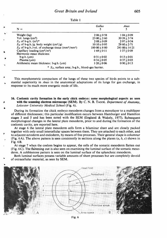
15. A morphometric comparison of the lungs of two species of bird of different exercise capacities.By J. N. MAINA (introduced by A. S. KING). Department of Veterinary Anatomy, Universityof Liverpool
Structural-functional correlations in the design of the mammalian lungs for gaseous exchangeare now well established (Gehr et al. Respir. Physiol. 44, 1981). The present lack of compre-hensive quantitative data on the structure of the avian lung does not enable such a correlation tobe attempted in birds, and makes comparison with mammals and other vertebrates unsatisfactory.Therefore a comparative morphometric investigation has been made of the gas exchange appa-ratus in the domestic fowl (Gallus domesticus), an inactive bird, and in the wild mallard (Anasplatyrhynchos), a powerful flyer.
The birds were killed by an intravenous injection of barbiturate and weighed. The lungs werefixed in situ by tracheal perfusion with buffered 2-3 % glutaraldehyde. Lung volumes were de-termined by a water displacement method. For each bird the left and right lungs were sampledby a stratified procedure for light and electron microscopy respectively, and processed by standardtechniques. Histological sections were analysed by point-counting to determine the volumedensities of the main functional components of the lung, notably the exchange tissue; absolutevolumes were calculated from the lung volume.From ultrathin resin sections a sufficient number of electron micrographs was analysed using
a quadratic lattice grid by standard morphometric methods (Weibel, Respir. Physiol. 11, 1970/1).The results are expressed as means (+S.D.) and appertain to the left and right lungs together(Table l).
604
Great Britain and Ireland
Table 1
Gallus AnasN= 5 5
Weight (kg) 2-06+0-58 1-04±0-09Vol. lungs (cm3) 25 00+ 2 60 3039 + 3 74SA of b-g.b. (m2)* 2-08 + 0-09 297 + 0 54SA of b-g.b./g. body weight (cm2/g) 10 10+0 95 28-60+ 2-71SA of b-g.b./vol. of exchange tissue (mm2/mm3) 180-00+ 8-80 241-00± 14-21Capillary loading (cm3/m2) 1-69+ 0 11 1-37 + 009Harmonic mean thickness
b-g.b. (,tm) 0-31+002 013+001Plasma (,um) 0 34+0°05 0 37+ 0 03
Arithmetic mean thickness: b-g.b. (/Lm) 1-20±0-06 0-90+0-13* SA, surface area; b-g.b., blood-gas barrier.
This morphometric comparison of the lungs of these two species of birds points to a sub-stantial superiority in Anas in the anatomical adaptations of its lungs for gas exchange, inresponse to its much more energetic mode of life.
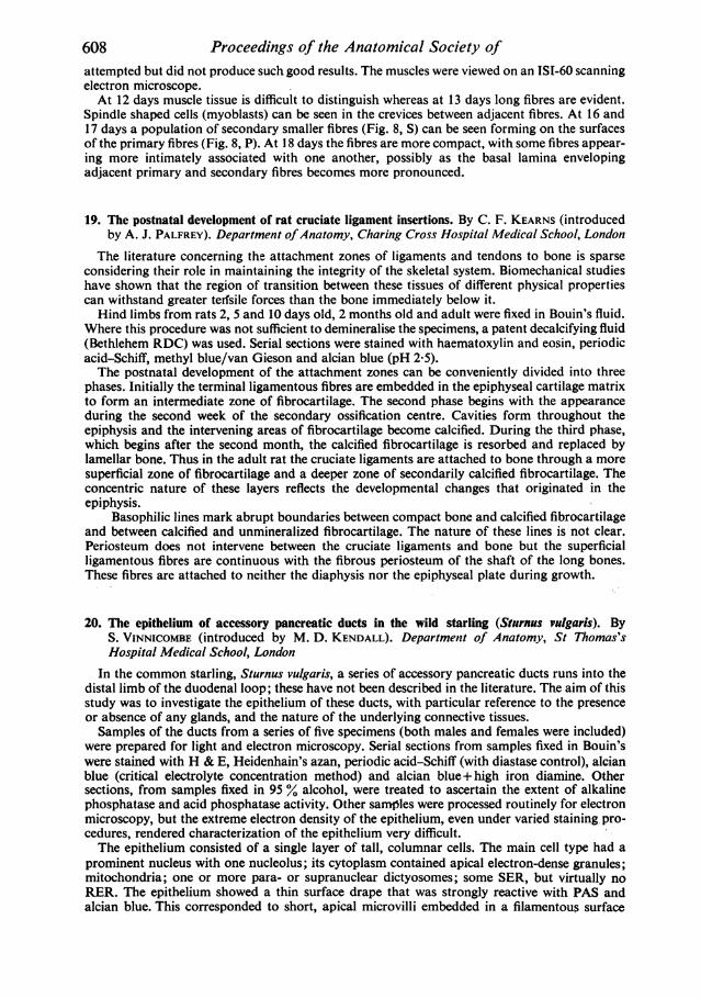
16. Coelomic cavity formation in the early chick embryo: some morphological aspects as seenwith the scanning electron microscope (SEM). By C. N. B. TAGOE. Department of Anatomy,Leicester University Medical School (Fig. 6).
During its formation the chick embryo mesoderm changes from a monolayer to a multilayerof different thicknesses; this particular modification occurs between Hamburger and Hamiltonstages 3 and 5 and has been noted with the SEM (England & Wakely, 1977). Subsequentmorphological changes in the lateral plate mesoderm, prior to and during the formation of thecoelomic cavity, are reported here.At stage 6 the lateral plate mesoderm cells form a bilaminar sheet and are closely packed
together with only small intercellular spaces between them. They are attached to each other, andto adjacent ectoderm and endoderm, by means of fine processes. Their general shape is columnar(Fig. 6A). The above pattern is seen consistently in sections along the planes (a, b, c) shown inFig. 6B.At stage 7 when the coelom begins to appear, the cells of the somatic mesoderm flatten out
(Fig. 6C). The flattening out is also seen on examining the luminal surface of the somatic meso-derm. A cobblestone pattern is seen on the luminal surface of the splanchnic mesoderm.Both luminal surfaces possess variable amounts of short processes but are completely devoid
of extracellular material, as seen by SEM.
Head fold~~~~~~~~~~~~~~~~~~~ f)L _
Fo~~~ ~~~~~~~
*,sswA~~~~~~~~~~~~~~~~lF - S; s ~ ~ ~ ~ I
Fig. 6
605

Proceedings of the Anatomical Society ofThe changes described take place in a cephalocaudal direction, and also from the periphery
towards the midline. This results in a horseshoe pattern of morphological changes, with a gapcephalad in the region of the proamnion.That the lateral plate mesoderm cells are rearranged into two definite layers at stage 6 is
significant, because it is from this stage onwards that the coelom begins to form between thesetwo layers. It is not immediately clear why somatic mesoderm cells flatten out while those of thesplanchnic mesoderm remain columnar.
17. Localisation of the sodium pump in the epiblast of the early chick embryo. By C. D. STERN(introduced by P. L. DEBBAGE). Department of Anatomy and Embryology, UniversityCollege London (Fig. 7).
Previous electrophysiological studies (Jaffe & Stern, Science 206, 1979) have shown thatstrong electrical currents flow out of the primitive streak of chick embryos during gastrulation.They are probably the result of ion pumping by the epiblast into the underlying intrablasto-dermic space. Sodium is likely to be the current-carrying ion (Howard, J. cell. comp. Physiol. 50,1957). These studies have led me to investigate the localisation of the sodium pump in the epi-blast of the early chick embryo by exposing early, middle and late gastrula blastoderms totritiated ouabain, which binds specifically to the sodium/potassium pump.Each of the embryos was exposed to 6 ,uCi tritiated ouabain in saline/albumen (1:1) (final
ouabain concentration, 2 x 10- M, specific activity, 17 Ci/mmol) for 30 min at 37 'C. Theembryos were grown on their vitelline membranes (New, J. Embryol. exp. Morph. 3, 1955) ona pool of the label-containing medium. The blastoderms were fixed in Bouin's fixative andembedded in paraffin wax, or briefly fixed in buffered formal saline and sectioned in a cryostatafter freezing in isopentane cooled with liquid nitrogen. The sections (8-20 #um) were coated withIlford Nuclear Research Emulsion K 2. The slides were exposed for 4 weeks at 4 'C and de-veloped in Kodak D 19b. The autoradiographs were photographed by dark-field optics.The results show that prior to the appearance of the primitive streak ouabain binding is
Fig. 7
606

Great Britain and Irelandlocalised near the basal surfaces of the epiblast cells (Fig. 7, arrowheads). After the emergence ofthe mesoderm from the streak, the labelling remains at the basal side in lateral regions but itshifts to the apical side of the tissue at the primitive streak (Fig. 7, PS). Both figures are trans-verse sections through the embryos. Control embryos incubated at 4 °C, or using a mediumcontaining 20 mm potassium, did not show the above localisation.From these experiments it can be concluded that ouabain binding sites are probably restricted
to the basal (ventral) surface of the epiblast cells during gastrulation in the early chick embryo,except at the primitive streak where they appear at the apical (dorsal) surface of the cells. Thereis thus a close correspondence between the pattern of current flow and the position of the pumps,suggesting that sodium may therefore be the major current-carrying ion.
18. A scanning electron microscopic study of prenatal muscle development in vivo. By N. C. STICK-LAND. Department of Anatomy, Royal (Dick) School of Veterinary Studies, Edinburgh(Fig. 8).
Early stages of myoblast fusion have been studied in tissue culture preparations and some ofthese studies have employed scanning electron microscopy. Structural aspects of muscle de-velopment in vivo, however, have been studied almost exclusively using sectioning techniques.Theories of muscle development, based on these latter studies, include deductions about thelongitudinal arrangement of cells and fibres in developing muscle. Scanning electron microscopyof muscle tissue in vivo provides a method whereby our knowledge of the three dimensionalarrangement of developing muscle may be extended.Mouse fetuses (C57 strain) were obtained at daily intervals from 12 days gestation to almost
full term (18 days). Whole forelimbs were dissected off the fetuses and fixed in 3 % glutaraldehyde.In the older fetuses. (> 15 days) m. biceps brachii was then removed entirely whereas in theyounger fetuses the muscle was left in situ. The muscles were pulled apart to expose the developingmuscle fibres and then processed and critical point dried. Freeze-drying techniques were also
Fig. 8
ANA 134
607
20
Proceedings of the Anatomical Society ofattempted but did not produce such good results. The muscles were viewed on an ISI-60 scanningelectron microscope.At 12 days muscle tissue is difficult to distinguish whereas at 13 days long fibres are evident.
Spindle shaped cells (myoblasts) can be seen in the crevices between adjacent fibres. At 16 and17 days a population of secondary smaller fibres (Fig. 8, S) can be seen forming on the surfacesof the primary fibres (Fig. 8, P). At 18 days the fibres are more compact, with some fibres appear-ing more intimately associated with one another, possibly as the basal lamina envelopingadjacent primary and secondary fibres becomes more pronounced.
19. The postnatal development of rat cruciate ligament insertions. By C. F. KEARNS (introducedby A. J. PALFREY). Department ofAnatomy, Charing Cross Hospital Medical School, London
The literature concerning the attachment zones of ligaments and tendons to bone is sparseconsidering their role in maintaining the integrity of the skeletal system. Biomechanical studieshave shown that the region of transition between these tissues of different physical propertiescan withstand greater terfsile forces than the bone immediately below it.Hind limbs from rats 2, 5 and 10 days old, 2 months old and adult were fixed in Bouin's fluid.
Where this procedure was not sufficient to demineralise the specimens, a patent decalcifying fluid(Bethlehem RDC) was used. Serial sections were stained with haematoxylin and eosin, periodicacid-Schiff, methyl blue/van Gieson and alcian blue (pH 2 5).The postnatal development of the attachment zones can be conveniently divided into three
phases. Initially the terminal ligamentous fibres are embedded in the epiphyseal cartilage matrixto form an intermediate zone of fibrocartilage. The second phase begins with the appearanceduring the second week of the secondary ossification centre. Cavities form throughout theepiphysis and the intervening areas of fibrocartilage become calcified. During the third phase,which begins after the second month, the calcified fibrocartilage is resorbed and replaced bylamellar bone. Thus in the adult rat the cruciate ligaments are attached to bone through a moresuperficial zone of fibrocartilage and a deeper zone of secondarily calcified fibrocartilage. Theconcentric nature of these layers reflects the developmental changes that originated in theepiphysis.
Basophilic lines mark abrupt boundaries between compact bone and calcified fibrocartilageand between calcified and unmineralized fibrocartilage. The nature of these lines is not clear.Periosteum does not intervene between the cruciate ligaments and bone but the superficialligamentous fibres are continuous with the fibrous periosteum of the shaft of the long bones.These fibres are attached to neither the diaphysis nor the epiphyseal plate during growth.
20. The epithelium of accessory pancreatic ducts in the wild starling (Sturnus vulgaris). ByS. VINNICOMBE (introduced by M. D. KENDALL). Department of Anatomy, St Thomas'sHospital Medical School, London
In the common starling, Sturnus vulgaris, a series of accessory pancreatic ducts runs into thedistal limb of the duodenal loop; these have not been described in the literature. The aim of thisstudy was to investigate the epithelium of these ducts, with particular reference to the presenceor absence of any glands, and the nature of the underlying connective tissues.Samples of the ducts from a series of five specimens (both males and females were included)
were prepared for light and electron microscopy. Serial sections from samples fixed in Bouin'swere stained with H & E, Heidenhain's azan, periodic acid-Schiff (with diastase control), alcianblue (critical electrolyte concentration method) and alcian blue+ high iron diamine. Othersections, from samples fixed in 95 % alcohol, were treated to ascertain the extent of alkalinephosphatase and acid phosphatase activity. Other samples were processed routinely for electronmicroscopy, but the extreme electron density of the epithelium, even under varied staining pro-cedures, rendered characterization of the epithelium very difficult.The epithelium consisted of a single layer of tall, columnar cells. The main cell type had a
prominent nucleus with one nucleolus; its cytoplasm contained apical electron-dense granules;mitochondria; one or more para- or supranuclear dictyosomes; some SER, but virtually noRER. The epithelium showed a thin surface drape that was strongly reactive with PAS andalcian blue. This corresponded to short, apical microvilli embedded in a filamentous surface
608

Great Britain and Irelandcoat that could be seen with the electron microscope. However, the apical granules appearnon-reactive to any stains for mucosubstances.A second, rarer, cell type, which appeared vesicular under the light microscope, had a large,
convoluted nucleus with sparse cytoplasm. Its function is unclear.
21. The distribution of chondrocytes in human tracheal cartilage. By N. E. MCCALLION, RUTHST. C. GILMORE and A. J. PALFREY.* Department of Physiology, The Queen's University ofBelfast and *Department of Anatomy, Charing Cross Hospital Medical School, London
In this study the number and distribution of chondrocytes in human tracheal cartilage havebeen investigated using a grid-counting method, planimetry and computer analysis. Pieces oftrachea incorporating several rings above the carina were obtained from cadavers aged from34 weeks gestation to 27 years. The tissue was fixed in neutral formol saline. Following routineparaffin processing, longitudinal sections were stained with haematoxylin and eosin or haemalum.
Sections were projected ( x 250) on to a screen marked out with grids of sides of 50 mm. Thenumber of cells per grid in the middle area of the image was counted and the area of matrix percell calculated. For planimetry, sections were projected on to a plain screen. Chondrocytes wereselected at random and the area of matrix surrounding them mapped out. The area of thesematrix 'domains', bounded by the cell's nearest neighbours, was measured.
Sections were analysed using a programmable flying spot microscope and picture preprocessor.Computer analysis of the image field enabled the number of cells and their nearest neighbourdistances to be calculated. Total cellularity declined with increasing age, and within age groupswas found to vary with position within the central area of the cartilage - moving from top tobottom of the rings minimum cellularity occurred at 50 % of total section length in neonates,30 % in prepubertal children and at 20 % in adults.Computer analysis of chondrocyte distribution identified cell clustering in all specimens.
Younger cartilages showed little evidence of grouping based on first and second nearest neighbourdistances, but third to fifth nearest neighbour distances indicated that the cells were notrandomly dispersed. This grouping may have some function in determining the limiting thick-ness of cartilage which can be nourished by the surrounding vascular tissue.Computer facilities were kindly made available by Dr D. Rosen, Department of Biophysics
and Bioengineering, Chelsea College, London.
22. The ureteric epithelium of the wild starling (Sturnus vulgaris). By G. R. WILLIAMS (introduced,by M. D. KENDALL). Department ofAnatomy, St Thomas's Hospital Medical School, London
Samples of ureter from five male and three female starlings (Sturnus vulgaris) were preparedfor light and electron microscopy by conventional methods. Paraffin-embedded sections werestained with H & E, periodic acid-Schiff (PAS) and alcian blue for light microscopy. Thinsections were stained with lead citrate and uranyl acetate for electron microscopy.At the light microscopic level individual cells were difficult to distinguish due to the presence
of thick mucus which was highly PAS- and alcian blue-positive. Alcian blue histochemistrydetermined the presence of an acidic mucus containing both neutral and, predominantly,carboxylated acidic mucopolysaccharides. The epithelium was of tall pseudostratified columnarform throughout its length.Under the electron microscope three cell populations were observed. A population of basal
electron-lucent cuboidal cells each containing a central leptochromatic irregular nucleus; granularcytoplasm; few organelles, and cytoplasmic extensions extending into dilated intercellular spaces.The second cell type was composed of large, irregularly shaped, electron-dense, mucin-secretingcells extending from the lumen to basal cytoplasmic extensions. They contained crenated nuclei,highly proliferated and dilated rough endoplasmic reticulum, Golgi apparatus and many mito-chondria, some of which were degenerate. They also contained many apical inclusions of mucusand these features were suggestive of holocrine secretion. A third population of intermediatecells with characters of both basal and mucin-secreting cells was also observed extending to thelumen.These relationships between the cell types and their internal structures indicate the presence
of a cell line from immature basal cells to the mature mucin-secreting cells with a variablepopulation of intermediate cells.
20-2
609
Proceedings of the Anatomical Society of
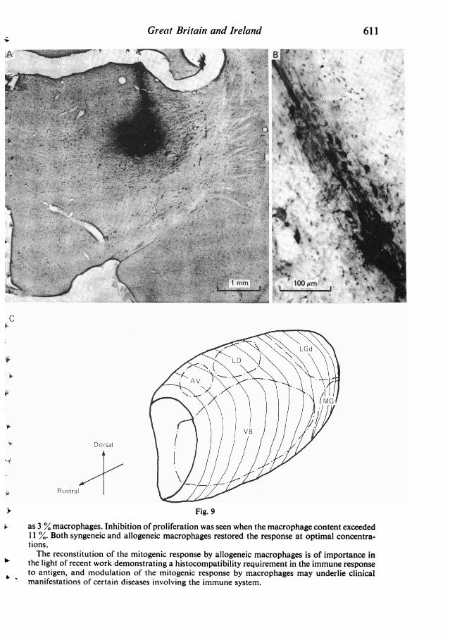
23. Connections of the thalamic reticular nucleus in the adult rat: a study using horseradishperoxidase (HRP) techniques. By M. O'CONNELL, P. T. OHARA and A. R. LIEBERMAN.Department of Anatomy, University College London (Fig. 9)
The thalamic reticular nucleus (TRN) is a thin, curved sheet of cells surrounding the lateraland anterior aspects of the dorsal thalamus. It is penetrated by, and receives input from, thefibre bundles interconnecting the dorsal thalamus and the cerebral cortex, sends its output pre-dominantly to the dorsal thalamus, and appears to play a role in monitoring and controlling theflow of information from the thalamus to the cortex.To gain further insight into the organization of the TRN, its afferent and efferent connections
were investigated by the HRP method, with special emphasis on the topographical organizationof reticulothalamic projections. HRP (15 % in tris buffer) was injected iontophoretically fromglass micropipettes (external diameter - 60 ,tm) positioned stereotaxically in various thalamicnuclei of anaesthetized adult albino rats. In several additional rats HRP was injected into thevisual or somatosensory cortices. Animals were perfused with fixative 16-24 hours after injection.The brain was removed and sectioned at 40-60,m in the frontal, horizontal or parasagittalplanes. Series of alternate sections were processed for visualization of HRP using the DAB(diaminobenzidine) and the TMB (tetramethylbenzidine) techniques. TheDAB material was usedto define the injection sites and to determine the positions of cell bodies labelled by retrogradetransport of HRP. The more sensitive TMB material was used both for this purpose and todetermine areas of terminal labelling resulting from anterograde transport of HRP from theinjection site. Fig. 9A shows an example of an injection site into the ventrobasal (VB) complex(frontal section, DAB) and Fig. 9B shows a patch of labelled cells in TRN following a similarinjection (horizontal section, DAB).Four major non-overlapping territories of cells projecting to ipsilateral dorsal thalamus were
defined and are summarized .in the simplified representation of TRN shown in Fig. 9C. Thedorsocaudal region projects to the dorsal lateral geniculate nucleus (LGd), the dorsorostralregion to the anteroventral (AV) and laterodorsal nuclei (LD), the ventrocaudal region to themedial geniculate nucleus (MG) and a large part of the ventral TRN anterior to the latterterritory projects to the ventrobasal complex (VB). Cell populations projecting to other dorsalthalamic nuclei (e.g. to the lateroposterior and ventrolateral nuclei) lie between these projectionterritories. Anterograde labelling of axon terminals in the neuropil surrounding the retrogradelylabelled cell bodies in TRN show reciprocal projections to TRN from the thalamic injectionsites with a topography corresponding to that of the reticulothalamic projections. Anterogradelabelling of terminals in TRN after HRP injections into cortex shows that the visual cortexprojects to the part of TRN which receives input from LGd and that the somatosensory cortexprojects to the part which receives input from VB.
24. Lymphocyte proliferative response to mitogen depends on macrophage content. By S. P.BARNARD and C. W. EVANS. Department of Anatomy, University of St Andrews
A requirement for macrophages has been demonstrated for various in vitro immune pheno-mena involving lymphocytes. These include the primary and secondary antibody responses andthe mixed lymphocyte reaction, although the dependency for macrophages in the lymphocytemitogenic response to lectins such as phytohaemagglutinin remains controversial. The problemappears to involve the identification and removal of residual macrophages in the lymphocytepopulation, so that the mitogenic response can be tested independently of macrophages.A mixed population of lymphocytes and macrophages was isolated from mouse lymph nodes.
Purification of the lymphocytes involved one or two nylon wool column adherence steps.A purified macrophage sample was obtained by allowing peritoneal exudate cells to adhere to
a serum-coated petri dish. The adherent macrophages were subsequently removed by treatmentwith 5 mm EDTA at 4°C. By centrifuging a sample of the cells and resuspending in 100 %serum, the staining of spread macrophages by the non-specific esterase histochemical techniquewas much improved.One nylon wool column purification of the mixed peripheral lymph node cell population did
not affect the PHA response, whereas two column purification steps abolished it. Loss of pro-liferation was not due to the isolation of an unresponsive subpopulation or to cell damage. Themitogenic response could be restored by reconstitution of the purified lymphocytes with as little
610
Great Britain and Ireland 611
./ V
-- _ . . ss
-~~~~~~~~~~~~~~~~~~~~.~~~~~ ~~~0
) 1mmk: ,. $S
t ~ ~~~ ~ ~ ~ ~~~~~~tOM
C *tX 0100~~~~~~~~~~~~~~~~~~~~~~~~~W17XX.
* ~~~Rostral / ///
> Fig. 9
as 3 %~macrophages. Inhibition of proliferation was seen when the macrophage content exceeded11 %. Both syngeneic and allogeneic macrophages restored the response at optimal concentra-tions.The reconstitution of the mitogenic response by allogeneic macrophages is of importance in
the light of recent work demonstrating a histocompatibility requirement in the immune responseto antigen, and modulation of the mitogenic response by macrophages may underlie clinicalmanifestations of certain diseases involving the immune system.
Proceedings of the Anatomical Society of
25. The fetal vasculature of the human term placenta: a morphological study. By S. HABASHI(introduced by G. J. BURTON). Department of Anatomy, University of Cambridge
Although scanning electron microscopy of corrosion casts has provided much information onthe microvasculature of many varied tissues (Hodde & Nowell, S.E.M. 11, 1980), to the author'sknowledge the technique has not previously been applied to the human placenta. In this study,casts of the fetal vasculature of 12 normal term placentae were produced by injecting chorionicvessels with an unsaturated polyester resin (Trylon Crystic Resin: Trylon Ltd, Wollaston,Northants). After corrosion of the placental tissues using concentrated hydrochloric acid, theresultant microvascular casts were dried, coated with gold/palladium and viewed in a PhilipsStereoscan 600 microscope.The most conspicuous feature of the casts was the presence of sinusoidally dilated capillaries,
which often formed complex interlocking loops, producing a knot-like structure. The generaldisposition and closed-loop nature of these capillary knots suggested that they represented thevessels of terminal villi. This supposition was confirmed by the observation of such a knotactually within a terminal villus in an incompletely corroded specimen. Only the tissue overlyingthe maximally dilated regions of the capillary loops had been removed, indicating that at thesepoints the vessels are normally overlain by a comparatively thin layer of trophoblast. Replicasof paravascular networks of capillaries surrounding larger villous vessels near the chorionicplate were also observed, as were casts of vasa vasorum.By comparing the findings of this study with those obtained by conventional histological
techniques, it was confirmed that the casts represented faithful replicas of the original luminalform of the fetal vessels. In conjunction with stereo-pair photography this technique enablesrapid and unequivocal interpretation of the three dimensional configuration of the villousvasculature. It is hoped it may prove of assistance in the investigation of changes in the fetalvasculature which may occur in abnormal pregnancies.
26. Recent improvements in endoscopic techniques. By J. C. NICHOLLS (introduced by A. J.PALFREY). Department of Surgery, Charing Cross Hospital, London and Hemel HempsteadHospital, Buckinghamshire
The 'modern' era of endoscopy began in 1958 with the introduction of the fibrescope byHirschowitz. From early times the desire to visualize the interior of organs had been strong, butthe physical limitations of the equipment made this impossible. The early part of this centurysaw the development of cystoscopy and gastroscopy.
Fibre-optic endoscopes rely upon the principle of total internal reflexion, which almost defiesthe principle of light travelling in straight lines. Recent improvements have been dependent uponrefinements of the equipment, and ingenuity of the operators. There is now a range of endoscopessmall enough to pass into the common bile duct, the ureters, the blood vessels, and large enoughto allow therapeutic manoeuvres. Their flexibility and field of vision are almost unlimited, andthey are long enough to reach from mouth to jejunum, and anus to caecum. Optical refinementshave improved the light carrying ability, and the resulting view can be recorded on film andclosed circuit television. The endoscopist now has the facility to pass biopsy forceps, cytologybrushes, snares, needles, baskets and laser beams.The techniques for performing upper and lower gastrointestinal endoscopy and for examining
the bile ducts are now standard, and the risk to the patient is at a very low level.The modern uses of endoscopy fall into two main categories. Diagnosis of diseases of oeso-
phagus, stomach and duodenum, rectum and colon can be made by direct observation and proofobtained by biopsy. In combination with radiological techniques the biliary tree and pancreascan be examined for disease. Therapeutic applications of endoscopy are constantly widening.Dilatation of oesophageal strictures, control of gastrointestinal haemorrhage, removal of bileduct stones and excision of colonic polyps are an example of such applications. The place ofendoscopy in screening for disease is not yet established.
612
Great Britain and Ireland
27. photogrammetric techniques. By L. F. H. BEARD (introduced by A. J. PALFREY). MedicalPhotography and Illustration Service, Addenbrooke's Hospital, Cambridge
A stereopair of photographs contains comprehensive and accurate information on the size andshape of the subject recorded. However, delays occur in gaining access to this information be-cause of the procedures involved in reorientation of the stereopairs in the plotting equipment.These delays, together with the scarcity of the skill required for taking measurements from a
spatial model, has hindered the replacement of traditional methods of measurement by stereo-photogrammetry. The design of a stereometric camera/plotter for clinical photography basedon non-metric cameras and lenses has minimised problems of reorientation. Methods of simplify-ing analysis have also been devised. The results of these developments, and their applications,were described and demonstrated.
28. The contribution of nuclear medicine. By E. S. WILLIAMS (introduced by A. J. PALFREY).Institute of Nuclear Medicine, The Middlesex Hospital Medical School, London
Radiation-emitting atoms can be incorporated into, or attached to, molecules, colloidparticles, cells, and so on, thus enabling the distribution of these materials in the body to bedelineated by suitable radiation detection instruments. It is important to note that the medicallyuseful information is provided by how the body handles such a radioactively labelled material.The choice of label, the radioactive isotope, is a complicated one, but is a largely technicalproblem.A common form of the presentation of results is in the form of an 'image' of the distribution
in the body of the radioactive material which has been administered. However, it must be empha-sised that this is a functional image representing an aspect of the physiological state of the organ
or part of the body being studied. For example, if a radioactive colloid is introduced by intra-venous injection the colloidal particles are rapidly cleared by the cells of the reticulo-endothelialsystem and, because of the concentration of these cells in the liver, an image of the liver can beobtained. If this cellular system has been damaged (e.g. after recent radiotherapy to the liver)then no image will be obtained. This is a dramatic illustration of imaging the functional natureof a nuclear medicine image.
This property sets nuclear medicine imaging apart from other imaging modalities, such as theuse of ultrasound, or X-rays which, while they demonstrate anatomical structures, give no
information on the functional state of those structures. An example is provided by disease of bone.If the bone is only slightly decalcified by the disease, then it could well present a normal X-rayimage. However, if its metabolic state is affected this might easily be detected early in the pro-
gress of the disease by the use of an appropriate bone-seeking agent and the production of a
nuclear medicine functional image.
29. Xeroradiography. By A. J. STACEY (introduced by A. J. PALFREY). Department of PhysicsApplied to Medicine, Charing Cross Hospital, London
Xeroradiography is a relatively new X-ray technique in which a specially prepared Xeroxplate is used instead of X-ray film. It is becoming accepted in mammography, and is establishingits place in the examination of other anatomical sites.The radiological image on the Xerox plate is characterized by the so called 'edge effect',
which results in pictures bearing remarkable resemblances to line drawings. It also exhibits a
wide exposure latitude and one X-ray exposure can be used to display tissues of widely differingradiation properties.The method of image formation was outlined and the limitations imposed by the radiation
dose to the patient were considered. Examples were shown to illustrate the significant propertiesof the Xeroradiographs and also to indicate the range of clinical application.Other electrostatic systems are being developed and the principal features were discussed.
Future improvements which may be expected of these methods were also reviewed.
613

Proceedings of the Anatomical Society of
30. Recent trends in interventional radiology. By J. MCIVOR (introduced by A. J. PALFREY).Department of Diagnostic Radiology, Charing Cross Hospital, London
It is now possible for the radiologist to pass needles and catheters into most large organs andinto the blood vessels which supply them. These procedures provide anatomical demonstrationswhich were unobtainable in the living until recently. A knowledge of normal anatomy and of thecommon variations is of course a pre-requisite for technical success and for diagnostic accuracy.
In addition to diagnostic procedures, therapeutic measures, such as drainage of obstructedbiliary and renal tracts, occlusion of the arteries supplying tumours and arteriovenous mal-formations and the dilatation of narrowed arteries, are now regularly undertaken.
This paper showed examples of these newer techniques and discussed their role in the manage-ment of patients.
31. Isolation of structures at the basement membrane zone of normal human skin. By J. NANCHAHAL,D. J. RICHES* and T. W. GLENISTEk. Department of Anatomy, Charing Cross HospitalMedical School, London, and *Department of Anatomy, Chinese University of Hong Kong
The isolation of human cutaneous basement membrane (BM) has previously been attemptedby Heaphy & Winkelmann (J. Invest. Derm. 68, 1977), but their final preparation showed con-siderable collagen contamination. All biochemical analyses of cutaneous BM have been per-formed on partial enzyme-digests of whole skin and so this study was undertaken to obtain anultrastructurally 'pure' sample of cutaneous BM for analysis.Areas of normal human skin, about 8 x 30 cm, were obtained from patients undergoing plastic
surgery and were soaked in 2M-NaSCN for 5 hours at 4 'C. This permitted the epidermis to bepeeled away, leaving the BM intact on the dermal surface. The latter was intermittently ultra-sonicated in the presence of 0-3 % Triton X-100 in phosphate-buffered saline (PBS) for 45 minin an ice-bath. A fraction enriched in BM was obtained from the PBS suspension using differ-ential centrifugation and discontinuous CsCl density gradients. All stages were monitored byelectron microscopy.The NaSCN appeared to disrupt selectively the structures at the lamina lucida (hemidesmo-
somes) whilst the desmosomes remained intact; this may reflect an intrinsic chemical differencebetween these two structures. The dermal cells were disrupted and the cytoplasm of the epidermalcells was very electron-dense whilst their nuclei became electron-lucent. In the presence of enzymeinhibitors, flocculent material (at least in part of nuclear origin) was attached to the BM and incentrifugates two types of BM could be identified:
(i) 'light BM' - similar to that in normal tissues, but which stained less intensely.(ii) 'dark BM' with flocculent material attached to it.In the absence of Triton X-100, very vigorous ultrasonication was required to release the BM,
which showed marked variation in thickness. During the purification steps the BM appeared tobreak up, but the bond between the BM and anchoring fibrils was found to be very resilient.However, in the absence of enzyme inhibitors profiles of BM without anchoring fibrils werecommon and occasionally the BM was seen to split into three layers.
Preliminary analyses have shown the final fraction to consist of c. 75 % basement membraneand anchoring fibrils by mass.
32. Studies on hemolymph nodes: the intrinsic vasculature of the renal hemolymph node of rats.By AVRIL M. PEARCE, 0. REID and R. J. SCOTHORNE. Department of Anatomy, Universityof Glasgow
Unlike those of 'typical' nodes, the lymph sinuses of hemolymph nodes contain abundanterythrocytes. Nopajaroonsri, Luk & Simon (J. Ultrastruct. Res. 1974) claimed that the hemo-lymph node was more vascular than typical nodes and that erythrocytes entered its sinuses bydiapedesis through intrinsic capillaries and venules. We have compared the intrinsic vasculatureof the renal hemolymph node with that of the facial node of the rat, using thick cleared sectionsafter intravascular perfusion with alcian blue, and semithin plastic embedded sections.The vascular pattern was essentially similar in the two types of node. Arterioles extended
radially from the hilum, to supply principally a deep plexus at the corticomedullary junction
614

Great Britain and Irelandand a richer subcapsular plexus. These plexuses drained into postcapillary venules and thence,via high endothelial venules (HEV), to segmental and collecting veins.
Differences between vascular patterns in the two nodes may be related to the fact that thefacial node is the more active immunologically, for:
(i) Arteriovenous anatomoses were more numerous in the facial node.(ii) HEVs were consistently more numerous and larger in the facial node.(iii) The subcapsular and corticomedullary plexuses were richer in the facial nodes, although
their patterns were distorted by more numerous and (avascular) germinal centres and by larger(and relatively avascular) paracortical nodules of the thymus-dependent zone.
Counts of cells migrating through the walls of HEV show comparable, very large, numbersof lymphocytes and very small numbers of erythrocytes in the two nodes. The results do notsupport the view that significant numbers of erythrocytes enter the lymph sinuses by an intrinsicroute.
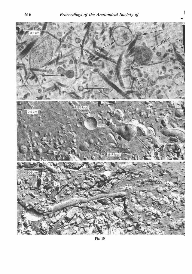
33. Origin of matrix vesicles of developing dentine investigated by freeze-fracture. By E. KATCH-BURIAN and N. J. SEVERS*. Department of AnatomY and Histology, The London HospitalMedical College, London, and *Department of Cardiac Medicine, Cardiothoracic Institute,London (Fig. 10)
Matrix vesicles are cell-derived membrane-bound bodies which have been demonstrated inultrathin sections by electron microscopy in the matrix of dentine during early dentinogenesis(Fig. 10, arrows). The vesicles lie free in the matrix and are limited by a membrane which exhibitsa typical trilaminar structure. When mineralization of dentine begins, the first mineral depositsappear as crystal-like inclusions inside the matrix vesicles, an observation which led to theproposition that matrix vesicles act, in some way, as promoters of calcium phosphate precipita-tion (Katchburian, J. Anat. 116, 1973). Although of cellular origin it is not known whethermatrix vesicles form by budding off from the surface of odontoblasts or by extrusion of someintracytoplasmic precursor. To investigate the origin of matrix vesicles we have examined de-veloping dentine in specimens prepared by freeze-fracture.
Molar tooth germs from 3 days old rats were fixed in cacodylate-buffered 4 % glutaraldehyde-4 % formaldehyde and were then treated with cacodylate-buffered 25 % glycerol. The specimenswere frozen in propane and fractured in a Balzers BAF 400T apparatus, and the replicas wereexamined in a Jeol JEM 1OOB electron microscope.
Early developing dentine shows typical criss-crossing collagen fibres which appear as plastic-ally deformed stubs in transverse fractures and as striated structures in longitudinal fractures(Fig. 10). Portions of odontoblast processes (Fig. 10, P) and matrix vesicles (Fig. 10, M) arepresent in the intercollagenous regions. The membranes of odontoblast processes reveal adensity and distribution of intramembrane particles on the P- and E-faces which is indistinguish-able from that observed on the membrane fracture face of matrix vesicles. This is consistentwith the idea that matrix vesicles probably arise by budding off from the surface of odontoblastprocesses. We are currently investigating the possible changes which may take place in themembrane of matrix vesicles during calcification.
34. Radiation-induced giant epithelial cells in small intestinal mucosa. By K. E. CARR, R. HAM-LETT*t and C. WATT. Department of Anatomy, Glasgow University and *Radiobiology Re-search Group, Belvidere Hospital, Glasgow
Structures which were interpreted as 'giant enterocytes' were identified in a scanning electronmicroscope study of the effects of irradiation on mouse small intestinal mucosa (Carr et al.J. Microsc. 123, 1981). Their histology and ultrastructure have now been studied in more detail.A sample of 22 adult C3H/He mice, made up of both sexes, were irradiated with either 10 GyX-rays or 5 Gy neutrons, and areas of mucosa were examined 5 days later by light microscopy,transmission electron microscopy (TEM) and scanning electron microscopy (SEM). In 8 of the22 mice, SEM revealed flat or pedunculated swellings, 25-60,um in width, situated at variouslevels of the surface of the villus. The surface was smooth, and showed neither cell boundaries
t Present address: University of Oxford Research Institute, Churchill Hospital, Oxford.
615
Proceedings of the Anatomical Society of
jie x./
ALS
T
Fig. 10
616

Great Britain and Irelandnor evidence of discharging goblet cells. in semithin sections, the swellings consisted of a multi-nucleate mass of cytoplasm lacking cell boundaries, containing large vacuoles and PAS-positivegranules which we have interpreted as mucus. Contact with stroma and adjacent epithelial cellsappeared complete.
Transmission electron microscopy showed apical microvilli to be sparser than on normalenterocytes. The cell membrane showed basal herniations, and exaggered lateral interdigitations.Intracytoplasmic vacuoles lined by trilaminar unit membranes were seen, as were occasionalintracytoplasmic desmosomes. Lipid inclusions were conspicuous, as they were in the radiationdamaged enterocytes.The swellings show some features of both absorptive cell and goblet cell differentiation. The
significance of their structural peculiarities is discussed in relation to irregularities of mitoticdivision and differentiation of the stem cell population.
35. The muscularis mucosae of the human urinary bladder. By J. S. DIXON and J. A. GOSLING.Department of Anatomy, University of Manchester
Standard histological textbooks (e.g. Ham's Histology) describe the mucosa of the urinarybladder as consisting of a layer of transitional epithelium and a supporting layer of loose con-nective tissue (the lamina propria) and make no mention of a muscularis mucosae. Indeed-sometexts state categorically that a muscularis mucosae is absent from the human urinary bladder(e.g. Gray's Anatomy). During studies on the human lower urinary tract we have recently had theopportunity to examine a number of normal bladder biopsy samples removed from a variety ofsites, including the fundus, the anterior and lateral walls and the trigone. In all cases the biopsysamples were examined using both light and electron microscopy.
Irrespective of site, the bladder wall was observed to possess an incomplete layer of smoothmuscle separated by loose connective tissue from the basal aspect of the urothelium and also theinner aspect of the detrusor muscle, thus corresponding to a rudimentary muscularis mucosae.The constituent cells of this muscle layer could be clearly distinguished both histochemicallyand ultrastructurally from detrusor smooth muscle cells. The cells are rich in non-specificcholinesterase and react positively when stained with the periodic acid-Schiff technique. In theelectron microscope the smooth muscle cells are extremely irregular in outline and contain largeareas of sarcoplasm packed with electron-dense glycogen granules. Numerous acetylcholin-esterase-containing nerves course between the smooth muscle cells of this muscularis mucosae;in the electron microscope, varicose axon terminals containing small (50 nm diameter) agranularand occasional large (100 nm diameter) granulated vesicles are observed in close proximity tothe smooth muscle cells.
36. Histological observations on the effects of localised hyperthermia on normal pig striated muscle.By D. J. RICHES, B. M. SOUTHCOTT*, P. B. DUNSCOMBEt and K. GAMMAPILAt. Departmentof Anatomy, The Chinese University ofHong Kong and Departments of *Radiotherapy andSurgery, and t Medical Physics, Charing Cross Hospital, London
The recent clinical application of hyperthermia produced by microwave radiation in the treat-ment of tumours has prompted an investigation to establish at what temperature damage occursin normal tissue. Preliminary results have shown that maintaining deep temperatures in excessof 45 °C for 30 minutes produced observable effects. As there is a temperature gradient acrossthe area covered by the applicator, approx. 10 cm x 6 cm, with a central peak, a method formeasuring the deep (i.e. subcutaneous fat/muscle interface) temperature at 1 cm intervals usingthermocouples was devised, thus enabling a study of the effects of varying temperature.
Hyperthermia was induced in the gluteal region of anaesthetized pigs, using microwave radia-tion at 434 MHz for 30 minutes. The skin was cooled by air blown between the applicator faceand the skin. Both skin and deep temperatures were continuously monitored. Punch biopsies ofskin and underlying muscle were taken across the treated area 1 and 6 weeks after treatment.The tissues were processed for both light and electron microscopy.At 1 week after treatment, muscle fibres that had been heated to 48 °C AVere obviously
damaged, with loss of nuclei and coagulation of myofibrils. Similar effects, but to a lesser degree,were seen at 47 and 45 'C. At lower- temperatures the muscle fibres appeared relatively normal,
617

Proceedings of the Anatomical Society ofwith only slight separation of the myofibril bundles. By 6 weeks after treatment, the area ofmuscle heated to 48 °C showed signs of recovery. The fibres were swollen, but nuclei were present,some being 'trapped' within the fibres. In the other areas, the fibres were also recovering, butthere was marked lymphocytic infiltration in all regions.Although the effects of hyperthermia described are non-specific, these preliminary results
demonstrate that muscle fibres can recover even after being heated to 48 'C. Further studies arein progress at the ultrastructural level to establish the detailed effects both on the myofibrils andthe microvasculature.
37. The physics of tomographic techniques. By J. S. ORR (introduced by A. J. PALFREY). Depart-ment of Medical Physics, Royal Postgraduate Medical School and Hammersmith Hospital,Du Cane Road, London
Tomography is the process of representing an object by a series of pictures in the form ofsections or slices. The process has three components. First, measurements of properties of theobject, either as a point, or as a small volume, must be made. Second, the point, or volume, mustbe localised in the slice. Third, interference from material elsewhere in the object, or slice, mustbe eliminated or reduced. Under certain conditions tomography is preferable to techniques whichproduce total projection images.A wide range of properties can be measured: radioisotope concentration, X-ray or particle
attenuation and magnetic resonance properties. Localisation commonly depends simply on thelinear propagation of rays. Reduction of interference is often produced by blurring detail fromelsewhere in the object, and physicists have devoted considerable efforts to refining the last pro-cedure. The modern techniques of magnetic resonance imaging illustrate most of the essentialfeatures of tomography.
38. Nuclear magnetic resonance imaging. By G. M. BYDDER and I. R. YOUNG (introduced byA. J. PALFREY). Royal Postgraduate Medical School, Hammersmith Hospital, London
A description of a nuclear magnetic resonance (NMR) imaging system was presented togetherwith some results from its clinical evaluation. The detection of proton magnetisation within astatic magnetic field is outlined, and the rotating frame is used to describe the motion of themagnetisation. Radiofrequency pulses are used to rotate the magnetisation and slice selectionis achieved using a 900 pulse and a magnetic field gradient. Three scanning modes are described:repeated free-induction decay (FID), inversion-recovery and spin-echo. These sequences pro-duce images whose pixel values have different dependencies on proton density, T1 and T2.
Inversion-recovery images show striking differentiation between grey and white matter in thebrain. The absence of bone artefact is a significant advantage over X-ray computed tomographyin the posterior fossa where rapid repeated FID sequences can also be used to demonstrate floweffects.Blood within the left ventricular cavities is differentiated from myocardium, and the para-
magnetic effects due to dissolved arterial oxygen can be demonstrated.The considerable soft tissue contrast available with NMR is of value in demonstrating disease
within the liver where T1 appears to be a sensitive, but relatively non-specific, diagnostic para-meter. The cortex and medulla of the kidney can also be distinguished. High resolution scansare of value in demonstrating the adrenal gland and spinal cord.Although the spatial resolution of NMR scanners is not as good as that of third and fourth
generation computerised tomography scanners, the high level of soft tissue contrast, and absenceof bone artefact, enable a variety of structures to be visualised in a way not possible with otherimaging techniques.
39. Computerised tomography (CT). By D. E. KATZ (introduced by A. J. PALFREY). Departmentof Radiology, Northwick Park Hospital and Clinical Research Centre, Harrow, Middlesex
Computerised tomography is a technique which utilizes a conventional X-ray beam and, withthe aid of highly sensitive detectors and computers, produces a cross sectional image at anydesired level of the body.The technique requires a tightly collimated X-ray beam to rotate around the patient, and,
for each section detectors obtain over 300000 absorption measurements at a variety of angles.
618

Great Britain and Ireland6The computer converts this raw data into a set of profiles and the main part of the computerprocessing then converts these profiles into information which can be displayed as a picture.
Computerised tomography overcomes many of the main limitations which have been imposedon conventional radiology for almost a century. By summating the cross sectional images it ispossible to present three dimensional information about the internal structure of the body. Thesensitivity of the recording apparatus makes it easy to distinguish between the soft tissues of thebody, and gives a clear anatomical display of organs such as liver, pancreas and spleen.
Computerised tomography also allows quantitative differentiation by measuring X-rayabsorptions of the different tissues. In addition it allows detailed examination of all tissues sothat in a single section tissues such as lung parenchyma, mediastinal soft tissues and bone can allbe scrutinised.The anatomical limits are imposed by the spatial resolution and density resolution and these
two are limited by the radiation dose.The greatest impact of computerised tomography has been in neuroradiology, but the ability
to display the whole body has created a new dimension in non-invasive investigative imaging ofthe body to be used in conjunction with other scanning techniques.
40. Microfocal radiography: a new X-ray technique for high resolution quantitative macroradio-graphy of human extremities. By J. C. BUCKLAND-WRIGHT. Department of Anatomy, Guy'sHospital Medical School, London
Fine X-ray sources (5-10,um diameter) have been used with some success in the detailed radio-graphic examination of the structural organisation of normal and diseased tissues. The relativelylow energy levels necessary for ultra-small X-ray sources results in long X-ray exposure timeswhich have hitherto limited its use to small live animals and post mortem animal and humanmaterial. However, recent advances in the design of microfocal X-ray equipment has resultedin a new high energy unit suitable for the routine radiographic examination of patients.
This new X-ray unit operates at 60 kV, 1 mA with an estimated X-ray source size of 6-8 umdiameter; this is, to date, the smallest known source at these energy levels. The operation of thisunit in conjunction with the new rare earth film/screen combinations (Trimax) permits macro-radiographic examination of the extremities of patients with an object resolution in the film of25-35 ,um at magnifications up to x 10. Exposure times are between 0 7 and 1 second. TheX-ray dose is within clinically acceptable limits of 34-70 mR/exposure. As penumbral blurringis minimal and all planes of the object are in focus, stereopair radiographs provide threedimensional assessment of structures with greater radiographic detail than obtained hitherto.The microfocal X-ray unit is currently used clinically for diagnosis and investigative work.
In the former, it is used in the precise identification of osseous changes associated with metabolicbone disorders and in the different types of arthritis. The latter consists of long term quantitativeanalysis of groups of patients with (a) early rheumatoid arthritis (to study the onset and enlarge-ment of erosions), (b) rheumatoid arthritis with well developed erosive arthropathies, to assessthe effectiveness of treatment, (c) osteoarthritis and (d) the levels of osteoporosis and the effectof phosphate binders in children with renal osteodystrophy. Because of the large magnificationand high resolution in the macroradiographs, quantitative assessment of the changes observedare readily obtained by direct measurement using an image analyser.
41. The sex of Alligator mississippiensis is determined by the temperature ot egg incubation. ByM. W. J. FERGUSON. Department ofAnatomy, The Queen's University ofBelfast (Fig. 11)
Heteromorphic sex chromosomes are absent in all crocodilians. The effect of egg incubationtemperature on sexual differentiation was investigated in laboratory and field experiments. 500alligator eggs were collected from wild nests in the swamps of Louisiana and incubated at either26, 28, 30, 32, 34 or 36 'C. After 60 days (alligators normally hatch at 65 days) eggs were fixedin 10% formalin. The alligators were examined under a dissecting microscope for the presence orabsence ofan oviduct or vas deferens and for the external shape and surface features of the gonad.The entire reproductive systems of 100 animals were serially sectioned at 8 ,sm in the horizontal,transverse and sagittal planes. In all cases where an oviduct was present the gonad was an ovary,when absent it was a testis. Sex is therefore fully determined in the embryonic period, and canbe assayed by the presence or absence of an oviduct. Eggs incubated at, or below, 26 'C and
619

Proceedings of the Anatomical Society of
N
'I .Layer 1
Layer 2
.. ,I -
Marsh Water
+1-
Fig. 11. P1, 34 °C; P2, 31 °C; P3, 32 °C; P4, 30 °C; P5, 31 °C; P6, 30 'C.
620

Great Britain and Irelandat, or above, 36 °C died. 100 % of eggs incubated between 28 and 30 °C were females and 100 %of eggs incubated at 34 °C were males. Eggs incubated at 32 °C yielded 86 % females and 14 %males. The average mortality was 6 % and there was no significant difference between the fourtemperature groups (28, 30, 32 and 34 °C).
Nest temperatures were continuously chart-recorded for eight wild alligator nests (four con-structed in the marsh and four on levees) with probes (PI-P6) at the top (P1), sides (P3-P6), andbottom (P2) of the egg cavity (Fig. 11). A map of egg position within the nest was drawn whenthe eggs were fixed at 60 days (Fig. 11). All levee nests recorded average temperatures of 34 °Cor above, at all locations in the nest and 100 % of the eggs developed into male alligators. Allwet marsh nests recorded temperatures of 30 'C or below and 100 % of the eggs yielded females.Intermediate dry marsh nests recorded varying temperatures (Fig. 1 1). Male alligators developedfrom the hottest location in the nests - top central layer (Fig. 11, eggs 12, 13 and 16) and femalealligators from the cooler locations - periphery and bottom (Fig. 11, eggs 1-11, 14, 15, 17-32).Laboratory experiments in which alligator eggs were incubated at a female-producing tempera-ture for the first x stages of development and then shifted to a male temperature for the remainderof development (or vice versa) showed that the temperature-sensitive period for sex determinationis within the first 3 weeks of incubation. A 4 years study of the sex of all hatchling alligators(n = 6000) within an extensive area of normal habitat revealed that the natural sex ratio athatching is 6 females: 1 male. Given this natural skewed sex ratio, theoretical calculations showthat genotypic influences probably have little effect on sex determination. Animals raised for4 years after hatching showed no sex reversal. In both laboratory and field experiments, theweight of males at hatching was significantly less than that of females. Given the reproductivebiology and natural history of alligators it can be shown that this constitutes a selective biologicaladvantage for temperature-dependent sex determination over possible genotypic mechanisms.Temperature-dependent sex determination in alligators has many implications for embryo-logical, teratological and molecular investigations as well as for conservation planning andfarming. It may also provide a clue to the mechanism of extinction of other archosaurs, e.g.dinosaurs.
This work was supported by M.R.C. Grant No. G979/386/C, E.H.S.S.B. grant no. EP109/74/75 and a Wellcome Trust Research Travel Grant.
42. Venous drainage of the ovine corpus cavernosum penis. By R. R. ASHDOWN, S. W. BARNETTand G. ARDALANI. Department of Anatomy, Royal Veterinary College, University ofLondon(Fig. 12)
Studies on various Artiodactyla suggest that drainage of the corpus cavernosum penis (ccp)by the v. dorsalis penis occurs only in young or impotent animals. In the goat, however, ana-stomoses between an intracorporeal 'v. trunci penis' and the corpus spongiosum glandis (csg),have been described draining the ccp (Starflinger, Anat. Histol. Embryol. 1, 1972). These anasto-moses would be of functional and clinical importance, and we therefore examined the angio-architecture of the excised penile bodies of 28 rams all aged 5 months or more, so that separationbetween penile integument and prepuce was completed.Erection was simulated by the injection of Colorpaque or Micropaque at 40°C into the
dorsal canal of the ccp at known reservoir pressures (23 specimens). Straightening of the peniswas completed at 210 x 103 Pa. At 350 x 103 Pa, mean total length had increased by 37-3 % andmean diameters by 3 0 %.Two small specimens showed traces of injected material in the v. dorsalis penis at, or just
distal to, the sigmoid flexure; these were the smallest specimens examined (total lengths beforeinjection - 28-8 and 30-1 cm). In all other specimens, radiography and clearing in methylsalicylate failed to demonstrate venous drainage from the ccp. Anastomoses with the corpusspongiosum penis (csp) or csg were not demonstrated in any specimen (Fig. 12).
Incremental injections into the ccp and csp (five specimens) showed that the csp drained viathe tuberculum spongiosum and csg into the v. dorsalis penis, but showed no connections withthe ccp. The 'v. trunci penis' was a secondary dorsal canal of the ccp, having no anastomoseswith vessels outside the tunica albuginea. Our experiments suggest that absence of venousdrainage within the penile body facilitates the rapid generation of high pressures in the ccp byminimal flow volumes. When the distal bend of the sigmoid flexure began to straighten, only afurther 7-12 ml of blood were needed to complete this straightening and then produce stiffeningwith maximum elongation of the organ.
621
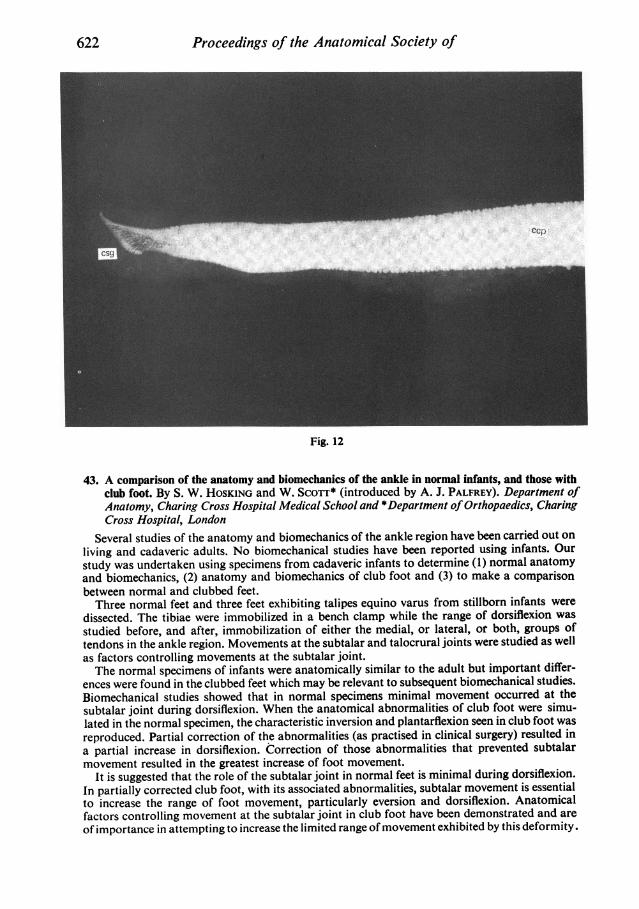
Proceedings of the Anatomical Society of
Fig. 12
43. A comparison of the anatomy and biomechanics of the ankle in normal infants, and those withclub foot. By S. W. HOSKING and W. SCOTT* (introduced by A. J. PALFREY). Department ofAnatomy, Charing Cross Hospital Medical School and *Department ofOrthopaedics, CharingCross Hospital, London
Several studies of the anatomy and biomechanics of the ankle region have been carried out onliving and cadaveric adults. No biomechanical studies have been reported using infants. Ourstudy was undertaken using specimens from cadaveric infants to determine (1) normal anatomyand biomechanics, (2) anatomy and biomechanics of club foot and (3) to make a comparisonbetween normal and clubbed feet.Three normal feet and three feet exhibiting talipes equino varus from stillborn infants were
dissected. The tibiae were immobilized in a bench clamp while the range of dorsiflexion wasstudied before, and after, immobilization of either the medial, or lateral, or both, groups oftendons in the ankle region. Movements at the subtalar and talocrural joints were studied as wellas factors controlling movements at the subtalar joint.The normal specimens of infants were anatomically similar to the adult but important differ-
ences were found in the clubbed feet which may be relevant to subsequent biomechanical studies.Biomechanical studies showed that in normal specimens minimal movement occurred at thesubtalar joint during dorsiflexion. When the anatomical abnormalities of club foot were simu-lated in the normal specimen, the characteristic inversion and plantarflexion seen in club foot wasreproduced. Partial correction of the abnormalities (as practised in clinical surgery) resulted ina partial increase in dorsiflexion. Correction of those abnormalities that prevented subtalarmovement resulted in the greatest increase of foot movement.
It is suggested that the role of the subtalar joint in normal feet is minimal during dorsiflexion.In partially corrected club foot, with its associated abnormalities, subtalar movement is essentialto increase the range of foot movement, particularly eversion and dorsiflexion. Anatomicalfactors controlling movement at the subtalar joint in club foot have been demonstrated and areof importance in attempting to increase the limited range of movement exhibited by this deformity.
622
Great Britain and Ireland 623
44. The use of ultrasound in medical diagnosis. By H. B. MEIRE (introduced by A. J. PALFREY).Division of Radiology, Northwich Park Hospital and Clinical Research Centre, Harrow,Middlesex
Diagnostic ultrasound techniques rely on the extraction of information from the echoesproduced by internal structures during insonation with high frequency sound. Although earlyclinical results were obtained over thirty years ago the technique has only found wide clinicalapplication during the past decade, with particularly rapid growth during the past five years.Modern ultrasound apparatus is rapidly diversifying and has now developed from a static
anatomical imaging technique to encompass the production of live, moving, images and measure-ment of a wide range of physiological functions.The basic physical principles of diagnostic ultrasound apparatus are reviewed and recent
advances in its clinical application presented. The latter include the use of high resolutionultrasound images to detect fetal abnormalities. Over 65 separate congenital anatomical abnor-malities of the fetus have now been identified by ultrasound and many centres throughout theworld have initiated routine screening programmes for the identification of unsuspected fetalanomalies. The recent improvement in image resolution and tissue differentiation has also per-mitted extension of ultrasound imaging to include many of the intra-abdominal organs. Theprogress in this field is best exemplified by reference to the localisation of the pancreas by virtueof its vascular anatomical relations. This organ can now be visualised almost routinely andrecent technical developments permit us to identify not only the gland but also the duct systemwithin it by an entirely non-invasive transcutaneous technique.An additional recent technical development is the identification of Doppler-shifted frequency
in the echoes returning from pulsed ultrasound systems. This facility has enabled the develop-ment of non-invasive vascular imaging systems which have recently been developed for the trans-cutaneous measurement of volume blood flow in accessible vessels.The value of ultrasound in the field of fetal, pancreatic and vascular imaging was discussed and
illustrated with selected clinical images.
45. The evaluation of risk to the patient. By E. E. POCHIN (introduced by A. J. PALFREY). NationalRadiological Production Board, Chilton, Oxfordshire
The risks of exposure to high doses of ionizing radiation were recognized within a few yearsof the discovery and first uses of X-rays and of radioactive tnaterials. Extensive radiobiologicalinvestigations in the subsequent eighty years have indicated that some risks of later malignantor hereditary effects are likely to follow even the lowest doses, without any threshold effect. Theapproximate magnitudes of these risks have been assessed in a substantial number of prolongedepidemiological studies following known radiation exposure of the body or of individual organsat moderate dose; and adequate inferences can be made of the size of the risks to be expectedfrom the low doses likely in diagnostic X-ray or radionuclide examinations.No investigations of any comparable scope or duration have been made on possible late effects
of ultrasound or nuclear magnetic resonance procedures. No indications have, however, beenobtained that either early or late harm is likely to result from the exposures at the levels neededin such tests. It is, on the other hand, clearly impossible yet to be sure that there might not befatal risks as low as those in the order of 1O-5 which may apply to typical radiological examina-tions.No extensive study appears to have been made of risks involved in imaging by endoscopic
methods but it seems unlikely that any risk of fatality would approach the value of 2 x 1O-4 thathas followed needle biopsy of the liver in two large series.The risks of different radionuclide and other radiological examinations can be tentatively
assessed in terms of the absorbed doses of radiation received by the various organs or tissues,and the epidemiological evidence of the size of the risks applicable to each. Conventionaldiagnostic procedures appear, on this basis, to imply fatal radiation risks of from 10-6 to 10-4for X-rays, and of 10-7 for radionuclide studies.

Proceedings of the Anatomical Society of
DEMONSTRATIONS
D.1. The epithelial-connective tissue junction in mucosae from different intra-oral sites: comparativemorphometric studies of attachment mechanisms. By ELIZABETH BALE and F. H. WHITE.Department ofHuman Biology and Anatomy, University of Sheffield
Stratified squamous epithelia are attached to the adjacent connective tissue by a specialisedbasal lamina complex. This comprises localised zones of attachment on the basal plasma mem-brane, the hemidesmosomes, and a lamina densa, which forms a thin sheet in the connectivetissue immediately below the epithelial basal plasma membrane. It has been suggested that thetopography of the interface between these tissues is important in modifying the attachment, withareas under high stress possessing a more irregular, rete-ridge type configuration. The purposeof this report is to clarify the morphological nature of the epithelial-connective tissue attachmentby comparing palatal and buccal mucosae in which the interfaces are convoluted and relativelyflat, respectively.Mucosal samples were obtained from five male ferrets from both cheek and hard palate.
Tissue blocks were routinely processed for electron microscopy and flat-embedded in Araldite;semithin sections were prepared and stained with toluidine blue. This material was used toobtain an index of the degree of irregularity of the epithelial-connective tissue junction bymeasuring the lengths of the granular cell/keratin (LGK) and epithelial-connective tissue (LEc)junctions using a MOP (Kontron) semi-automatic image analysing system. The ratio of LEc/LOKprovided such an index. From ultrathin sections obtained from the same blocks, a strict samplingprocedure was used to acquire micrographs which included epithelial-connective tissue junction,at a final magnification of x 12100. Using stereological intersection counting, the followingparameters were estimated: the relative surfaces of hemidesmosomes to basal plasma membrane(Sshd, bpm); the mean hemidesmosomal diameter (Alid); and the number of hemidesmosomes perunit surface of basal plasma membrane (Nshd, bpm).
Results suggest that in palatal mucosa, with a prominent rete-ridge pattern, values forSShd, bpm and Ahd were substantially increased when compared with cheek mucosa, whereasNShd, bpm values were similar in both tissues. Since mucosae from both sites have a similarhemidesmosomal frequency, it appears that palatal tissue which is subjected to higher stresspossesses larger hemidesmosomes, resulting in an increase in Sshd. bpm thus providing a greaterspecialised surface for adhesion between the epithelium and underlying lamina propria.
D.2. Some morphometric aspects of epidermal carcinogenesis. By BUSHRA AL-AZZAWI and F. H.WHITE. Department ofHuman Biology and Anatomy, University of Sheffield
The histopathological diagnosis of malignant lesions of the oral mucosa is usually relativelystraightforward, but this is not the case for pre-malignant lesions. Since the early detection ofmalignancy is accompanied by greatly improved survival, there is a requirement for assistingdiagnosis, which is normally performed by predominantly subjective methods. The aim of thepresent report is to evaluate the application of objective histological methods to tissues subjectedto chemical carcinogenesis which are known to be, or to become, malignant, with the long termview of improving the diagnosis of pre-malignant lesions. The parameters selected for evaluationare ones which can be detected readily on routine histological material.The dorsal skin of 15 male Syrian golden hamsters was shaved and a 0 5 % solution of 7,12-
dimethylbenz(a) anthracene (DMBA) was applied three times per week for periods up to 7 weeks.Samples of skin were removed at 9, 12 and 16 weeks after commencement ofDMBA applicationfrom five animals at each interval. Five untreated animals served as controls. Using a semi-automatic image analysing system, the following parameters were estimated for each group:epidermal thickness (T), and the ratios epidermis/dermis (EP/DERM), epidermis/hypodermis(EP/HYP) and dermis/hypodermis (DERM/HYP).Our results suggest that epidermal carcinogenesis is accompanied by increases in T, EP/DERM
and DERM/HYP whereas the parameter DERM/HYP decreases at 9 weeks but returns to normalvalues by 12 weeks. These observations implicate previously undescribed volumetric alterationsin the connective tissues. Since by far the majority of reports on experimental and human skincancer are concerned with epithelial changes, our results lead us to suggest that the quantitativeevaluation of the connective tissues might be beneficial in improving our understanding of thepathogenesis of these lesions. The specificity of these changes will require further investigationbefore they can be implicated as indicators of malignant change.
624

Great Britain and Ireland
D.3. Volumetric alterations in the collagen content of the lamina propria during experimentaloral carcinogenesis. By S. G. TARPEY and F. H. WHITE. Department ofHuman Biology andAnatomy, University of Sheffield
A variety of morphological changes accompanying the development of cancer in epithelialtissues has been reported. Studies on cancer development in epithelia have suggested thatalterations in the collagenous component of the underlying dermis or lamina propria mayfacilitate the invasion of cancer cells. The aim of the present report is to quantify the volume ofcollagen fibres within the lamina propria of normal hamster cheek pouch mucosa. These dataare compared with similar data obtained from similar sites during defined stages of carcino-genesis induced by the potent carcinogen, 7,12-dimethylbenz(a)anthracene (DMBA). In addition,we have introduced a non-malignant inflammatory control to assess the specificity of anyalterations.A 0-5 % solution of DMBA was applied to hamster cheek pouches. Tissue samples which
could be diagnosed histopathologically as hyperplasia, dysplasia and carcinoma were obtainedfrom five animals for each stage. Five untreated animals were used as normal controls. Theinflammatory control was induced by a single topical sensitizing application of 5 % dinitro-chlorobenzene (DNCB) in acetone to the dorsal skin of five animals followed by secondarytopical application to the cheek pouches 10 days later. This procedure induced a delayed hyper-sensitivity reaction in the lamina propria of the pouches.
All tissue was immersed in a glutaraldehyde/formaldehyde fixative for 2 hours and routinelyprocessed for electron microscopy. Following a strict sampling regime, electron micrographswere recorded from the epithelial-connective tissue junction at a final magnification of x 37 500.Using stereological point counting, the volume density of collagen per unit volume of extra-cellular lamina propria was determined from the prints.Our results describe a progressive decrease in the volume density of collagen during chemical
carcinogenesis. The values obtained were 0-82, 0 54, 0 45 and 0-15 for normal, hyperplasia,dysplasia and carcinoma stages respectively. However, data for the inflammatory control pro-vided a value of 0 43 which is similar to the dysplasia stage. These changes may reflect funda-mental alterations in the lamina propria, and the extensive reduction in the collagenous com-ponent may provide a less restrictive environment through which invading cancer cells may pass.The results of our inflammatory control lead us to conclude that the loss of collagen in carcinomasmay not be entirely the result of the inflammatory infiltrate which invariably accompanies suchlesions.
D.4. The in vitro effects of histamine dihydrochloride on the sub-epithelial nerves of the ratureter. By PAULINE BARBER and A. D. HOYES. Department ofAnatomy, St Mary's HospitalMedical School, London
It has been suggested that histamine is a chemical mediator for cutaneous pain. There isevidence that the axons in the sub-epithelial nerves of the rat ureter are pain afferents. Twostudies were undertaken to assess the effects of histamine on these axons in adult male Wistarrats. In the first study, specimens of ureter from each of six rats incubated in vitro in oxygenatedKrebs Ringer bicarbonate buffer were exposed for 2 minutes to histamine at concentrations of10-, 10-6, 10-7 and 10-9 g/ml. One specimen from each rat was incubated in buffer alone andwas used as a control. The specimens were then fixed with glutaraldehyde. In the second study,specimens from each of six rats were exposed in vitro to histamine at a final concentration of10-3 g/ml for i, 1, 2, 5 and 10 minutes before fixation in glutaraldehyde. A control specimenincubated in buffer alone was also processed from each rat. Electron micrographs were takenfrom sections of two blocks from each specimen. Measurements of axon and vesicle diameterand counts of the no./100 axon profiles of clear and large dense-cored vesicles were made onprints of the micrographs. No increase in either axon diameter or vesicle size or the number ofvesicles per 100 axons was found, either after incubation in different concentrations of histamineor incubation for different times in the same concentration of histamine. These results suggestthat histamine does not stimulate pain afferents. Further studies are now being undertaken todetermine whether it has a sensitising effect on these axons.
625
Proceedings of the Anatomical Society of
D.5. Vasoactive intestinal polypeptide (VIP)-like immunoreactivity in the mammalian ureter.By A. D. HOYES, K. L. SIKRI, PAULINE BARBER and H. JAGESSAR. Department of Anatomy,St Mary's Hospital Medical School, London (Fig. 13)
VIP-like immunoreactivity has recently been demonstrated by immunofluorescence and gold-labelled antibody techniques in the intramural nerves of the cat and the guinea-pig ureter(Wharton et al. Neuroscience 6, 1981). To provide further evidence on the distribution of VIP-like immunoreactivity in the ureter of these species, light and electron microscopical studies wereundertaken on material processed by the peroxidase-antiperoxidase antibody technique.Specimens of ureter were fixed in periodate-lysine-paraformaldehyde. Those from the cat werefixed by immersion and those from the guinea-pig were fixed by vascular perfusion. Cryostatsections were treated with VIP antibodies and peroxidase-antiperoxidase complex, stained withdiaminobenzidine and processed for electron microscopy.
Light microscopical studies were undertaken on cryostat sections and on sections1,um thickof material embedded in resin. In the cat, there were considerable amounts of immunoreactivityin nerves in the muscle coat (Fig. 13 A, m) and in nerves associated with arterioles (Fig. 13 A, a).There were also small amounts of immunoreactivity in nerves in the submucous and sub-epithelial connective tissue (Fig. 13 A,s). In the guinea-pig, there were similar amounts ofimmunoreactivity in nerves in the submucosa and beneath the epithelium, but there was lessreaction in nerves in the muscle coat (Fig. 13 B, m) and in nerves related to blood vessels.In ultrathin sections, reaction product was seen in only a proportion of the axons in the nerves.In both the cat (Fig. 13 C) and the guinea-pig (Fig. 13 D), it was present in axon terminals con-taining mainly small vesicles. Large vesicles containing reaction product were only occasionallyseen in these terminals, and the immunoreactivity was located mainly on the membranes of thesmall vesicles (Fig. 13 C, D). There was no evidence of reaction product interminals containingmainly large vesicles.The findings of the present study differed from those reported by Wharton et al. (1981) in
that, in the guinea-pig, there were larger amounts of immunoreactivity in the plexuses and that,at the ultrastructural level, there was evidence of reaction product only in axons with terminalscontaining mainly small vesicles. These differences may be due to technical factors, and inparticular to the use of different fixation and immunocytochemical procedures to those employedby these authors.We are indebted to Dr J. M. Polak for the supply of VIP antibodies used in this study and to
the Wellcome Trust for financial support.
D.6. Collagen Type I in the early chick embryo. By MARJORIE A. ENGLAND, D. R. CRITCHLEY*and G. SHELLSWELLt. Departments of Anatomy and *Biochemistry, University of Leicesterand t Agriculture Research Council, Meat Research Institute, Nr. Langford, Bristol (Fig. 14)
An arc of extracellular fibres has been described on the anterior area pellucida/area opacaborder of the gastrulating chick embryo. This fibrous band is present on the ventral surface ofthe ectoblast. It is exposed by fixing the chick embryo in 4% formalin in phosphate-bufferedsaline (PBS)/or Karnovsky's fluid and subsequently dissecting off the endoblast and mesoblastlayers. This leaves the ventral ectoblast exposed for examination. In the present study the stage 4chick embryo was examined by scanning electron microscopy as a control and by immuno-chemistry. It was previously reported that this arc of fibres stained prominently for the presenceof fibronectin in a concentration of 1:25 using PBS. This was also used as a control for the pre-sent study. The present communication reports the findings of staining with anti-chick collagenType I.The fibrous band stains prominently with anti-collagen I (Fig. 14A) as does the expanding
edge of the blastoderm in contact with the vitelline membrane (Fig. 14B). The fibres in the bandare prominently stained, while the expanding edge cells are outlined.
These findings further clarify the composition of the fibrous band. As this band is functionallyof importance in normal morphogenetic movements in the embryo and in primordial germ cellmigration its composition is relevant.
626

Great Britain and Ireland
. '2?.S , ME, K .gR'j:i
ru aX >;} if '
i,.
?4:-::
,....
* 4-
......4.R
..9.;s. :
W
I
D
Fig. 13
627m .~~~~~~~~~~~~~~~~~~~~~~~~~... ....
4 1
.. .C."
..
ALr - ?B
Vl
d - >f z
w'}-,|r%s: Sp~~~~~~~~~~~~~~~~~~~~~~ng
*:# -.

Proceedings of the Anaiomical Societv of
Fig. 14
D.7. Extracellular materials in the early quail embryo. By MARJORIE A. ENGLAND and CAROLE *PERCIVAL. Department ofAnatomy, University ofLeicester (Fig. 15)
Patterns of extracellular materials which alter during gastrulation have been described in theearly chick embryo. In the present study the gastrulating quail embryo has been examined by sscanning electron microscopy to determine if the same patterns occur. Stage 4 embryos werefixed in a mixture of aldehydes and subsequently the mesoblast and endoblast layers were dis-sected off exposing the ventral surface of the ectoblast for examination.The same three patterns of extracellular materials present in the chick embryo are also present
on the quail ventral ectoblast layer. On the anterior area pellucida/area opaca border is an arcof several fibres. The fibres are similar in appearance to those found in the chick embryo (Fig.15 A), but they form a wider band in the quail. Fibres are present radiating from the primitivestreak region but are shorter in length and not as numerous as those found in the gastrulatingchick. A small area of vertically orientated rods of extracellular materials is present immediately vin front of the arc of fibres in the direction of the primitive node, but is much smaller than thatfound in the chick.The three patterns of extracellular materials in the quail are similar to those described in the
chick embryo but differ in degree and quantity.
628

Great Britain and Ireland 629
A~~~~~~~~~~~~~~~~~~~~~~~~~~~~~~.
- .-~~~~~~~~~~~~~~~~~~~~~~~~~~~~~~~~~~~T,{ -. >
X;-~~~~~~X- trk
; ~~t tEI T,II^ ;
4~ 4
Fig. 15
D.8. Gastrulatingchickembryoprimordialgerm cells in an ectopic site. By MARJORIE A ENGLAND.Department of Anatomy, University ofLeicester (Fig. 15)
Chick embryo primordial germ cells (PGC) are present in the endoblast and subsequentlyseparate from this layer and migrate caudally between the ectoblast and endoblast. This active
- migration is contrary to other morphogenetic movements in the gastrulating embryo. In thepresent study the PGC were observed on the outer surface of the endoblast.
Stage 4 chick embryos were fixed in Karnovsky's fluid and subsequently prepared for scanningelectron microscopy. The PGCs were located in the anterior half of the embryo in the area
* pellucida. Their morphological appearance was similar to their normal appearance when locatedbetween the ectoblast and endoblast. Cells were rounded with long pseudopodial extensions withruffled membranes at their tips (Fig. 15B). Several cells were located in naturally occurringspaces in the endoblast where new cells are normally added to the endoblast layer.The finding of PGCs on the outer surface of the endoblast suggests that they are invasive by
nature, and that several do not reach their normal caudal destination but migrate in ectopicsites.

Proceedings of the Anatomical Society of
D.9. Some numerical characteristics of the discrete helical axes determined in a roentgen-photogrammetric analysis of tarsal movements. By E. J. VAN LANGELAAN (introducedby A. HuSON). Department of Anatomy and Embryology, University of Leiden, TheNetherlands (Fig. 16)
We reported in earlier communications on the method and some preliminary results of aroentgenphotogrammetric analysis of the supinatory tarsal movements following tibial lateralrotation (J. Anat. 117, 1974; 126, 1978). Since that time data from experiments in 10 more legspecimens have become available, and these are presented.
In each specimen we calculated from six positions of the tibia and the tarsal bones five discretehelical axes as approximations of the instantaneous axis of motion. The position of these axes ischaracterised by a deviation angle 7T (angle between axis and sagittal plane) and an inclinationangle 0 (angle between axis and horizontal plane). In each of the ten specimens we determinedthe mean value of 7 and 0 for five positions. They are presented as absolute axes (ABS) for themovements of each separate bone, and as relative axes (REL) for the intertarsal movements.The range of values is as follows:
(a) Abs TIBIA xr max. 78.8° rT min. 4.30' max. 87.6°0 min. 82.90(b) Abs TALUS irmax. 765° o min. 45jlq max. 86.70 min. 55.80(c) Abs CALCANEUS or max. 22.70 i7 min. 4.00 q max. 60.00 q min. 25.60(d) Abs NAVICULAR ir max. 23.90 fr min. 2,60 3 max. 17.90 min. 5.00(e) Abs CUBOID 7T max. 15-2' 7Tmin. 2 5° ¢ max. 16.70 S min. 3.00(f) Rel TACA ir max. 40.30 7 min. 8.40 q max. 54.20 min. 23.20(g) Rel CUCA n max. 33 60 7r min. 3.40 q max. 72.10I' min. 43.20(h) Rel TANA or max. 23 7° 7T min. 4.20 q max. 49.6°0 min. 24.30The angle between two successive discrete axes is called y. These angles y have been summed
(Xy). The range of values is given below:(1) Rel TACA Xy max. 88-9' Ey min. 10ioi(2) Rel CUCA Ey max. 58.40 Ey min. 10.00(3) Rel TANA Ey max. 32.90 Ey min. 13.10
In the demonstration we also present the paths of six intersection points of the helicalaxes with a chosen plane and the 3-D projection of the successive axes. Fig. 16 is an example ofthe relative talocalcaneal (subtalar) movement. Note the oblique axis, moving in the course ofsupination in a posteromedial direction along the canalis tarsi.A summary of the rotations around and translations along these helical axes is presented.
D.10. Cellular and nuclear volume changes during the development of experimental oral cancer.By F. H. WHITE, R. M. CODD and K. GOHARI. Departments ofOralPathology, andHumanBiology and Anatomy, University of Sheffield.
One of the characteristic cytological features of squamous cell carcinoma is the heterogeneityof the individual cell components. It has been suggested that cells within pre-malignant epitheliaalso demonstrate cellular and nuclear pleomorphism, and indeed these features are commonlyused by histopathologists when making their assessment of the malignant potential of a particularlesion. We decided to evaluate the reliability of this alteration by estimating cellular and nuclearvolumes in a controlled experimental carcinogenesis system.
Following application of 0f5 % 7,12-dimethylbenz(a)anthracene (DMBA) to cheek pouchmucosa, the epithelial lining passes through a series of histopathological pre-malignant stageswhich can be classified as hyperplasia and dysplasia, before becoming malignant. Mucosalspecimens were obtained from five animals from each of these stages and five untreated animalsserved as controls. Using a systematic stratified sampling procedure, electron micrographs wereobtained from defined basal, spinous and granular layers for each of normal and hyperplasia,dysplasia and carcinoma groups. Direct nuclear measurement was used to obtain estimates ofmean nuclear diameters which were then converted to those of circles of equivalent area. Thesetransformed diameters were then used to determine nuclear volumes (VN) for each layer at eachstage. Stereological point-counting procedures were used to determine the nuclear-cytoplasmicratio (N/C) for all the layers and stages. By substituting VN into N/C it was possible to derive
630
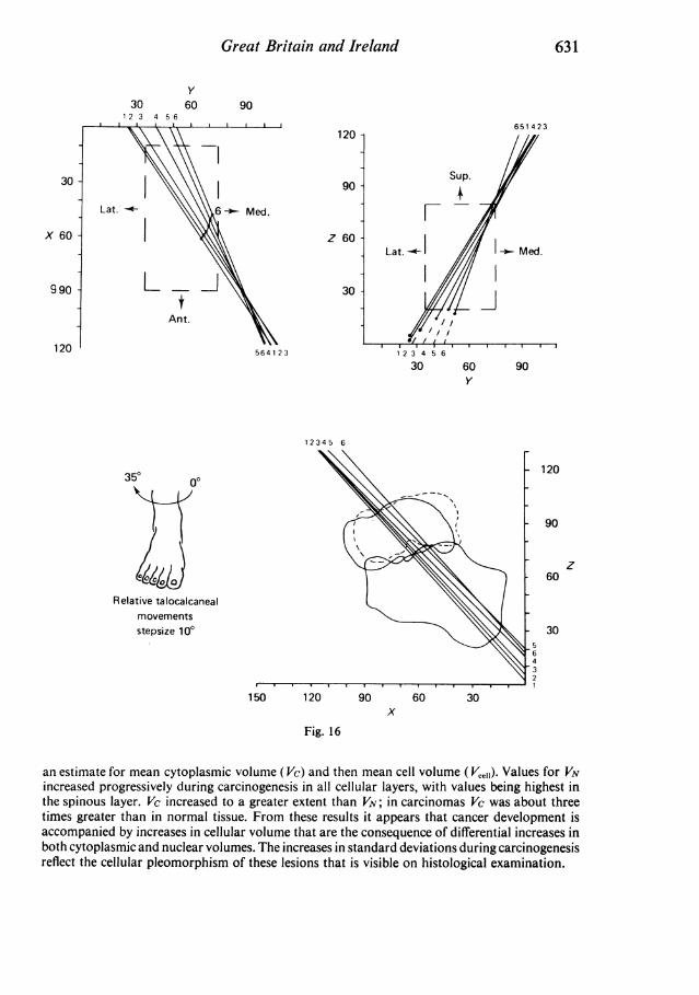
3012 3 4 56
An
Great Britain and Ireland
y60 90
120 -
- Z U | Lat.906-- Med.
Z 60-Lat.
1 ~~~~~30It.
564123
30 60 90y
12345 6
350 00
Relative talocalcanealmovementsstepsize 100
150 120 90 60 30x
Fig. 16
an estimate for mean cytoplasmic volume (Vc) and then mean cell volume (Vce,i). Values for VNincreased progressively during carcinogenesis in all cellular layers, with values being highest inthe spinous layer. Vc increased to a greater extent than VN; in carcinomas Vc was about threetimes greater than in normal tissue. From these results it appears that cancer development isaccompanied by increases in cellular volume that are the consequence of differential increases inboth cytoplasmic and nuclear volumes. The increases in standard deviations during carcinogenesisreflect the cellular pleomorphism of these lesions that is visible on histological examination.
631
651423
30
X 60
990
120
Sup.
FI- Med.
120
90
z60
30- 564321