microglia and brain macrophages in the molecular age: from ... · microglia are tissue-resident...
TRANSCRIPT
Microglia are tissue-resident macrophages in the CNS. They belong to a group of mononuclear phagocytes that comprises peripheral tissue macrophages, CNS-associated macrophages, dendritic cells and monocyte-derived cells1,2. As such, they are critical effectors and regulators of changes in CNS homeostasis during development and in health and disease.
All mononuclear cells originate from haematopoietic stem cells (HSCs) and develop along distinct differen-tiation pathways in response to endogenous and envi-ronmental cues3,4. The body’s mononuclear phagocyte system was for a long time believed to derive from a subgroup of white blood cells called leukocytes that are produced from HSCs. It was assumed that bone marrow-derived monocytes, a subgroup of leukocytes, circulate in the blood and enter the tissues (where they differentiate into tissue-resident macrophages) in non-pathological conditions and during inflammation. However, this view has changed in recent years as a result of the discovery of new subtypes of mononuclear phagocytes that have different origins and distinct roles in CNS disorders5. Furthermore, the findings of recent population-based gene expression studies have highlighted macrophage heterogeneity6–8.
Emerging evidence from such studies suggests that microglia differ considerably from the macrophages that reside in other tissues6. Indeed, some genes that
distinguish them from their peripheral relatives have been identified9. Moreover, recent fate-mapping stud-ies of several macrophage populations in the body have provided elegant evidence that, under homeostatic con-ditions, microglia are not derived from the bone marrow but originate from HSCs in the yolk sac10–12. Thus, the existing model of tissue macrophage development needs to be extended to highlight the different origins of many macrophage populations, including microglia.
Several other important distinctions between differ-ent macrophage populations have been identified. Novel myeloid-specific gene targeting techniques targeting CX3C chemokine receptor 1 (Cx3cr1; also known as the fractalkine receptor) have enabled researchers to examine the kinetics of myeloid cell turnover during homeostasis and disease13–15. These new transgenic approaches have helped to establish the major kinetic features of micro-glia that distinguish them from other macrophages: namely, that they are long-lived, that they are not nor-mally replaced by peripheral cells from the circulation and that they are able to self-renew in a context-dependent manner to ensure cell expansion. Previous studies used genes such as Cd11b (also known as Itgam), lysozyme M (Lysm; also known as Lyz2) and ionized calcium-binding adaptor molecule 1 (Iba1; also known as Aif1) to target myeloid cells in the brain16–18. However, these experiments either used transgenic mouse lines that showed low levels
1Institute of Neuropathology, University of Freiburg, Breisacherstraße 64, 79106 Freiburg, Germany.2BIOSS Centre for Biological Signalling Studies, University of Freiburg, 79104 Freiburg, Germany.3Department of Neuropsychiatry and Laboratory of Molecular Psychiatry, Charité – Universitätsmedizin Berlin, Charitéplatz 1, 10117 Berlin, Germany.4Cluster of Excellence NeuroCure, Charitéplatz 1, 10117 Berlin, Germany.e‑mails: marco.prinz@uniklinik‑freiburg.de; [email protected]:10.1038/nrn3722 Published online 9 April 2014
MacrophagesTissue-resident cells of the mononuclear phagocyte system that are characterized by their ability to phagocytose foreign particulate material, debris and colloidal material.
Microglia and brain macrophages in the molecular age: from origin to neuropsychiatric diseaseMarco Prinz1,2 and Josef Priller3,4
Abstract | Mononuclear phagocytic cells in the CNS used to be defined according to their anatomical location and surface marker expression. Recently, this concept has been challenged by the results of developmental and gene expression profiling studies that have used novel molecular biological tools to unravel the origin of microglia and to define their role as specialized tissue macrophages with long lifespans. Here, we describe how these results have redefined microglia and helped us to understand how different myeloid cell populations operate in the CNS based on their cell-specific gene expression signatures, distinct ontogeny and differential functions. Moreover, we describe the vulnerability of microglia to dysfunction and propose that myelomonocytic cells might be used in the treatment of neurological and psychiatric disorders that are characterized by primary or secondary ‘microgliopathy’.
R E V I E W S
NATURE REVIEWS | NEUROSCIENCE ADVANCE ONLINE PUBLICATION | 1
© 2014 Macmillan Publishers Limited. All rights reserved
Choroid plexus
a
Microglial cell
Blood vessel
Perivascular spacec
Meningealmacrophage
b
Dendritic cell
e
Nature Reviews | Neuroscience
d
Perivascularmacrophage
Choroid plexus macrophage
Meninges
Mononuclear phagocytesA mononuclear cell type of the myeloid lineage (macrophages, monocytes or dendritic cells) that have the ability to phagocytose.
Dendritic cellsAlso known as an interdigitating reticular cells because of their branched morphology. Dendritic cells are the most potent stimulators of T cell responses.
MonocyteA type of mononuclear leukocyte that is derived from the bone marrow and circulates in the bloodstream. Monocytes typically migrate into tissues, where they can differentiate into various types of macrophages.
Haematopoietic stem cells(HSCs). Rare multipotent cells that give rise to all blood cells, including myeloid and lymphoid lineages.
of recombination in microglia compared with circulating myeloid cells (such as in the Lysm-Cre line)13 or used the antiviral drug ganciclovir, which has tremendous drug-induced effects on microglia proliferation in vivo19.
Bone marrow-derived phagocytes often have dis-ease-related functions that are distinct from those of yolk sac-derived microglia1. The differential roles of microglia and other brain macrophages have tremen-dous clinical implications for the treatment of severe brain diseases such as Alzheimer’s disease, Parkinson’s disease, amyotrophic lateral sclerosis (ALS; also known as Lou Gehrig’s disease), multiple sclerosis and several psychiatric disorders1,20,21. In theory, if practical hurdles can be overcome, specific myeloid populations — such as phagocytes derived from the yolk sac, bone marrow or blood — might be used to deliver therapeutic molecules to the CNS in order to ameliorate disease.
In this Review, we provide an overview of the progress in our understanding of the origin, fate and function of microglia and compare this with other brain macrophage populations. This information may help us to design new strategies to promote restoration of tissue homeostasis in the most complex organ of the body, the brain.
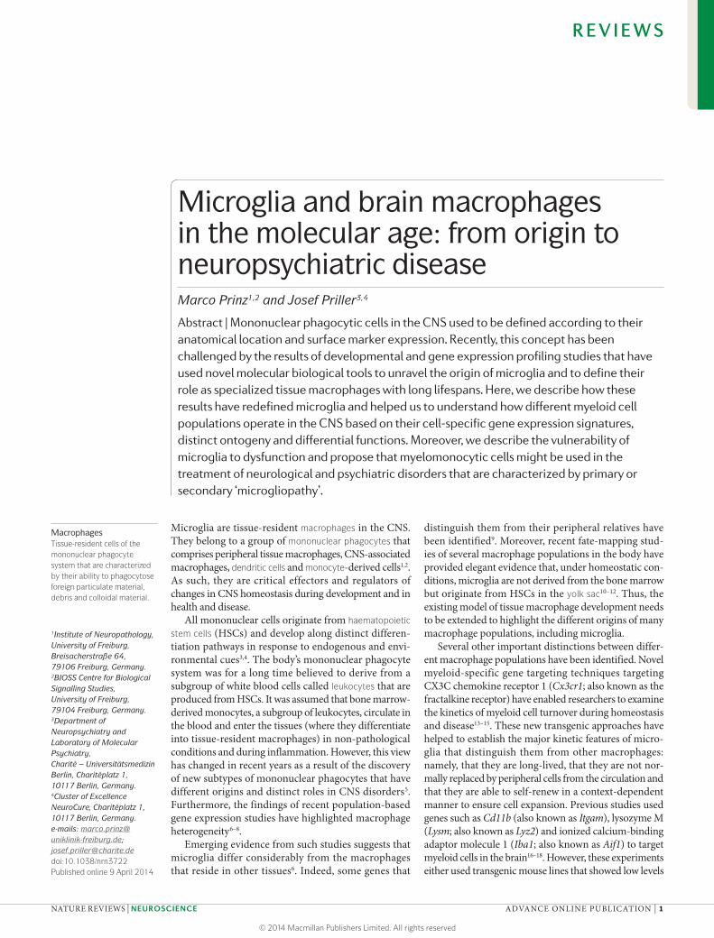
Diversity of CNS myeloid cell originsThe brain hosts several myeloid populations. In addi-tion to the parenchymal microglia, these cells include perivascular cells, meningeal macrophages and cho-roid plexus macrophages (FIG. 1). Despite the fact that all of these macrophage populations share numerous
myeloid- and macrophage-specific markers (such as IBA1, F4/80 (also known as EMR1) and CX3CR1 (REF. 1)) and exhibit similar immune regulatory func-tions (such as local immune surveillance and removal of debris), early results suggested that they have distinct ontogenesis22,23.
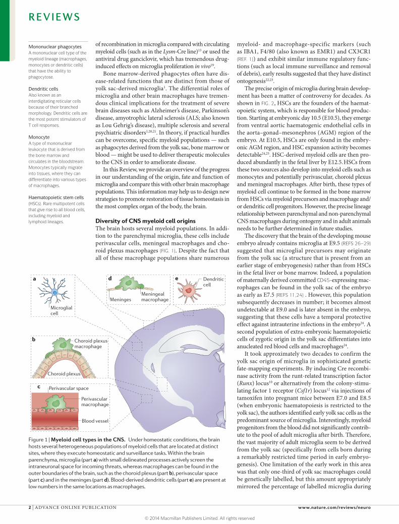
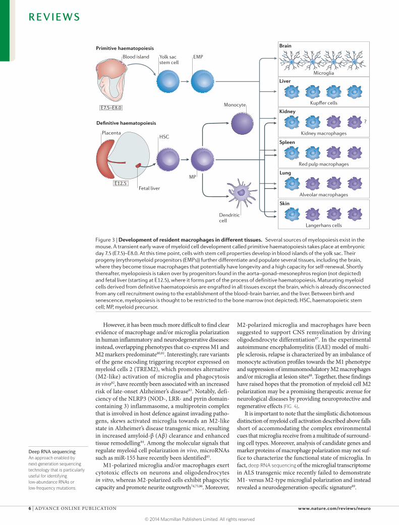
The precise origin of microglia during brain develop-ment has been a matter of controversy for decades. As shown in FIG. 2, HSCs are the founders of the haemat-opoietic system, which is responsible for blood produc-tion. Starting at embryonic day 10.5 (E10.5), they emerge from ventral aortic haematogenic endothelial cells in the aorta–gonad–mesonephros (AGM) region of the embryo. At E10.5, HSCs are only found in the embry-onic AGM region, and HSC expansion activity becomes detectable24,25. HSC-derived myeloid cells are then pro-duced abundantly in the fetal liver by E12.5. HSCs from these two sources also develop into myeloid cells such as monocytes and potentially perivascular, choroid plexus and meningeal macrophages. After birth, these types of myeloid cell continue to be formed in the bone marrow from HSCs via myeloid precursors and macrophage and/or dendritic cell progenitors. However, the precise lineage relationship between parenchymal and non-parenchymal CNS macrophages during ontogeny and in adult animals needs to be further determined in future studies.
The discovery that the brain of the developing mouse embryo already contains microglia at E9.5 (REFS 26–29) suggested that microglial precursors may originate from the yolk sac (a structure that is present from an earlier stage of embryogenesis) rather than from HSCs in the fetal liver or bone marrow. Indeed, a population of maternally derived committed CD45-expressing mac-rophages can be found in the yolk sac of the embryo as early as E7.5 (REFS 11,24) . However, this population subsequently decreases in number; it becomes almost undetectable at E9.0 and is later absent in the embryo, suggesting that these cells have a temporal protective effect against intrauterine infections in the embryo24. A second population of extra-embryonic haematopoietic cells of zygotic origin in the yolk sac differentiates into anucleated red blood cells and macrophages24.
It took approximately two decades to confirm the yolk sac origin of microglia in sophisticated genetic fate-mapping experiments. By inducing Cre recombi-nase activity from the runt-related transcription factor (Runx) locus10 or alternatively from the colony-stimu-lating factor 1 receptor (Csf1r) locus12 via injections of tamoxifen into pregnant mice between E7.0 and E8.5 (when embryonic haematopoiesis is restricted to the yolk sac), the authors identified early yolk sac cells as the predominant source of microglia. Interestingly, myeloid progenitors from the blood did not significantly contrib-ute to the pool of adult microglia after birth. Therefore, the vast majority of adult microglia seem to be derived from the yolk sac (specifically from cells born during a remarkably restricted time period in early embryo-genesis). One limitation of the early work in this area was that only one-third of yolk sac macrophages could be genetically labelled, but this amount appropriately mirrored the percentage of labelled microglia during
Figure 1 | Myeloid cell types in the CNS. Under homeostatic conditions, the brain hosts several heterogeneous populations of myeloid cells that are located at distinct sites, where they execute homeostatic and surveillance tasks. Within the brain parenchyma, microglia (part a) with small delineated processes actively screen the intraneuronal space for incoming threats, whereas macrophages can be found in the outer boundaries of the brain, such as the choroid plexus (part b), perivascular space (part c) and in the meninges (part d). Blood-derived dendritic cells (part e) are present at low numbers in the same locations as macrophages.
R E V I E W S
2 | ADVANCE ONLINE PUBLICATION www.nature.com/reviews/neuro
© 2014 Macmillan Publishers Limited. All rights reserved
Other CNS macrophage development
Nature Reviews | Neuroscience
CD45
KIT
CX3CR1
CSF1R
MMP8 or MMP9
EMP A1 A2
HSC
Ramified microglial cell
MDP
F4/80
RUNX1
IRF8
PU.1
MYB
CSF1
IL-34
Fetal liver (E12.5)
AGM (E10.5)
Microglial development
E7.5–E8.0
Yolk sac blood islands
E9.0
Yolk sac subpopulations
Embryonic microglia
a
b
Bone marrow(postnatal)
MP
• Perivascular macrophage• Choroid plexus macrophage• Meningeal macrophage
E9.5
Blood vessel
Monocyte
Macrophage precursor
LeukocytesWhite blood cells derived from multipotent haematopoietic stem cells in the bone marrow. Leukocytes are of myeloid or lymphoid lineage and are found in the blood and lymphatic system.
Yolk sacA membranous sac attached to the embryo that provides early nourishment in the form of yolk. It functions as the developmental circulatory system of the embryo before internal circulation begins.
CD45(Also known as leukocyte common antigen and PTPRC). A type I transmembrane protein present on all haematopoietic cells that assists in cell activation and the levels of which are reduced in mature parenchymal microglia.
NeuroepitheliumThe ectodermal epithelium in the embryo from which the CNS and its main cellular constituents (neurons, astrocytes, oligodendrocytes and ependymal cells) are derived.
adulthood10. In a more recent study12, mice lacking the transcriptional activator MYB, which do not develop HSCs or their progeny, were used. Yolk sac macrophages from these mice gave rise to a MYB-independent nor-mal population of microglia in the adult. By contrast, MYB was required for stem cell development in the bone marrow. Further global transcriptional analysis showed a common signature of gene expression in embryonic microglia and F4/80+ yolk sac macrophages12.
A similar pattern of microglial cell development occurs in humans. In human foetuses, microglia-like cells with a range of morphologies can be detected as early as 3 weeks of estimated gestational age30. However, it seems that maturation of the microglial compart-ment is ongoing during most of the gestation period: colonization of the spinal cord begins at around 9 weeks, the major influx and distribution of microglia com-mences at about 16 weeks and ramified microglia take up to 22 weeks to become widely distributed within the intermediate zone31,32. In fact, it is only close to term, at
35 weeks, that well-differentiated microglial populations can be detected within the developing human brain33 (for reviews, see REFS 31,32,34).
A recent study characterized the early yolk sac progenitors that give rise to microglia in the brain as KIT-positive, lineage marker-negative (mean-ing that no markers of mature haematopoietic cells were expressed) progenitor cells that have the abil-ity to differentiate into CX3CR1-expressing microglia in vitro as well as in vivo11,35. These cells also generate Ter119-expressing erythrocytes, indicating that there is a common erythromyeloid progenitor (EMP) in the yolk sac for both lineages11. The uncommitted EMPs subse-quently disappear and immature F4/80+CX3CR1− (A1) and F4/80+CX3CR1+ (A2) macrophages develop and can be located on the surface of the developing brain at E9.0 in mice11. These macrophage-like cells, which have an amoeboid shape, were previously described in the rodent neuroepithelium28,36,37. At E13.5, when the fetal liver is already the primary haematopoietic organ and
Figure 2 | Embryonic and postnatal development of microglia in mice. The figure illustrates the precursor cells, transcription factors (black symbols), cytokines (purple symbols) and cytokine receptors (red symbols) that are expressed by each cell population during the development of microglia and other CNS macrophages. a | Microglia derive from immature, uncommitted KIT+ erythromyeloid progenitors (EMPs): that is, stem cells that are formed during primitive haematopoiesis, which begins at embryonic day 7.5 (E7.5)–E8.0 in the yolk sac in mice11. These cells start to upregulate the CD antigen CD45 but do not yet express myeloid markers (A1 stage). Later, myeloid cell markers, such as F4/80, CX3C chemokine receptor 1 (CX3CR1) and colony-stimulating factor 1 receptor (CSF1R) are expressed by migrating A2 cells (differentiated from A1 cells) that populate the brain mesenchyme. These early microglia reside in the brain throughout life and are thought to sustain the microglial population locally. b | By contrast, other CNS macrophages found in the meninges, choroid plexus and perivascular spaces are thought to be derived from the definitive haematopoiesis that starts at E10.5, first in the aorta–gonad–mesonephros (AGM) region or later, at E12.5, in the fetal liver. Postnatally, monocytes are formed in the bone marrow. Haematopoietic stem cells (HSCs) generate monocytes from myeloid precursors (MPs) and macrophage and/or dendritic cell progenitors (MDPs). IL-34, interleukin-34; IRF8, interferon regulatory factor 8; MMP, matrix metalloproteinase; MYB, transcriptional activator MYB; PU.1, transcription factor PU.1; RUNX1, runt-related transcription factor 1.
R E V I E W S
NATURE REVIEWS | NEUROSCIENCE ADVANCE ONLINE PUBLICATION | 3
© 2014 Macmillan Publishers Limited. All rights reserved
Natural killer cellsA type of cytotoxic lymphocyte that are crucial for the innate immune system.
the main site of HSC expansion and differentiation38, A2 macrophages can be detected in significant num-bers within the lining of the fourth ventricle37. There is a 20-fold increase in the number of CD11b+F4/80+ micro-glial cells during the early postnatal period (between postnatal day 0 (P0) and P11) in rodents27. However, evidence suggests that this increase in microglial cell number is not induced by the recruitment of peripheral myeloid cells but instead results from the expansion of resident microglial cells10,12.
Microglia and CNS macrophages thus represent two ontogenetically distinct myeloid populations. These differences imply that microglia and infiltrating mac-rophages have different functions, which are increasingly apparent in mouse models of disease (see below)1.
Microglial developmentThe transcriptional programme that controls the differ-entiation of yolk sac progenitors into microglia is only partially understood. Indeed, many studies of the tran-scriptional control of macrophage differentiation have been carried out in vitro using progenitor-enriched cell populations. Thus, the precise role of the factors identi-fied in these studies in driving the differentiation of yolk sac macrophages into typical microglia in vivo remains to be determined. This is particularly important given that functional macrophage specialization is likely to be regulated at the tissue level — that is, in the brain. Nevertheless, some molecules that are essential for the regulation of microglial cell development have already been described35 (TABLE 1).
A dramatic reduction in the number of tissue mac-rophages, including microglia, has been observed in Csf1r-knockout mice10,39,40 and in Csf1op/op-mutant mice, which have a natural null mutation in Csf1 (REFS 41,42). Thus, CSF1 and its receptor have a key role in microglial homeostasis in mice in vivo43. However, the mechanisms by which CSF1 and its receptor promote microglia com-mitment remain controversial. One hypothesis suggests that CSF1 drives the differentiation of phagocytic yolk sac macrophages that have entered the embryo into micro-glia44, whereas a different theory proposes that CSF1 pro-vides a survival signal for the differentiating macrophages
and that surviving cells utilize an intrinsic developmental programme to become mature microglia45,46. Interestingly, microglia are more profoundly affected by the absence of CSF1R than by the absence of CSF1 (REF. 10). Of note, a second CSF1R ligand, interleukin-34 (IL-34), has recently been identified47 and has been found to be highly expressed in the postnatal mouse brain48. In order to explore the role of this cytokine in microgliogenesis, mice deficient in IL-34 were generated49,50. Interestingly, the number of microglia was reduced in distinct regions of the brains of these mutants, suggesting that there is regional heterogeneity of CSF1R and/ or IL-34 expres-sion49,50. Similar results were obtained in zebrafish, in which yolk sac-derived macrophages enter the developing brain and form early microglia that have high endocytic activity. This process was shown to depend on CSF1R expression: the Panther mutation of CSF1R led to a com-plete loss of brain macrophages51. Similarly, microglial cell numbers were reduced in mice deficient in an adap-tor protein of the CSF1R, DNAX-activation protein 12 (DAP12; also known as TYROBP)52. DAP12, which has an important role in the human brain (see below), con-tains an immunoreceptor tyrosine-based activation motif (ITAM) in its cytoplasmic domain and is highly expressed in natural killer cells and myeloid cells.
PU.1 is another transcription factor that is exclu-sively expressed in haematopoietic cells and is involved in microglial cell development. The gene encoding PU.1, SFPI1 (also known as SPI1), is a member of the ETS fam-ily of transcription factors53. Its targeted disruption in mice leads to multiple haematopoietic abnormalities, including a lack of mature B cells and macrophages54. In fact, PU.1-deficient mice are devoid not only of circu-lating monocytes and tissue macrophages54 but also of parenchymal microglia in the brain55.
Additional transcription factors such as RUNX1 and interferon regulatory factor 8 (IRF8) are also indispen-sable regulators of the differentiation of microglia dur-ing embryonic development in mice10,11. A recent study carried out a detailed analysis of the function of RUNX1 in postnatal microglia56 and showed that it not only regu-lates the differentiation but also the proliferation and homeostasis of postnatal microglia. The authors further suggested that RUNX1 might modulate the transition of activated amoeboid microglia into deactivated ramified microglia56. The transcription factors PU.1 and RUNX1 were shown to act in a negative feedback loop that governs the equilibrium between distinct myeloid fates by assuring an appropriate PU.1 dosage57. These results suggest that RUNX1 is a non-redundant transcription factor that is important for the activation of microglia.
The transcriptional programming of microglial cell development is also tightly regulated by transcription fac-tors expressed in the myeloid lineage, such as IRF8. IRF8 is a heterodimeric partner of PU.1 and has known roles in the development of B cells and myeloid cells in the bone marrow58. A recent study investigated the role of IRF8 in microglial cell development in mice and found that yolk sac-derived A2 macrophages were particularly depend-ent on the presence of IRF8, whereas other myeloid tran-scription factors, such as MYB, DNA-binding protein
Table 1 | Microglia phenotypes in animals lacking specific molecules
Absent molecule Microglia morphology Microglia number Refs
CSF1 ↓ ↓ 41,169
CSF1 receptor ↓ ↓ 10,40
DAP12 ND ↓ 52
Interleukin-34 ND ↓ 49,50
IRF8 ↓ → or ↓ 11,59–61
Transcription factor PU.1
NA ↓ 51,55
RUNX1 ↓ ND 56,57
↓ indicates a either dysmorphic or reduced number of microglia; → indicates no change. CSF1, colony-stimulating factor 1; DAP12, DNAX-activation protein 12; IRF8, interferon regulatory factor 8; NA, non-applicable; ND, not determined; RUNX1, runt-related transcription factor 1.
R E V I E W S
4 | ADVANCE ONLINE PUBLICATION www.nature.com/reviews/neuro
© 2014 Macmillan Publishers Limited. All rights reserved
MyelopoiesisThe regulated formation of myeloid cells, including macrophages, monocytes, dendritic cells and granulocytes. Myelopoiesis takes place in the bone marrow or the yolk sac.
inhibitor ID2, basic leucine zipper transcriptional factor ATF-like 3 (BATF3) and Krüppel-like factor 4 (KLF4), were not essential for their development11. Consequently, the number of microglia is strongly reduced during adult-hood in animals lacking IRF8 (REF. 11). Moreover, recent studies have indicated a role for IRF8 in the activation of adult microglia59–61.
In summary, these data provide evidence that microglia develop from EMPs in the yolk sac in an IRF8- and PU.1-dependent manner but independently of MYB, ID2, BATF3 and KLF4. This unique genetic profile distinguishes microglia from other myeloid cells, such as circulating monocytes, CD11b+ dendritic cells, CD8+CD103+CD11b− dendritic cells, endogenous brain macrophages and other tissue macrophages62
(BOX 1; FIG. 3).
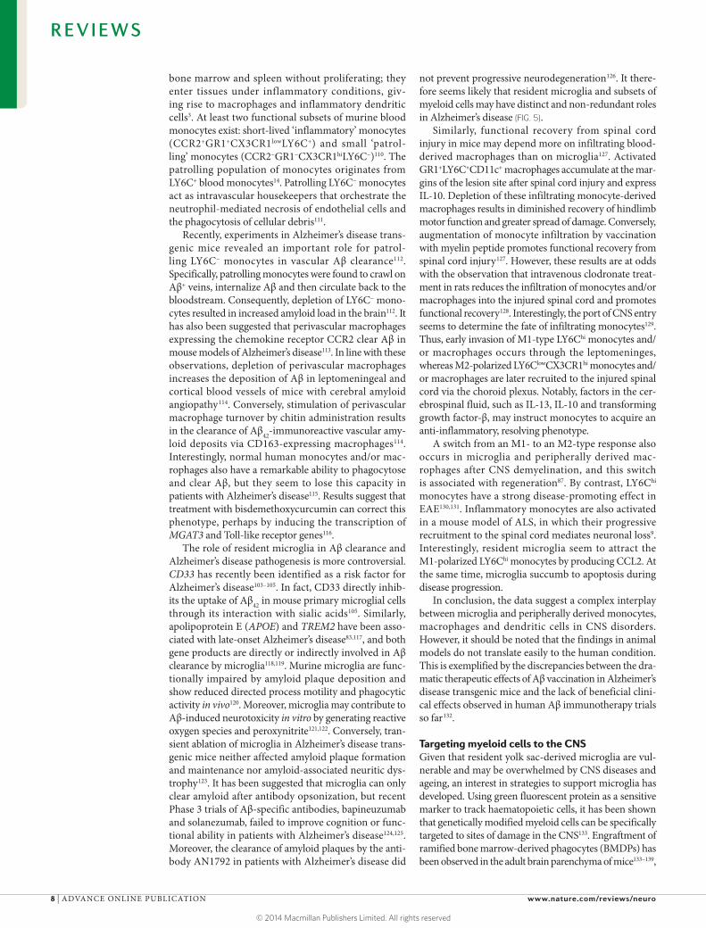
Microglial polarizationLike macrophages in the periphery, microglia act as the first line of defence in the nervous system. Every few hours, so-called ‘resting’ microglia screen the brain parenchyma with their highly motile processes63. Upon detection of signs of injury, such as extracellular calcium waves and release of adenosine triphosphate from neigh-bouring cells, microglial processes rapidly move towards the lesion site64,65. This is followed by the transforma-tion of resting microglia into an activated state (FIG. 4). Depending on the particular signals detected and on the actions of modulators of microglial activation, a diversity of reactive microglial phenotypes can be generated66.
Macrophages in non-neural tissues reprogramme their function in response to pathogens, tissue dam-age and lymphocyte interactions. This process, termed polarization, enables the adaptive responses of innate immunity to take place67. At least two distinct states of macrophage polarization have been recognized: M1 and M2 polarization. The T helper 1 (TH1) cytokine, interferon-γ (IFNγ), and bacterial lipopolysaccharide
(LPS) polarize macrophages towards the M1 pheno-type. By contrast, the TH2 cytokine, IL-4, drives M2 polarization. Notably, the chemokine CCL2 (also known as monocyte chemoattractant protein 1), which is strongly induced in neurodegenerative and neuro-inflammatory conditions, also drives M2 macrophage polarization68. Macrophages activated in these ways are functionally distinct. M1-polarized macrophages pro-duce pro-inflammatory cytokines such as IL-12, IL-23 and tumour necrosis factor-α (TNFα). They attract TH1 cells by releasing the chemokines CXCL9 and CXCL10, and show enhanced antigen presentation capacity67. Moreover, they generate reactive oxygen and nitrogen species through activity of inducible nitric oxide syn-thase67,69. By contrast, M2-polarized macrophages pro-duce anti-inflammatory cytokines such as IL-10 and promote TH2 responses. They show increased phagocytic activity and expression of scavenger receptors such as CD163 (REF. 67). In addition, M2-polarized macrophages support tissue remodelling and promote fibrosis through increased arginase 1 activity69,70. However, as appealing as this dichotomous view of macrophage polarization may seem to be, it cannot account for the plasticity of myelomonocytic cells that has been observed67,71. In fact, various overlapping and novel phenotypes have been observed. Among M2-polarized macrophages, three subsets with different functional properties have been defined: M2a, M2b and M2c macrophages67,71. M2a and M2c macrophages dampen inflammation and promote tissue repair, whereas M2b macrophages (char-acterized by their downregulation of IL-12) have both pro- and anti-inflammatory functions67,71. In fact, there is probably a spectrum of plastic functional conditions of mononuclear phagocytes rather than a set of discrete activation states.
Despite their different ontogeny, microglia may also have the capacity to become polarized into M1-like and M2-like phenotypes. In an early in vitro study, TH1- or TH2-polarized T cells isolated from patients with multi-ple sclerosis differentially modulated human microglia and monocytes to become type 1 (M1-like) or type 2 (M2-like) antigen-presenting cells72. Interestingly, human microglia seem to be more restricted in their capacity to adopt the M2-like phenotype in vitro than monocyte-derived macrophages73.
In animal models of stroke, traumatic brain injury or spinal cord injury, microglia and/or macrophages have been suggested to shift from a transient M2 phenotype to become M1-like phagocytes74–76. The NADPH oxidase-dependent redox state of the microenvironment plays a crucial part in the modulation of microglial pheno-type in vivo. Inhibition or deletion of NADPH oxidase switches microglial activation from a classical (M1-like) to an alternative (M2-like) state in response to an inflam-matory challenge77. Similarly, human gliomas instruct macrophages to shift towards an M2-like phenotype, as determined by immunostaining in glioma samples and in vitro78. Interestingly, CSF1R inhibitors were recently found to slow the growth of patient-derived glioma xen-ografts in mice by reducing the M2-like polarization of microglia and/or macrophages79.
Box 1 | Microglia in the universe of mononuclear phagocytic cells
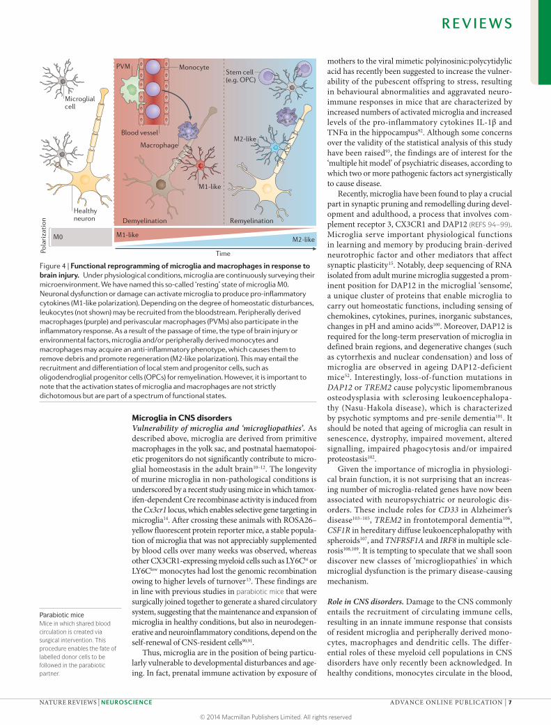
Our knowledge of macrophage lineages was dramatically expanded by recent studies using both mutant mice and fate-mapping approaches. The first fate-mapping studies focused on microglia and Langerhans cells in the skin. Microglia are exclusively derived from KIT+ erythromyeloid progenitors in the yolk sac as part of primitive haematopoiesis10–12, whereas Langerhans cells predominantly originate in the fetal liver, with a smaller contribution from yolk sac macrophages12,161. Yolk sac-derived tissue macrophages were found to have a long lifespan, to be able to self-renew and to be present in several tissues such as brain (microglia), skin (Langerhans cells), liver (Kupffer cells), pancreas, lung (alveolar macrophages), spleen (red pulp macrophages) and kidney12,14,161–165 (FIG. 3). In most tissues, except the brain and potentially also the liver, these yolk sac-derived tissue macrophages coexist with macrophages derived from definitive myelopoiesis in the fetal liver. It is not yet clear whether yolk sac-derived macrophages or fetal liver-derived populations are more dominant in normal adult tissues. Accordingly, it is also not known whether these ontogenetically different macrophages have distinct roles and functions. Microglia are an obvious exception in that they exclusively derive from one source (the yolk sac) before birth. More recently, mutations in GATA-binding protein 2 (GATA2)166 and interferon regulatory factor 8 (IRF8)167 have been associated with severe defects in bone marrow-derived myeloid cells but did not affect many tissue macrophages167,168. These data clearly indicate that different pathways regulate the development of myeloid cells by primitive and definitive haematopoiesis.
R E V I E W S
NATURE REVIEWS | NEUROSCIENCE ADVANCE ONLINE PUBLICATION | 5
© 2014 Macmillan Publishers Limited. All rights reserved
Monocyte
Nature Reviews | Neuroscience
Primitive haematopoiesis
Definitive haematopoiesis
E7.5–E8.0
Blood island
Fetal liver
Yolk sac stem cell
EMP
MP
HSC
Dendritic cell
Brain
Microglia
Kidney
Kidney macrophages
?
Spleen
Red pulp macrophages
Lung
Alveolar macrophages
Skin
Langerhans cells
Liver
Kupffer cells
E12.5
Placenta
Deep RNA sequencingAn approach enabled by next-generation sequencing technology that is particularly useful for identifying low-abundance RNAs or low-frequency mutations.
However, it has been much more difficult to find clear evidence of macrophage and/or microglia polarization in human inflammatory and neurodegenerative diseases: instead, overlapping phenotypes that co-express M1 and M2 markers predominate80,81. Interestingly, rare variants of the gene encoding triggering receptor expressed on myeloid cells 2 (TREM2), which promotes alternative (M2-like) activation of microglia and phagocytosis in vivo82, have recently been associated with an increased risk of late-onset Alzheimer’s disease83. Notably, defi-ciency of the NLRP3 (NOD-, LRR- and pyrin domain-containing 3) inflammasome, a multiprotein complex that is involved in host defence against invading patho-gens, skews activated microglia towards an M2-like state in Alzheimer’s disease transgenic mice, resulting in increased amyloid-β (Aβ) clearance and enhanced tissue remodelling84. Among the molecular signals that regulate myeloid cell polarization in vivo, microRNAs such as miR-155 have recently been identified85.
M1-polarized microglia and/or macrophages exert cytotoxic effects on neurons and oligodendrocytes in vitro, whereas M2-polarized cells exhibit phagocytic capacity and promote neurite outgrowth74,75,86. Moreover,
M2-polarized microglia and macrophages have been suggested to support CNS remyelination by driving oligodendrocyte differentiation87. In the experimental autoimmune encephalomyelitis (EAE) model of multi-ple sclerosis, relapse is characterized by an imbalance of monocyte activation profiles towards the M1 phenotype and suppression of immunomodulatory M2 macrophages and/or microglia at lesion sites88. Together, these findings have raised hopes that the promotion of myeloid cell M2 polarization may be a promising therapeutic avenue for neurological diseases by providing neuroprotective and regenerative effects (FIG. 4).
It is important to note that the simplistic dichotomous distinction of myeloid cell activation described above falls short of accommodating the complex environmental cues that microglia receive from a multitude of surround-ing cell types. Moreover, analysis of candidate genes and marker proteins of macrophage polarization may not suf-fice to characterize the functional state of microglia. In fact, deep RNA sequencing of the microglial transcriptome in ALS transgenic mice recently failed to demonstrate M1- versus M2-type microglial polarization and instead revealed a neurodegeneration-specific signature89.
Figure 3 | Development of resident macrophages in different tissues. Several sources of myelopoiesis exist in the mouse. A transient early wave of myeloid cell development called primitive haematopoiesis takes place at embryonic day 7.5 (E7.5)–E8.0. At this time point, cells with stem cell properties develop in blood islands of the yolk sac. Their progeny (erythromyeloid progenitors (EMPs)) further differentiate and populate several tissues, including the brain, where they become tissue macrophages that potentially have longevity and a high capacity for self-renewal. Shortly thereafter, myelopoiesis is taken over by progenitors found in the aorta–gonad–mesonephros region (not depicted) and fetal liver (starting at E12.5), where it forms part of the process of definitive haematopoiesis. Maturating myeloid cells derived from definitive haematopoiesis are engrafted in all tissues except the brain, which is already disconnected from any cell recruitment owing to the establishment of the blood–brain barrier, and the liver. Between birth and senescence, myelopoiesis is thought to be restricted to the bone marrow (not depicted). HSC, haematopoietic stem cell; MP, myeloid precursor.
R E V I E W S
6 | ADVANCE ONLINE PUBLICATION www.nature.com/reviews/neuro
© 2014 Macmillan Publishers Limited. All rights reserved
Nature Reviews | Neuroscience
M1-likeM2-like
TimePola
riza
tion
M0
M1-like
M2-like
Healthy neuron
Blood vessel
PVM
Macrophage
Demyelination
Stem cell (e.g. OPC)
Remyelination
Monocyte
Microglial cell
Parabiotic miceMice in which shared blood circulation is created via surgical intervention. This procedure enables the fate of labelled donor cells to be followed in the parabiotic partner.
Microglia in CNS disordersVulnerability of microglia and ‘microgliopathies’. As described above, microglia are derived from primitive macrophages in the yolk sac, and postnatal haematopoi-etic progenitors do not significantly contribute to micro-glial homeostasis in the adult brain10–12. The longevity of murine microglia in non-pathological conditions is underscored by a recent study using mice in which tamox-ifen-dependent Cre recombinase activity is induced from the Cx3cr1 locus, which enables selective gene targeting in microglia14. After crossing these animals with ROSA26–yellow fluorescent protein reporter mice, a stable popula-tion of microglia that was not appreciably supplemented by blood cells over many weeks was observed, whereas other CX3CR1-expressing myeloid cells such as LY6Chi or LY6Clow monocytes had lost the genomic recombination owing to higher levels of turnover13. These findings are in line with previous studies in parabiotic mice that were surgically joined together to generate a shared circulatory system, suggesting that the maintenance and expansion of microglia in healthy conditions, but also in neurodegen-erative and neuroinflammatory conditions, depend on the self-renewal of CNS-resident cells90,91.
Thus, microglia are in the position of being particu-larly vulnerable to developmental disturbances and age-ing. In fact, prenatal immune activation by exposure of
mothers to the viral mimetic polyinosinic:polycytidylic acid has recently been suggested to increase the vulner-ability of the pubescent offspring to stress, resulting in behavioural abnormalities and aggravated neuro-immune responses in mice that are characterized by increased numbers of activated microglia and increased levels of the pro-inflammatory cytokines IL-1β and TNFα in the hippocampus92. Although some concerns over the validity of the statistical analysis of this study have been raised93, the findings are of interest for the ‘multiple hit model’ of psychiatric diseases, according to which two or more pathogenic factors act synergistically to cause disease.
Recently, microglia have been found to play a crucial part in synaptic pruning and remodelling during devel-opment and adulthood, a process that involves com-plement receptor 3, CX3CR1 and DAP12 (REFS 94–99). Microglia serve important physiological functions in learning and memory by producing brain-derived neurotrophic factor and other mediators that affect synaptic plasticity15. Notably, deep sequencing of RNA isolated from adult murine microglia suggested a prom-inent position for DAP12 in the microglial ‘sensome’, a unique cluster of proteins that enable microglia to carry out homeostatic functions, including sensing of chemokines, cytokines, purines, inorganic substances, changes in pH and amino acids100. Moreover, DAP12 is required for the long-term preservation of microglia in defined brain regions, and degenerative changes (such as cytorrhexis and nuclear condensation) and loss of microglia are observed in ageing DAP12-deficient mice52. Interestingly, loss-of-function mutations in DAP12 or TREM2 cause polycystic lipomembranous osteodysplasia with sclerosing leukoencephalopa-thy (Nasu-Hakola disease), which is characterized by psychotic symptoms and pre-senile dementia101. It should be noted that ageing of microglia can result in senescence, dystrophy, impaired movement, altered signalling, impaired phagocytosis and/or impaired proteostasis102.
Given the importance of microglia in physiologi-cal brain function, it is not surprising that an increas-ing number of microglia-related genes have now been associated with neuropsychiatric or neurologic dis-orders. These include roles for CD33 in Alzheimer’s disease103–105, TREM2 in frontotemporal dementia106, CSF1R in hereditary diffuse leukoencephalopathy with spheroids107, and TNFRSF1A and IRF8 in multiple scle-rosis108,109. It is tempting to speculate that we shall soon discover new classes of ‘microgliopathies’ in which microglial dysfunction is the primary disease-causing mechanism.
Role in CNS disorders. Damage to the CNS commonly entails the recruitment of circulating immune cells, resulting in an innate immune response that consists of resident microglia and peripherally derived mono-cytes, macrophages and dendritic cells. The differ-ential roles of these myeloid cell populations in CNS disorders have only recently been acknowledged. In healthy conditions, monocytes circulate in the blood,
Figure 4 | Functional reprogramming of microglia and macrophages in response to brain injury. Under physiological conditions, microglia are continuously surveying their microenvironment. We have named this so-called ‘resting’ state of microglia M0. Neuronal dysfunction or damage can activate microglia to produce pro-inflammatory cytokines (M1-like polarization). Depending on the degree of homeostatic disturbances, leukocytes (not shown) may be recruited from the bloodstream. Peripherally derived macrophages (purple) and perivascular macrophages (PVMs) also participate in the inflammatory response. As a result of the passage of time, the type of brain injury or environmental factors, microglia and/or peripherally derived monocytes and macrophages may acquire an anti-inflammatory phenotype, which causes them to remove debris and promote regeneration (M2-like polarization). This may entail the recruitment and differentiation of local stem and progenitor cells, such as oligodendroglial progenitor cells (OPCs) for remyelination. However, it is important to note that the activation states of microglia and macrophages are not strictly dichotomous but are part of a spectrum of functional states.
R E V I E W S
NATURE REVIEWS | NEUROSCIENCE ADVANCE ONLINE PUBLICATION | 7
© 2014 Macmillan Publishers Limited. All rights reserved
bone marrow and spleen without proliferating; they enter tissues under inflammatory conditions, giv-ing rise to macrophages and inflammatory dendritic cells3. At least two functional subsets of murine blood monocytes exist: short-lived ‘inflammatory’ monocytes (CCR2+GR1+CX3CR1lowLY6C+) and small ‘patrol-ling’ monocytes (CCR2−GR1−CX3CR1hiLY6C−)110. The patrolling population of monocytes originates from LY6C+ blood monocytes14. Patrolling LY6C− monocytes act as intravascular housekeepers that orchestrate the neutrophil-mediated necrosis of endothelial cells and the phagocytosis of cellular debris111.
Recently, experiments in Alzheimer’s disease trans-genic mice revealed an important role for patrol-ling LY6C− monocytes in vascular Aβ clearance112. Specifically, patrolling monocytes were found to crawl on Aβ+ veins, internalize Aβ and then circulate back to the bloodstream. Consequently, depletion of LY6C− mono-cytes resulted in increased amyloid load in the brain112. It has also been suggested that perivascular macrophages expressing the chemokine receptor CCR2 clear Aβ in mouse models of Alzheimer’s disease113. In line with these observations, depletion of perivascular macrophages increases the deposition of Aβ in leptomeningeal and cortical blood vessels of mice with cerebral amyloid angiopathy114. Conversely, stimulation of perivascular macrophage turnover by chitin administration results in the clearance of Aβ42-immunoreactive vascular amy-loid deposits via CD163-expressing macrophages114. Interestingly, normal human monocytes and/or mac-rophages also have a remarkable ability to phagocytose and clear Aβ, but they seem to lose this capacity in patients with Alzheimer’s disease115. Results suggest that treatment with bisdemethoxycurcumin can correct this phenotype, perhaps by inducing the transcription of MGAT3 and Toll-like receptor genes116.
The role of resident microglia in Aβ clearance and Alzheimer’s disease pathogenesis is more controversial. CD33 has recently been identified as a risk factor for Alzheimer’s disease103–105. In fact, CD33 directly inhib-its the uptake of Aβ42 in mouse primary microglial cells through its interaction with sialic acids105. Similarly, apolipoprotein E (APOE) and TREM2 have been asso-ciated with late-onset Alzheimer’s disease83,117, and both gene products are directly or indirectly involved in Aβ clearance by microglia118,119. Murine microglia are func-tionally impaired by amyloid plaque deposition and show reduced directed process motility and phagocytic activity in vivo120. Moreover, microglia may contribute to Aβ-induced neurotoxicity in vitro by generating reactive oxygen species and peroxynitrite121,122. Conversely, tran-sient ablation of microglia in Alzheimer’s disease trans-genic mice neither affected amyloid plaque formation and maintenance nor amyloid-associated neuritic dys-trophy123. It has been suggested that microglia can only clear amyloid after antibody opsonization, but recent Phase 3 trials of Aβ-specific antibodies, bapineuzumab and solanezumab, failed to improve cognition or func-tional ability in patients with Alzheimer’s disease124,125. Moreover, the clearance of amyloid plaques by the anti-body AN1792 in patients with Alzheimer’s disease did
not prevent progressive neurodegeneration126. It there-fore seems likely that resident microglia and subsets of myeloid cells may have distinct and non-redundant roles in Alzheimer’s disease (FIG. 5).
Similarly, functional recovery from spinal cord injury in mice may depend more on infiltrating blood-derived macrophages than on microglia127. Activated GR1+LY6C+CD11c+ macrophages accumulate at the mar-gins of the lesion site after spinal cord injury and express IL-10. Depletion of these infiltrating monocyte-derived macrophages results in diminished recovery of hindlimb motor function and greater spread of damage. Conversely, augmentation of monocyte infiltration by vaccination with myelin peptide promotes functional recovery from spinal cord injury127. However, these results are at odds with the observation that intravenous clodronate treat-ment in rats reduces the infiltration of monocytes and/or macrophages into the injured spinal cord and promotes functional recovery128. Interestingly, the port of CNS entry seems to determine the fate of infiltrating monocytes129. Thus, early invasion of M1-type LY6Chi monocytes and/or macrophages occurs through the leptomeninges, whereas M2-polarized LY6ClowCX3CR1hi monocytes and/or macrophages are later recruited to the injured spinal cord via the choroid plexus. Notably, factors in the cer-ebrospinal fluid, such as IL-13, IL-10 and transforming growth factor-β, may instruct monocytes to acquire an anti-inflammatory, resolving phenotype.
A switch from an M1- to an M2-type response also occurs in microglia and peripherally derived mac-rophages after CNS demyelination, and this switch is associated with regeneration87. By contrast, LY6Chi monocytes have a strong disease-promoting effect in EAE130,131. Inflammatory monocytes are also activated in a mouse model of ALS, in which their progressive recruitment to the spinal cord mediates neuronal loss9. Interestingly, resident microglia seem to attract the M1-polarized LY6Chi monocytes by producing CCL2. At the same time, microglia succumb to apoptosis during disease progression.
In conclusion, the data suggest a complex interplay between microglia and peripherally derived monocytes, macrophages and dendritic cells in CNS disorders. However, it should be noted that the findings in animal models do not translate easily to the human condition. This is exemplified by the discrepancies between the dra-matic therapeutic effects of Aβ vaccination in Alzheimer’s disease transgenic mice and the lack of beneficial clini-cal effects observed in human Aβ immunotherapy trials so far132.
Targeting myeloid cells to the CNSGiven that resident yolk sac-derived microglia are vul-nerable and may be overwhelmed by CNS diseases and ageing, an interest in strategies to support microglia has developed. Using green fluorescent protein as a sensitive marker to track haematopoietic cells, it has been shown that genetically modified myeloid cells can be specifically targeted to sites of damage in the CNS133. Engraftment of ramified bone marrow-derived phagocytes (BMDPs) has been observed in the adult brain parenchyma of mice133–139,
R E V I E W S
8 | ADVANCE ONLINE PUBLICATION www.nature.com/reviews/neuro
© 2014 Macmillan Publishers Limited. All rights reserved

Nature Reviews | Neuroscience
a Alzheimer’s disease
Monocyte
Blood vessel
Activated microglial cell
Amyloid plaque
Damaged endogenous microglial cell
CNS conditioning
PVM
Diminished Aβ plaque
Aβ
BMDP
BMDP
b Rett syndrome
CNS conditioning
Bone marrow
DebrisMicroglia fail to phagocytose debris
BMDP (full of debris)
• Reduced axonal boutons
• Reduced number of microglia• Reduced dendritic branching and spines
rats140 and humans141 after bone marrow transplantation (BMT). However, the vast majority of bone marrow-derived cells in these studies populated the perivascular spaces rather than the brain parenchyma. This is in line with earlier observations suggesting a high turnover of perivascular cells in rodents and humans22,142–145. It soon became apparent that the engraftment of BMDPs in the adult brain parenchyma is facilitated by the conditioning regimens used to prepare the recipients for BMT, such as total body irradiation. In fact, protecting the head from irradiation and the resulting CNS inflammation during BMT abrogated the engraftment of ramified BMDPs in the brain parenchyma146.
However, irradiation is not sufficient to recruit BMDPs into the CNS, as demonstrated in parabiotic mice that were surgically joined together to generate blood chimerism without irradiation or BMT. Notably, no engraftment of ramified BMDPs was observed in the brain parenchyma of parabiotic mice more than 1 year after the surgery10,90. Moreover, neither irradiation nor neurodegeneration promoted the CNS engraftment of ramified BMDPs in parabiotic mice90. Together, these findings suggest that the BMDPs that are recruited to
the brain parenchyma — in contrast to perivascular macrophages — do not derive from circulating precur-sors such as monocytes. Two recent studies lend support to this conclusion. First, EAE experiments in parabi-otic mice revealed that inflammatory monocytes and/or macrophages are recruited from the bloodstream to the spinal cord in a CCR2-dependent manner and trig-ger disease progression91. Notably, the presence of these myelomonocytic cells in the CNS was transient, and only uncommitted lineage-negative KIT+SCA1+ stem or pro-genitor cells were capable of generating long-lived BMDPs in irradiated recipients. Second, HSC transplantation in mice revealed a short-term wave of brain infiltration by a fraction of the donor cells independently of whether irradiation or chemotherapy (using the agents busulphan or treosulphan) was used as a conditioning regimen147. However, only lethal irradiation and myeloablation with busulphan, which are capable of ablating brain-resident myeloid precursors (treosulphan does not cross the blood–brain barrier), enabled turnover of microglia with the donor. This was mediated by local proliferation of early immigrants rather than entrance of mature cells from the circulation. In line with these results, it was
Figure 5 | Disease-modulating roles of bone marrow-derived myeloid cells in the CNS. Neuropsychiatric disorders are often associated with microglial dysfunction or demise. a | In Alzheimer’s disease, activated microglia have been suggested to contribute to amyloid-β (Aβ)-induced neurotoxicity. At the same time, microglia are damaged by amyloid species. Transplantation of wild-type bone marrow cells (green) in transgenic mouse models of Alzheimer’s disease results in the recruitment of bone marrow-derived phagocyte (BMDP) populations to the neurovascular unit and brain parenchyma after previous CNS conditioning (for example, as a result of irradiation)113,156. As a result, amyloid load in the brain may be reduced through enhanced clearance of Aβ from the CNS by perivascular monocytes and/or perivascular macrophages (PVMs). b | In Rett syndrome, microglia are reduced in numbers and fail to phagocytose debris160. This may contribute to neuronal dysfunction and structural abnormalities of neurons (reduced dendritic branching, spine density and axonal boutons). Transplantation of wild-type bone marrow cells in the methyl-CpG-binding protein 2 (Mecp2)-null mouse models of Rett syndrome results in the recruitment of BMDP populations to the brain and rescues the phenotype160.
R E V I E W S
NATURE REVIEWS | NEUROSCIENCE ADVANCE ONLINE PUBLICATION | 9
© 2014 Macmillan Publishers Limited. All rights reserved
Graft-versus-host diseaseA complication following an allogeneic tissue transplant in which immune cells (white blood cells) in the tissue (the graft) recognize the recipient (the host) as ‘foreign’.
X-linked adrenoleukodystrophyA rare X chromosome-linked disorder resulting from mutations in ABCD1 (ATP-binding cassette subfamily D member 1) that cause defects in peroxisomal β-oxidation and lead to the accumulation of very-long-chain fatty acids, particularly in the CNS and adrenal cortex.
recently shown that myeloid cells can be targeted to sites of brain damage even in the presence of very low levels of peripheral blood chimerism148. To date, the cellular ori-gins of the precursors of BMDPs in the adult brain paren-chyma remain unclear. Nevertheless, myelosuppressive conditioning using busulphan is sufficient to trigger the recruitment of BMDPs to the CNS149,150. This is an impor-tant observation, as myeloablation with busulphan has been successfully used in clinical trials151.
There is growing interest in long-term engraftment of myeloid cells in the mature CNS because these cells harbour great therapeutic potential for neurological and psychiatric disorders. This is particularly true for disorders in which microglia fail to support normal brain function or even ‘turn against’ their environment. Allogeneic HSC transplantation and autologous stem cell-based gene therapies have been tested in models of inborn errors of metabolism, such as lysosomal storage diseases152,153. The aim is to repopulate recipient haemat-opoietic and lymphoid compartments with cells express-ing functional enzymes. However, HSC transplantation seems to benefit only a subset of patients who have not yet developed overt neurological symptoms, suggesting that BMDP engraftment in the CNS occurs at a slower pace than disease progression. In addition, graft fail-ures and incomplete chimerism may limit the success of allogeneic HSC transplantation. Autologous HSC-based gene therapies offer immunological advantages in relation to graft rejection and graft-versus-host disease, and may benefit even those patients with early-onset and rapid disease progression. In the case of X-linked adrenoleukodystrophy, zones devoid of microglia and the occurrence of microglial apoptosis in perilesional white matter suggest that microgliopathy is an early pathogenic event154. HSC transplantation can arrest the neuroin-flammatory demyelinating process with a characteristic delay of 12–18 months, which has been attributed to the slow replacement of microglia with BMDPs153.
Non-cell-autonomous neurodegeneration has also been described for ‘classical’ neurodegenerative diseases. In ALS, upper and lower motor neurons are lost, which results in progressive tetraparalysis and death. The most commonly inherited form of ALS is caused by mutations in superoxide dismutase 1 (SOD1). Microglia derived from transgenic mice overexpressing mutant SOD1 pro-duce more free radicals and induce more neuronal cell death in vitro than wild-type microglia55. Notably, expres-sion of mutant SOD1 in motor neurons is not sufficient to trigger their degeneration, and SOD1-mutant mice survive longer when surrounded by wild-type microglia or BMDPs55,155.
As described above, dysfunction of microglia has also been described in animal models of Alzheimer’s disease120. Although resident microglia are recruited to sites of amyloid deposition in the brain, they may ulti-mately fail to restrict amyloid plaque formation (FIG. 5). However, peripheral myeloid cells may still possess the capacity to remove Aβ from the CNS. Following irra-diation and BMT, peripherally derived monocytes and/or macrophages populate the perivascular spaces, and BMDPs are engrafted into the brain parenchyma, where
they accumulate around amyloid plaques (FIG. 5). It has been suggested that BMDPs are specifically attracted to Aβ in vivo and are more efficient than resident microglia in eliminating this protein by phagocytosis156. Moreover, CCR2-expressing perivascular monocytes and/or mac-rophages clear Aβ from the brain113, which is line with previous observations that CCR2 deficiency accelerates disease progression and promotes cerebral amyloid angiopathy in Alzheimer’s disease transgenic mice157.
Even behavioural disorders with less overt neuro-pathological changes may benefit from the engraftment of BMDPs in the brain20,21. Thus, the compulsive groom-ing and hair removal in Hoxb8-mutant mice has been linked to a defect in resident microglia, and the behav-ioural deficit is corrected by the transplantation of wild-type bone marrow cells158. Rett syndrome is an X-linked neurodevelopmental disorder with phenotypic overlap with autism spectrum disorders159. The patients are gen-erally female and develop normally during the first year of their life, after which they start to suffer from deficits in language and communication, impaired social interac-tions, stereotypic behaviours and autonomic dysfunction. Rett syndrome is mainly caused by mutations in the gene encoding methyl-CpG-binding protein 2 (MECP2). The symptoms are believed to result from deficits at the micro-circuit level in the brain, involving synaptic transmission and plasticity (FIG. 5). Recent experiments in Mecp2−/y male mice (in which the single X chromosome copy of Mecp2 is mutated) and Mecp2−/+ female mice (which carry only one functional copy of Mecp2) suggested that resident micro-glia may also be responsible for the disorder160. In fact, microglia are reduced in number in Mecp2-null mice and fail to phagocytose debris as effectively as those in wild-type mice (FIG. 5). Importantly, engraftment of wild-type BMDPs after BMT increased the lifespan of Mecp2-null mice and ameliorated the behavioural and autonomic phenotypes160.
Conclusions and future directionsThe advent of new transgenic animal models, intravital imaging and transcriptomic tools has enabled research-ers to unequivocally determine the origin of microglia as well as to study their development and fate in the CNS. Microglia belong to the mononuclear phagocyte system of the body. They are more closely related to mac-rophages that reside in peripheral tissues than they are to the neuroectodermal-derived neurons, astroglia and oligodendroglia in the brain.
We are now able to study microglia as they develop in the embryo, acquire specialized functions, participate in neuronal network formation and activity, respond to damage and succumb to disease. Although many of the insights have come from studying laboratory animals and remain to be translated to the human condition, there is an increasing awareness of the diversity and plasticity of innate immune cells in the brain. Novel tran-scriptomic and epigenomic techniques will improve our knowledge even further.
Microglia are not a homogeneous population, and they do not respond uniformly to microenvironmental changes. Instead, they meticulously survey and weigh
R E V I E W S
10 | ADVANCE ONLINE PUBLICATION www.nature.com/reviews/neuro
© 2014 Macmillan Publishers Limited. All rights reserved
the signals that instruct them to acquire specific func-tions. The same is true for brain macrophages and peripherally derived monocytes, macrophages and den-dritic cells. All of these innate immune cells rarely act in isolation, and it may turn out to be a dangerous oversim-plification to distinguish ‘detrimental’ M1- and ‘healing’ M2-type responses when there is actually a spectrum of spatiotemporally defined graded activation patterns of microglia and brain macrophages.
One of the most exciting findings of the latest research on myeloid cells in the brain is the changing concept of microglia from culprit to victim. We are beginning to rec-ognize that disturbances or loss of microglial function may be a driving force in CNS disorders. It is tempting to speculate that we shall see an increasing number of primary or secondary ‘microgliopathies’, which will open up new avenues for the treatment of neurological and psychiatric disorders.
1. Prinz, M., Priller, J., Sisodia, S. S. & Ransohoff, R. M. Heterogeneity of CNS myeloid cells and their roles in neurodegeneration. Nature Neurosci. 14, 1227–1235 (2011).
2. Gomez, P. E., Schulz, C. & Geissmann, F. Development and homeostasis of “resident” myeloid cells: the case of the microglia. Glia 61, 112–120 (2013).
3. Geissmann, F. et al. Development of monocytes, macrophages, and dendritic cells. Science 327, 656–661 (2010).
4. Sieweke, M. H. & Allen, J. E. Beyond stem cells: self-renewal of differentiated macrophages. Science 342, 1242974 (2013).
5. Prinz, M. & Mildner, A. Microglia in the CNS: immigrants from another world. Glia 59, 177–187 (2011).
6. Gautier, E. L. et al. Gene-expression profiles and transcriptional regulatory pathways that underlie the identity and diversity of mouse tissue macrophages. Nature Immunol. 13, 1118–1128 (2012).
7. Chow, A., Brown, B. D. & Merad, M. Studying the mononuclear phagocyte system in the molecular age. Nature Rev. Immunol. 11, 788–798 (2011).
8. Davies, L. C., Jenkins, S. J., Allen, J. E. & Taylor, P. R. Tissue-resident macrophages. Nature Immunol. 14, 986–995 (2013).
9. Butovsky, O. et al. Modulating inflammatory monocytes with a unique microRNA gene signature ameliorates murine ALS. J. Clin. Invest. 122, 3063–3087 (2012).
10. Ginhoux, F. et al. Fate mapping analysis reveals that adult microglia derive from primitive macrophages. Science 330, 841–845 (2010).
11. Kierdorf, K. et al. Microglia emerge from erythromyeloid precursors via Pu.1- and Irf8-dependent pathways. Nature Neurosci. 16, 273–280 (2013).
12. Schulz, C. et al. A lineage of myeloid cells independent of Myb and hematopoietic stem cells. Science 336, 86–90 (2012).
13. Goldmann, T. et al. A new type of microglia gene targeting shows TAK1 to be pivotal in CNS autoimmune inflammation. Nature Neurosci. 16, 1618–1626 (2013).References 10–13 use fate-mapping and genetic tools to show that microglia are derived from the yolk sac.
14. Yona, S. et al. Fate mapping reveals origins and dynamics of monocytes and tissue macrophages under homeostasis. Immunity 38, 79–91 (2013).This study establishes a new line of transgenic mice in which Cre is expressed specifically in microglia.
15. Parkhurst, C. N. et al. Microglia promote learning-dependent synapse formation through brain-derived neurotrophic factor. Cell 155, 1596–1609 (2013).
16. Prinz, M. et al. Distinct and nonredundant in vivo functions of IFNAR on myeloid cells limit autoimmunity in the central nervous system. Immunity 28, 675–686 (2008).
17. Heppner, F. L. et al. Experimental autoimmune encephalomyelitis repressed by microglial paralysis. Nature Med. 11, 146–152 (2005).
18. Pfrieger, F. W. & Slezak, M. Genetic approaches to study glial cells in the rodent brain. Glia 60, 681–701 (2012).
19. Ding, Z. et al. Antiviral drug ganciclovir is a potent inhibitor of microglial proliferation and neuroinflammation. J. Exp. Med. 211, 189–198 (2014).
20. Blank, T. & Prinz, M. Microglia as modulators of cognition and neuropsychiatric disorders. Glia 61, 62–70 (2013).
21. Priller, J. in Neuroglia 3rd edn (eds Kettenmann, H. & Ransom, B. R.) 906–916 (Oxford Univ. Press, 2013).
22. Hickey, W. F. & Kimura, H. Perivascular microglial cells of the CNS are bone marrow-derived and present antigen in vivo. Science 239, 290–292 (1988).
23. Hickey, W. F., Vass, K. & Lassmann, H. Bone marrow-derived elements in the central nervous system: an immunohistochemical and ultrastructural survey of rat chimeras. J. Neuropathol. Exp. Neurol. 51, 246–256 (1992).
24. Bertrand, J. Y. et al. Three pathways to mature macrophages in the early mouse yolk sac. Blood 106, 3004–3011 (2005).
25. Cumano, A. & Godin, I. Ontogeny of the hematopoietic system. Annu. Rev. Immunol. 25, 745–785 (2007).
26. Alliot, F., Lecain, E., Grima, B. & Pessac, B. Microglial progenitors with a high proliferative potential in the embryonic and adult mouse brain. Proc. Natl Acad. Sci. USA 88, 1541–1545 (1991).
27. Alliot, F., Godin, I. & Pessac, B. Microglia derive from progenitors, originating from the yolk sac, and which proliferate in the brain. Brain Res. Dev. Brain Res. 117, 145–152 (1999).
28. Ashwell, K. The distribution of microglia and cell death in the fetal rat forebrain. Brain Res. Dev. Brain Res. 58, 1–12 (1991).
29. Lawson, L. J., Perry, V. H. & Gordon, S. Turnover of resident microglia in the normal adult mouse brain. Neuroscience 48, 405–415 (1992).
30. Hutchins, K. D., Dickson, D. W., Rashbaum, W. K. & Lyman, W. D. Localization of morphologically distinct microglial populations in the developing human fetal brain: implications for ontogeny. Brain Res. Dev. Brain Res. 55, 95–102 (1990).
31. Rezaie, P. & Male, D. Colonisation of the developing human brain and spinal cord by microglia: a review. Microsc. Res. Tech. 45, 359–382 (1999).
32. Rezaie, P., Dean, A., Male, D. & Ulfig, N. Microglia in the cerebral wall of the human telencephalon at second trimester. Cereb. Cortex 15, 938–949 (2005).
33. Esiri, M. M., al Izzi, M. S. & Reading, M. C. Macrophages, microglial cells, and HLA-DR antigens in fetal and infant brain. J. Clin. Pathol. 44, 102–106 (1991).
34. Verney, C., Monier, A., Fallet-Bianco, C. & Gressens, P. Early microglial colonization of the human forebrain and possible involvement in periventricular white-matter injury of preterm infants. J. Anat. 217, 436–448 (2010).
35. Kierdorf, K. & Prinz, M. Factors regulating microglia activation. Front. Cell Neurosci. 7, 44 (2013).
36. Ashwell, K. Microglia and cell death in the developing mouse cerebellum. Brain Res. Dev. Brain Res. 55, 219–230 (1990).
37. Chan, W. Y., Kohsaka, S. & Rezaie, P. The origin and cell lineage of microglia: new concepts. Brain Res. Rev. 53, 344–354 (2007).
38. Lichanska, A. M. & Hume, D. A. Origins and functions of phagocytes in the embryo. Exp. Hematol. 28, 601–611 (2000).
39. Dai, X. M. et al. Targeted disruption of the mouse colony-stimulating factor 1 receptor gene results in osteopetrosis, mononuclear phagocyte deficiency, increased primitive progenitor cell frequencies, and reproductive defects. Blood 99, 111–120 (2002).
40. Erblich, B., Zhu, L., Etgen, A. M., Dobrenis, K. & Pollard, J. W. Absence of colony stimulation factor-1 receptor results in loss of microglia, disrupted brain development and olfactory deficits. PLoS ONE 6, e26317 (2011).
41. Wegiel, J. et al. Reduced number and altered morphology of microglial cells in colony stimulating factor-1-deficient osteopetrotic op/op mice. Brain Res. 804, 135–139 (1998).
42. Yoshida, H. et al. The murine mutation osteopetrosis is in the coding region of the macrophage colony
stimulating factor gene. Nature 345, 442–444 (1990).
43. Pixley, F. J. & Stanley, E. R. CSF-1 regulation of the wandering macrophage: complexity in action. Trends Cell Biol. 14, 628–638 (2004).
44. Metcalf, D. The granulocyte-macrophage colony stimulating factors. Cell 43, 5–6 (1985).
45. Lagasse, E. & Weissman, I. L. Enforced expression of Bcl-2 in monocytes rescues macrophages and partially reverses osteopetrosis in op/op mice. Cell 89, 1021–1031 (1997).
46. Nakahata, T., Gross, A. J. & Ogawa, M. A stochastic model of self-renewal and commitment to differentiation of the primitive hemopoietic stem cells in culture. J. Cell. Physiol. 113, 455–458 (1982).
47. Lin, H. et al. Discovery of a cytokine and its receptor by functional screening of the extracellular proteome. Science 320, 807–811 (2008).
48. Wei, S. et al. Functional overlap but differential expression of CSF-1 and IL-34 in their CSF-1 receptor-mediated regulation of myeloid cells. J. Leukoc. Biol. 88, 495–505 (2010).
49. Greter, M. et al. Stroma-derived interleukin-34 controls the development and maintenance of langerhans cells and the maintenance of microglia. Immunity 37, 1050–1060 (2012).
50. Wang, Y. et al. IL-34 is a tissue-restricted ligand of CSF1R required for the development of Langerhans cells and microglia. Nature Immunol. 13, 753–760 (2012).
51. Herbomel, P., Thisse, B. & Thisse, C. Zebrafish early macrophages colonize cephalic mesenchyme and developing brain, retina, and epidermis through a M-CSF receptor-dependent invasive process. Dev. Biol. 238, 274–288 (2001).
52. Otero, K. et al. Macrophage colony-stimulating factor induces the proliferation and survival of macrophages via a pathway involving DAP12 and β-catenin. Nature Immunol. 10, 734–743 (2009).
53. Rosenbauer, F. & Tenen, D. G. Transcription factors in myeloid development: balancing differentiation with transformation. Nature Rev. Immunol. 7, 105–117 (2007).
54. McKercher, S. R. et al. Targeted disruption of the PU.1 gene results in multiple hematopoietic abnormalities. EMBO J. 15, 5647–5658 (1996).
55. Beers, D. R. et al. Wild-type microglia extend survival in PU.1 knockout mice with familial amyotrophic lateral sclerosis. Proc. Natl Acad. Sci. USA 103, 16021–16026 (2006).
56. Zusso, M. et al. Regulation of postnatal forebrain amoeboid microglial cell proliferation and development by the transcription factor Runx1. J. Neurosci. 32, 11285–11298 (2012).
57. Jin, H. et al. Runx1 regulates embryonic myeloid fate choice in zebrafish through a negative feedback loop inhibiting Pu.1 expression. Blood 119, 5239–5249 (2012).
58. Holtschke, T. et al. Immunodeficiency and chronic myelogenous leukemia-like syndrome in mice with a targeted mutation of the ICSBP gene. Cell 87, 307–317 (1996).
59. Masuda, T. et al. IRF8 is a critical transcription factor for transforming microglia into a reactive phenotype. Cell Rep. 1, 334–340 (2012).
60. Horiuchi, M. et al. Interferon regulatory factor 8/interferon consensus sequence binding protein is a critical transcription factor for the physiological phenotype of microglia. J. Neuroinflammation 9, 227 (2012).
61. Minten, C., Terry, R., Deffrasnes, C., King, N. J. & Campbell, I. L. IFN regulatory factor 8 is a key constitutive determinant of the morphological and molecular properties of microglia in the CNS. PLoS ONE 7, e49851 (2012).
R E V I E W S
NATURE REVIEWS | NEUROSCIENCE ADVANCE ONLINE PUBLICATION | 11
© 2014 Macmillan Publishers Limited. All rights reserved
62. Hashimoto, D., Miller, J. & Merad, M. Dendritic cell and macrophage heterogeneity in vivo. Immunity 35, 323–335 (2011).
63. Nimmerjahn, A., Kirchhoff, F. & Helmchen, F. Resting microglial cells are highly dynamic surveillants of brain parenchyma in vivo. Science 308, 1314–1318 (2005).
64. Davalos, D. et al. ATP mediates rapid microglial response to local brain injury in vivo. Nature Neurosci. 8, 752–758 (2005).
65. Sieger, D., Moritz, C., Ziegenhals, T., Prykhozhij, S. & Peri, F. Long-range Ca2+ waves transmit brain-damage signals to microglia. Dev. Cell 22, 1138–1148 (2012).
66. Hanisch, U. K. & Kettenmann, H. Microglia: active sensor and versatile effector cells in the normal and pathologic brain. Nature Neurosci. 10, 1387–1394 (2007).
67. Biswas, S. K. & Mantovani, A. Macrophage plasticity and interaction with lymphocyte subsets: cancer as a paradigm. Nature Immunol. 11, 889–896 (2010).
68. Roca, H. et al. CCL2 and interleukin-6 promote survival of human CD11b+ peripheral blood mononuclear cells and induce M2-type macrophage polarization. J. Biol. Chem. 284, 34342–34354 (2009).
69. Hesse, M. et al. Differential regulation of nitric oxide synthase-2 and arginase-1 by type 1/type 2 cytokines in vivo: granulomatous pathology is shaped by the pattern of L-arginine metabolism. J. Immunol. 167, 6533–6544 (2001).
70. Wynn, T. A. Fibrotic disease and the TH1/TH2 paradigm. Nature Rev. Immunol. 4, 583–594 (2004).
71. David, S. & Kroner, A. Repertoire of microglial and macrophage responses after spinal cord injury. Nature Rev. Neurosci. 12, 388–399 (2011).
72. Kim, H. J. et al. Type 2 monocyte and microglia differentiation mediated by glatiramer acetate therapy in patients with multiple sclerosis. J. Immunol. 172, 7144–7153 (2004).
73. Durafourt, B. A. et al. Comparison of polarization properties of human adult microglia and blood-derived macrophages. Glia 60, 717–727 (2012).
74. Kigerl, K. A. et al. Identification of two distinct macrophage subsets with divergent effects causing either neurotoxicity or regeneration in the injured mouse spinal cord. J. Neurosci. 29, 13435–13444 (2009).
75. Hu, X. et al. Microglia/macrophage polarization dynamics reveal novel mechanism of injury expansion after focal cerebral ischemia. Stroke 43, 3063–3070 (2012).
76. Wang, G. et al. Microglia/macrophage polarization dynamics in white matter after traumatic brain injury. J. Cereb. Blood Flow Metab. 33, 1864–1874 (2013).
77. Choi, S. H., Aid, S., Kim, H. W., Jackson, S. H. & Bosetti, F. Inhibition of NADPH oxidase promotes alternative and anti-inflammatory microglial activation during neuroinflammation. J. Neurochem. 120, 292–301 (2012).
78. Komohara, Y., Ohnishi, K., Kuratsu, J. & Takeya, M. Possible involvement of the M2 anti-inflammatory macrophage phenotype in growth of human gliomas. J. Pathol. 216, 15–24 (2008).
79. Pyonteck, S. M. et al. CSF-1R inhibition alters macrophage polarization and blocks glioma progression. Nature Med. 19, 1264–1272 (2013).
80. Dal, B. A. et al. Multiple sclerosis and Alzheimer’s disease. Ann. Neurol. 63, 174–183 (2008).
81. Vogel, D. Y. et al. Macrophages in inflammatory multiple sclerosis lesions have an intermediate activation status. J. Neuroinflammation 10, 35 (2013).
82. Takahashi, K., Prinz, M., Stagi, M., Chechneva, O. & Neumann, H. TREM2-transduced myeloid precursors mediate nervous tissue debris clearance and facilitate recovery in an animal model of multiple sclerosis. PLoS Med. 4, e124 (2007).
83. Guerreiro, R. et al. TREM2 variants in Alzheimer’s disease. N. Engl. J. Med. 368, 117–127 (2013).
84. Heneka, M. T. et al. NLRP3 is activated in Alzheimer’s disease and contributes to pathology in APP/PS1 mice. Nature 493, 674–678 (2012).
85. Moore, C. S. et al. miR-155 as a multiple sclerosis-relevant regulator of myeloid cell polarization. Ann. Neurol. 74, 709–720 (2013).
86. Wang, Y. et al. Transforming growth factor beta-activated kinase 1 (TAK1)-dependent checkpoint in the survival of dendritic cells promotes immune homeostasis and function. Proc. Natl Acad. Sci. USA 109, E343–E352 (2012).
87. Miron, V. E. et al. M2 microglia and macrophages drive oligodendrocyte differentiation during CNS remyelination. Nature Neurosci. 16, 1211–1218 (2013).
88. Mikita, J. et al. Altered M1/M2 activation patterns of monocytes in severe relapsing experimental rat model of multiple sclerosis. Amelioration of clinical status by M2 activated monocyte administration. Mult. Scler. 17, 2–15 (2011).
89. Chiu, I. M. et al. A neurodegeneration-specific gene-expression signature of acutely isolated microglia from an amyotrophic lateral sclerosis mouse model. Cell Rep. 4, 385–401 (2013).
90. Ajami, B., Bennett, J. L., Krieger, C., Tetzlaff, W. & Rossi, F. M. Local self-renewal can sustain CNS microglia maintenance and function throughout adult life. Nature Neurosci. 10, 1538–1543 (2007).
91. Ajami, B., Bennett, J. L., Krieger, C., McNagny, K. M. & Rossi, F. M. Infiltrating monocytes trigger EAE progression, but do not contribute to the resident microglia pool. Nature Neurosci. 14, 1142–1149 (2011).
92. Giovanoli, S. et al. Stress in puberty unmasks latent neuropathological consequences of prenatal immune activation in mice. Science 339, 1095–1099 (2013).
93. Lazic, S. E. Comment on “Stress in puberty unmasks latent neuropathological consequences of prenatal immune activation in mice”. Science 340, 811 (2013).
94. Roumier, A. et al. Impaired synaptic function in the microglial KARAP/DAP12-deficient mouse. J. Neurosci. 24, 11421–11428 (2004).
95. Wake, H., Moorhouse, A. J., Jinno, S., Kohsaka, S. & Nabekura, J. Resting microglia directly monitor the functional state of synapses in vivo and determine the fate of ischemic terminals. J. Neurosci. 29, 3974–3980 (2009).
96. Paolicelli, R. C. et al. Synaptic pruning by microglia is necessary for normal brain development. Science 333, 1456–1458 (2011).
97. Tremblay, M. E., Lowery, R. L. & Majewska, A. K. Microglial interactions with synapses are modulated by visual experience. PLoS Biol. 8, e1000527 (2010).
98. Schafer, D. P. et al. Microglia sculpt postnatal neural circuits in an activity and complement-dependent manner. Neuron 74, 691–705 (2012).
99. Schafer, D. P. & Stevens, B. Phagocytic glial cells: sculpting synaptic circuits in the developing nervous system. Curr. Opin. Neurobiol. 23, 1034–1040 (2013).
100. Hickman, S. E. et al. The microglial sensome revealed by direct RNA sequencing. Nature Neurosci.16, 1896–1905 (2013).
101. Paloneva, J. et al. Mutations in two genes encoding different subunits of a receptor signaling complex result in an identical disease phenotype. Am. J. Hum. Genet. 71, 656–662 (2002).
102. Mosher, K. I. & Wyss-Coray, T. Microglial dysfunction in brain aging and Alzheimer’s disease. Biochem. Pharmacol. http://dx.doi.org/10.1016/j.bcp.2014.01.008 (2014).
103. Hollingworth, P. et al. Common variants at ABCA7, MS4A6A/MS4A4E, EPHA1, CD33 and CD2AP are associated with Alzheimer’s disease. Nature Genet. 43, 429–435 (2011).
104. Naj, A. C. et al. Common variants at MS4A4/MS4A6E, CD2AP, CD33 and EPHA1 are associated with late-onset Alzheimer’s disease. Nature Genet. 43, 436–441 (2011).
105. Griciuc, A. et al. Alzheimer’s disease risk gene CD33 inhibits microglial uptake of amyloid beta. Neuron 78, 631–643 (2013).
106. Guerreiro, R. J. et al. Using exome sequencing to reveal mutations in TREM2 presenting as a frontotemporal dementia-like syndrome without bone involvement. JAMA Neurol. 70, 78–84 (2013).
107. Rademakers, R. et al. Mutations in the colony stimulating factor 1 receptor (CSF1R) gene cause hereditary diffuse leukoencephalopathy with spheroids. Nature Genet. 44, 200–205 (2012).The first description of a primary ‘microgliopathy’ in humans.
108. De Jager, P. L. et al. Meta-analysis of genome scans and replication identify CD6, IRF8 and TNFRSF1A as new multiple sclerosis susceptibility loci. Nature Genet. 41, 776–782 (2009).
109. International Multiple Sclerosis Genetics Consortium. The genetic association of variants in CD6, TNFRSF1A and IRF8 to multiple sclerosis: a multicenter case-control study. PLoS ONE 6, e18813 (2011).
110. Geissmann, F., Jung, S. & Littman, D. R. Blood monocytes consist of two principal subsets with
distinct migratory properties. Immunity 19, 71–82 (2003).
111. Carlin, L. M. et al. Nr4a1-dependent Ly6Clow monocytes monitor endothelial cells and orchestrate their disposal. Cell 153, 362–375 (2013).
112. Michaud, J. P., Bellavance, M. A., Prefontaine, P. & Rivest, S. Real-time in vivo imaging reveals the ability of monocytes to clear vascular amyloid β. Cell Rep. 5, 646–653 (2013).
113. Mildner, A. et al. Distinct and non-redundant roles of microglia and myeloid subsets in mouse models of Alzheimer’s disease. J. Neurosci. 31, 11159–11171 (2011).
114. Hawkes, C. A. & McLaurin, J. Selective targeting of perivascular macrophages for clearance of β-amyloid in cerebral amyloid angiopathy. Proc. Natl Acad. Sci. USA 106, 1261–1266 (2009).
115. Fiala, M. et al. Ineffective phagocytosis of amyloid-beta by macrophages of Alzheimer’s disease patients. J. Alzheimers. Dis. 7, 221–232 (2005).
116. Fiala, M. et al. Innate immunity and transcription of MGAT-III and Toll-like receptors in Alzheimer’s disease patients are improved by bisdemethoxycurcumin. Proc. Natl Acad. Sci. USA 104, 12849–12854 (2007).
117. Saunders, A. M. et al. Association of apolipoprotein E allele ε 4 with late-onset familial and sporadic Alzheimer’s disease. Neurology 43, 1467–1472 (1993).
118. Jiang, Q. et al. ApoE promotes the proteolytic degradation of Aβ. Neuron 58, 681–693 (2008).
119. Lucin, K. M. et al. Microglial beclin 1 regulates retromer trafficking and phagocytosis and is impaired in Alzheimer’s disease. Neuron 79, 873–886 (2013).
120. Krabbe, G. et al. Functional impairment of microglia coincides with β-amyloid deposition in mice with Alzheimer-like pathology. PLoS ONE 8, e60921 (2013).
121. Yan, S. D. et al. RAGE and amyloid-β peptide neurotoxicity in Alzheimer’s disease. Nature 382, 685–691 (1996).
122. Xie, Z. et al. Peroxynitrite mediates neurotoxicity of amyloid β-peptide1–42- and lipopolysaccharide-activated microglia. J. Neurosci. 22, 3484–3492 (2002).
123. Grathwohl, S. A. et al. Formation and maintenance of Alzheimer’s disease β-amyloid plaques in the absence of microglia. Nature Neurosci. 12, 1361–1363 (2009).
124. Doody, R. S. et al. Phase 3 trials of solanezumab for mild-to-moderate Alzheimer’s disease. N. Engl. J. Med. 370, 311–321 (2014).
125. Salloway, S. et al. Two phase 3 trials of bapineuzumab in mild-to-moderate Alzheimer’s disease. N. Engl. J. Med. 370, 322–333 (2014).
126. Holmes, C. et al. Long-term effects of Aβ42 immunisation in Alzheimer’s disease: follow-up of a randomised, placebo-controlled phase I trial. Lancet 372, 216–223 (2008).
127. Shechter, R. et al. Infiltrating blood-derived macrophages are vital cells playing an anti-inflammatory role in recovery from spinal cord injury in mice. PLoS Med. 6, e1000113 (2009).
128. Popovich, P. G. et al. Depletion of hematogenous macrophages promotes partial hindlimb recovery and neuroanatomical repair after experimental spinal cord injury. Exp. Neurol. 158, 351–365 (1999).
129. Shechter, R. et al. Recruitment of beneficial M2 macrophages to injured spinal cord is orchestrated by remote brain choroid plexus. Immunity 38, 555–569 (2013).
130. King, I. L., Dickendesher, T. L. & Segal, B. M. Circulating Ly-6C+ myeloid precursors migrate to the CNS and play a pathogenic role during autoimmune demyelinating disease. Blood 113, 3190–3197 (2009).
131. Mildner, A. et al. CCR2+Ly-6Chi monocytes are crucial for the effector phase of autoimmunity in the central nervous system. Brain 132, 2487–2500 (2009).
132. Boche, D., Denham, N., Holmes, C. & Nicoll, J. A. Neuropathology after active Aβ42 immunotherapy: implications for Alzheimer’s disease pathogenesis. Acta Neuropathol. 120, 369–384 (2010).
133. Priller, J. et al. Targeting gene-modified hematopoietic cells to the central nervous system: use of green fluorescent protein uncovers microglial engraftment. Nature Med. 7, 1356–1361 (2001).
134. Djukic, M. et al. Circulating monocytes engraft in the brain, differentiate into microglia and contribute to the pathology following meningitis in mice. Brain 129, 2394–2403 (2006).
R E V I E W S
12 | ADVANCE ONLINE PUBLICATION www.nature.com/reviews/neuro
© 2014 Macmillan Publishers Limited. All rights reserved
135. Priller, J. et al. Early and rapid engraftment of bone marrow-derived microglia in scrapie. J. Neurosci. 26, 11753–11762 (2006).
136. Simard, A. R. & Rivest, S. Bone marrow stem cells have the ability to populate the entire central nervous system into fully differentiated parenchymal microglia. FASEB J. 18, 998–1000 (2004).
137. Nakano, K., Migita, M., Mochizuki, H. & Shimada, T. Differentiation of transplanted bone marrow cells in the adult mouse brain. Transplantation 71, 1735–1740 (2001).
138. Corti, S. et al. Neuroectodermal and microglial differentiation of bone marrow cells in the mouse spinal cord and sensory ganglia. J. Neurosci. Res. 70, 721–733 (2002).
139. Vallieres, L. & Sawchenko, P. E. Bone marrow-derived cells that populate the adult mouse brain preserve their hematopoietic identity. J. Neurosci. 23, 5197–5207 (2003).
140. Flugel, A., Bradl, M., Kreutzberg, G. W. & Graeber, M. B. Transformation of donor-derived bone marrow precursors into host microglia during autoimmune CNS inflammation and during the retrograde response to axotomy. J. Neurosci. Res. 66, 74–82 (2001).
141. Cogle, C. R. et al. Bone marrow transdifferentiation in brain after transplantation: a retrospective study. Lancet 363, 1432–1437 (2004).
142. Unger, E. R. et al. Male donor-derived cells in the brains of female sex-mismatched bone marrow transplant recipients: a Y-chromosome specific in situ hybridization study. J. Neuropathol. Exp. Neurol. 52, 460–470 (1993).
143. Kennedy, D. W. & Abkowitz, J. L. Kinetics of central nervous system microglial and macrophage engraftment: analysis using a transgenic bone marrow transplantation model. Blood 90, 986–993 (1997).
144. Lassmann, H. & Hickey, W. F. Radiation bone marrow chimeras as a tool to study microglia turnover in normal brain and inflammation. Clin. Neuropathol. 12, 284–285 (1993).
145. Krall, W. J., Challita, P. M., Perlmutter, L. S., Skelton, D. C. & Kohn, D. B. Cells expressing human glucocerebrosidase from a retroviral vector repopulate macrophages and central nervous system microglia after murine bone marrow transplantation. Blood 83, 2737–2748 (1994).
146. Mildner, A. et al. Microglia in the adult brain arise from Ly-6ChiCCR2+ monocytes only under defined host conditions. Nature Neurosci. 10, 1544–1553 (2007).Together with reference 90, this study provides the first description of microglial self-renewal occurring independently of circulating myeloid cells.
147. Capotondo, A. et al. Brain conditioning is instrumental for successful microglia reconstitution following hematopoietic stem cell transplantation. Proc. Natl Acad. Sci. USA 109, 15018–15023 (2012).
148. Bottcher, C., Fernandez-Klett, F., Gladow, N., Rolfes, S. & Priller, J. Targeting myeloid cells to the brain using non-myeloablative conditioning. PLoS ONE 8, e80260 (2013).
149. Wilkinson, F. L. et al. Busulfan conditioning enhances engraftment of hematopoietic donor-derived cells in the brain compared with irradiation. Mol. Ther. 21, 868–876 (2013).
150. Kierdorf, K., Katzmarski, N., Haas, C. A. & Prinz, M. Bone marrow cell recruitment to the brain in the absence of irradiation or parabiosis bias. PLoS ONE 8, e58544 (2013).
151. Cartier, N. et al. Hematopoietic stem cell gene therapy with a lentiviral vector in X-linked adrenoleukodystrophy. Science 326, 818–823 (2009).
152. Biffi, A. et al. Lentiviral hematopoietic stem cell gene therapy benefits metachromatic leukodystrophy. Science 341, 1233158 (2013).
153. Biffi, A., Aubourg, P. & Cartier, N. Gene therapy for leukodystrophies. Hum. Mol. Genet. 20, R42–R53 (2011).
154. Eichler, F. S. et al. Is microglial apoptosis an early pathogenic change in cerebral X-linked adrenoleukodystrophy? Ann. Neurol. 63, 729–742 (2008).
155. Boillee, S. et al. Onset and progression in inherited ALS determined by motor neurons and microglia. Science 312, 1389–1392 (2006).
156. Simard, A. R., Soulet, D., Gowing, G., Julien, J. P. & Rivest, S. Bone marrow-derived microglia play a critical role in restricting senile plaque formation in Alzheimer’s disease. Neuron 49, 489–502 (2006).
157. El Khoury, J. et al. Ccr2 deficiency impairs microglial accumulation and accelerates progression of Alzheimer-like disease. Nature Med. 13, 432–438 (2007).
158. Chen, S. K. et al. Hematopoietic origin of pathological grooming in Hoxb8 mutant mice. Cell 141, 775–785 (2010).
159. Castro, J., Mellios, N. & Sur, M. Mechanisms and therapeutic challenges in autism spectrum disorders: insights from Rett syndrome. Curr. Opin. Neurol. 26, 154–159 (2013).
160. Derecki, N. C. et al. Wild-type microglia arrest pathology in a mouse model of Rett syndrome. Nature 484, 105–109 (2012).Together with reference 158, this report suggests that dysfunction of microglia may induce behavioural disturbances.
161. Hoeffel, G. et al. Adult Langerhans cells derive predominantly from embryonic fetal liver monocytes with a minor contribution of yolk sac-derived macrophages. J. Exp. Med. 209, 1167–1181 (2012).
162. Chorro, L. et al. Langerhans cell (LC) proliferation mediates neonatal development, homeostasis, and inflammation-associated expansion of the epidermal LC network. J. Exp. Med. 206, 3089–3100 (2009).
163. Hashimoto, D. et al. Tissue-resident macrophages self-maintain locally throughout adult life with minimal contribution from circulating monocytes. Immunity 38, 792–804 (2013).
164. Guilliams, M. et al. Alveolar macrophages develop from fetal monocytes that differentiate into long-lived cells in the first week of life via GM-CSF. J. Exp. Med. 210, 1977–1992 (2013).
165. Tamoutounour, S. et al. Origins and functional specialization of macrophages and of conventional and monocyte-derived dendritic cells in mouse skin. Immunity 39, 925–938 (2013).
166. Hsu, A. P. et al. GATA2 haploinsufficiency caused by mutations in a conserved intronic element leads to MonoMAC syndrome. Blood 121, 3830–3837 (2013).
167. Hambleton, S. et al. IRF8 mutations and human dendritic-cell immunodeficiency. N. Engl. J. Med. 365, 127–138 (2011).
168. Bigley, V. & Collin, M. Dendritic cell, monocyte, B and NK lymphoid deficiency defines the lost lineages of a new GATA-2 dependent myelodysplastic syndrome. Haematologica 96, 1081–1083 (2011).
169. Blevins, G. & Fedoroff, S. Microglia in colony-stimulating factor 1-deficient op/op mice. J. Neurosci. Res. 40, 535–544 (1995).
AcknowledgementsThe authors apologize to all of those colleagues whose work was discussed without proper quotation owing space con-straints. The authors thank S. M. Brendecke for critically reading the manuscript and M. Knust for help with the art-work. M.P. and J.P. are joint coordinators of the DFG-funded research unit (FOR) 1336. In addition, M.P. is supported by the BMBF-funded competence network of multiple sclerosis (KKNMS), the Gemeinnützige Hertie-Stiftung (GHST), the Fritz Thyssen Stiftung, the competence network of neurode-generative disorders (KNDD) and the DFG (SFB 992). J.P. receives additional funding from the DFG (SFB/TRR43 and the Cluster of Excel lence NeuroCure), the BMBF (Forschungsnetz zu psychischen Erkrankungen) and the Berlin Institute of Health.
Competing interests statementThe authors declare no competing interests.
R E V I E W S
NATURE REVIEWS | NEUROSCIENCE ADVANCE ONLINE PUBLICATION | 13
© 2014 Macmillan Publishers Limited. All rights reserved